Downregulation of leaf flavin content induces early flowering and photoperiod gene expression in Arabidopsis
Bạn đang xem bản rút gọn của tài liệu. Xem và tải ngay bản đầy đủ của tài liệu tại đây (3.41 MB, 13 trang )
Ji et al. BMC Plant Biology 2014, 14:237
/>
RESEARCH ARTICLE
Open Access
Downregulation of leaf flavin content induces
early flowering and photoperiod gene expression
in Arabidopsis
Hongtao Ji†, Yueyue Zhu†, Shan Tian, Manyu Xu, Yimin Tian, Liang Li, Huan Wang, Li Hu, Yu Ji, Jun Ge,
Weigang Wen and Hansong Dong*
Abstract
Background: Riboflavin is the precursor of flavin mononucleotide (FMN) and flavin adenine dinucleotide (FAD),
essential cofactors for many metabolic enzymes that catalyze a variety of biochemical reactions. Previously we
showed that free flavin (riboflavin, FMN, and FAD) concentrations were decreased in leaves of transgenic Arabidopsis
plants expressing a turtle riboflavin-binding protein (RfBP). Here, we report that flavin downregulation by RfBP induces
the early flowering phenotype and enhances expression of floral promoting photoperiod genes.
Results: Early flowering was a serendipitous phenomenon and was prudently characterized as a constant phenotype
of RfBP-expressing transgenic Arabidopsis plants in both long days and short days. The phenotype was eliminated
when leaf free flavins were brought back to the steady-state levels either by the RfBP gene silencing and consequently
nullified production of the RfBP protein, or by external riboflavin feeding treatment. RfBP-induced early flowering was
correlated with enhanced expression of floral promoting photoperiod genes and the florigen gene FT in leaves but not
related to genes assigned to vernalization, autonomous, and gibberellin pathways, which provide flowering regulation
mechanisms alternative to the photoperiod. RfBP-induced early flowering was further correlated with increased
expression of the FD gene encoding bZIP transcription factor FD essential for flowering time control and the floral
meristem identity gene AP1 in the shoot apex. By contrast, the expression of FT and photoperiod genes in leaves
and the expression of FD and AP1 in the shoot apex were no longer enhanced when the RfBP gene was silenced,
RfBP protein production canceled, and flavin concentrations were elevated to the steady-state levels inside plant
leaves.
Conclusions: Token together, our results provide circumstantial evidence that downregulation of leaf flavin
content by RfBP induces early flowering and coincident enhancements of genes that promote flowering through
the photoperiod pathway.
Background
Riboflavin (vitamin B2) is the precursor of flavin mononucleotide (FMN) and flavin adenine dinucleotide (FAD), essential cofactors for many metabolic enzymes implicated
in multiple cellular processes [1-3]. Plants can synthesize
riboflavin while the levels vary widely in different organs
and during different stages of development, suggesting
that changes in riboflavin levels may cause physiological
* Correspondence:
†
Equal contributors
Plant Growth and Defense Signaling Laboratory, State Ministry of Education
Key Laboratory of Integrated Management of Crop Pathogens and Insect
Pests, Nanjing Agricultural University, Nanjing 210095, China
effects [2,4,5]. Foliar application of riboflavin increases the
intrinsic concentrations of all flavins (riboflavin, FMN, and
FAD), alters cellular redox, and induces defense responses
to pathogens [6-10]. The foliar flavin content can be also
modulated by transgenic expression of the turtle (Trionyx
sinensis japonicus) gene encoding riboflavin-binding protein (RfBP) [11]. The protein contains a nitroxyl-terminal
(N-terminal) ligand-binding domain, which is implicated
in molecular interactions, and a carboxyl-terminal (Cterminal) phosphorylated domain, which accommodates
the riboflavin molecule [12-15]. In the RfBP-expressing
(RfBP+) Arabidopsis thaliana line, the RfBP protein localizes to chloroplasts, binds with riboflavin to decrease free
© 2014 Ji et al.; licensee BioMed Central Ltd. This is an Open Access article distributed under the terms of the Creative
Commons Attribution License ( which permits unrestricted use, distribution, and
reproduction in any medium, provided the original work is properly credited. The Creative Commons Public Domain
Dedication waiver ( applies to the data made available in this article,
unless otherwise stated.
Ji et al. BMC Plant Biology 2014, 14:237
/>
flavin concentrations in leaves, and enhances the plant
resistance to diseases [11]. The induction of disease resistance accompanies elevated cytosolic levels of hydrogen
peroxide (H2O2), a cellular signal that can regulate defense
responses [7,10,11,16]. All of these RfBP-conferred responses can be eliminated by nullifying RfBP expression
and abolishing production of the RfBP protein. The RfBPsilenced (RfBP−) Arabidopsis line generated under RfBP+
background resembles the wild-type (WT) plant in the leaf
flavin content, disease resistance, and H2O2 production
[11]. These findings support the notion that changing flavin
concentrations has biological consequences [7,10,11].
RfBP is a phosphoglycoprotein that was first isolated
from the white of chicken egg [17] and then identified in
different species of both ovipara and mammals, such as
emu [18], amphibian [19], fish [20], and humans [21]. In
ovipara, the RfBP gene is expressed in the liver and oviduct in an estrogen-dependent manner, and is also
expressed in oocytes subsequent to fecundation [12,18,22].
The estrogen-dependent and fecundation-induced expression patterns are also found in mammals [21]. Regarding
to the RfBP protein, it is mainly produced in the blood
plasma of podocyte and localizes to the plasma membrane
via the N-terminal ligand-binding domain [23,24]. RfBP
also employs the C-terminal phosphorylated domain to
tightly bind riboflavin in a 1:1 molar ratio [24-26]. Owing
to these features, RfBP functions to mediate the cellular
translocation of riboflavin in the animals [27,28]. The
animals absorb riboflavin directly from dietary sources
[29] or produce this vitamin through conversions from
ingested FMN and FAD [1,30]. In both cases, RfBP acts
to redistribute riboflavin between cells and organs
[13,27]. Moreover, RfBP adopts a ligand-receptor binding manner [13,31,32] to mediate riboflavin translocation into the growing embryo [25]. Either riboflavin
deficit or insufficient decomposition of the riboflavinRfBP complex is fatal to embryogenesis [33]. These
findings suggest that RfBP plays an important role in
the animal development. In agreement with this role,
we unexpectedly found that the Arabidopsis RfBP+ line
flowered earlier than WT and RfBP− plants [11]. This
serendipitous phenomenon suggests that the de novo
expression of RfBP may affect the regulation of flowering time in the plant.
Plant flowering time is mainly controlled by four genetic pathways that are well characterized in Arabidopsis
[34,35]. The photoperiod and vernalization pathways
regulate flowering in response to the length of the day
and a long period of cold, respectively [36,37]. The
gibberellin (GA) pathway refers to the requirement of
GA for normal flowering patterns [35,36]. The autonomous pathway indicates flowering regulation in a
photoperiod and GA independent manner [37]. These
pathways may interact [34,35] through multiple regulators,
Page 2 of 13
such as the putative zinc finger transcription factor CO
(CONSTANS) [38], the florigen protein FT (FLOWERING LOCUS T) [39], and the circadian clock oscillators
TOC1 (TIMING OF CAB EXPRESSION1) and CCA1
(CIRCADIAN CLOCK-ASSOCIATED1) [40]. As a result,
the expression of floral meristem identity (FMI) genes,
such as AP1 (APETALA1) [41], is induced at the shoot
apex to promote the growth of floral organ primordia,
which form flowers in the subsequent days [42,43]. A
main purpose of this study was to elucidate which of the
four floral pathways is related to the early flowering
phenotype associated with downregulation of free flavin
concentrations by RfBP.
Results
RfBP reduces leaf flavin content in long days and short
days
Recently we showed that leaf flavin (riboflavin, FMN,
and FAD) concentrations were significantly reduced in
the Arabidopsis RfBP+ (synonym REAT11) line than in
WT or RfBP− (synonym RfBPi11) plants under a 12hour light/12-hour dark cycle [11]. This photoperiod is
not well suited for the study of flowering regulation, but
instead, short day is specified to be an 8-hour light/16hour dark cycle while long day indicates 16-hour light
[34]. Therefore, we changed to grow WT, RfBP+, and
RfBP− plants under long day (16-hour light) and short
day (8-hour) conditions, respectively. We retested the
RfBP gene expression, RfBP protein production, and free
flavin concentrations in the two youngest expanded
leaves of 10-day-old plants from long days and 25-dayold plants from short days according to flowering time
of the different plants (see below).
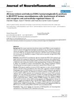
In parallel tests of plants under long days or short
days, the RfBP gene was highly expressed (Figure 1a)
and a substantial amount of the RfBP protein was produced (Figure 1b) in leaves of RfBP+ in contrast to the
absence of gene expression and protein production in
the WT plant. The gene expression and protein production were highly reduced in the RfBP− plant (Figure 1a,b).
In long days, free riboflavin, FMN, and FAD concentrations were decreased by 60%, 52%, and 69%, respectively,
in leaves of RfBP+ compared to WT, but in RfBP−, flavins
were retrieved to approximations of WT levels (Figure 1c).
Similar differences were found in RfBP expression
(Figure 1a), the protein production (Figure 1b), and
flavin concentrations (Figure 1c) among WT, RfBP+,
and RfBP− under short days. Leaf flavin concentrations
were decreased approximately by 20% in all plants
grown in long days compared to short days (Figure 1c).
These analyses suggest that downregulation of free
flavin concentrations in leaves is a constant character
of the RfBP+ plant under short day and long day
conditions.
Ji et al. BMC Plant Biology 2014, 14:237
/>
Page 3 of 13
the early flowering phenotype (Additional file 1: Figure S1).
Thus, early flowering is a constant character of RfBPexpressing plants.
To elucidate the effect of leaf flavin concentrations on
flowering, we performed a pharmacological study in
which plants under long days were fed with an aqueous
solution of riboflavin or treated with ultrapure water as
a control. Riboflavin feeding caused substantial increases
in the leaf content of all flavins, and flavin concentrations in riboflavin-fed RfBP+ were retrieved to the approximations in water-treated WT plants (Figure 3a).
RfBP− resembled WT in the riboflavin-feeding effects on
leaf flavin content (Figure 3a). All plants flowered later
and had more rosette leaves following riboflavin feeding
compared to control while riboflavin-fed RfBP+ plants
lost the early flowering phenotype (Figure 3b). These
observations are in agreement with the RfBP silencing
effect and both lines of evidence attribute the early
flowering phenotype to the reduction of leaf flavin
concentrations.
Flavin content downregulation enhances foliar expression
of floral promoting photoperiod genes
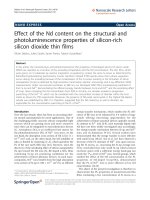
Figure 1 RfBP expression and flavin content in leaves of the
wild-type (WT) plant and RfBP-expressing (RfBP+) or RfBPsilencing (RfBP−) line of Arabidopsis. Plants were grown for
10 days in long days (16 hour light) or 25 days in short days (8 hour)
before use in the following analyses. (a) Northern blotting with the
probe specific to the RfBP gene or the constitutively expressed EF1α
gene used as a reference. (b) Analysis of plant proteins by the gel
electrophoresis. Protein bands were visualized by gel staining with
Coomassie G-250. Molecular makers are indicated. (c) Quantification
of flavin concentrations. Data shown are mean values ± standard
deviation bars of results from three independent experiments each
containing three repeats and 15 plants per repeat. Different letter
on bar graphs indicate significant differences by analysis of variance
and least significant difference test (P < 0.01).
Downregulation of leaf flavin content causes early
flowering
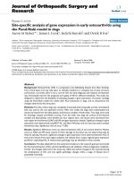
The WT plant took 24 and 47 days to flower with 20
and 43 rosette leaves in long days (Figure 2a) and short
days (Figure 2b), respectively. RfBP− resembled WT in
flowering time and rosette leaf number but RfBP+ flowered 6 days earlier with a reduction of 11 rosette leaves
in long days (Figure 2a) and flowered 15 days earlier with
a shortage of 15 rosette leaves in short days (Figure 2b).
Like RfBP+, other RfBP-expressing lines [11] also acquired
To infer the molecular basis of RfBP-induced early flowering, we compared WT, RfBP+, and RfBP− plants in
terms of the expression of 14 flowering regulatory genes
assigned to photoperiod (PHYA, PHYB, CRY1, CRY2,
CCA1, TOC1, and CO), vernalization (FLC, FRI, and
VIN3), GA (GA1 and GAI), and autonomous (FLC
shared with vernalization, FLM, and LD) pathways. Plants
were grown in long days and sampled during 10–30 days
after seed germination. Gene expression was analyzed by
quantitative real-time reverse transcriptase-polymerase
chain reaction (RT-PCR) using constitutively expressed
EF1α and Actin2 genes as references. RNAs used in the
analysis were isolated from the two youngest expanded
leaves at 13 hours in light (three hours to dark), a time
point at which floral promoting genes are highly expressed
under regulation of the circadian clock, a central player in
the photoperiod pathway [34,35].
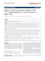
Chronological patterns of gene expression analyzed every
other day during 10–30 days of plant growth are provided
in Figure 4. The seven genes assigned to the vernalization,
GA, or autonomous pathway were little expressed in all
plants while the seven photoperiod genes behaved differently. Regarding to photoperiod, red/far red light receptor
phytochromes PHYA and PHYB [44,45] and blue light receptor cryptochromes CRY1 and CRY2 [46,47] serve as the
entry of the clock [40], which employs the negative CCA1
and TOC1 transcriptional feedback loop to control daynight rhythm of photoperiod gene expression [40,48,49].
RfBP did not cause evident effect on CCA1 as its expression levels were similar in all plants through out the course
of time. PHYB expression was decreased with time in all
Ji et al. BMC Plant Biology 2014, 14:237
/>
Page 4 of 13
Figure 2 Flowering characters of WT, RfBP+, and RfBP− plants. Plants were grown in long days (a) and short days (b), respectively. Data
shown in bar graphs are mean values ± standard deviation bars of results from three independent experiments each containing three repeats
and 30 plants per repeat. Observed values are shown on deviation bars. Different letters in bar graphs indicate significant differences by analysis
of variance and least significant difference test (P < 0.01).
plants but decreasing extents were significantly (P < 0.01)
smaller in RfBP+ compared to WT or RfBP−. Expression
levels of five other photoperiod genes (PHYA, CRY1,
CRY2, TOC1, and CO), which are flowering activators
[34,38,40,44,48,50], were highly elevated as compared
to controls (Actin2 to EF1α transcript ratios) and sharp
elevations were detected approximately four days before
flowering in all plants. However, RfBP+ was more vigorous
than WT and RfBP− in chronologically increased expression of the photoperiod genes. Their expression was highly
enhanced in RfBP+ compared to WT or RfBP− at every
time point during 10–30 days. During this period multiples
Figure 3 The effects of riboflavin feeding on leaf flavin content and plant flowering time under long days. Ten-day-old plants grown in
long days were fed with riboflavin or treated with water in control. Two days later, leaf flavin content was determined (a). Subsequently, plant
flowering time and rosette leaf number were scored (b). Data shown in bar graphs are mean values ± standard deviation bars of results from
three independent experiments each containing three repeats and 15 plants per repeat. Different letters on top indicate significant differences by
analysis of variance and least significant difference test.
Ji et al. BMC Plant Biology 2014, 14:237
/>
Page 5 of 13
Figure 4 The expression of flowering regulatory genes in leaves of WT, RfBP+, and RfBP− plants grown in long days. Chronological patterns of
gene expression were analyzed by quantitative real-time RT-PCR with RNAs isolated from the two youngest leaves of plants at the indicated
times. The constitutively expressed EF1α and Actin2 genes were used as references. Data shown in curves are mean values ± standard bars of
results from three independent experiments each containing three repeats and five plants per repeat. Gray dashed and bidirectional arrowheads indicate
significant differences between RfBP+ and WT or RfBP− at the range of time intervals based on analysis of variance and least significant difference test
(P < 0.01).
of expression enhancements by RfBP were 1.4–3.2 for PHYA,
1.6–4.4 for CRY1, 1.4–4.0 for CRY2, 1.5–2.8 for TOC1, and
1.9–4.5 for CO. Clearly, RfBP+ enhances the foliar expression
of floral promoting photoperiod genes (Figure 4).
The effect of RfBP on gene expression was cancelled
by the riboflavin feeding treatment (Additional file 2:
Figure S2), which annulled the early flowering phenotype
and also eliminated approximate RfBP-reduced parts of
the intrinsic flavin content in RfBP+ leaves (Figure 3b).
The endogenous flavin concentrations were increased
(Figure 3a) and expression levels of the five floral
promoting photoperiod genes were decreased significantly (P < 0.01) in leaves of WT and RfBP− plants fed
with riboflavin compared to water (Additional file 2:
Figure S2). Therefore, free flavin concentrations negatively affect RfBP-enhanced expression of the floral
promoting photoperiod genes in leaves with long days.
RfBP enhances FT expression in leaves and coordinate FD
and AP1 expression in the shoot apex
The circadian clock exit gene CO [48] is one of RfBPinduced photoperiod genes (Figure 4). In response to the
Ji et al. BMC Plant Biology 2014, 14:237
/>
photoperiod signal, CO is produced as an output of the
circadian clock and acts in turn to activate the expression of the florigen gene FT in leaves [48,50]. As shown
in Figure 5a, marked expression of FT was detected in
leaves of 12- and 18-day-old plants with greater quantities
in RfBP+ than in WT or RfBP− under long day condition.
Interestingly, FT still displayed substantial expression
in RfBP+ on the flowering day (Figure 5a compared to
Figure 2a). As shown in Figure 5b, quantities of the FT
transcript in the different plants with long days were
markedly increasing since 10 days of growth, reached
the highest values on two days before flowering, and
started to decline gradually after flowering. Thus,
chronological patterns of FT expression were similar
in all plants during 10–30 days of growth in long days.
However, FT expression levels kept greater at every
time point and was increased earlier with significantly
(P < 0.01) higher extents in leaves of RfBP+ compared
to WT and RfBP− (Figure 5b).
As a result of the photoperiod regulation, the florigen
FT protein moves from leaves to the shoot apex [51,52],
where it functions with FD to activate AP1 [12,13], which
marks the beginning of floral organ formation [34]. At the
transcription level, the FD and AP1 genes are coordinately
expressed at the shoot apex to initiate flowering by promoting the growth of floral organ primordia [42,43]. To
elucidate the role of FD and AP1 in RfBP-induced flowering, we analyzed their expression in shoot apices of
12- and 18-day-old plants. We detected concomitant
expression of FD and AP1 from all plants (Figure 6a)
and significantly (P < 0.01) higher amounts of gene
transcripts in RfBP+ than in WT or RfBP− (Figure 6b).
Clearly, the de novo expression of RfBP affects the
Page 6 of 13
synchronized expression of FD and AP1 at the shoot
apex.
RfBP enhances expression of photoperiod and FT genes
in leaves and expression of FD and AP1 in shoot apices
under inductive photoperiod
To further elucidate the molecular basis of RfBP-induced
early flowering, we tested the expression of FT and flowering regulatory genes in leaves and the expression of FD
and AP1 in shoot apices of WT, RfBP+, and RfBP− plants
under inductive photoperiod. This condition was devised
by considering: (i) RfBP+ flowers after 32 days while WT
and RfBP− flower after 47 and 46 days of growth in short
days (Figure 2b); and (ii) floral organ primordia can well
grow within five days and differentiate into floral organs in
the subsequent days under inductive photoperiod [43].
Therefore, we employed the inductive photoperiod by
growing plants in short days for 23 days and transferred
them to long days. We analyzed gene expression immediately (zero day) after inductive photoperiod and in the
subsequent nine days.
As shown in Figure 7, inductive photoperiod caused
different effects on the foliar expression of genes
assigned to different floral pathways and the effects were
also different in RfBP+ from WT and RfBP−. In all plants,
inductive photoperiod did not cause evident effect on
CCA1 or genes assigned to vernalization, GA, and autonomous pathways in comparison with transcript ratios
between reference genes Actin2 and EF1α. Inductive
photoperiod repressed the expression of PHYB and repression extents were significantly (P < 0.01) lower in RfBP+
leaves than in leaves of WT or RfBP−. In comparison to
transcript ratios between Actin2 and EF1α, expression levels
Figure 5 Expression of the florigen gene FT in leaves of the different plants grown in long days. Gene expression was analyzed by Northern
blotting (a) and quantitative real-time RT-PCR (b). Both analyses were performed on RNAs isolated from the two youngest leaves of plants at the
indicated times and using EF1α and Actin2 genes as references. Data shown in curves (b) are mean values ± standard bars of results from three
independent experiments each containing three repeats and five plants per repeat. Gray dashed and bidirectional arrowheads indicate significant
differences between RfBP+ and WT or RfBP− at the range of time intervals based on analysis of variance and least significant difference test (P < 0.01).
Ji et al. BMC Plant Biology 2014, 14:237
/>
Page 7 of 13
Figure 6 Expression of floral meristem identity genes FD and AP1 in shoot apices of plants grown in long days. Northern blotting (a) and
real-time RT-PCR (b) analyses were performed with RNAs isolated from shoot apices of plants at the indicated times. Data shown in (b) are mean
values ± standard deviation bars of results from three independent experiments each with three repeats and five plants per repeat. Different letters in
bar graphs indicate significant differences by analysis of variance and least significant difference test (P < 0.01).
of floral promoting photoperiod genes PHYA, CRY1, CRY2,
TOC1, and CO in leaves of all plants were increased by inductive photoperiod. These genes were expressed in a similar chronological pattern. Expression levels were increased
slightly in 3 days in RfBP+ and 5 days in WT and RfBP−,
reached the highest levels in the next two days, and then
declined in all plants. At every time point, extents by
which inductive photoperiod acted to enhance the expression of PHYA, CRY1, CRY2, TOC1, and CO were
significantly (P < 0.01) greater in RfBP+ leaves than in
leaves of WT or RfBP−.
In all plants, inductive photoperiod caused enhancements in the foliar expression of FT (Figure 8a) and the
expression of FD and AP1 in shoot apices (Figure 8b).
Nevertheless, enhancement extents were significantly
(P < 0.01) greater in RfBP+ than in WT or RfBP−. In all
plants, moreover, expression levels of FT in leaves and
expression levels of FD and AP1 in shoot apices were increased in six days and then the foliar expression of FT
was continuously increased (Figure 8a) but the apical expression of FD and AP1 remained stable till the ninth
day (Figure 8b).
Taken together, these analyses suggest that the de
novo expression of RfBP in Arabidopsis enhances
the expression of FT and floral promoting photoperiod genes in leaves and also enhances the expression of FD and AP1 in the shoot apex under
inductive photoperiod. Gene expression enhancements
are significant in the RfBP+ plant compared to WT or
RfBP− background.
Reduction of leaf flavin content is responsible for
enhancements of the gene expression under inductive
photoperiod
To correlate leaf flavin content with RfBP-enhanced
gene expression under inductive photoperiod, we
tried to increase flavin levels by feeding plants with
riboflavin and analyzed PHYA, CRY1, CRY2, CCA1,
TOC1, CO, FT, FD, and AP1 expression at the fifth
day after inductive photoperiod, a time point at which
these genes are highly expressed in leaves or shoot
apices in the absence of riboflavin feeding (Figures 7
and 8). Under inductive photoperiod, feeding plants
with riboflavin caused substantial increases in leaf
concentrations of all flavins, and flavin levels in
riboflavin-fed RfBP+ were retrieved to the approximations in water-treated WT plants (Additional file 3:
Figure S3). RfBP− resembled WT in the riboflavinfeeding effects on leaf flavin content (Additional file 3:
Figure S3). In all plants, CCA1 expression in leaves
was unaffected, but the foliar expression of PHYA,
CRY1, CRY2, TOC1, CO, and FT in leaves (Figure 9a)
and the expression of FD and AP1 in shoot apices
(Figure 9b) were decreased by the riboflavin feeding
treatment compared to water. Riboflavin-fed RfBP+
plants performed similarly to water-treated WT or
RfBP− plants in gene expression. In RfBP+, therefore,
enhancements of FT and photoperiod gene expression
in leaves, and enhancements of FD and AP1 expression in the shoot apex, are caused by the reduction of
leaf flavin concentrations.
Ji et al. BMC Plant Biology 2014, 14:237
/>
Page 8 of 13
Figure 7 The expression of flowering regulatory genes in leaves of WT, RfBP+, and RfBP− plants grown under inductive photoperiod.
Gene expression in the two youngest leaves was analyzed by real-time RT-PCR at the indicated times. Data shown in curves are mean values ±
standard deviation bars of results from three independent experiments each containing three repeats and five plants per repeat. Gray dashed
and bidirectional arrowheads indicate significant differences between RfBP+ and WT or RfBP− at the range of time intervals based on analysis of
variance and least significant difference test (P < 0.01).
Discussion
The well-demonstrated developmental role of oviparous
RfBP in riboflavin binding and redistribution [13,28,32]
inspired the idea to manipulate plant riboflavin content
by engineering with the turtle RfBP [11]. Its activity in
riboflavin binding allows for the function in modulating
free flavin concentrations in transgenic plants [11]. On
this basis, in the present study we have characterized the
serendipitous role of the RfBP protein in affecting flowering
time after de novo expression in Arabidopsis. We investigated Arabidopsis RfBP+ and RfBP− lines in comparison
with the WT plant (Figure 1) and demonstrated that
RfBP-caused downregulation of free flavin content in
leaves (Figure 1) induced the early flowering phenotype (Figure 2). By feeding plants with riboflavin to
increase the intrinsic content of free flavins and determining the subsequent effect on flowering time, analyzing the pharmacological data together with those about
the RfBP+ vs. RfBP− effects, we were able to attribute the
early flowering phenotype to the reduction of free flavin
concentrations in leaves (Figure 3) on the basis of RfBP
binding with riboflavin inside leaf cells [11].
Ji et al. BMC Plant Biology 2014, 14:237
/>
Page 9 of 13
Figure 8 The expression of FT, FD, and AP1 under inductive photoperiod. Gene expression in the two youngest leaves (a) and shoot apices
(b) was analyzed by real-time RT-PCR at the indicated times. Data shown in curves are mean values ± standard deviation bars of results from three
independent experiments each containing three repeats and five plants per repeat. Gray dashed and bidirectional arrowheads indicate significant
differences between RfBP+ and WT or RfBP− at the range of time intervals based on analysis of variance and least significant difference test (P < 0.01).
Riboflavin is a venerable multifaceted player in tremendous biochemical processes and frequently receives
renascent attentions with newly discovered functions
[1-11]. Since its discovery in 1879 and biochemical
characterization in 1933, a variety of physiological roles
that flavins play in plants have been extensively studied
[3,53]. In particular, previously unappreciated functions
have been often reported in recent 10 years. For example,
genetic modification of the riboflavin biosynthesis pathway
alters some aspects of plant development, such as leaf senescence regulated by the COS1 protein, an essential component of the jasmonic acid signaling pathway [54]. In
fact, COS1 is the lumazine synthase [54], which catalyzes
the penultimate step of the riboflavin biosynthesis pathway
[55]. Arabidopsis mutants that have partial defect in COS1
and partial decrease in riboflavin content compromise the
regulatory role of jasmonic acid in leaf senescence [54]. In
plants, moreover, externally applied riboflavin induces resistance to pathogens by priming of defense responses in a
manner of salicylic acid dependence or independence according to the type of pathogens, biotrophic or necrotrophic [6,10]. Externally applied riboflavin also induces
plant growth enhancement by activating the ethylene signaling pathway [56]. These findings suggest that changes
in riboflavin content cause physiological and pathological
responses by affecting phytohormone signaling pathways.
Through studies detailed here, novel functions of flavins
have been extended from cellular signaling to flowering
time control in relation to the expression of photoperiod
and flowering time genes.
The expression of photoperiod and flowering time
genes is implicated in RfBP-induced flowering based on
several lines of evidence (Figures 4, 5, 6, 7, 8 and 9). First,
RfBP+ causes enhanced expression of five photoperiod
genes (PHYA, CRY1, CRY2, TOC1, and CO), which are
flowering activators [34,35,40], in leaves under long day
(Figure 4) or inductive photoperiod (Figure 7) conditions. By contrast, PHYB is a flowering repressor [44,57]
and PHYB expression is repressed by RfBP in contrast
to the early flowering phenotype and RfBP-enhanced
expression of the floral promoting photoperiod genes
(Figures 4 and 7). In addition, CCA1 is highly expressed
in the early phase and its expression declines in the late
phase of day (46,49), explaining why RfBP is unable to
affect CCA1 expression. Similarly, genes assigned to autonomous, gibberellin, and vernalization pathways are
not related to RfBP-induced early flowering (Figure 4).
Second, enhanced expression of the photoperiod genes
Ji et al. BMC Plant Biology 2014, 14:237
/>
Page 10 of 13
Figure 9 The effects of riboflavin feeding on the expression of flowering regulatory genes under inductive photoperiod. Plants were
grown in short days for 23 days and transferred to long days. Immediately after plant transfer, H2O or an aqueous riboflavin solution was applied
by spraying over plant tops. Four days later, the expression of FT and photoperiod genes in leaves (a) and the expression of FD and AP1 in shoot
apices (b) were analyzed by real-time RT-PCR using EF1α and Actin2 as reference genes. Data shown are mean values ± standard deviation bars of
results from three independent experiments each containing three repeats and 15 plants per repeat. Different letters in bar graphs indicate significant
differences by analysis of variance and least significant difference test (P < 0.01).
was correlated with enhanced expression of FT in leaves
(Figures 5 and 8). The FT protein is the florigen that can
moves from leaves to shoot apices [51,52], where it functions with FD to activate AP1 for the growth of floral organs [42,43]. Third, concomitantly enhanced expression of
FT and photoperiod genes was further correlated with the
synchronized expression of FD and AP1 in the shoot apex
(Figures 6, 8, and 9), while synchronized expression of FD
and AP1 in the shoot apex initiates floral organ formation
[42,43,58,59]. The role of RfBP in gene expression is
attributable to reduction of free flavin levels in leaves
(Figure 9).
As flavins anticipate in numerous biochemical processes,
it is difficult to elucidate the functional relationship between
downregulated flavin concentrations and the photoperiod
pathway. A possible mediator is H2O2, a cellular signal that
can be induced by the de novo RfBP expression and downregulation of free flavin content inside Arabidopsis leaves
[11]. H2O2 has been implicated in crosstalk with flowering
regulators [60] and actually participates in the regulation of
flowering time [61-64]. For example, flowering is promoted
when cytosolic H2O2 levels are elevated by the activity of
chloroplastic lipoxygenase or ascorbate peroxidase in Arabidopsis [61,62]. As downregulation of free flavin concentrations in leaves by RfBP induces the production of the H2O2
signal and its translocation from the apoplast to the cytosol
[11], the signal may act in turn to promote flowering
[61,62]. Alternatively, H2O2 may be generated through
electron leakage from the mitochondrial electron transport chain due to shortage of FMN and FAD, which
serve as redox centers in the chain [65-68].
Conclusions
Meticulous phenotypic observations indicate that early
flowering is a constant character conferred by the de novo
expression of RfBP in transgenic Arabidopsis plants grown
Ji et al. BMC Plant Biology 2014, 14:237
/>
in short days and long days. The phenotype is caused indirectly by downregulation of free flavin concentrations in
leaves based on pertinent analyses of the RfBP+ vs. RfBP−
effects, as well as the pharmacological consequence from
the riboflavin feeding treatment, performed under long
days and inductive photoperiod. Under both conditions,
reduction of leaf flavin content induces the expression of
floral promoting photoperiod genes in leaves, coincident
expression of the florigen gene FT in leaves, and synchronized expression of the flowering regulatory gene FD and
the floral meristem identity gene AP1 in the shoot apex.
We don’t have evidence to show the connection between
changes in flavin concentrations and any of the floral regulators. In fact, we found the early flowering phenomenon
by accident, but we don’t know what it means with respect
to photoperiod gene expression and flowering time control.
Methods
Plant growth conditions and flowering observations
Plants were grown in pots containing potting soil [69]
under the environment-controlled conditions: 22 ± 1°C,
55% humidity, short days or long days, and light at
200 μM quanta/m2/s. The flowering phenotype was
characterized by two criteria: days to flowering and
rosette leaf number [70].
Gene expression analyses
Total RNA was isolated from the two youngest expanded leaves or shoot apices and subjected to real-time
RT-PCR or Northern (RNA) blotting analyses using the
constitutively expressed EF1α and/or Actin2 genes as
references. Real-time RT-PCR was performed with specific primers (Additional file 4: Table S1) as previously
described [71,72]. The expression level of a tested gene
was quantified as the ratio between transcript amounts of
the gene and EF1α. Northern blots were hybridized to the
RfBP-specific probe labeled with digoxigenin (Novagen,
EMD Biosci., Inc., WI, USA).
Protein analyses
A histidine (His) tag had been added to the C-terminus
of RfBP in the transformation construction and was used
to facilitate purification of plant protein preparations by
nickel chromatography [11]. The two youngest expanded
leaves were excised and used in isolation of total proteins
from 10 mg fresh leaves as previously described [73]. Isolated proteins were bound to nickel-polystyrene beads
according to the manufacturer’s instruction (Amersham
Biosciences Corp., Piscataway, NJ, USA), eluted with
aqueous solutions of imidazole at 100, 150, and 300 mM,
respectively. The 200-mM imidazole eluent was treated
with the Novagen Enterokinase Cleavage Capture Kit
(EMD Biosciences Inc., Darmstadt, Germany) to remove
the His tag and analyzed by tricine sodium dodecyl sulfate
Page 11 of 13
polyacrylamide gel electrophoresis [71]. Proteins were visualized by gel staining with Coomassie G-250.
Flavin measurements
All operations were in subdued light. Riboflavin, FMN,
and FAD were extracted using a previously described
method [11,74]. Leaf samples (1 g/treatment) were ground
on the ice with 2 ml cold extraction buffer A (pH6.9) containing 5 mM NaH2PO4. 2H2O, 5 mM Na2HPO4. 12H2O,
0.2 M NaCl, 0.5 mM phenylmethylsulfonyl fluoride, and
1 mM ethylene dianetetra-acetic acid. Homogenate was
centrifugated at 4°C and 12,000 g for 10 minutes. Supernatant was divided into two groups. In the first group,
200 μl supernatant was supplemented with 1 ml buffer B
made of 10% trichloroacetic acid in 0.1 M ammonium
acetate (pH6.1). The mixture was centrifuged at room
temperature (12,000 g, 10 minutes) and the new supernatant was regarded as a preparation of total flavins [74].
In the second group, 500 μl supernatant was loaded into a
Microcon YM-3 (3 kDa NMWL) ultrafiltration spin column (Millipore, Billerica, MA, USA). The column was
spun at 4°C and 14,000 g for 15 minutes. Filtrate of 200 μl
was shifted into an Eppendorf tube, supplemented with
1 ml buffer B. The mixture was centrifuged at room
temperature (12,000 g, 10 minutes) and the final supernatant was regarded as a preparation of free flavins. The
preparations of total and free flavins were filtrated separately with 0.22 μm blend cellulose ester filters. Each filtrate
of 20 μl was analyzed by high performance liquid chromatography [75] with the Agilent 1200 HPLC system (Agilent
Tech. Inc., Santa Clara, CA, USA). Concentrations of riboflavin, FMN, and FAD in the preparations were determined
by reference to similar analysis of the inner standards [75]
and quantified in contrast to plant weight.
Riboflavin feeding experiments
The riboflavin (EMD Biosci., Inc., Darmstadt, Germany)
feeding experiments were performed on plants grown
under long day and inductive photoperiod conditions,
respectively. Plants were treated by spraying over tops
with an aqueous solution of 0.2 mM riboflavin, made in
ultrapure water produced by the EliX10/Milli-Q Synthesis
A10 ultrapure water system (Merck Millipore Corporation, Billerica, MA, USA), and treated similarly with ultrapure water in the experimental control group. Flavin
measurements and gene expression analyses were performed on the two youngest expanded leaves. Flowering
time and the rosette leaf number were monitored.
Data treatment
All experiments were carried out at least three times with
similar results. Quantitative data were analyzed with the IBM
SPSS19.0 software package (IBM Corporation, Armonk,
NY, USA; />
Ji et al. BMC Plant Biology 2014, 14:237
/>
Page 12 of 13
according to instructions in a text book that describes
in details analysis methods using IBM SPSS19.0 [76].
Homogeneity-of-variance in data was determined by
Levene test, and formal distribution pattern of the data
was confirmed by Kolmogorov-Smirnov test and P-P
Plots [76]. Then, data were analyzed by analysis of variance and least significant difference test [77].
6.
Additional files
9.
Additional file 1: Figure S1. Flowering characteristics of different RfBPexpressing Arabidopsis lines in comparison with the WT plant in long days.
Additional file 2: Figure S2. The effects of riboflavin feeding treatment
on expression of photoperiod genes in long days.
7.
8.
10.
11.
Additional file 3: Figure S3. The effects of riboflavin feeding treatment
on flavin concentrations in leaves under inductive photoperiod.
Additional file 4: Table S1. Information on genes tested and primers
used in this study.
12.
13.
Abbreviations
AP1: APETALA1; CCA1: CIRCADIAN CLOCK-ASSOCIATED1; CO: CONSTANS;
CRY: Cryptochrome; FAD: Flavin adenine dinucleotide; FLC: FLOWERING
LOCUS C; FMN: Flavin mononucleotide; FLM: FLOWERING LOCUS M;
FRY: FRIGIDA; FT: FLOWERING LOCUS T; GA: Gibberellin; GAI: GA INSENSITIVE;
GA1: GA REQUIRING 1; LD: LUMINIDEPENDENS; PHY: Phytochrome;
RfBP: riboflavin-binding protein; RfBP+: RfBP-expressing transgenic
Arabidopsis line; RfBP−: RfBP-silenced Arabidopsis line generated under RfBP+
background; SOC1: Suppressor of overexpression of CO1; TOC1: Timing of
cab expression1; VIN3: Vernalization insensitive 3.
Competing interests
The authors declared that they have no competing interests.
Authors’ contributions
HJ and YZ performed the experiments, analyzed the data, and wrote the
paper. ST and MX performed the experiments and wrote the paper. YT, LL,
HW, LH, YJ, JG, and WW performed the experiments. HD designed the
experiments and wrote the paper. All authors read and approved the final
manuscript.
Acknowledgements
This study was supported by NSFC (31171830 and 31272072), National Key
Basic Research Program of China (973 plan 2012CB114003), Novel Transgenic
Organisms Breeding Project (2013ZX08002-001), and Ministry of Education
111 Project of China and Academic Priority Program of High Education in
Jiangsu Province.
Received: 12 February 2014 Accepted: 20 August 2014
Published: 9 September 2014
References
1. Powers HJ: Riboflavin (vitamin B-2) and health. Am J Clin Nutr 2003,
77:1352–1360.
2. Weimar WR, Neims AH: Physical And Chemical Properties Of Flavin.
Edited by Rivlin RS. Riboflavin: Plenum Press; 1975:2–36.
3. Jordan DB, Bacot KO, Carlson TJ, Kessel M, Viitanen PV: Plant riboflavin
biosynthesis. Cloning, chloroplast localization, expression, purification,
and partial characterization of spinach lumazine synthase. J Biol Chem
1999, 274:22114–22121.
4. Sierra I, Vidal-Valverde C: Kinetics of free and glycosylated B6 vitamers,
thiamin and riboflavin during germination of pea seeds. J Sci Food Agr
1999, 79:307–310.
5. Mori T, Sakurai E: Riboflavin affects anthocyanin synthesis in nitrogen
culture using strawberry suspended cells. J Food Sci 1996, 61:698–702.
14.
15.
16.
17.
18.
19.
20.
21.
22.
23.
24.
25.
26.
27.
28.
29.
30.
Dong HS, Beer SV: Riboflavin induces disease resistance in plants by
activating a novel signal transduction pathway. Phytopathology 2000,
90:801–811.
de Souza AC, Kodach L, Gadelha FR, Bos CL, Cavagis AD, Aoyama H,
Peppelenbosch MP, Ferreira CV: A promising action of riboflavin as a
mediator of leukaemia cell death. Apoptosis 2006, 11:1761–1771.
Taheri P, Höfte M: Riboflavin induces resistance in rice against
Rhizoctonia sheath diseases by activating signal transduction
pathways leading to upregulation of rice cationic peroxidase and
formation of lignin as a structural barrier. Commun Agr Appl Biol Sci
2006, 71:255–258.
Taheri P, Tarighi S: Riboflavin induces resistance in rice against
Rhizoctonia solani via jasmonate-mediated priming of phenylpropanoid
pathway. J Plant Physiol 2010, 167:201–208.
Zhang SJ, Yang X, Sun MW, Sun F, Deng S, Dong HS: Riboflavin-induced
priming for pathogen defense in Arabidopsis thaliana. J Integr Plant Biol
2009, 51:167–174.
Deng BL, Deng S, Sun F, Zhang SJ, Dong HS: Down-regulation of free
riboflavin content induces hydrogen peroxide and a pathogen defense
in Arabidopsis. Plant Mol Biol 2011, 77:185–201.
Hamajima S, Ono S: Sequence of a cDNA encoding turtle riboflavinbinding protein: a comparison with avian riboflavin-binding protein.
Gene 1995, 164:279–282.
Bangaru ML, Karande AA: Biochemical characterization of recombinant
chicken riboflavin carrier protein. Mol Cell Biochem 2008, 308:1–7.
Bedhomme M, Hoffmann M, McCarthy EA, Gambonnet B, Moran RG,
Rébeillé F, Ravanel S: Folate metabolism in plants: an Arabidopsis
homolog of the mammalian mitochondrial folate transporter mediates
folate import into chloroplasts. J Biol Chem 2005, 280:34823–34831.
Sabharanjak S, Mayor S: Folate receptor endocytosis and trafficking.
Adv Drug Deliv Rev 2004, 56:1099–1109.
Torres MA: ROS in biotic interactions. Physiol Plant 2010, 138:414–429.
Rhodes MB, Bennett N, Feeney RE: The flavoprotein-apoprotein system of
egg white. J Biol Chem 1959, 234:2054–2060.
Maehashi K, Matano M, Uchino M, Yamamoto Y, Takano K, Watanabe T: The
primary structure of a novel riboflavin-binding protein of emu (Dromaius
novaehollandiae). Comp Biochem Physiol (Part B) 2009, 153:95–100.
Storey KB, Dent ME, Storey JM: Gene expression during estivation in
spadefoot toads, Scaphiopus couchii: Upregulation of riboflavin binding
protein in liver. J Exp Zool 1999, 284:325–333.
Wang DS, Senthilkumaran B, Kobayashi T, Kajiura-Kobayashi H, Matsuda M,
Yoshikuni M, Nagahama Y: Molecular cloning and gene expression of the
riboflavin-binding protein in the Nile tilapia, Oreochromis niloticus. Fish
Physiol Biochem 2003, 28:225–226.
Natraj U, George S, Kadam PA: Isolation and partial characterization of
human riboflavin carrier protein and the estimation of this protein
during human pregnancy. J Reprod Immunol 1988, 13:1–16.
Zheng DB, Lim HM, Pene JJ, White HB: Chicken riboflavin-binding protein
cDNA sequence and homology with milk folate-binding protein. J Biol
Chem 1988, 263:11126–11129.
Pattanaik P, Sooryanarayana, Adiga PR, Visweswariah SS: Refolding of
native and recombinant chicken riboflavin carrier (or binding)
protein: evidence for the formation of non-native intermediates
during the generation of active protein. Eur J Biochem 1998,
258:411–418.
Monaco HL: Crystal structure of chicken riboflavin-binding protein. EMBO
J 1997, 16:1475–1483.
Kozik A: Disulfide bonds in egg-white riboflavin-binding protein: chemical
reduction studies. Eur J Biochem 1982, 121:395–400.
Bartosík M, Ostatná V, Palecek E: Electrochemistry of riboflavin-binding protein
and its interaction with riboflavin. Bioelectrochemistry 2009, 76:70–75.
Huang SN, Swaan PW: Involvement of a receptor-mediated component in
cellular translocation of riboflavin. J Pharmacol Exp Ther 2000, 294:117–125.
Foraker AB, Khantwal CM, Swaan PW: Current perspectives on the cellular
uptake and trafficking of riboflavin. Adv Drug Deliv Rev 2003, 55:1467–1483.
Gastaldi G, Laforenza U, Casirola D, Ferrari G, Tosco M, Rindi G: Energy
depletion differently affects membrane transport and intracellular
metabolism of riboflavin taken up by isolated rat enterocytes.
J Nutr 1999, 129:406–409.
Said HM, Mohammed ZM: Intestinal absorption of water-soluble vitamins:
An update. Curr Opin Gastroen 2006, 22:140–146.
Ji et al. BMC Plant Biology 2014, 14:237
/>
31. Adiga PR, Visweswariah SS, Karande AA, Velu NK: Biochemical and
immunological aspects of riboflavin carrier protein. J Biosciences 1988,
13:87–104.
32. Wasylewski M: Binding study of riboflavin-binding protein with riboflavin
and its analogues by differential scanning calorimetry. J Protein Chem
2000, 19:523–528.
33. Sooryanarayana Sarkar S, Adiga PR, Visweswariah SS: Identification and
characterization of receptors for riboflavin carrier protein in the chicken
oocyte. Role of the phosphopeptide in mediating receptor interaction.
Biochim Biophys Acta 1998, 1382:230–242.
34. Srikanth A, Schmid M: Regulation of flowering time: all roads lead to
Rome. Cell Mol Life Sci 2011, 68:2013–2037.
35. Jung C, Müller AE: Flowering time control and applications in plant
breeding. Trends Plant Sci 2009, 14:563–573.
36. Mutasa-Göttgens E, Hedden P: Gibberellin as a factor in floral regulatory
networks. J Exp Bot 2009, 60:1979–1989.
37. Simpson GG: The autonomous pathway: epigenetic and posttranscriptional gene regulation in the control of Arabidopsis flowering
time. Curr Opin Plant Biol 2004, 7:570–574.
38. Putterill J, Robson F, Lee K, Simon R, Coupland G: The CONSTANS gene of
Arabidopsis promotes flowering and encodes a protein showing
similarities to zinc finger transcription factors. Cell 1995, 80:847–857.
39. Turck F, Fornara F, Coupland G: Regulation and identity of florigen:
FLOWERING LOCUS T moves center stage. Annu Rev Plant Biol 2008,
59:573–594.
40. Más P, Yanovsky MJ: Time for circadian rhythms: plants get synchronized.
Curr Opini Plant Biol 2009, 12:574–579.
41. Kaufmann K, Wellmer F, Muiño J, Ferrier T, Wuest S, Kumar V, SerranoMislata A, Madueño F, Krajewski P, Meyerowitz E, Angenent G, Riechmann J:
Orchestration of floral initiation by APETALA1. Science 2010, 328:85–89.
42. Abe M, Kobayashi Y, Yamamoto S, Daimon Y, Yamaguchi A, Ikeda Y, Ichinoki
H, Notaguchi M, Goto K, Araki T: FD, a bZIP protein mediating signals
from the floral pathway integrator FT at the shoot apex. Science 2005,
309:1052–1056.
43. Wigge PA, Kim MC, Jaeger KE, Busch W, Schmid M, Lohmann JU, Weigel D:
Integration of spatial and temporal information during floral induction
in Arabidopsis. Science 2005, 309:1056–1059.
44. Chen M, Chory J: Phytochrome signaling mechanisms and the control of
plant development. Trends Cell Biol 2011, 21:664–671.
45. Kircher S, Terecskei K, Wolf I, Sipos M, Adam E: Phytochrome A-specific
signaling in Arabidopsis thaliana. Plant Signal Behav 2011, 6:1714–1719.
46. Thomas B: Light signals and flowering. J Exp Bot 2006, 57:3387–3393.
47. Van Buskirk EK, Decker PV, Chen M: Photobodies in light signaling.
Plant Physiol 2012, 158:52–60.
48. Suárez-López P, Wheatley K, Robson F, Onouchi H, Valverde F, Coupland G:
CONSTANS mediates between the circadian clock and the control of
flowering in Arabidopsis. Nature 2001, 410:1116–1120.
49. Martinez-Garcia JF, Huq E, Quail PH: Direct targeting of light signals to a
promoter element-bound transcription factor. Science 2000, 288:859–863.
50. Yoo SK, Chung KS, Kim J, Lee JH, Hong SM, Yoo SJ, Yoo SY, Lee JS, Ahn JH:
CONSTANS activates SUPPRESSOR OF OVEREXPRESSION OF CONSTANS 1
through FLOWERING LOCUS T to promote flowering in Arabidopsis.
Plant Physiol 2005, 139:770–778.
51. Corbesier L, Vincent C, Jang S, Fornara F, Fan Q, Searle I, Giakountis A,
Farrona S, Gissot L, Turnbull C, Coupland G: FT protein movement
contributes to long-distance signaling in floral induction of Arabidopsis.
Science 2007, 316:1030–1033.
52. Jäger K, Wigge P: FT protein acts as a long-range signal in Arabidopsis.
Curr Biol 2007, 17:1050–1054.
53. Fernandez AP, Strand A: Retrograde signaling and plant stress: plastid signals
initiate cellular stress responses. Curr Opin Plant Biol 2008, 11:509–513.
54. Xiao S, Dai L, Liu F, Wang Z, Peng W, Xie D: COS1: An Arabidopsis
coronatine insensitive1 suppressor essential for regulation of jasmonatemediated plant defense and senescence. Plant Cell 2004, 16:1132–1142.
55. Roje S: Vitamin B biosynthesis in plants. Phytochemistry 2007, 68:1904–1921.
56. Peng JL, Zhao J, Pan XM, Zhao JS, Dong HS, Wang JS, Liu BX, Liu GY, Cheng
YJ: Riboflavin activates growth signal transduction pathway in plants.
J Nanjing Agric Univ 2002, 25:33–36.
57. Reed JW, Nagatani A, Elich TD, Fagan M, Chory J: Phytochrome A and
phytochrome B have overlapping but distinct functions in Arabidopsis
development. Plant Physiol 2007, 104:1139–1149.
Page 13 of 13
58. Corbesier L, Coupland G: The quest for florigen: a review of recent
progress. J Exp Bot 2006, 57:3395–3403.
59. Jack T: Molecular and genetic mechanisms of floral control. Plant Cell
2004, 16:S1–S17.
60. Chai L, Wang J, Fan Z, Liu Z, Wen G, Li X, Yang Y: Regulation of the
flowering time of Arabidopsis thaliana by thylakoid ascorbate
peroxidase. Afr J Biotechnol 2012, 11:7151–7157.
61. Bañuelos GR, Argumedo R, Patel K, Ng V, Zhou F, Vellanoweth RL: The
developmental transition to flowering in Arabidopsis is associated with an
increase in leaf chloroplastic lipoxygenase activity. Plant Sci 2008, 174:366–373.
62. Lokhande SD, Ogawa K, Tanaka A, Hara T: Effect of temperature on
ascorbate peroxidase activity and flowering of Arabidopsis thaliana
ecotypes under different light conditions. J Plant Physiol 2003, 160:57–64.
63. Shen CH, Krishnamurthy R, Yeh KW: Decreased L-ascorbate content
mediating bolting is mainly regulated by the galacturonate pathway in
Oncidium. Plant Cell Physiol 2009, 50:935–946.
64. Zafra A, Rodríguez-García MI, Alché JD: Cellular localization of ROS and NO
in olive reproductive tissues during flower development. BMC Plant Biol
2010, 10:36.
65. Blokhina O, Fagerstedt KV: Reactive oxygen species and nitric oxide in
plant mitochondria: origin and redundant regulatory systems. Physiol
Plant 2009, 138:447–462.
66. Gill SS, Tuteja N: Reactive oxygen species and antioxidant machinery in
abiotic stress tolerance in crop plants. Plant Physiol Biochem 2010, 48:909–930.
67. Gleason C, Huang S, Thatcher LF, Foley RC, Anderson CR, Carroll AJ, Millar AH,
Singh KB: Mitochondrial complex II has a key role in mitochondrial-derived
reactive oxygen species influence on plant stress gene regulation and
defense. Proc Natl Acad Sci U S A 2011, 108:10768–10773.
68. Puente-Maestu L, Tejedor A, Lázaro A, de Miguel J, Alvarez-Sala L, GonzálezAragoneses F, Simón C, Agustí A: Site of mitochondrial reactive oxygen
species production in skeletal muscle of chronic obstructive pulmonary
disease and its relationship with exercise oxidative stress. Am J Respir Cell
Mol Biol 2012, 47:358–362.
69. Dong HP, Yu HY, Bao ZL, Guo XJ, Peng JL, Yao Z, Chen GY, Qu SP, Dong HS:
The ABI2-dependent abscisic acid signalling controls HrpN-induced
drought tolerance in Arabidopsis. Planta 2005, 221:313–327.
70. He Y, Tang RH, Hao Y, Stevens RD, Cook CW, Ahn SM, Jing L, Yang Z, Chen
L, Guo F, Fiorani F, Jackson RB, Crawford NM, Pei ZM: Nitric oxide represses
the Arabidopsis floral transition. Science 2004, 305:1968–1971.
71. Chen L, Qian J, Qu SP, Long JY, Yin Q, Zhang CL, Wu XJ, Sun F, Wu TQ,
Hayes M, Beer SV, Dong HS: Identification of specific fragments of
HpaGXooc, a harpin from Xanthomonas oryzae pv. oryzicola, that induce
disease resistance and enhance growth in plants. Phytopathology 2008,
98:781–791.
72. Liu RX, Chen L, Jia ZH, Lü BB, Shi HJ, Shao WL, Dong HS: Transcription
factor AtMYB44 regulates induced expression of the ETHYLENE
INSENSITIVE2 gene in Arabidopsis responding to a harpin protein. Mol
Plant Microbe Interact 2011, 24:377–389.
73. Sang SL, Li XJ, Gao R, You ZZ, Lü BB, Liu PQ, Dong HS: Apoplastic and
cytoplasmic location of harpin protein Hpa1Xoo plays different roles in
H2O2 generation and pathogen resistance in Arabidopsis. Plant Mol Biol
2012, 79:375–391.
74. Vorwieger A, Gryczka C, Czihal A, Douchkov D, Tiedemann J, Mock HP,
Jakoby M, Weisshaar B, Saalbach I, Bäumlein H: Iron assimilation and
transcription factor controlled synthesis of riboflavin in plants. Planta
2007, 226:147–158.
75. Dawson KR, Unklesbay NF, Hedrick HB: HPLC determination of riboflavin,
niacin, and thiamin in beef, pork, and lamb after alternate heatprocessing methods. J Agric Food Chem 1988, 36:1176–1179.
76. Shi LW: SPSS19.0 Statistical Analysis from Accidence to Conversance (in
Chinese). Beijing: Tsinghua University Press 2012, 19:109–143.
77. Li XJ, Han B, Xu MY, Han LP, Zhao YY, Liu ZL, Dong HS, Zhang CL: Plant
growth enhancement and associated physiological responses are
coregulated by ethylene and gibberellin in response to harpin protein
Hpa1. Planta 2014, 239:831–846.
doi:10.1186/s12870-014-0237-z
Cite this article as: Ji et al.: Downregulation of leaf flavin content induces
early flowering and photoperiod gene expression in Arabidopsis. BMC
Plant Biology 2014 14:237.