Sites of metastasis and association with clinical outcome in advanced stage cancer patients treated with immunotherapy
Bạn đang xem bản rút gọn của tài liệu. Xem và tải ngay bản đầy đủ của tài liệu tại đây (730.17 KB, 8 trang )
Bilen et al. BMC Cancer
(2019) 19:857
/>
RESEARCH ARTICLE
Open Access
Sites of metastasis and association with
clinical outcome in advanced stage cancer
patients treated with immunotherapy
Mehmet Asim Bilen1,2*† , Julie M. Shabto1,2†, Dylan J. Martini1,2, Yuan Liu3, Colleen Lewis2, Hannah Collins2,
Mehmet Akce1,2, Haydn Kissick2,4, Bradley C. Carthon1,2, Walid L. Shaib1,2, Olatunji B. Alese1,2, Conor E. Steuer1,2,
Christina Wu1,2, David H. Lawson1,2, Ragini Kudchadkar1,2, Viraj A. Master4, Bassel El-Rayes1,2,
Suresh S. Ramalingam1,2, Taofeek K. Owonikoko1,2 and R. Donald Harvey1,2,5
Abstract
Background: Selecting the appropriate patients to receive immunotherapy (IO) remains a challenge due to the lack
of optimal biomarkers. The presence of liver metastases has been implicated as a poor prognostic factor in patients
with metastatic cancer. We investigated the association between sites of metastatic disease and clinical outcomes
in patients receiving IO.
Methods: We conducted a retrospective review of 90 patients treated on IO-based phase 1 clinical trials at Winship
Cancer Institute of Emory University between 2009 and 2017. Overall survival (OS) and progression-free survival
(PFS) were measured from the first dose of IO to date of death or hospice referral and clinical or radiographic
progression, respectively. Clinical benefit (CB) was defined as a best response of complete response (CR), partial
response (PR), or stable disease (SD). Univariate analysis (UVA) and Multivariate analysis (MVA) were carried out
using Cox proportional hazard model or logistic regression model. Covariates included age, whether IO is indicated
for the patient’s histology, ECOG performance status, Royal Marsden Hospital (RMH) risk group, number of
metastatic sites, and histology.
Results: The median age was 63 years and 53% of patients were men. The most common histologies were
melanoma (33%) and gastrointestinal cancers (22%). Most patients (73.3%) had more than one site of distant
metastasis. Sites of metastasis collected were lymph node (n = 58), liver (n = 40), lung (n = 37), bone (n = 24), and
brain (n = 8). Most patients (80.7%) were RMH good risk. Most patients (n = 62) had received 2+ prior lines of
systemic treatment before receiving IO on trial; 27 patients (30.0%) received prior ICB. Liver metastases were
associated with significantly shorter OS (HR: 0.38, CI: 0.17–0.84, p = 0.017). Patients with liver metastasis also trended
towards having shorter PFS (HR: 0.70, CI: 0.41–1.19, p = 0.188). The median OS was substantially longer for patients
without liver metastases (21.9 vs. 8.1 months, p = 0.0048).
Conclusions: Liver metastases may be a poor prognostic factor in patients receiving IO on phase 1 clinical trials.
The presence of liver metastases may warrant consideration in updated prognostic models if these findings are
validated in a larger prospective cohort.
Keywords: Immunotherapy, Phase 1 clinical trials, Sites of metastasis, Liver metastasis, Clinical outcomes, Tumor
immunology, Tumor microenvironment, Immune checkpoint blockade
* Correspondence:
†
Mehmet Asim Bilen and Julie M. Shabto contributed equally to this work.
1
Department of Hematology and Medical Oncology, Emory University School
of Medicine, Atlanta, GA, USA
2
Department of Hematology and Medical Oncology, Winship Cancer Institute
of Emory University, 1365 Clifton Rd, Atlanta, GA, USA
Full list of author information is available at the end of the article
© The Author(s). 2019 Open Access This article is distributed under the terms of the Creative Commons Attribution 4.0
International License ( which permits unrestricted use, distribution, and
reproduction in any medium, provided you give appropriate credit to the original author(s) and the source, provide a link to
the Creative Commons license, and indicate if changes were made. The Creative Commons Public Domain Dedication waiver
( applies to the data made available in this article, unless otherwise stated.
Bilen et al. BMC Cancer
(2019) 19:857
Background
The emergence of immunotherapy (IO) has transformed
the clinical landscape for the treatment of patients with
advanced cancers of various histologies [1–5]. As of July
2018, the US Food and Drug Administration (FDA) has
approved six immune checkpoint blockers (ICB) for advanced cancer patients. These agents target CTLA-4 (ipilimumab), PD-1 (nivolumab, pembrolizumab), or PD-L1
(atezolizumab, avelumab, and durvalumab) and are used
as monotherapy as well as in combination with other anticancer drugs [2, 6–8]. These agents have a more favorable
toxicity profile than chemotherapy or targeted therapies
and offer the promise of durable clinical benefit, albeit
only for a minority of patients [9–13].
As the list of IO options continues to expand [14], selecting the appropriate patients to receive IO represents a critical area of research. Biomarkers of response previously
explored include angiopoietin-2 (ANGPT2) in melanoma
and polybromo-1 (PBRM1) and polybromo-associated barrier-to-autointegration factor (PBAF) in renal cell carcinoma
(RCC) [6, 15]. In lung cancer, bladder cancer, and RCC, PDL1 expression has been associated with response to ICB
[16–19]. Additionally, in lung cancer, tumor mutational burden has been investigated as a potential biomarker for responsiveness to IO-based therapies [20, 21]. In breast
cancer, levels of tumor-infiltrating lymphocytes may be
prognostic [22, 23]. The identification of a uniform prognostic and predictive biomarker of response to IO across various cancer types remains an unmet need in oncology.
Royal Marsden Hospital (RMH) risk scoring, which incorporates albumin < 3.5 g/dL, lactate dehydrogenase >
the upper limit of normal, and > two sites of metastasis,
has been shown to accurately predict survival in patients
treated on phase 1 clinical trials across various cancer
types [24–26]. While the RMH scoring system predicts
that the number of metastatic sites affects clinical outcomes, investigation into differential prognosis between
specific metastatic sites in IO therapy is lacking.
Previous studies have established that prognosis for
patients with liver metastasis is poor in those with primary colorectal, bladder, and breast cancer [27–31].
Based on the literature that liver metastases point to a
worse prognosis in various cancers, we hypothesized that
the specific sites of metastatic disease may affect survival
in patients enrolled onto IO-based phase 1 clinical trials.
In this study, we investigated the association between
sites of metastatic disease of various primary histologies
and clinical outcomes in patients enrolled on IO-based
phase 1 clinical trials.
Methods
We retrospectively reviewed the electronic medical records
of 90 patients with advanced cancer treated on IO-based
phase 1 clinical trials between 2009 and 2017 at the Winship
Page 2 of 8
Table 1 Baseline Characteristics and Demographics of Patients
n (%)
Gender
Male
53 (58.9)
Female
37 (41.1)
Race
White
70 (77.8)
Black
16 (17.8)
Asian/Unknown
4 (4.4)
Histology
Melanoma
30 (33.3)
Gastrointestinal
20 (22.2)
Lung, Head & Neck
18 (20.0)
Breast
11 (12.2)
Gynecological cancers
3 (3.3)
Genitourinary cancers
3 (3.3)
Others
5 (5.6)
Number of metastatic sites
1
24 (26.7)
2
33 (36.7)
3+
33 (36.7)
Sites of metastases
Lymph node
58 (64.4)
Liver
40 (44.4)
Lung
37 (41.1)
Bone
24 (26.7)
Brain
8 (8.9)
ECOG PS
0
34 (38.2)
1
55 (61.8)
RMH Risk Group
Good
71 (80.7)
Poor
17 (19.3)
Checkpoint Indication
Yes
49 (54.4)
No
41 (45.6)
Treatment Regimen
Anti-PD-L1 Monotherapy
25 (27.8)
FDA-approved IO + Experimental IO
46 (51.1)
Experimental IO Monotherapy
19 (21.1)
Number of prior systemic therapies in the metastatic setting
0–1
28 (31.1)
2+
62 (68.9)
Prior treatment with ICB
Yes
27 (30.0)
No
63 (70.0)
ECOG PS Eastern Cooperative Oncology Group performance status, RMH
Royal Marsden Hospital, IO Immunotherapy, PD-L1 Programmed death
ligand 1, ICB Immune checkpoint blocker
Bilen et al. BMC Cancer
(2019) 19:857
Page 3 of 8
Table 2 UVA of number of metastases with clinical outcome
OS
PFS
CB
Number of Metastases
HR (CI)
p-value
HR (CI)
p-value
OR (CI)
p-value
1 (n = 24)
0.47 (0.22–1.01)
0.054
0.60 (0.35–1.05)
0.072
4.37 (1.40–13.64)
0.011*
2 (n = 33)
0.39 (0.20–0.78)
0.007*
0.45 (0.27–0.77)
0.003*
4.24 (1.48–12.17)
0.007*
3+ (n = 33)
–
–
–
–
–
–
UVA Univariate analysis, OS overall survival, PFS progression-free survival, CB clinical benefit, HR Hazard Ratio, CI Confidence Interval, OR Odds Ratio
*statistical significance at alpha < 0.05
Cancer Institute of Emory University. Data collected from
electronic medical records included: demographic information, medication allergies, Eastern Cooperative Oncology
Group (ECOG) performance status (PS), histology, number
and site of distant metastases, number and type of prior
lines of systemic therapy, prior treatment with ICB, best response to IO on trial, date of radiographic or clinical progression, immune-related adverse events, date of death or
last follow-up, and RMH risk factors. Response to treatment
was determined by using Response Evaluation Criteria in
Solid Tumor version 1.1 by centralized review. The sites of
distant metastases that were collected from review of clinic
notes and baseline radiology reports included brain, lung,
liver, lymph node, and bone.
This data review and analysis was approved by the
Emory University Institutional Review Board (IRB), and
waiver of consent was granted due to the retrospective
nature of this study. All patients provided written informed consent for the phase 1 clinical trial to which
they were enrolled, which were also reviewed and approved by the Emory University IRB.
Statistical analysis
Clinical outcomes were measured using three variables:
overall survival (OS), progression-free survival (PFS), and
clinical benefit (CB). OS and PFS were measured from the
first dose of IO to date of death and clinical or radiographic
progression, respectively. For patients who were referred to
hospice but did not have confirmed dates of death, date of
hospice referral was used in place of date of death. In this
cohort, 54 patients had confirmed dates of death, while 9 patients had a documented date of hospice referral without a
confirmed date of death. Clinical benefit (CB) was defined as
a best response of complete response (CR), partial response
(PR), or stable disease (SD) for at least one restaging scan.
Median duration of SD for patients in this cohort was 6.7
weeks, with a range of 3.3 to 70.6 weeks. Progressive disease
(PD) was defined as a patient coming off trial for declining
performance status due to clinical progression.
Statistical analysis was conducted using SAS Version 9.4
and SAS macros developed by the Biostatistics and Bioinformatics Shared Resource at Winship Cancer Institute
[32]. The significance level was set at p < 0.05. The univariate association (UVA) with different sites of metastasis of
each covariate used the chi-square test or Fisher’s exact
for categorical covariates and ANOVA for numerical covariates. The Multivariate analysis (MVA) of OS or PFS
was tested by proportional hazard model, with hazard ratio (HR) and its 95% confidence interval (CI) being reported. The multivariable model was built by controlling
for age, gender, allergies, race, the patient’s primary histology, ECOG PS, RMH risk group, history of diabetes,
prior IO, number of prior therapies, and number of distant metastatic sites following by a backward selection
Table 3 UVA of sites of metastases with clinical outcome
Site of Metastasis
OS
PFS
CB
HR (CI)
p-value
HR (CI)
p-value
OR (CI)
p-value
No lymph node metastases (n = 32)
1.42 (0.79–2.54)
0.244
1.16 (0.74–1.83)
0.524
0.73 (0.31–1.76)
0.486
Lymph node metastases (n = 58)
–
–
–
–
–
–
No bone metastases (n = 66)
0.61 (0.32–1.17)
0.135
0.80 (0.48–1.32)
0.376
2.00 (0.75–5.31)
0.164
Bone metastases (n = 24)
–
–
–
–
–
–
No liver metastases (n = 50)
0.42 (0.23–0.78)
0.006*
0.60 (0.39–0.93)
0.024*
2.64 (1.11–6.28)
0.028*
Liver metastases (n = 40)
–
–
–
–
–
–
No brain metastases (n = 82)
0.69 (0.29–1.64)
0.406
0.86 (0.40–1.88)
0.712
1.44 (0.32–6.42)
0.633
Brain metastases
–
–
–
–
–
–
No lung (n = 53)
1.02 (0.57–1.82)
0.944
1.20 (0.76–1.87)
0.433
1.17 (0.50–2.73)
0.713
Lung metastases (n = 37)
–
–
–
–
–
–
UVA Univariate analysis, OS overall survival, PFS progression-free survival, CB clinical benefit, HR Hazard Ratio, CI Confidence Interval, OR Odds Ratio
*statistical significance at alpha < 0.05
Bilen et al. BMC Cancer
(2019) 19:857
Page 4 of 8
Table 4 MVA† of liver metastases with clinical outcome
OS
HR (CI)
No liver metastases (n = 50)
Liver metastases (n = 40)
0.38 (0.17–0.84)
PFS
p-value
0.017*
CB
p-value
HR (CI)
0.70 (0.41–1.19)
0.188
OR (CI)
p-value
1.42 (0.39–5.21)
0.597
Median: 21.9 months 12 month
survival: 60%
Median: 3.6 months 12 month
survival: 13%
Rate: 56% (0 CR, 6 PR, 22 SD, 17 PD, 5 NE) 0.026*
Median: 8.1 months 12 month
survival: 19%
Median: 1.8 months 12 month
survival: 5%
Rate: 33% (1 CR, 1 PR, 11 SD, 24 PD, 3 NE) –
MVA Multivariate analysis, OS overall survival, PFS progression-free survival, CB clinical benefit
† Covariates considered in MVA initially include age, gender, ECOG PS, prior IO, number of prior therapies, RMH risk group, race, number of metastatic sites and
primary histology. Backward selection procedure was implemented by removal criterial of p > 0.05. The final controlled variables are primary histology and RMH
risk group for OS and PFS and primary histology, race, and number of prior therapies for CB. MVA Multivariate analysis, OS overall survival, PFS progression-free
survival, CB clinical benefit, HR Hazard Ratio, CI Confidence Interval, OR Odds Ratio
*statistical significance at alpha < 0.05 by Chi-square test
procedure with a removal criterial of alpha > 0.05. Similar
strategy was used to fit logistic regression model for CB.
Results
Patient demographic information and disease characteristics are presented in Table 1. The majority of patients
(58.9%) in this retrospective cohort of 90 patients were
men. The most common histology was melanoma (33.3%),
followed by gastrointestinal (GI) cancers (22.2%), and lung
and head & neck cancers (20.0%). More than half of the patients (n = 46, 51.1%) received an FDA-approved ICB combined with an experimental IO agent, 27.8% (n = 25) of
patients received anti-PD-L1 monotherapy, and 21.1% (n =
19) received an experimental IO agent as monotherapy.
Most patients (n = 62, 68.9%) had received two or more
prior lines of systemic treatment before receiving IO on
trial; 27 patients (30.0%) received prior ICB. The majority
of patients (80.7%) were RMH good risk while 17 patients
were RMH poor risk at the start of IO.
Most patients (73.3%) had more than one site of distant metastasis. Sites of metastasis recorded were lymph
nodes (n = 58), liver (n = 40), lung (n = 37), bone (n = 24)
and brain (n = 8). Metastasis to each of these sites was
analyzed for association with OS, PFS, and CB.
UVA of total number of and sites of metastatic disease
with clinical outcome are provided in Tables 2 and 3, respectively. The presence of liver metastasis was significantly associated with shorter OS, PFS, and lower rate of
CB in UVA (all p < 0.03). Other sites of metastatic disease
were not significant in UVA. Therefore, we built an MVA
using liver metastases as a risk factor, provided in Table 4.
In MVA, patients with liver metastases had significantly
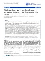
Fig. 1 Kaplan-Meier plot of overall survival (OS) stratified by presence of liver metastases
Bilen et al. BMC Cancer
(2019) 19:857
Page 5 of 8
Fig. 2 Kaplan-Meier plot of progression-free survival (PFS) stratified by presence of liver metastases
shorter OS (HR: 0.38, CI: 0.17–0.84, p = 0.017) and trended
towards having shorter PFS (HR: 0.70, CI: 0.41–1.19, p =
0.188), regardless of patients’ primary histologies. The median OS was substantially longer for patients without liver
metastases (21.9 vs. 8.1 months, p = 0.0048). The KaplanMeier plot of the association between liver metastases and
OS and PFS are shown in Fig. 1 and Fig. 2, respectively.
Patients with reported liver metastasis most commonly
had primary GI tumors (47.5%); non-GI tumors included
melanoma (27.5%), lung and head & neck (10%), breast
(7.5%), and gynecologic (2.5%). Patients without reported
liver metastasis most commonly had primary melanoma
(38%) and lung and head & neck tumors (28%). Of the
patients with liver metastases, 71.8% were RMH good
risk at the start of IO. Most patients with liver metastases (72.5%) had received two or more lines of systemic
therapy prior to treatment with IO. Patients with metastatic disease in the liver were more likely to have a
greater total number of sites of metastatic disease. One
half (50%) of the patients with liver metastases had a
total of three or more distant metastases while only 26%
of patients without liver metastases had three or more
distant metastatic sites.
Discussion
In this study, we demonstrated that metastasis to the liver
is associated with worse clinical outcomes in advanced
stage cancer patients treated on IO-based phase 1 clinical
trials. Regardless of tumor histology, patients in this cohort
with documented metastasis to the liver had shorter OS
and PFS and a lower rate of CB. The results from this study
build upon previous studies that have explored the predictive value of metastatic sites in cancers treated with chemotherapy, particularly in breast, bladder, and colon cancer
[27–31, 33]. In this study we assessed different sites of
metastatic disease and clinical outcomes in patients treated
with IO-based regimens as part of phase 1 clinical trials,
which has not been investigated previously. The results
support the Pires da Silva et al. study findings that in melanoma patients who receive combination immunotherapy,
different metastatic sites exhibit different effects on survival,
and patients with liver metastases experience inferior clinical responses [34]. Our cohort of patients receiving IObased therapy in phase 1 clinical trials is a unique population. The cohort includes patients with several different primary cancer types rather than just one. Furthermore,
patients enrolled onto phase 1 clinical trials receive novel
IO agents, which is another reason to investigate this cohort of patients.
Evidence suggests that primary tumor histology influences prognosis for patients with metastasis to the liver
who are treated with chemotherapy. Jaffe et al. (1968) found
that primary tumor site influences prognosis for patients
with hepatic metastases [35]. Furthermore, Soni et al.
(2015) found that subtypes of breast cancer differ in their
metastatic behavior [36]. The results of our study, however,
suggest that for patients on IO-based phase 1 clinical trials,
regardless of primary tumor site, liver metastases are a poor
prognostic indicator. This may be explained biologically by
the liver’s immuno-regulatory behavior [37]. The liver,
Bilen et al. BMC Cancer
(2019) 19:857
notably located between the genitourinary circulation and
systemic circulation, functions as a secondary lymphoid
organ. It contains a high density of natural killer T-cells as
well as T-regulatory cells [37, 38]. Therefore, metastases to
the liver may interfere with the immune-regulatory behavior of the organ, which in turn affects the response of cancer patients on IO. The mechanism by which this occurs
should be explored further.
The presence of metastatic disease in the liver has
been established as a poor predictive factor for patients
receiving chemotherapy-based treatment and has thus
merited different or more aggressive treatment for patients with liver metastases. Previous studies have found
that patients with breast and colorectal cancer with metastases to the liver may receive clinical benefit from
liver resection [39–43]. Given these previous findings in
cohorts treated with chemotherapy, patients with solitary
liver metastases may benefit from liver resection prior to
starting IO. However, many patients in our study cohort
with advanced stage cancers of various primary tumor
histologies had multiple liver metastases, making liver
resection not clinically appropriate. Priestman and Hanham (1972) found that combination chemotherapy produces longer overall survival rates than single
chemotherapy in treating patients with breast or colorectal cancer with liver metastases [44]. Using these results in chemotherapy-based treatment as a model,
clinical outcomes for patients on IO-based therapy may
improve with combination chemotherapy or targeted
therapy to the liver prior to or in addition to IO. Additionally, radiation therapy to the liver prior to initiating
IO could improve clinical outcomes in patients with
hepatic metastases, as per the abscopal effect [45–47].
Our analysis has limitations to note. This is a retrospective study, which is inherently subject to selection bias. We
attempted to mitigate this bias by including all patients
who received at least one dose of IO on a phase 1 clinical
trial at our institution. Due to our lenient inclusion criteria, the patient population was very heterogeneous in
primary tumor histology and in type of IO received. We
accounted for this by controlling for primary tumor histology and other baseline disease characteristics. Though
the size of our patient cohort may limit the impact of this
study, given our lenient inclusion criteria, the study cohort
was the largest cohort of patients receiving immunotherapy as part of phase 1 clinical trials at our institution.
Additionally, only the five most common sites of metastasis were captured and analyzed independently. We did not
differentiate between isolated metastases to the liver versus widespread metastatic disease. There were very few patients with brain metastases, so the predictive value of
brain metastases could not be adequately analyzed. Finally,
patients enrolled onto phase 1 clinical trials likely have
further advanced disease than patients who receive
Page 6 of 8
immunotherapy in the first or second line, which limits the
generalizability of this study.
Conclusions
Liver metastases are a poor predictive factor in this
cohort of patients treated on IO-based phase 1 clinical trials. Patients in the retrospective cohort with
hepatic metastases had shorter OS, PFS and lower
rate of CB. If these findings are validated in a larger
study, this baseline disease characteristic may warrant consideration in updated prognostic models for
stratification of patients enrolled onto IO-based
phase 1 clinical trials. The presence of liver metastases should not preclude patients from enrolling onto
phase 1 trials. Rather, the results of this study reveal
an important area for improvement in IO-based
therapies for advanced stage cancer patients with
hepatic metastases. Further advancements in treating
these patients are needed. The detection of liver metastasis in advanced stage cancer patients may be especially
useful in determining whether these patients should receive
novel combination therapy or should receive liver-targeted
therapy prior to or in combination with IO, given the
unique microenvironment around metastatic tumors in the
liver.
Abbreviations
CB: Clinical benefit; CI: Confidence interval; CR: Complete response; CTLA4: Cytotoxic T-lymphocyte associated protein 4; ECOG: Eastern Cooperative
Oncology Group; FDA: Food and Drug Administration; GI: Gastrointestinal;
HR: Hazard ratio; ICB: Immune checkpoint blocker; IO: Immunotherapy;
MVA: Multivariate analysis; OS: Overall survival; PD-1: Programmed cell death
protein 1; PD-L1: Programmed death ligand 1; PFS: Progression-free survival;
PR: Partial response; PS: Performance status; RCC: Renal cell carcinoma;
RMH: Royal Marsden Hospital; SD: Stable disease; UVA: Univariate analysis
Acknowledgments
The content is solely the responsibility of the authors and does not
necessarily represent the official views of the National Institutes of Health.
Part of data in this study was presented at the ESMO 2018 Congress in
Munich, Germany.
Authors’ contributions
MAB was involved in the identification and selection of patients,
construction of the database, caring for the patients included in the study,
study design and methodology, interpretation and analysis of study results,
and the writing of the manuscript. JMS was involved in data acquisition,
interpretation and analysis of study results, writing the manuscript, and
administrative support. DJM was involved in construction of the database,
data acquisition, interpretation and analysis of study results, writing of the
manuscript, and administrative support. YL was involved in the design and
methodology of the study, all statistical analysis, interpretation and analysis
of study results, and writing of the manuscript. MAB and RDH supervised the
study. CL, HC, MA, HK, BCC, WLS, OBA, CES, CW, DHL, RK, VAM, BE, SSR, TKO
were involved in the care of the patients in this study, interpretation and
analysis of study results, and editing the manuscript. All authors reviewed
and accepted the final version of the manuscript.
Funding
Research reported in this publication was supported in part by the
Biostatistics and Bioinformatics Shared Resource of the Winship Cancer
Institute of Emory University and NIH/NCI under award number
P30CA138292. The content is solely the work and responsibility of the
Bilen et al. BMC Cancer
(2019) 19:857
Page 7 of 8
authors and does not necessarily represent the official views of the National
Institutes of Health.
5.
Availability of data and materials
The datasets used and/or analyzed during the current study are available
from the corresponding author on reasonable request.
Ethics approval and consent to participate
This data review and analysis was approved by the Emory University
Institutional Review Board (IRB), and waiver of consent was granted due to
the retrospective nature of this study. All patients provided written informed
consent for the phase 1 clinical trial to which they were enrolled, also
reviewed and approved by the Emory University IRB.
6.
7.
8.
9.
Consent for publication
Not applicable.
10.
Competing interests
BCC has a consulting/advisory role with Astellas Medivation, Pfizer, and Blue
Earth Diagnostics and receives travel accommodations from Bristol-Myers
Squibb. WLS receives research funding from ArQule and Lilly. RP has a consulting/advisory role with Natera and AstraZeneca and receives travel accommodations from Genentech/Roche, Takeda, Novartis, and Clovis Oncology.
She also receives research funding from Bristol-Myers Squibb. CW receives
honorarium from BioTheranostics and research funding from Amgen, BristolMyers Squibb, Vaccinex, and Boston Biomedical. RRK has a consulting/advisory role with Bristol-Myers Squibb, Novartis, and Array BioPharma. She also
receives honorarium from Bristol-Myers Squibb and research funding from
Merck. BFE has a consulting/advisory role with Merrimack, BTG, Bayer, Loxo,
and RTI Health Solutions. He is a member of the speakers’ bureau of Lexicon
and Bristol-Myers Squibb. He also receives honorarium from Lexicon, RTI
Health Solutions, and Bayer and received research funding from Taiho
Pharmaceutical, Bristol-Myers Squibb, Boston Biomedical, Cleave Biosciences,
Genentech, AVEO, Pfizer, Novartis, Hoosier Cancer Research Network, Five
Prime Therapeutics, PPD Inc., Merck, and ICON Clinical Research. SSR has a
consulting/advisory role with Amgen, Boehringer Ingelheim, Celgene, Genetech/Roche, Lilly/ImClone, Bristol-Myers Squibb, AstraZeneca, Abbvie, Merck,
and Takeda and receives travel accommodations from EMD Serono, Pfizer,
and AstraZeneca. TKO has a consulting/advisory role with Novartis, BristolMyers Squibb, and MedImmune. MAB has a consulting/advisory role with
Exelixis, Sanofi and Nektar and receives research funding from Bayer, BristolMyers Squibb, Genentech/Roche, Incyte, Nektar, AstraZeneca, Tricon Pharmaceuticals, Peleton, and Pfizer.
11.
12.
13.
14.
15.
16.
17.
18.
19.
Author details
1
Department of Hematology and Medical Oncology, Emory University School
of Medicine, Atlanta, GA, USA. 2Department of Hematology and Medical
Oncology, Winship Cancer Institute of Emory University, 1365 Clifton Rd,
Atlanta, GA, USA. 3Departments of Biostatistics and Bioinformatics, Emory
University, 1518 Clifton Rd, Atlanta, GA, USA. 4Department of Urology, Emory
University, 5673 Peachtree, Dunwoody Rd, Atlanta, GA, USA. 5Department of
Pharmacology, Emory University School of Medicine, 1365 Clifton Rd, Atlanta,
GA, USA.
20.
21.
22.
Received: 22 September 2018 Accepted: 22 August 2019
References
1. Emens LA, Kok M, Ojalvo LS. Targeting the programmed cell death-1
pathway in breast and ovarian cancer. Curr Opin Obstet Gynecol. 2016;
28(2):142–7.
2. Chae YK, Arya A, Iams W, Cruz MR, Chandra S, Choi J, et al. Current
landscape and future of dual anti-CTLA4 and PD-1/PD-L1 blockade
immunotherapy in cancer; lessons learned from clinical trials with
melanoma and non-small cell lung cancer (NSCLC). J Immunother Cancer.
2018;6(1):39.
3. La-Beck NM, Jean GW, Huynh C, Alzghari SK, Lowe DB. Immune checkpoint
inhibitors: new insights and current place in Cancer therapy.
Pharmacotherapy. 2015;35(10):963–76.
4. Balar AV, Galsky MD, Rosenberg JE, Powles T, Petrylak DP, Bellmunt J, et al.
Atezolizumab as first-line treatment in cisplatin-ineligible patients with
23.
24.
25.
locally advanced and metastatic urothelial carcinoma: a single-arm,
multicentre, phase 2 trial. Lancet. 2017;389(10064):67–76.
Motzer RJ, Escudier B, McDermott DF, George S, Hammers HJ, Srinivas S,
et al. Nivolumab versus Everolimus in advanced renal-cell carcinoma. N Engl
J Med. 2015;373(19):1803–13.
Miao D, Margolis CA, Gao W, Voss MH, Li W, Martini DJ, et al. Genomic
correlates of response to immune checkpoint therapies in clear cell renal
cell carcinoma. Science. 2018;359(6377):801–6.
Chism DD. Urothelial carcinoma of the bladder and the rise of immunotherapy.
J Natl Compr Cancer Netw. 2017;15(10):1277–84.
Das R, Verma R, Sznol M, Boddupalli CS, Gettinger SN, Kluger H, et al.
Combination therapy with anti-CTLA-4 and anti-PD-1 leads to distinct
immunologic changes in vivo. J Immunol. 2015;194(3):950–9.
Michot JM, Bigenwald C, Champiat S, Collins M, Carbonnel F, Postel-Vinay S,
et al. Immune-related adverse events with immune checkpoint blockade: a
comprehensive review. Eur J Cancer. 2016;54:139–48.
Tay R, Prelaj A, Califano R. Immune checkpoint blockade for advanced nonsmall cell lung cancer: challenging clinical scenarios. J Thorac Dis. 2018;
10(Suppl 13):S1494–S502.
Wang C, Yu X, Wang W. A meta-analysis of efficacy and safety of antibodies
targeting PD-1/PD-L1 in treatment of advanced nonsmall cell lung cancer.
Medicine (Baltimore). 2016;95(52):e5539.
McDermott D, Lebbe C, Hodi FS, Maio M, Weber JS, Wolchok JD, et al. Durable
benefit and the potential for long-term survival with immunotherapy in
advanced melanoma. Cancer Treat Rev. 2014;40(9):1056–64.
Martini DJ, Hamieh L, McKay RR, Harshman LC, Brandao R, Norton CK, et al.
Durable clinical benefit in metastatic renal cell carcinoma patients who
discontinue PD-1/PD-L1 therapy for immune-related adverse events. Cancer
Immunol Res. 2018;6(4):402–8.
ClinicalTrials.gov. [Available from: Accessed 10 Sept 2018.
Wu X, Giobbie-Hurder A, Liao X, Connelly C, Connolly EM, Li J, et al.
Angiopoietin-2 as a biomarker and target for immune checkpoint therapy.
Cancer Immunol Res. 2017;5(1):17–28.
Mu CY, Huang JA, Chen Y, Chen C, Zhang XG. High expression of PD-L1 in
lung cancer may contribute to poor prognosis and tumor cells immune
escape through suppressing tumor infiltrating dendritic cells maturation.
Med Oncol. 2011;28(3):682–8.
Thompson RH, Gillett MD, Cheville JC, Lohse CM, Dong H, Webster WS, et
al. Costimulatory B7-H1 in renal cell carcinoma patients: Indicator of tumor
aggressiveness and potential therapeutic target. Proc Natl Acad Sci U S A.
2004;101(49):17174–9.
Huang Y, Zhang SD, McCrudden C, Chan KW, Lin Y, Kwok HF. The prognostic
significance of PD-L1 in bladder cancer. Oncol Rep. 2015;33(6):3075–84.
Herbst RS, Soria JC, Kowanetz M, Fine GD, Hamid O, Gordon MS, et al.
Predictive correlates of response to the anti-PD-L1 antibody MPDL3280A in
cancer patients. Nature. 2014;515(7528):563–7.
Hellmann MD, Ciuleanu TE, Pluzanski A, Lee JS, Otterson GA, AudigierValette C, et al. Nivolumab plus Ipilimumab in lung Cancer with a high
tumor mutational burden. N Engl J Med. 2018;378(22):2093–104.
Ahmadzada T, Kao S, Reid G, Boyer M, Mahar A, Cooper WA. An Update on
Predictive Biomarkers for Treatment Selection in Non-Small Cell Lung
Cancer. J Clin Med. 2018;7(6):153.
Hendry S, Salgado R, Gevaert T, Russell PA, John T, Thapa B, et al. Assessing
Tumor-Infiltrating Lymphocytes in Solid Tumors: A Practical Review for
Pathologists and Proposal for a Standardized Method from the International
Immuno-Oncology Biomarkers Working Group: Part 2: TILs in Melanoma,
Gastrointestinal Tract Carcinomas, Non-Small Cell Lung Carcinoma and
Mesothelioma, Endometrial and Ovarian Carcinomas, Squamous Cell
Carcinoma of the Head and Neck, Genitourinary Carcinomas, and Primary
Brain Tumors. Adv Anat Pathol. 2017;24(6):311–35.
Salgado R, Denkert C, Demaria S, Sirtaine N, Klauschen F, Pruneri G, et al. The
evaluation of tumor-infiltrating lymphocytes (TILs) in breast cancer:
recommendations by an international TILs working group 2014. Ann Oncol. 2015;
26(2):259–71.
Arkenau HT, Olmos D, Ang JE, de Bono J, Judson I, Kaye S. Clinical outcome
and prognostic factors for patients treated within the context of a phase I
study: the Royal Marsden Hospital experience. Br J Cancer. 2008;98(6):1029–33.
Garrido-Laguna I, Janku F, Vaklavas C, Falchook GS, Fu S, Hong DS, et al.
Validation of the Royal Marsden Hospital prognostic score in patients
treated in the phase I clinical trials program at the MD Anderson Cancer
Center. Cancer. 2012;118(5):1422–8.
Bilen et al. BMC Cancer
(2019) 19:857
26. Arkenau HT, Barriuso J, Olmos D, Ang JE, de Bono J, Judson I, et al.
Prospective validation of a prognostic score to improve patient selection for
oncology phase I trials. J Clin Oncol. 2009;27(16):2692–6.
27. Wyld L, Gutteridge E, Pinder SE, James JJ, Chan SY, Cheung KL, et al. Prognostic
factors for patients with hepatic metastases from breast cancer. Br J Cancer.
2003;89(2):284–90.
28. Ma R, Feng Y, Lin S, Chen J, Lin H, Liang X, et al. Mechanisms involved in
breast cancer liver metastasis. J Transl Med. 2015;13:64.
29. Ye LC, Liu TS, Ren L, Wei Y, Zhu DX, Zai SY, et al. Randomized controlled trial of
cetuximab plus chemotherapy for patients with KRAS wild-type unresectable
colorectal liver-limited metastases. J Clin Oncol. 2013;31(16):1931–8.
30. Zarour LR, Anand S, Billingsley KG, Bisson WH, Cercek A, Clarke MF, et al.
Colorectal Cancer liver metastasis: evolving paradigms and future directions.
Cell Mol Gastroenterol Hepatol. 2017;3(2):163–73.
31. Rosenberg JE, Hoffman-Censits J, Powles T, van der Heijden MS, Balar AV,
Necchi A, et al. Atezolizumab in patients with locally advanced and
metastatic urothelial carcinoma who have progressed following treatment
with platinum-based chemotherapy: a single-arm, multicentre, phase 2 trial.
Lancet. 2016;387(10031):1909–20.
32. Mandrekar JN, Mandrekar SJ, Cha SS. Cutpoint Determination Methods in
Survival Analysis using SAS. Proceedings of the 28th SAS Users Group
International Conference (SUGI). 2003;261:28.
33. Bellmunt J, de Wit R, Vaughn DJ, Fradet Y, Lee JL, Fong L, et al.
Pembrolizumab as second-line therapy for advanced urothelial carcinoma.
N Engl J Med. 2017;376(11):1015–26.
34. Pires da Silva I, Lo S, Gonzalez M, Guminski AD, Long GV, Menzies AM.
Distinct patterns of response and toxicity (tox) by sites of metastases (mets)
in patients (pts) treated with ipilimumab combined with PD-1 antibodies
(ipi+PD1). J Clin Oncol. 2018;36(15):9553.
35. Jaffe BM, Donegan WL, Watson F, Spratt JS Jr. Factors influencing survival in
patients with untreated hepatic metastases. Surg Gynecol Obstet. 1968;
127(1):1–11.
36. Soni A, Ren Z, Hameed O, Chanda D, Morgan CJ, Siegal GP, et al. Breast
cancer subtypes predispose the site of distant metastases. Am J Clin Pathol.
2015;143(4):471–8.
37. Mazzolini G, Ochoa MC, Morales-Kastresana A, Sanmamed MF, Melero I. The
liver, liver metastasis and liver cancer: a special case for immunotherapy
with cytokines and immunostimulatory monoclonal antibodies.
Immunotherapy. 2012;4(11):1081–5.
38. Dolina JS, Braciale TJ, Hahn YS. Liver-primed CD8+ T cells suppress antiviral
adaptive immunity through galectin-9-independent T-cell immunoglobulin
and mucin 3 engagement of high-mobility group box 1 in mice.
Hepatology. 2014;59(4):1351–65.
39. Van Cutsem E, Cervantes A, Adam R, Sobrero A, Van Krieken JH, Aderka D,
et al. ESMO consensus guidelines for the management of patients with
metastatic colorectal cancer. Ann Oncol. 2016;27(8):1386–422.
40. Hughes KS, Rosenstein RB, Songhorabodi S, Adson MA, Ilstrup DM, Fortner
JG, et al. Resection of the liver for colorectal carcinoma metastases. A multiinstitutional study of long-term survivors. Dis Colon Rectum. 1988;31(1):1–4.
41. Fong Y, Fortner J, Sun RL, Brennan MF, Blumgart LH. Clinical score for predicting
recurrence after hepatic resection for metastatic colorectal cancer: analysis of
1001 consecutive cases. Ann Surg. 1999;230(3):309–18 discussion 18-21.
42. van Walsum GA, de Ridder JA, Verhoef C, Bosscha K, van Gulik TM, Hesselink
EJ, et al. Resection of liver metastases in patients with breast cancer: survival
and prognostic factors. Eur J Surg Oncol. 2012;38(10):910–7.
43. Maksan SM, Lehnert T, Bastert G, Herfarth C. Curative liver resection for
metastatic breast cancer. Eur J Surg Oncol. 2000;26(3):209–12.
44. Priestman TJ, Hanham IW. Results of 27 cases with hepatic metastases
treated by combination chemotherapy. Br J Cancer. 1972;26(6):466–72.
45. Mole RH. Whole body irradiation; radiobiology or medicine? Br J Radiol.
1953;26(305):234–41.
46. Gong J, Le TQ, Massarelli E, Hendifar AE, Tuli R. Radiation therapy and PD-1/
PD-L1 blockade: the clinical development of an evolving anticancer
combination. J Immunother Cancer. 2018;6(1):46.
47. Reynders K, Illidge T, Siva S, Chang JY, De Ruysscher D. The abscopal effect
of local radiotherapy: using immunotherapy to make a rare event clinically
relevant. Cancer Treat Rev. 2015;41(6):503–10.
Publisher’s Note
Springer Nature remains neutral with regard to jurisdictional claims in
published maps and institutional affiliations.
Page 8 of 8