Nghiên cứu kết quả sớm và trung hạn của phương pháp bít thông liên thất phần quanh màng qua ống thông bằng dụng cụ hai đĩa đồng tâm tt tiếng anh
Bạn đang xem bản rút gọn của tài liệu. Xem và tải ngay bản đầy đủ của tài liệu tại đây (199.19 KB, 27 trang )
MINISTRY OF EDUCATION
MINISTRY OF HEALTH
AND TRAINING
HANOI MEDICAL UNIVERSITY
NGUYEN CONG HA
Research about short and medium-term results
of transcatheter closure of perimembranous VSD
using the symmetrical double-disc device
Major
Code
: Internal Cardiology
: 62720141
MEDICAL DOCTORATE THESIS
HANOI - 2020
THE THESIS WAS FULFILLED AT
HANOI MEDICAL UNIVERSITY
Scientific Supervisor: Professor Nguyen Lan Viet
1st Peer-reviewer: Assoc. Prof. PhD. Pham Huu Hoa
2nd Peer-reviewer: Assoc. Prof. PhD. Nguyen Ngoc Quang
3nd Peer-reviewer: Assoc. Prof. PhD. Ta Manh Cuong
Ph.D. Thesis will be evaluated by the Hanoi medical University
Thesis Board.
At h / / /2020.
The thesis can be found at:
- National Library
- Hanoi medical University Library
3
BACKGROUND
Ventricular septal defect (VSD) is when there is
communication between the left ventricle chamber (LV) and the
right ventricle (RV) with each other. VSD is one of the most
common congenital heart diseases (CHDs) accounting for 20 30% of CHDs.
In the treatment of VSD, the classic method is open surgery
with the support of cardiopulmonary bypass (CPB) technique, which
is considered as the gold standard method, but still, have some
complications of CPB, anesthetic resuscitation, infection, and
neurological complication …
The first device designed to close of perimembranous VSDs
(pVSD) is called Amplatzer muscular VSD occluder (AVSO)
manufactured by AGA. In 2002, Hijazi et al reported that this device
was used for 6 patients with the result that there were no cases of
residual shunt and significant complications. After that, many studies
applied Amplatzer instruments, but the rate of atrioventricular block
(AVB) was still high, with a study of 5.7% which is much higher than
that open surgery so now this AVSO device has stopped being applied
due to this complication.
To increase efficiency and minimize complications, some new
devices have recently been launched to overcome the disadvantages
of Amplatzer. Nit-Occlud® Lê VSD-Coils (Lê VSD Coils) have been
applied and have been highly successful, despite the reduction of
complications such as ABV, AVR but the rate of residual shunt and
hemolysis is still high. Dr. Nguyen Lan Hieu and other authors also
used devices that use for the closure of ductus arteriosus (DO) to
close VSDs and had good short- and medium-term outcomes but
there are still some complications.
4
The symmetrical double-disc device was also created for the
same purpose, this device has improved the design of Amplatzer with
two symmetrical discs, the smaller left disc had larger thickness. This
device has been studied and applied clinically for high success and
low rate of complications in the long-term follow-up.
In Vietnam, Nguyen Lan Hieu and other colleagues have
applied various types of devices to close perimembranous VSDs such
as Le VSD Coils, PDA closure devices and have some reports for
good outcomes in short and medium-term. The symmetrical doubledisk devices have also been used and have good clinical results but
there have been no specific studies on the safety and efficacy of this
device. Therefore, we carried out the study: "Evaluate the Short and
Medium-term outcomes of Transcatheter Closure of Perimembranous
VSDs using Symmetrical Double-disc Devices" to:
1. Evaluate the feasibility, short and medium-term results of
transcatheter closure of perimembranous VSDs using the symmetrical
double-disc device at Hanoi Heart Hospital.
2. Find out some factors that affect the outcome of the technique.
NEW CONTRIBUTIONS OF THE THESIS
The study had 84 patients with perimembranous VSDs who
had transcatheter closure of perimembranous VSDs using the
symmetrical double-disc device from January 2012 - December
2015. 81 patients successfully performed closure procedure ( 96.4%).
The follow-up of patients after the procedure was the longest
61 months (≈ 5 years), the shortest was 20 months, during the followup time, none of the patients left the study.
5
The study showed that high efficacy, low complications,
safety in patients with pVSDs selected with the size of defects ≤
8mm, aortic edge ≥ 2,0 mm. After the procedure, we evaluated
clinical symptoms such as delayed weight gain, recurrent pneumonia,
heart failure and absent of typical systolic murmur of VSD. The
parameters on cardioechography such as Left Ventricular End
Diastolic Dimension (LVEDd), Pulmonary Arterial pressure (PAP)
also decreased significantly after the procedure. Major complications
were mild complications and recovered, had 1 patient (1.2%) had
worsened TVR (3/4) after the follow-up time, especially no patients
with grade III AVB which is one of the critical complications but we
did not have any case in our study.
Factors affecting the outcome of the procedure are
inappropriate anatomy size of defect such as large defects, lack of
aortic edge are factors that directly affect 3 failure cases of the
procedure. Other difficult obstacles such as difficulty in passing
devices through the defects, re-taking the snare, redo the procedure
steps, exchanging larger devices due to the assessment of the defect
on cardioechography and the incorrect image of the LV chamber; are
limitations of the procedure. The trans-thoracic Doppler
cardioechography during the procedure also enhances the procedure.
LAYOUT OF THE THESIS
The thesis consists of 153 pages consisting of 4 chapters; 45
pages of overview, 21 pages of subjects and research methods, 50
pages of results, 31 pages for discussion, 2 pages of conclusion, 1
page of recommendations. Tables; there are 57 tables; 14 charts; 24
figures; 180 references; 7 references in Vietnamese; 173 references in
English.
6
CHAPTER 1: OVERVIEW
1.1 . Prevalence and anatomy of VSDs
VSD is the most common CHD, accounting for about 20-30%
of CHD, of which pVSD accounts for about 70-80% of the total VSD
types. Hoffman's meta-analysis of 22 statistics found that the
prevalence of VSD was 31% in CHDs.
An important anatomical feature is that the VSD is not aligned
as a wall to separate the two ventricular chambers from which the VSD
has a curved structure because of the round shape of the LV and the
crescent shape of the RV hug the right front of the LV. It must also be
remembered that VW's structure varies with the position of the wall.
pVSD is when the membrane part of the ventricular septum is
not fully formed, the opening is near the anterior leaf edge and the
leaf wall of TV. Not only is the defect in the membrane part but it
seems to be surrounded by the fibrous tissue, membrane and tends to
close. This fibrous tissue is also called the TI auxiliary organization,
which can form a bulging sac structure. Van Praagh differentiated
true pVSD, which is only a small hole in the membrane's septum. It
is more accurate to call VSD with the perimeter around the
membrane, because some VSDs have intact membrane but defects
are around the membrane part, so the name pVSD is used more often.
This VSD is closely related to the AV conduction path, which passes
through the TI ring and follows the lower posterior edge of the defect
then divides into the left and right branches. Therefore, when surgery
to patch or close the defect with a device, there is a risk of damage to
this transmission line.
7
Figure 1.1: Illustration of atrioventricular conduction pathway;
and related to pVSD.
1.2. Pathophysiology and clinical characteristics
VSD which has no more cardiac defect causes left and right
ventricular shunts that increase circulation to the lungs, increase left
ventricular volume, and increase PA pressure. The degree of shunting
depends on the diameter of the defect and the resistance of PA.
In newborns, the PA resistance is high and decreases
gradually from the first days after birth and decreases rapidly in the
first 4-6 weeks and returns to normal 2-3 months later. However, due
to the high PA resistance, it is often not detected by clinical
examination in the first months because the left-right shunts are not
large enough to generate systolic murmur as well as other clinical
symptoms. After 4-6 weeks of birth, the resistance to PA decreases,
the shunt will grow then the murmur and symptoms of heart failure
will be more significant.
1.3. Diagnosis
Patients with small defects are usually diagnosed when a systolic
murmur is heard at the left sternum. When the resistance of the PA
system increases, the murmur is weak and shorter.
The Doppler cardioechography is the best diagnostic tool for VSD.
Cardioechography can detect very small defects, locate very precisely
because it is possible to cut many different cross-sections, this is an
8
advantage over cardiography when limited to a few angles and using
limited contrast agent.
1.4. Treatment
1.4.1. Disease natural course and prognosis
Most patients with small defects of VSD grow up normally.
Some studies have found that VSD self-closing rate is up to ¾ cases.
The size of the defects tends to get smaller and the highest selfclosing ratio in the first years. In adults with small defects, Qp / Qs
<2, without PAH, the prognosis is very good. The rate of AVR and
endocarditis is very low, if there is arrhythmia, it is benign.
About 1/6 of VSD cases present with congestive heart
failure requiring medical treatment and most likely occur within 6
months after birth. Some respond to medical treatment and do not
need surgery right away, these patients often have PAH due to
large defects and are at risk of developing lung disease, possibly
before 2 years of age.
1.4.2. Medical treatment
For medical treatment, if symptoms of heart failure occur, the
medications used are digoxin, diuretic and ACE inhibitors or
Angiotensin receptor inhibitors. Medical treatments only postpone
surgery or follow up because the defect may close or may shrink. If
the defect is small, without increasing PA pressure, there is no need to
limit physical activity. Prevention of endocarditis is very important.
1.4.3. Surgical treatment of closing defects
This is the classic treatment method, considered the gold
standard method, especially VSD in newborns, multi-hole VSD,
broad-hole VSD, receiving chamber VSD, under two arterial valves
type and VSD with other lesions. The surgical mortality rate is <1%.
Complications like grade 3 ABV about 1-2%, residual shunts are ≈
9
5%. MI and TI may also be rare. Besides, there may be complications
such as infections, neurological complications due to CPB...
1.4.4. Transcatheter closure of perimembranous
VSDs using devices
Since the application of VSD by devices through the catheter
has been controversy about the safety, the effectiveness of this
method compared to the surgical method. The development of VSD
closure devices has undergone many changes to increase efficiency
and reduce complications, especially pVSD closing devices.
The asymmetrical double-disc device designed for pVSD
(Amplatzer) with a high ABV rate in the short-medium-term, with
research up to 5.7% so this device is not applied anymore.
To increase efficiency and minimize accidents, recently several
choices have been applied. Le VSD Coils has the advantage of
reducing the ABV catastrophe, but the residual shunt and hemolysis
are still high.
Nguyen Lan Hieu and other authors used PDA closing devices
to seal VSD and reported very good medium-term results, the success
rate of about 96%.
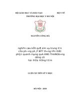
The symmetrical double-disk design is based on the Amplatzer
AGA (should also be called a modified 2-disc device), the design
changes to overcome the Amplatzer's disadvantages (high ABV rate).
These changes are two equal concentric discs with a diameter larger
than waist circumference 4mm (shrinking from eccentric disc larger
than 6mm waist to concentric disc larger than 4mm waist), 3mm of
the thickness (1.5 mm larger than with Amplatzer). The size name
stands for the diameter of the waist (Figure 1.2).
10
2
mmm
2
mmm
3
mmm
Figure 1.2: Artwork of the symmetrical double-disc
design to close pVSD.
In 2012, Wang et al reported experience for 8 years (20022011), multicenter, over 524 pVSD patients, the success rate was
95.6%, the average follow-up time was 45 months, 3 patients had
severe complications (0.6%) related to valve incomplete and were
under 3 years old, 104 patients had minor complications. The study
concludes that pVSD closing with this device has high success and
long-term follow-up and less invasive than surgery.
In 2014, Yang et al reported a prospective, randomized,
controlled clinical trial study of percutaneous pVSD closing or
surgery. Criteria to assess the safety and effectiveness of the two
above treatments. The data collected tracked over 2 years and
compared the two groups and concluded that: the two methods above
have very good results in the short and mid-term. Percutaneous VSD
has the advantage over surgery as it causes less damage to the heart
muscle, less blood transfusion, and shorter procedure and hospital
stay. It costs less.
In 2014, Yang et al. summarized the effectiveness and
complications of pVSD closing methods. The systematic review of
studies published in English-language world journals, they searched
PubMed from 2003-5 / 2012, excluding small studies, studies with
acquired VSD. As a result, 37 published studies included 4406 VSD
patients (including 3754 pVSD patients, 419 mVSD, 47 VSD under
the aortic valve, 36 VSD under two great arteries, 16 patients with
multi-hole VSD, 7 cases without differential type), devices are used
with many different types (Amplatzer, Coils, PDO, 2 symmetrical
double discs ...). Analysis of these 37 studies concluded that closing
11
VSD by devices is effective and safe. The limitation of this analysis
is that it is difficult to analyze different types of devices and it is
difficult to isolate VSD forms.
In 2018, Krishna A. Mandal et al reported the long-term result
of pVSD closing by symmetrical double-disk devices on 186
patients. The success rate is 96.8%, with no more shunts of 99.5%
after 6 months. There were 16 (8.9%) patients with stroke right after
the procedure in which 1 (0.5%) patients with ABV grade 3 and 1
(0.5%) had completely left bundle branch block, these 2 patients
recovered after treatment with steroid. During the mean follow-up of
18.4 months, no major complications or BAV occurred. The study
concluded that the severity of the stroke was low, no patient had lateonset ABV, the success rate was high, so this method could replace
the surgical method in selected patients.
CHAPTER 2: SUBJECTS AND METHODS OF THE STUDY
2.1. Criteria for selecting patients
Patients diagnosed at Hanoi Heart Hospital were diagnosed
with pVSD, diameter more than 2mm with left-right shunt on cardiac
Doppler ultrasound, weighing ≥ 8 kg or ≥ 1 year old, with aortic
valve margin ≥ 2 mm or a membrane with aneurysm, plus one of the
following: recurrent pneumonia, delayed weight gain, symptoms of
heart failure, dilated LV on echocardiography or Qp / Qs ≥ 1.5.
2.2. Exclusion criteria
VSDs had large defects or very small, no longer had
indications of closing. Moderately-severe AI, with erupted
Valsalva sinus aneurysm, acquired pVSD or other surgicalrequired lesions. Patients with contraindications to aspirin,
patients with ABV, patients, family members who do not agree to
do the procedure nor participate in the study.
2.3. Time and place of research
12
The patients were eligible for the study and had procedures
done from January 2012 - December 2015, then followed up,
collected and processed data.
The study was conducted at Hanoi Heart Hospital.
2.4. Type of research, devices used in research
Prospective research, single center, clinical intervention,
comparison before and after, no control, convenient sample selection,
follow up for at least 12 months after the procedure.
Selected devices are symmetrical double-disk devices with
names: HEART symmetric membranous VSD Occluder của hãng
Lifetech Scientific
2.5. Study process
The patient was researched according to the medical records,
collected clinical data, ECG, CXR, Doppler echocardiography, the
results of the procedure through 8 main steps. The results monitored for
complications, ECG and echocardiography within the first 24 hours and
before discharge. Appointment for follow-up visits after procedures 1, 3,
12, 18 months and annually or with any unusual manifestations. Patients
were re-examined clinically, performed ECG and echocardiogram. The
study selected 84 patients with pVSD and performed the procedure.
Successful 81 procedures, collected the data of procedures and tracked
data over time, finished tracking in December 2017, then analyzed and
processed the data, wrote the thesis.
2.6. Data processing
Using SPSS 20.0 software to process and analyze common
medical statistics.
2.7. Research ethics
The study was approved by the Ethics Council and Science
Council of Hanoi Heart Hospital.
CHAPTER 3: RESULTS
13
3.1. General characteristics of the study subjects
84 patients with pVSD were selected, and 81 patients
successfully had the procedure (81/84 ≈ 96.4%). The average duration
of follow-up was 42.7 months (20-61 months). No patients had left the
study. The average duration of hospital stay was 8.28 days.
Gender, age, weight characteristics: the study included 43
males (53.1%) and 38 females (46.9%). The average age is 9.9 years
(11 months - 55 years), ages <16 accounts for 81.5%. The average
weight is 23.8 kg (7-67 kg).
3.2. Characteristics of VSD defect and aortic edge on
echocardiography
Table 3.1. Size of the defect and aortic edge on TTE
n
Size of defect (mm)
Aortic edge (mm)
81
81
X
5,1
5,0
SD
Min
Max
1,5
2,0
2,5
1,5
11,5
11,5
Comments: Size of VSD defect is mostly medium size, the average
size is 5.1 mm (2.5-11.5 mm). Aortic edge average size is 5.0 mm
(1.5-11.5 mm).
3.3. Results of the VSD closing procedure
Methods of analgesia: In the study, 65/81 (80%) patients
underwent intravenous propofol and 16/81 (20%) patients under local
anesthesia. The minimum age for anesthesia is 14 years old, the
oldest age for anesthesia is 32 years old.
Access: in the study, 71/81 (87.6%) patients had access to the
right femoral artery and vein and 10 (12.4%) patients entered with
the left femoral artery and vein, no patients use other access routes.
The systolic PAP has a mean value of 27.4 mmHg (17-40).
The average Qp / Qs result is 2.33 (1.7 - 3.5).
14
The VSD defect morphology was assessed on LV images: the
funnel-shaped proportion had the highest rate of 69/81 (85.19%) of
patients, often accompanied by membrane aneurysm. Tubes are
common after funnel-shaped, 9/81 (11.11%) patients. Window
pattern is the least common with 3.7%.
VSD size measured on LV chamber images: 3.86 mm on
average, 3mm minimum, 8mm maximum. Aortic valve edge result:
mean 3.0 mm (2.0 - 5.0 mm).
Method of passing VSD from LV to RV: Directly through
0.035 wire, successfully bend the water at the tip of 58%, then by IM
catheter is 28%, pigtail cut is 11%, JR catheter is 3%. The position of
catching the wire (snare) to create A-V rings, in the study, from the
vena cava was 66.7%, the remaining 33.3% were at the pulmonary
artery. The start position of device drop: drop from the aorta up to
75.3%, also drop from the LV chamber in 24.7% of patients.
Device sizes used are from 4 to 10, sizes 4, 5 and 6 accounts
for 78% of the devices used. There is a size of 10 used for patients
with VSD = 8 mm. The average device size is 5.63 ± 1,269 mm (4 10 mm).
Stopping shunting immediately after releasing the device is
21%, while the low shunt is 79%, after 3 months follow up is 97,5%,
18 months is 98,8% ( small shunt after 18 month is 1,2%).
AVR: No patients had any change in AVR level, 4 (4.9%)
patients with mild AVR had it before the procedure.
The average procedure time is 47.3 minutes (37 - 121
minutes). And the average X-ray exposing time is 22.9 minutes (11 54 minutes).
3.4. The difficulties and obstacles during the procedure
Table 3.2. The difficulties during the procedure
15
The difficulties during the procedure
n
%
Through VSD have to change catheters
Re-catch wire (snare)
Change to larger size
Dropping the set into RV must repeat the steps
Cardioechography during procedure
20
18
13
9
31
24,7
22,2
16,0
11,1
38,3
Comments: 24.7% of patients had difficulty passing through
the vas from the LV side where they had to change another catheter.
Catching snare accounted for 22.2% while exchanging devices was
greater than 16.0% of patients. The whole system dropped into RV
when releasing had a rate of 11.1%. Support echocardiography
during the procedure was 38.3%.
3.5. Cardiac arrhythmia events encountered during the procedure
Table 3.3. Cardiac arrhythmia during the procedure
Cardiac arrhythmia
Sinus tachycardia
Ventricular ectopic
Short ventricular tachycardia
Sinus bradycardia
A-V Block grade 1,2
Others
n
8
81
17
3
2
0
%
9,9
100
21,
9
3,7
2,5
0
Comments: The most common arrhythmia during work is
ventricular ectopic (100%). Sinus tachycardia with 9.9%, short
ventricular tachycardia met 21.9%. Sinus bradycardia met 3.7%,
AVB grade 1.2 met 2/81 (2.5%). These arrhythmias recover as soon
as the stimulation stops, ending the procedure. In the study, there
were no patients with ABV grade 3.
16
3.6. Other complications and causes of failure of the procedure
Respiratory complications met 6.2% of patients, manifested as
increased respiratory secretion, wheezing, reduced SpO2 only
managed to suction and cleanthe airways, bag-via-mask ventilation,
then the patient was stable again, no case need intubation.
Complications on the pathway only manifested as a small
hematoma at the site of access in 8.6% of patients, only medical
treatment by compressing the position of the vascular access.
There are 9.9% of patients with allergic manifestations such as
skin rash, but do not cause severe respiratory and circulation
symptoms, only treated with corticoids and antihistamines.
There are 3 patients (3.6%) fail due to anatomical factors such
as large defect, defect located next to the aortic valve.
3.7. Clinical symptoms, ECG, CXR before and after the
procedure
Clinical: before the procedure, the symptom of delayed weight
gain accounted for 25.9%, recurrent pneumonia was 33.3%, NYHA 2
was 13.6% and systolic murmur intensity ≥3 / 6 accounted for 80, 2%.
Following the procedure, these symptoms gradually disappear.
ECG: After the procedure, there was a significant change in LV
overload signs, before procedure 28/81 (34.6%), after 18 months, only
3/81 (3.7%). Especially, no patients with ABV and other arrhythmias
after 18 months.
On the plain CXR before the procedure, 53.1% have a cardiacchest index ≥ 50%. Signs of pulmonary congestion were 76.5% of
patients.
3.8. Doppler echocardiography before and after the procedure
Left Ventricular End Diastolic Dimension (LVEDd) before
the procedure was 39.3 ± 8.0 mm, after 24 hours the procedure was
17
36.4 ± 8.2 mm after 1 month was 34.4 ± 7.2 mm. Comparing the
mean Dd after the 24-hour and 1-month procedure saw a decrease
compared to the previous procedure with statistical significance
(p = 0.001, t-test).
Systolic PAP measured by TI spectrum before the procedure
is 31.2 ± 6.5 mmHg, after 24 hours procedure is 2.50 ± 3.5 mmHg,
after 1 month is 24.6 ± 3.0 mmHg. Comparison of systolic PAP after
procedure 24 hours and 1 month showed a statistically significant
decrease compared to before the procedure (p <0.05, t-test).
TI changes on ultrasound before and after the procedure
Table 3.4. Compare TI before and after the procedure
TI
grade
1/4
2/4
3/4
Total
TI before procedure
n
%
74
91,4
3
3,7
4
4,9
81
100,0
After 18 months
n
%
79
97,5
2
2,5
0
0,0
81
100,0
Comments: The mild TI group after the procedure had 5 more
patients because these patients before the procedure were moderate or
high MI, in which before the procedure, there were 4 patients with
high TI (3/4) with right atrial LV shunt, after the procedure no longer
right atrial LV shunt. In the mild TI group, before the procedure,
there were 3 patients, after the procedure, there were 2 patients
(decreased 1 patient), of which 1 patient did not change the
incomplete level (still 2/4) and 1 patient had an incomplete level
increased from mild TI. With aortic valve, there was no change
before and after the procedure with 4 (4.9%) patients with mild TI.
3.9. Compare defect size measured on US v.s cardiac catheterization
18
Table 3.5. Compare defect size measured on US v.s cardiac
catheterization
Size of defect
(mm)
Cardioechography
On catheterization
X
SD
5,1
3,9
1,5
0,9
95%CI
4,8
3,7
5,7
4,1
p (t-test)
0,001
Comment: The mean size of VSD measured on
echocardiography was significantly greater than the size measured on
cardiac catheterization (p <0.05).
CHAPTER 4: DISCUSSION
4.1. General characteristics of patients studied
The follow-up time for our study was 42.7 ± 15.5 months (20 61 months). According to Nguyen Lan Hieu's research, the follow-up
time is from 2 to 7 years. Lei Wang's study had a follow-up period of
up to 8 years (1-96 months).
The average hospitalization time is 8.28 days (3-28 days). Our
study had a longer hospital stay than other studies because some
patients were hospitalized with pneumonia requiring prolonged
treatment. According to research by Lei Wang TB is 3.4 days.
The gender distribution of other studies and we are quite similar,
male / female ratio ≈ 1, epidemiological equivalent rate of VSD.
The average age of our patients was 9.9 ± 11.1 years (11 months
- 55 years old). According to the EUREVECO study, the age range is
from 8 months to 67 years old, while the study by Lei Wang is from 2
to 12 years old, the study of Nguyen Lan Hieu is all over 1 year old.
Regarding the body weight of patients, the studies all chose to
19
weigh over 7 kg, our study was the lowest of 7.0 kg in 1-year-old
patients. According to research by Nikolaus A. Haas, the lightest
patient is 7.18 kg, Lei Wang's study is ≥ 9.5 kg.
4.2. History, clinical symptoms, ECG and CXR characteristics
History of recurrent pneumonia: our study is 33.3%. According
to Nguyen Lan Hieu, the Coils group had 19.7%, the PDA-Device
group had 27.0%. According to Lei Wang, recurrent VPQ accounted
for 25.9%.
Slow weight gain: our study is 25.9%. According to Nguyen
Lan Hieu, the Coils group is 25.4%, the PDA-Device group is 32.1%.
Functional symptoms of heart failure assessment (NYHA):
According to Nguyen Lan Hieu, the Coils group is 19.7%, and the
PDA-Device group is 22.5% (NYHA ≥ 2). According to Lei Wang,
9.1% (8.6% NYHA2; 0.5% NYHA3; 90.9% HYHA1). Our data is
13.6% NYHA2; 76.4% NYHA1.History of endometriosis, the Lei
Wang study met 0.4% (4 patients), we did not have any cases.
Systolic murmur on the left sternal edge we encountered 100% of
patients and the intensity of murmur ≥ 3/6 met in 80.2% of patients.
In our study, ECG signs of LV overload were 34.6%, right
bundle branch blocks completely were 2.5%, and the lung-heart
index ≥ 50% was 53.1%. According to Lei Wang, the signs of
hemodynamic changes, enlarged heart shape, increased blood flow to
the lungs, and heart overload on the ECG were 73.3%.
20
4.3. Echocardiography characteristics
Left Ventricular End Diastolic Dimension: our data are 39.3 ±
8.0 mm. Nguyen Lan Hieu: Coils group is 42.1 ± 8.7 mm, PDADevice group is 43.3 ± 8.5 mm.
Systolic PAP: our data is 30.9 ± 6.4 mmHg (20 - 50mmHg).
According to Nguyen Lan Hieu, the Coils group is 34.8 ± 6.6 mmHg,
the PDA-Device group is 35.9 ± 8.9 mmHg. Our PAP is lower than
that of Nguyen Lan Hieu.
TI: we had 4 patients with severe TI with shunt LV-RA and 3
patients with moderate TI, the rest were mild TI (91.36%). In the
report of Gunter Kerst in 2015 in Germany in 4 patients with LV-RA
shunt, these patients were VSD closing by 2nd generation arterial
closure device (PDO II) recorded no LV-RA shunt after the
procedure.
4.4. Characteristics of VSD on echocardiography
Our results have a size of defect of 5.1 ± 1.5 mm (2.5 - 11.5
mm), a diaphragm aneurysm of 45%, aortic valve edge of size 5.0 ±
2.0 mm (1.5 - 11.5). VSD characteristics of our study are quite
similar to those of other authors. According to Nguyen Lan Hieu, the
VSD of the DO group is 4.4 ± 1.2 mm, the Coils group is 4.1 ± 1.5
mm. The DO group aneurysm is 48.3%, the Coils group is 66.2%, the
aortic edge of the DO group is 3.5 ± 1.4 mm, the Coils group is 3.7 ±
2.9 mm. According to Lei Wang, VSD is 4.4 mm (2.3 - 10.3 mm), the
aorta edge is 3.2 mm (1.0 - 18.3 mm).
4.5. Characteristics of VSD closing procedure
In our study, we selected the femoral artery and vein for all
cases. 20% local anesthesia and 80% IV anesthesia. According to
21
EUREVECO's study of 111 patients, 15.3% of the way through the
carotid vein, the rest has performed by the artery and femoral vein,
this is the basic and convenient access. For general anesthesia and
analgesia, 49.5% had mild sedation and 50.5% used general
anesthesia.
Systolic PAP is 27.4 ± 4.7 mmHg (17-40 mmHg), Qp / Qs is
2.33 ± 0.43 (1.7-3.5). Our results are similar to those of other authors.
According to Nguyen Lan Hieu, the Qp / Qs of the DO group is 2.2 ±
0.7, the Coils group is 2.1 ± 0.7. According to Lei Wang, the average
value of systolic PAP is 25.6 mmHg (18-55 mmHg), Qp / Qs has an
average value of 2.5 (1.7-6.8).
VSD characteristics on LV chamber image: Our result, the
size of defects were 3.86 ± 0.93 mm (3.0-8.0). The morphology is
the funnel-shaped and diaphragm-shaped aneurysm of 85.19%
followed by the 11.11% tubular and 3.7% window-shaped. Aortic
edge is 3.0 ± 0.7 mm (2.0 - 5.0), our characteristics are similar to
other authors. According to Nguyen Lan Hieu, the size of defect of
DO group is 4.7 ± 2.0 mm, Coils group is 4.4 ± 1.7 mm, DO's
aortic valve edge is 3.3 ± 1.1 mm, group Coils are 3.1 ± 0.9 mm.
According to Lei Wang, the average size of the defect is 4.6 mm
(1.9-21.6), the tubular form accounts for 23.2%, the window shape
accounts for 2.9%, the diaphragm aneurysm accounts for 9.2%,
funnel-shaped for 64.7%. Tools: our results are 5.6 ± 1.3 mm (410), the numbers 5 and 6 account for 58%, so our device size is
smaller than our research Lei Wang (TB is 6.5 mm, the smallest is
4 mm, the largest is 18 mm). In our study through VSD with a
wire of 58% success, then an IM catheter of 28%, with a pigtail
22
cut 11% and finally a 3% JR. The direct method of wire is more
convenient, less arrhythmic when doing the procedure than using
the catheter.
The position of catching the wire (snare) in the superior or
inferior vena cava accounts for 66.7%, the rest is at the pulmonary
artery. The rate at which the device drop usually starts from the
aorta is 75.3%, the remainder from the LV. According to the
EUREVECO study, passing through the common hole is the JR
catheter then caught the snare in the pulmonary artery, and the
starting point for releasing the device is from the aorta.
4.6. Characteristics of success, causes of failure
Our success rate is 96.4%, according to Nguyen Lan Hieu of the
Coils group, which has a higher success rate than the DO group (DO:
95.6%; Coils: 97.2%).
Cause of device failure caused by AI: according to Lei Wang's
research is 2.3%, according to EUREVECO's research, 1.8%, and
Nguyen Lan Hieu's research in DO group is 0,3%, our results showed
1 patient (1.2%) had AI so he had to stop the procedure.
Shunt residue group: according to EUREVECO's research,
1.8%, while Nguyen Lan Hieu's research in DO group is 0.6%, Coils
group is 1.4%, their results I met 1 patient (1.2%) with large residual
shunt who had to stop the procedure.
The fourth group was for TI, according to Lei Wang's study, 3
patients (0.6%) had TI so they had to stop the procedure. We did not
record any cases where the procedure was stopped due to TI.
The group of causes of arrhythmia, especially ABV: there was
no patient in Lei Wang's study, according to EUREVECO study, 1
23
patient (0.9%) had grade 3 ABV when doing so he had to stop the
procedure, and Nguyen Lan Hieu's research in DO group was 0.3%
severe arrhythmia, Coils group did not have any patients, our results
did not see any patients with arrhythmia who had to stop.
4.7. Severe complications of the procedure
Mortality events: in our study, other authors such as Nguyen Lan
Hieu, Lei Wang, Nikolaus A. Haas, the mortality rate was 0%.
ABV: According to our research, there were no patients who had
ABV during follow-up. According to Nguyen Lan Hieu's study, ABV
needed to place pacemakers in DO group of 2 (0.7%) patients, Coils
1 group (1.4%) patients. According to Lei Wang's research, 1 patient
who had level 3 ABV had to have a permanent pacemaker after 1
month due to no recovery.
Our study had 1 patient (1.2%) causing moderate-severere TI
during follow-up. According to Lei Wang's research on the same
device, 1 (0.2%) of patients with severe TI had surgery to repair
valves after 12 months of follow-up.
Endocarditis, Nguyen Lan Hieu's study had 1 (0.3%) of patients
in DO group, the Coils group did not have any cases. In the
EUREVECO study, there were 4 (3.6%) hemolytic patients, of which
1 patient dislocated the device causing residual shunt and hemolysis
required surgery in the second year, 1 hemolytic patient had to use
the second device, 2 patients Hemolytic transfusion and autolysis, no
cases of endocarditis. According to our study, there were no cases of
infectious endocarditis, hemolysis, and other severe events.
24
4.8. Mild complications of the procedure
In our study, the rate of mild complications was 38.3%: 5
patients (6.2%) had ABV grade 1, 2 and transient sinus bradycardia.
Mild hematoma at the puncture site occurred in 7 (8.6%) patients.
Rash, allergic to contrast agent were in 8 (9.9%) patients, fever> 38.5
C was in 6 (7.4%) patients, respiratory failure at anesthesia was
6.2%. Nguyen Lan Hieu's Coils group had hemolysis at 7.3% higher
than that of DO group, and the rate of arrhythmia of DO group is
higher than that of the Coils group. According to Lei Wang, the rate
of mild complications is 19.8%.
4.9. Ability to close VSD with no residual shunt of the procedure
Our study recorded that the rate of no shunting immediately
after releasing the device was 21.0%, after 24 hours was 87.6%,
after 18 months was 98.8% (and 1 patient with a small shunt) was
quite high and similar to that of Nguyen Lan Hieu and higher than
other authors.
The study of Nguyen Lan Hieu noted that the rate of no shunt
after 24 hours in DO group was 85.2%, Coils was 73.9%, after 6 months
DO group was 91.3%, Coils group was 84.1% , after 24 months the DO
group was 99.0%, the Coils group was 98.6%. According to Lei Wang
on symmetrical double discs, after 24 hours they were 54.9%, but after
24 months, 99.24% were completely closed
4.10. Difficulties and obstacles of procedure
In our study, there were 3 (3.6%) failed surgical procedures, all
of which were related to the surgical anatomy factor, in which 2
patients had large defect despite using the largest device but shunt
persisted and need to stop the procedure and 1 patient, though the
25
hole is small, the short aortic valve edge causes AI, so also need to
stop the procedure.
Nguyen Lan Hieu's research in DO group had 14 (4.4%)
failed cases, of which 6 patients due to lack of aortic edge, 1 patient
with severe AI, 1 patient with severe arrhythmia, 2 patients with
large residual shunt. According to Lei Wang's research on
symmetrical double-disk, 23 (4.4%) fail cases including 8 patients
who failed to deliver the device via VSD, 12 patients caused AI, 3
patients caused TI.
Some other difficulties when doing the procedure we
encountered such as difficulty through VSD is 24.7%, having to resnare is 22.2%, changing larger devices is 16.0%, having to repeat
the procedure steps from the beginning is 11.1%, assisted
echocardiography during procedures is 38.3%.
CONCLUSION
1. Transcatheter closure of perimembranous VSDs using the
symmetrical double-disc device is feasible and gives good results
in selected patients, namely:
The success rate of the procedure: 96.4%
The four clinical manifestations before the procedure no longer
after the procedure. Signs such as increased LV workload on ECG,
Dd, PAP all decreased significantly after the follow-up.
The rate of residual shunts after 18 months is only 1.2%.
Incidence of complications: TI worsened (from ¼ to ¾) in 1
patient (1.2%). Atrioventricular block grade I, II, transient sinus
bradycardia 6.2%. Hematoma in the vascular access (femoral artery)
8.6%. No patients had ABV during follow-up after 18 months.