Comparison of adenocarcinoma and adenosquamous carcinoma prognoses in Chinese patients with FIGO stage IB-IIA cervical cancer following radical surgery
Bạn đang xem bản rút gọn của tài liệu. Xem và tải ngay bản đầy đủ của tài liệu tại đây (858.74 KB, 10 trang )
Zhang et al. BMC Cancer
(2020) 20:664
/>
RESEARCH ARTICLE
Open Access
Comparison of adenocarcinoma and
adenosquamous carcinoma prognoses in
Chinese patients with FIGO stage IB-IIA
cervical cancer following radical surgery
Xiaojing Zhang1, Zunfu Lv2, Xiaoxian Xu1, Zhuomin Yin1 and Hanmei Lou1*
Abstract
Background: To compare adenocarcinoma (AC) and adenosquamous carcinoma (ASC) prognoses in patients with
FIGO stage IB–IIA cervical cancer who underwent radical hysterectomy.
Methods: We performed a retrospective analysis of 240 patients with AC and 130 patients with ASC. Kaplan–Meier
curves, Cox regression models, and log-rank tests were used for statistical analysis.
Results: Patients with ASC had higher frequencies of lymphovascular space invasion (LVSI) and serum squamous
cell carcinoma antigen (SCC-Ag) > 5 ng/ml (p = 0.049 and p = 0.013, respectively); moreover, they were much older
(P = 0.029) than patients with AC. There were no clinically significant differences in overall survival (OS) between the
groups. When stratified into three risk groups based on clinicopathological features, survival outcomes did not differ
between patients with AC and those with ASC in any risk group. Multivariate analysis showed that lymph node
metastasis (LNM) was an independent risk factor for recurrence-free survival (RFS) and OS in patients with AC and in
patients with ASC. Carcinoembryonic antigen (CEA) > 5 ng/ml and SCC-Ag > 5 ng/ml were independent predictors
of RFS and OS in patients with AC. In addition, among those stratified as intermediate-risk, patients with ASC who
received concurrent chemoradiotherapy (CCRT) had significantly better RFS and OS (P = 0.036 and P = 0.047,
respectively).
Conclusions: We did not find evidence to suggest that AC and ASC subtypes of cervical cancer were associated
with different survival outcomes. CCRT is beneficial for survival in intermediate-risk patients with ASC, but not in
those with AC. Serum tumour markers can assist in evaluating prognosis and in providing additional information for
patient-tailored therapy for cervical AC.
Keywords: Adenocarcinoma, Adenosquamous carcinoma, Survival, Radiotherapy, Concurrent chemoradiotherapy
* Correspondence:
1
Department of Gynecological Oncology, Cancer Hospital of University of
Chinese Academy of Sciences (Zhejiang Cancer Hospital), Institute ofCancer
Research and Basic Medicine (IBMC), Chinese Academy of Sciences, 1
Banshan East Road, Hangzhou 310022, P. R. China
Full list of author information is available at the end of the article
© The Author(s). 2020 Open Access This article is licensed under a Creative Commons Attribution 4.0 International License,
which permits use, sharing, adaptation, distribution and reproduction in any medium or format, as long as you give
appropriate credit to the original author(s) and the source, provide a link to the Creative Commons licence, and indicate if
changes were made. The images or other third party material in this article are included in the article's Creative Commons
licence, unless indicated otherwise in a credit line to the material. If material is not included in the article's Creative Commons
licence and your intended use is not permitted by statutory regulation or exceeds the permitted use, you will need to obtain
permission directly from the copyright holder. To view a copy of this licence, visit />The Creative Commons Public Domain Dedication waiver ( applies to the
data made available in this article, unless otherwise stated in a credit line to the data.
Zhang et al. BMC Cancer
(2020) 20:664
Background
There were an estimated 570,000 cases of cervical cancer,
including 311,000 deaths, worldwide in 2018. Cervical
cancer is the fourth most frequently diagnosed cancer and
the fourth leading cause of cancer death in women [1]. If
cervical cancer is caught in the early stages [International
Federation of Gynecology and Obstetrics (FIGO) stage I–
II], the 5-year survival rate is generally at least 80% [2].
For patients with FIGO stage IB–IIA cervical cancer,
radical radiation therapy or radical hysterectomy plus
pelvic lymphadenectomy (RH-PLND) are the primary
treatments. Primary radical surgery for most early stage
cervical cancers is preferred, particularly for adenocarcinoma (AC) [3]. This is because it allows for more accurate surgical staging and avoids chronic radiation
injury. After surgical resection, adjuvant radiotherapy
(RT) or concurrent chemoradiotherapy (CCRT) is recommended depending on the patient-specific pathologic
risk factors [4]. In cervical cancer, the most common
sites of distant metastasis are the lung, bone, and liver.
Cervical cancer comprises three common histologic
subtypes: squamous carcinoma (SCC), AC, and adenosquamous carcinoma (ASC). While the most common
histologic type of cervical cancer is SCC, which constitutes approximately 75% of all cases, it is progressively
decreasing in incidence [5]. Approximately 20–25% of
cervical carcinomas are AC, the second most common
histologic type [6, 7]; its incidence is increasing, particularly in women aged 20–40 years [8]. Due to the relative
rarity of AC and ASC, optimal management and prognostic factors for early-stage patients have not been
clearly established. Currently, ACs and ASCs (AC/ASCs)
are treated similarly to SCC [9, 10].
Controversy exists regarding whether histologic type
can have an impact on the prognosis of cervical cancer.
Previous studies identified similar outcomes among patients with AC, ASC, or SCC [11–18]. However, some
studies have shown that ASC histology is associated with
a worse prognosis, compared to that of AC histology
[19, 20], other studies found that early-stage AC and
ASC are more aggressive and have worse prognoses,
compared to SCC [9, 21–24]. Given that only a few
studies have directly compared outcomes between patients with AC and those with ASC [11, 20], the relationship between histology findings (AC or ASC) and the
outcome of cervical cancer remains unclear. We therefore evaluated outcomes and prognostic factors in patients with FIGO stage IB-IIA AC or ASC, after radical
hysterectomy followed by tailored adjuvant therapy.
Methods
Study population
We examined the records of Chinese patients with stage
IB-IIA AC or ASC, who received primary radical
Page 2 of 10
treatment and RH-PLND at Zhejiang Cancer Hospital
from January 2010 to December 2016.No patients received neoadjuvant chemotherapy or RT prior to surgery. There were 435 patients with complete clinical
data and 65 patients were excluded due to a lack of
follow-up information.
Pathologic characteristics and adjuvant therapy
Clinicopathologic data were collected, including
tumour size, histotype, grade of differentiation, lymph
node metastasis (LNM), depth of cervical stromal invasion (DSI), lymphovascular space invasion (LVSI),
parametrial invasion (PI), resection margin status, and
distant metastasis. Recurrence-free survival (RFS) was
calculated as the number of months from the date of
surgery to either the date of recurrence or the date of
censoring. Overall survival (OS) was calculated as the
number of months from the date of surgery to either
the date of death or the date of censoring. Preoperative serum levels of squamous cell carcinoma antigen
(SCC-Ag), CA125, CEA, and CA19–9 were detected
using an automatic chemiluminescence immunoassay
analyser. Cut-off levels for cancer antigens recommended by detection kit manufacturers were 1.5 ng/
ml for SCC-Ag, 5 ng/ml for carcinoembryonic antigen
(CEA), 37 U/ml for carbohydrate antigen (CA)19–9,
and 35 U/ml for CA 125. The clinical cut-off value
applied for SCC-Ag in this study was 5 ng/ml, defined
by maximising the log-rank statistics for OS in the
total population.
High-risk patients were defined as those with pathological findings, including LNM, PI, and positive results in the margin of the vagina. LVSI, DSI, and a
tumour size ≥4 cm were the criteria for intermediaterisk status. CCRT was generally administered to such
high-risk patients, while the low-risk group were observed only. Intermediate-risk patients generally
underwent CCRT or conventional external beam
radiotherapy (EBRT) of the pelvis (1.8–2.0 Gy for 25–
27 days. No patient received brachytherapy. The RT
regimen was the same for CCRT. The chemotherapy
regimen consisted of weekly cisplatin (40 mg/m2) for
4–5 cycles, or paclitaxel (135 mg/m2) with cisplatin
(60 mg/m2) every 3 weeks for 1–2 cycles.
Statistical analysis
To identify prognostic factors for RFS and OS, the
correlation between clinicopathologic factors and RFS
or OS were analysed and compared between the AC
and ASC groups. Survival rates and differential survival were estimated using Kaplan–Meier curves and
log-rank tests. Univariate Cox regression and stepwise
multivariate Cox regression using the forward Wald
method were performed to determine independent
Zhang et al. BMC Cancer
(2020) 20:664
Page 3 of 10
prognostic factors for survival. The proportional hazards assumption was tested based on the Schoenfeld
residual. All P values in this study were two-sided,
and P-values < 0.05 were considered statistically significant. All data were analysed using SPSS statistical
software (version 22.0; IBM Corp., Armonk, NY,
USA).
Results
A total of 370 patients met the eligibility criteria for
this study, including 240 (64.9%) with AC and 130
(35.1%) with ASC. The maximum follow-up period
was 110 months. The treatment regimen for patients
included cancer-directed surgery alone and cancerdirected surgery with adjuvant treatment (RT or
CCRT). The mean follow-up period was 81 months
(range: 8–110 months) for the AC group and 79
months (range: 13–96 months), for the ASC group.
The 3- and 5-year OS rates for all patients were 78.2
and 70.5%, respectively, compared to 76.4 and 68.1%,
respectively, for patients with AC, and 80.6 and
74.7%, respectively, for those with ASC.
Characteristics and clinicopathological features of the
patients
The clinicopathological features of the 370 eligible patients are summarised in Table 1. Cox regression analyses revealed that FIGO stage, tumour size, DSI ≥ 1/
2, LNM, SCC-Ag, and CEA were significantly associated with OS. When the patients were stratified by
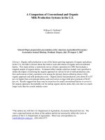
histology with AC and ASC, no statistically significant
differences were found between the groups in terms
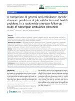
of OS (P = 0.145, Fig. 1). After adjustment for factors
that were significant in univariate analysis, multivariate analysis showed that FIGO stage (HR = 1.83, 95%
CI = 1.12–2.95) and LNM were significantly associated
with shorter OS (HR = 2.29, 95% CI = 1.90–4.32).
Clinicopathological features and OS were compared
between patients with AC and those with ASC. As
shown in Table 2, LVSI (P = 0.049) and SCC-Ag > 5
Table 1 Clinicopathological features associated with overall survival
Characteristics
Age
FIGO
Size
LNM
LVSI
DSI
SCC-Ag
CEA
CA 19–9
CA 125
RT
Histology
No.
Overall survival
P
Multivariate analysis
HR (95% CI)
HR (95% CI)
≤40
88
1
>40
282
1.33 (0.84–2.11)
IB
262
1
IIA
108
2.11 (1.45–3.07)
<4 cm
247
1
≥4 cm
123
1.70 (1.17–2.48)
No
267
1
Yes
103
3.43 (2.36–4.99)
No
200
1
Yes
170
1.32 (0.91–1.92)
<1/2
195
1
≥1/2
175
2.23 (1.51–3.28)
≤5 ng/ml
351
1
>5 ng/ml
19
2.12 (1.07–4.21)
≤5 ng/ml
285
1
>5 ng/ml
85
1.83 (1.22–2.74)
0.220
1
<0.001
2.29 (1.90–4.32)
0.083
<0.001
1
<0.001
2.16 (0.73–2.78)
0.031
1.44 (0.71–3.56)
0.004
1.13 (0.66–1.51)
0.079
1
0.113
1
311
1
1.38 (0.86–2.22)
≤35 U/ml
283
1
>35 U/ml
87
1.71 (0.98–3.00)
0.061
No
165
Yes
205
0.80 (0.55–1.71)
0.251
0.725 (0.478–1.10)
0.145
240
1.56 (0.94–2.09)
0.142
59
130
0.003
1
<0.001
>37 U/ml
AC
1.83 (1.12–2.95)
1
0.006
≤37 U/ml
ASC
P
Univariate analysis
0.183
0.076
Zhang et al. BMC Cancer
(2020) 20:664
Page 4 of 10
Fig. 1 Kaplan-Meier curves of overall survival for patients with adenocarcinoma (AC) and adenosquamous carcinoma (ASC)
ng/ml (P = 0.013) were more common in the ASC
group than in the AC group. Patients with ASC were
older (> 40 years) than patients with AC (83.1% vs.
72.5%, P = 0.029). The differences in OS between patients with otherwise similar clinicopathological features were not statistically significant.
Table 2 Clinicopathologic features in the AC and ASC groups
Characteristics
AC
ASC
(n = 240) (n = 130)
Pa
Overall survival
HR (95% CI)
Pb
Age
≤40
>40
174
108
Size
<4 cm
162
85
≥4 cm
78
45
0.65 (0.34–1.24) 0.192
LNM
No
172
95
0.809 0.64 (0.35–1.18) 0.153
Yes
68
35
0.88 (0.50–1.56) 0.660
LVSI
No
139
61
0.049 0.71 (0.38–1.33) 0.289
Yes
101
69
0.71 (0.40–1.24) 0.223
DSI
<1/2
135
60
0.065 0.57 (0.26–1.24) 0.158
≥1/2
105
70
66
SCC-Ag ≤5 ng/ml 233
>5 ng/ml 7
22
118
12
0.029 0.67 (0.23–1.98) 0.471
0.70 (0.45–1.11) 0.129
0.729 0.77 (0.44–1.33) 0.345
0.71 (0.43–1.18) 0.187
0.013 0.72 (0.46–1.11) 0.138
0.34 (0.09–1.28) 0.110
Pa-value, clinicopathological features were compared between patients with
AC and those with ASC; HR and Pb-value, OS were compared between patients
with AC and those with ASC using log-rank tests
Survival analysis of patients with AC and ASC
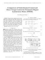
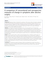
As shown in Tables 3 and 4, univariate Cox regression analyses revealed that, as FIGO stage and DSI
increased, and lymph node metastasis, whereas RFS
and OS significantly decreased, among patients with
ASC and AC (Fig. 2). In the AC group, RFS and OS
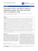
were significantly associated with tumour size (P =
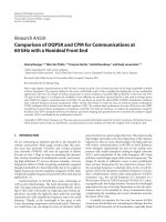
0.011 and P = 0.06, respectively), CEA (P = 0.023 and
P = 0.001, respectively; Fig. 3), SCC-Ag (P = 0.012 and
P = 0.001, respectively; Fig. 3), and CA 125 (P = 0.036
and P = 0.060, respectively); the associations were not
significant in the ASC group. CA 125 (P = 0.0036)
was associated with OS in patients with AC, but not
with RFS (P = 0.060).
Multivariate Cox regression analysis revealed that the
combination of CEA > 5 ng/ml (P = 0.042 and P = 0.033
for RFS and OS, respectively), SCC-Ag > 5 ng/ml (P =
0.027 and P = 0.018 for RFS and OS, respectively), and
LNM (P = 0.001, and P = 0.001 for RFS and OS, respectively) was a significant predictor of poor survival in patients with AC. Only LNM (P = 0.026 and P = 0.001 for
RFS and OS, respectively) was a significant predictor of
poor survival in patients with ASC.
The 5-year RFS and OS rates in the low-, intermediate-, and high-risk groups were 77.2 and 80.8%; 75.1 and
77.4%; and 35.1 and 41.8%, respectively, for the AC
group, and 83.7 and 85.4%, 80.5 and 83.7%, and 39.9 and
Zhang et al. BMC Cancer
(2020) 20:664
Page 5 of 10
Table 3 Clinicopathological features associated with survival outcomes of AC
Characteristics
No.
RFS
≤40
66
69.9
>40
174
63.4
IB
176
78.1
IIA
64
59.7
Size
<4 cm
162
71.3
≥4 cm
78
55.2
LNM
No
172
70.5
Yes
68
40.4
No
139
69.9
Yes
101
60.2
<1/2
135
75.9
≥1/2
105
52.4
≤5 ng/ml
233
67.7
P
5-year rate (%)
Age
FIGO Stage
LVSI
DSI
SCC-Ag
CEA
CA 19–9
CA 125
>5 ng/ml
7
25.0
≤5 ng/ml
199
70.2
>5 ng/ml
41
47.6
≤37 U/ml
198
67.7
>37 U/ml
42
58.6
≤35 U/ml
185
70.3
>35 U/ml
55
53.8
OS
P
5-year rate (%)
0.262
74.0
0.229
66.1
0.003
72.7
0.002
56.0
0.011
73.2
0.006
57.7
<0.001
73.0
<0.001
41.9
0.149
72.4
0.159
62.8
<0.001
79.9
<0.001
53.5
0.012
69.7
0.023
71.8
0.001
25.0
0.001
50.1
0.248
70.1
0.195
59.3
0.060
71.5
0.036
54.2
Table 4 Clinicopathological features associated with survival outcomes of ASC
Characteristics
No.
RFS
P
5-year rate (%)
Age
FIGO Stage
Size
LNM
LVSI
DSI
SCC-Ag
CEA
CA 19–9
CA 125
≤40
22
78.0
>40
108
71.2
IB
86
77.6
IIA
44
58.2
<4 cm
85
75.5
≥4 cm
45
66.8
No
95
81.7
Yes
35
42.4
No
61
76.9
Yes
69
71.3
<1/2
60
81.3
≥1/2
70
63.7
≤5 ng/ml
118
72.8
>5 ng/ml
12
62.6
≤5 ng/ml
86
75.9
>5 ng/ml
44
67.1
≤37 U/ml
113
71.6
>37 U/ml
17
72.5
≤35 U/ml
98
72.4
>35 U/ml
32
60.7
OS
P
5-year rate (%)
0.374
80.0
0.459
73.6
0.035
81.0
0.013
62.0
0.413
77.5
0.282
69.6
<0.001
84.4
<0.001
47.8
0.156
77.6
0.453
72.0
0.039
83.2
0.012
65.1
0.595
75.8
0.291
64.2
0.441
77.4
0.205
69.0
0.536
74.5
0.886
75.3
0.256
78.5
62.2
0.070
Zhang et al. BMC Cancer
(2020) 20:664
Page 6 of 10
Fig. 2 Kaplan-Meier curves of overall survival for patients with adenocarcinoma (a, b and c) and adenosquamous carcinoma (d, e and f) by FIGO
stage, LNM and DSI
Fig. 3 Kaplan-Meier curves of overall survival for patients with adenocarcinoma by CEA and SCC-Ag
Zhang et al. BMC Cancer
(2020) 20:664
Page 7 of 10
47.8% respectively, for the ASC group (Table 5). There
was no statistically significant difference in RFS or OS
between patients with AC and those with ASC in any
risk group (P > 0.05, Table 6).
Effect of adjuvant therapy on intermediate-risk-group
patients
According to the univariate analysis, intermediate-risk
ASC patients who received CCRT had significantly better RFS and OS than those who received no further
treatment (NFT) (HR = 0.101, 95% CI = 0.011–0.939; and
HR = 0.108, 95% CI = 0.012–0.972 for RFS and OS, respectively). Although the hazard ratio for RFS with RT
alone was statistically significant (HR = 0.691, 95%CI =
0.192–0.981), that was not true of OS (HR = 0.760, 95%
CI = 0.204–1.434). Patients with AC who received RT or
CCRT did not have better RFS or OS than those who received NFT (Table 7).
Discussion
The main histological type of cervical cancer is SCC
[25]. However, the incidences of AC and ASC of the
uterine cervix have increased over the past 40 years, especially among younger women [26–29]. In this retrospective cohort study, we examined the records of
Chinese patients with FIGO stage IB-IIA AC or ASC to
evaluate potential prognostic factors among these patients. All patients underwent surgery as the primary
treatment. Multivariate analyses showed that FIGO stage
and LNM were independent prognostic factors for OS.
Previous studies showed that FIGO stage, tumour size,
and LNM were independent prognostic factors for survival [13, 30, 31]. Shu et al. [2] reported that in patients
with AC/ASC, differentiation was an independent predictor of OS, and LVSI was an independent predictor of
DFS. We investigated whether histology (AC vs. ASC) is
a prognostic factor in patients with cervical cancer.
There were no differences, in terms of clinical impact on
OS, between the two histological groups in early-stage
Table 5 Stratified analysis of risk group associated with survival
outcomes of AC and ASC
No.
AC
5-year (%)
RFS
P
<0.001
High risk group
68
38.5
Intermediate risk group
112
74.2
Low risk group
60
High risk group
OS
P
41.9
<0.001
77.4
79.1
80.8
ASC
5-year (%)
No.
RFS
P
OS
P
35
44.6
<0.001
47.8
<0.001
Intermediate risk group
65
81.2
83.7
Low risk group
30
83.9
85.4
Table 6 Survival analysis by histologic type and risk group
No.
RFS
P
5-year (%)
High risk group |
OS
P
5-year (%)
0.563
0.675
AC
15
38.5
41.9
ASC
4
44.6
47.8
Intermediate risk group
AC
14
74.2
ASC
9
81.2
0.549
77.4
0.647
83.7
Low Risk Group
AC
89
79.1
ASC
28
83.9
0.485
80.8
0.437
85.4
cervical cancer, although a greater proportion of patients
with ASC had LVSI and SCC-Ag > 5 ng/ml; moreover,
patients with ASC were much older than those with AC.
Multivariate Cox regression analysis revealed that CEA >
5 ng/ml and SCC-Ag > 5 ng/ml were independent risk
factors for RFS and OS in patients with AC, but not in
patients with ASC. This suggests that pre-treatment
levels of CEA > 5 ng/ml and SCC-Ag > 5 ng/ml can be
regarded as risk factors for AC, providing additional information for patient-tailored therapy, and should be
analysed in prospective studies. Previous studies reported that elevated pre-treatment serum SCC-Ag levels
were associated with poor prognosis [32, 33], but the
histologic type of most patients was cervical squamous
cell carcinoma a few patients had AC. Nakamura et al.
[34] showed that AC, DOI, tumour size, and LVSI were
significantly associated with disease recurrence.
The respective 5-year survival rates for patients with
stages IB and IIA were 72.7 and 56% for the AC group
and 81.0 and 62% for the ASC group. Saigo et al. [35] reported that 5-year survival rates for patients with stages
I and II (IIA, IIB) AC were 79 and 37%, respectively. Presumably because the latter group also included patients
with IIB cancer, the 5-year survival rate for patients with
stage II cancer was lower than the rate observed in our
study. These results suggest that as FIGO stage increases, the survival time is reduced accordingly. Our results demonstrated that FIGO stage (IB vs. IIA) was
significantly associated with survival time (P < 0.05).
Similarly, Noh et al. [30] reported that ASC histology
was associated with more favourable survival outcomes,
compared to AC histology, although the differences were
not statistically significant. Lai et al. [12] found no differences between the ASC and AC subtypes in RFS or
cancer-specific survival (CSS).
Wang et al. [36] demonstrated that higher tumour
grade and more vascular invasion were present in patients with ASC, compared to patients with AC. Reis
et al. [11] found that Grade III histology and LVSI were
Zhang et al. BMC Cancer
(2020) 20:664
Page 8 of 10
Table 7 Stratified analysis of treatment associated with survival of AC and ASC
AC
ASC
RFS
OS
RFS
OS
Treatment
No.
HR(95% CI)
P
HR(95% CI)
P
No.
HR(95% CI)
P
HR(95% CI)
P
NFT
53
1
0.745
1
0.951
15
1
0.043
1
0.134
RT
21
1.068 (0.314–3.017)
1.095 (0.387–3.104)
22
0.691 (0.192–0.981)
0.760 (0.204–1.434)
CRRT
38
0.764 (0.334–2.063)
0.920 (0.393–2.154)
28
0.101 (0.011–0.939)
0.108 (0.012–0.972)
more common in patients with ASC than in patients
with AC. In addition, they demonstrated that although
the time to recurrence was shorter for patients with
ASC (7.9 months vs. 15 months; P = 0.01), differences in
OS or recurrence rates between patients with AC and
patients with ASC were not statistically significant. Baek
et al. [18] reported greater mean tumour size and more
frequent LVSI in patients with ASC, but found that
histologic type did not influence RFS or OS in multivariate analyses, following adjustment for significant prognostic factors. In contrast, several studies reported poor
survival for patients with ASC. A meta-analysis by Lee
et al. [16] demonstrated that ASC patients may have
poorer outcomes than those with AC of the cervix. Farley et al. [20] observed an increased risk of death among
patients with ASC histology compared to those with AC
histology. Twu et al. [37] found that survival for ASC
was slightly worse than that for AC in a univariate analysis, but the RFS and CSS of the two subtypes were not
significantly different in multivariate analyses.
Our study demonstrated a tendency for better RFS
and OS in patients with ASC than in those with AC, in
both the low-risk and intermediate/high-risk groups, although the difference was not statistically significant.
The prognosis for the ASC subtype appears to be intermediate (i.e. between those of the SCC and AC subtypes) [31]. Previous studies showed no statistically
significant differences between patients with AC and
those with ASC in low-, intermediate-, or high-risk
groups (P > 0.05) [9, 18]. However, Lea et al. reported
that ASC histology was associated with reduced diseasefree survival relative to AC histology, among patients
with low-risk stage IB1 cancer [38].
We also examined the effect of treatment on OS in
intermediate-risk ASC and AC patients. Univariate analysis indicated that in patients with ASC, CCRT was associated with significantly better RFS and OS. RT alone
was related to RFS but not OS. This indicated that RT
alone may be effective for local control, while CCRT is
advantageous for control of distant metastasis. In
addition, RT and CCRT did not confer any survival
benefit in patients with AC. This may have been because
there is greater radio resistance and more aggressive behaviour of tumours in patients with AC relative to those
with ASC. A retrospective study suggested that RT and
CCRT after radical hysterectomy were not beneficial in
intermediate-risk patients. In particular, RT and CCRT
appeared to increase the incidence of lymphedema, and
even led to RT-related morbidities such as small-bowel
obstruction and leg oedema [34, 39]. Twu et al. confirmed that adjuvant therapy (radiotherapy with or without chemotherapy) following RH-PLND, for early stage
AC/ASC patients with a low prognostic score, may not
improve survival. Therefore, omitting adjuvant therapy
could decrease morbidity [37]. We suspected that systemic CT alone could confer a survival benefit for patients with AC. Takekuma et al. [40] reported that
chemotherapy alone after surgery for high-risk patients
had similar efficacy to CCRT, but with less toxicity. Further prospective randomized studies including larger patient populations are needed to confirm our findings.
Our study was limited by its retrospective design. Furthermore, since most patients in the high-risk group received CCRT, while most patients in the low-risk group
underwent observation only, the statistical power may not
have been sufficient to detect a statistical difference in the
impact of adjuvant therapies on survival. Finally, systemic
CT alone, i.e., without RT, might confer a survival benefit.
However, we did not investigate the effects of chemotherapy because no patient received systemic CT alone. Despite these limitations, to our knowledge this study
included the largest number of FIGO stage IB–IIA cervical AC/ASC patients undergoing radical hysterectomy.
It also provided sufficient data on prognosis and adjuvant
treatment efficacy, given the long follow-up period.
Conclusion
In conclusion, there were no differences, in terms of OS,
between early stage AC and ASC cervical cancers. Patients
with ASC were older (> 40 years) and more likely to have
LVSI and SCC-Ag > 5 ng/ml, compared to patients with
AC. LNM, CEA > 5 ng/ml, and SCC-Ag > 5 ng/ml were
independent risk factors for poor RFS and OS in patients
with AC, whereas only LNM was an independent risk factor for poor RFS and OS in patients with ASC. In addition,
within an intermediate-risk-stratified group, patients with
ASC who received CCRT experienced significantly better
survival outcomes. Our findings may facilitate improvements in clinical diagnostics and therapeutic applications
for patients with cervical cancer.
Zhang et al. BMC Cancer
(2020) 20:664
Abbreviations
AC: adenocarcinoma; ASC: adenosquamous carcinoma; RFS: recurrence-free
survival; OS: overall survival; LNM: lymph node metastasis; DSI: depth of
stromal invasion; LVSI: lymph–vascular space invasion; MST: the median
survival time; RT: radiotherapy; CCRT: concurrent chemoradiotherapy;
CEA: Carcinoembryonic antigen; kSCC: squamous cell carcinoma antigen;
CA: carbohydrate antigen (CA); HR: hazard ratio
Page 9 of 10
8.
9.
10.
Acknowledgements
Not applicable.
Authors’ contributions
XJZ and HML conceived and designed the study. XJZ and ZFL collected
patient data. XXX and ZMY analyzed and interpreted the patient data. XJZ
was a major contributor in writing the manuscript. HML reviewed the
manuscript. All authors read and approved the final manuscript.
11.
12.
13.
Funding
The study was supported by a grant from the Zhejiang Medical Science and
Technology Foundation (No.2018254294). The funders will not have a role in
the study design, data collection, analysis, interpretation of results or the
manuscript.
14.
15.
Availability of data and materials
The datasets used and analyzed during the current study are available from
the corresponding author on reasonable request.
Ethics approval and consent to participate
The present study was approved by the Ethics Committee of Zhejiang
Cancer Hospital, and all participants gave written informed consent.
16.
17.
Consent for publication
Not applicable.
18.
Competing interests
The authors declare that they have no conflict of interest.
19.
Author details
1
Department of Gynecological Oncology, Cancer Hospital of University of
Chinese Academy of Sciences (Zhejiang Cancer Hospital), Institute ofCancer
Research and Basic Medicine (IBMC), Chinese Academy of Sciences, 1
Banshan East Road, Hangzhou 310022, P. R. China. 2Department of
Agriculture and Food Science, Zhejiang A&F University, Lin’an 311300, P. R.
China.
Received: 30 March 2020 Accepted: 7 July 2020
References
1. Bray F, Ferlay J, Soerjomataram I, Siegel RL, Torre LA, Jemal A, et-al. Global
cancer statistics 2018: globocan estimates of incidence and mortality
worldwide for 36 cancers in 185 countries. Ca A Cancer J Clin. 2018;68(6):
394–424.
2. Shu T, Zhao D, Li B, Wang Y, Liu S, Li P, et al. Prognostic evaluation of
postoperative adjuvant therapy for operable cervical cancer: 10 years’
experience of National Cancer Center in China. Chin J Cancer Res. 2017;
29(6):510–20.
3. Baalbergen A, Veenstra Y, Stalpers L. Primary surgery versus primary
radiotherapy with or without chemotherapy for early adenocarcinoma of
the uterine cervix. Cochrane Database Syst. 2013;1:CD006248.
4. Ryu SY, Kim MH, Nam BH, Lee TS, Song ES, Park CY, et al. Intermediate-risk
grouping of cervical cancer patients treated with radical hysterectomy: a
korean gynecologic oncology group study. Br J Cancer. 2014;110(2):278–85.
5. Fujiwara H, Yokota H, Monk B, Treilleux I, Devouassoux-Shisheboran M,
Davis A, et al. Gynecologic cancer intergroup (GCIG) consensus review for
cervical adenocarcinoma. Int J Gynecol Cancer. 2014;24(9):S96–S101.
6. Young RH, Clement PB. Endocervical adenocarcinoma and its variants: their
morphology and differential diagnosis. Histopathology. 2002;41:185–207.
7. Chan PG, Sung HY, Sawaya GF. Changes in cervical cancer incidence after
three decades of screening US women less than 30 years old. Obstet
Gynecol. 2003;102:765–73.
20.
21.
22.
23.
24.
25.
26.
27.
28.
Vinh-Hung V, Bourgain C, Vlastos G, Gábor Cserni, Ridder, MD, Storme G,
et al. Prognostic value of histopathology and trends in cervical cancer: a
seer population study. BMC Cancer; 2007: 7(1), 164–0.
Mabuchi S, Okazawa M, Matsuo K, Kawano M, Suzuki O, Miyatake T, et al.
Impact of histological subtype on survival of patients with surgically-treated
stage ia2–iib cervical cancer: adenocarcinoma versus squamous cell
carcinoma. Gynecol Oncol. 2012;127(1):114–20.
National Comprehensive Cancer Network NCCN Clinical Practice Guidelines
in Oncology, Cervical Cancer Version 2 (2015).
Reis RD, Frumovitz M, Milam MR, Capp E, Sun CC, Coleman RL, et al.
Adenosquamous carcinoma versus adenocarcinoma in early-stage cervical
cancer patients undergoing radical hysterectomy: an outcomes analysis.
Gynecol Oncol. 2007;107(3):458–63.
Lai CH, Chou HH, Chang CJ, Wang CC, Hsueh S, Huang YT, et al. Clinical
implications of human papillomavirus genotype in cervical adenoadenosquamous carcinoma. China J Modern Med. 2013;49(3):633–41.
Kasamatsu T, Onda T, Sawada M, Kato T, Ikeda S, Sasajima Y, et al. Radical
hysterectomy for FIGO stage I–IIB adenocarcinoma of the uterine cervix. Br J
Cancer. 2009;100:1400–5.
Park JY, Kim DY, Kim JH, Kim YM, Kim YT, Nam JH. Outcomes after radical
hysterectomy in patients with early-stage adenocarcinoma of uterine cervix.
Br J Cancer. 2010;102:1692–8.
Mabuchi S, Suzuki O, Kamiura S, Ogawa K, Kimura T, Okazawa M, et al.
Impact of the addition of concurrent chemotherapy to pelvic radiotherapy
in surgically treated stage IB1-IIB cervical cancer patients with intermediaterisk or high-risk factors: a 13-year experience. Int J Gynecol Cancer. 2013;
23(3):567–75.
Lee KBM, Lee JM, Park CY, Lee KB, Cho HY, Ha SY. What is the difference
between squamous cell carcinoma and adenocarcinoma of the cervix? A
matched case–control study. Int J Gynecol Cancer. 2006;16:1569–73.
Chen JL, Cheng JC, Kuo SH, Chen CA, Lin MC, Huang CY. Outcome analysis
of cervical adenosquamous carcinoma compared with adenocarcinoma.
Acta Obstet Gynecol Scand. 2012;91:1158–66.
Baek MH, Park JY, Kim D, Suh DS, Nam JH. Comparison of adenocarcinoma
and adenosquamous carcinoma in patients with early-stage cervical cancer
after radical surgery. Gynecologic Oncol. 2014;135(3):462–7.
Lee JY, Lee C, Hahn S, Kim MA, Kim HS, Chung HH, et al. Prognosis of
Adenosquamous carcinoma compared with adenocarcinoma in uterine
cervical Cancer: a systematic review and meta-analysis of observational
studies. Int J Gynecol Cancer Official J Int Gynecol Cancer Soc. 2014;24(2):
289.
Farley JH, Hickey KW, Carlson JW, Rose GS, Kost ER, Harrison TA.
Adenosquamous histology predicts a poor outcome for patients with
advanced-stage, but not early-stage, cervical carcinoma. Cancer. 2003;97:
2196–202.
Nakanishi T, Ishikawa H, Suzuki Y, Inoue T, Nakamura S, Kuzuya K. A
comparison of prognoses of pathologic stage Ib adenocarcinoma and
squamous cell carcinoma of the uterine cervix. Gynecol Oncol. 2000;79:289–
93.
Galic V, Herzog TJ, Lewin SN, Neugut AI, Burke WM, Lu YS, et al. Prognostic
significance of adenocarcinoma histology in women with cervical cancer.
Gynecol Oncol. 2012;125(2):287–91.
Irie T, Kigawa J, Minagawa Y, Itamochi H, Sato S, Akeshima R, et al.
Prognosis and clinicopathological characteristics of Ib-IIb adenocarcinoma
of the uterine cervix in patients who have had radical hysterectomy. Eur J
Surg Oncol. 2000;26:464–7.
Huang YT, Wang CC, Tsai CS, Lai CH, Chang TC, Chou HH, et al. Long-term
outcome and prognostic factors for adenocarcinoma/adenosquamous
carcinoma of cervix after definitive radiotherapy. Int J Radiat Oncol Biol
Phys. 2011;80:429–36.
Gien LT, Beauchemin MC, Thomas G. Adenocarcinoma: a unique cervical
cancer. Gynecol Oncol. 2010;116:140–6.
Sherman ME, Wang SS, Carreon J, Devesa SS. Mortality trends for cervical
squamous and adenocarcinoma in the United States. Cancer. 2005;103(6):
1258–64.
Castellsagué X, Díaz M, De Sanjosé S, Muñoz N, Herrero R, Franceschi S,
et al. Worldwide human papillomavirus etiology of cervical adenocarcinoma
and its cofactors: implications for screening and prevention. J Natl Cancer
Inst. 2006;98:303–15.
Sasieni P, Adams J. Changing rates of adenocarcinoma and adenosquamous
carcinoma of the cervix in England. Lancet. 2001;357:1490–3.
Zhang et al. BMC Cancer
(2020) 20:664
29. Huang CY, You SL, Chen CJ, Cheng WF, Luo HC, Hsieh CY. Incidence of
cervical cancer and age-specific survival of small cell cervical carcinoma in
Taiwan. Acta Obstet Gynecol Scand. 2011;90:1342–9.
30. Noh JM, Park W, Kim YS, Kim JY, Kim HJ, Kim J, et al. Comparison of clinical
outcomes of adenocarcinoma and adenosquamous carcinoma in uterine
cervical cancer patients receiving surgical resection followed by
radiotherapy: a multicenter retrospective study (KROG 13-10). Gynecol
Oncol. 2014;132(3):618–23.
31. Chen JL‐Y, Cheng JC‐H, Kuo S‐H, Chen C‐A, Lin M‐C, Huang C‐Y. Outcome
analysis of cervical adenosquamous carcinoma compared
withadenocarcinoma[J]. Acta Obstetricia Et Gynecologica Scandinavica,
2012; 91(10):1158–1166.
32. Davelaar EM, Lande JVD, Mensdorff-Pouilly SV, Blankenstein MA, Kenemans
PA. Combination of serum tumor markers identifies high-risk patients with
early-stage squamous cervical Cancer. Tumor Biol. 2008;29(1):9–17.
33. Reesink-Peters N, van der Velden J, ten Hoor KA, Boezem HM, de Vries EGE,
Schilthuis MS, Mourits MJE, Nijman HW, Aalders JG, Hollema H, Pras E, Duk
JM, van der Zee AGJ. Preoperative serum squamous cell carcinoma antigen
levels in clinical decision making for patients with early-stage cervical
cancer. J Clin Oncol. 2005;23:1455–62.
34. Nakamura K, Kitahara Y, Satoh T, Takei Y, Takano M, Nagao S, et al. Analysis
of the effect of adjuvant radiotherapy on outcomes and complications after
radical hysterectomy in FIGO stage IB1 cervical cancer patients with
intermediate risk factors (GOTIC study). Gynecol Oncol. 2016;14(1):61–2.
35. Saigo PE, Cain JM, Kim WS, Gaynor JJ, Johnson K, Jr LJ, et al. prognostic factors
in adenocarcinoma of the uterine cervix. Cancer. 2004; 92(1):262–267.
36. Wang SS, Sherman ME, Silverberg SG, Carreon JD, Lacey JV Jr, Zaino R, et al.
Pathological characteristics of cervical adenocarcinoma in a multi-center USbased study. Gynecol Oncol. 2006;103:541–6.
37. Twu NF, Ou YC, Liao CI, Chang WY, Yang LY, Tang YH, et al. Prognostic factors
and adjuvant therapy on survival in early-stage cervical adenocarcinoma/
adenosquamous carcinoma after primary radical surgery: a Taiwanese
gynecologic oncology group (TGOG) study. Surg Oncol. 2016;25:229–35.
38. Lea JS, Coleman RL, Garner EO, Duska LR, Miller DS, Schorge JO.
Adenosquamous histology predicts poor outcome in low-risk stage IB1
cervical adenocarcinoma. Gynecol Oncol. 2003;91:558–62.
39. Lee KB, Lee JM, Ki KD, Lee SK, Park CY, Ha SY. Comparison of adjuvant
chemotherapy and radiation in patients with intermediate risk factors after
radical surgery in FIGO stage IB-IIA cervical cancer. Int J Gynecol Cancer.
2008;18:1027–31.
40. Takekuma M, Kasamatsu Y, Kado N, et al. Adjuvant chemotherapy versus
concurrent chemoradiotherapy for high-risk cervical cancer after radical
hysterectomy and systematic lymphadenectomy. Int J Clin Oncol. 2016;21:741–7.
Publisher’s Note
Springer Nature remains neutral with regard to jurisdictional claims in
published maps and institutional affiliations.
Page 10 of 10