A prediction model for lymph node metastases using pathologic features in patients intraoperatively diagnosed as stage I non-small cell lung cancer
Bạn đang xem bản rút gọn của tài liệu. Xem và tải ngay bản đầy đủ của tài liệu tại đây (506.5 KB, 8 trang )
Zhao et al. BMC Cancer (2017) 17:267
DOI 10.1186/s12885-017-3273-x
RESEARCH ARTICLE
Open Access
A prediction model for lymph node
metastases using pathologic features in
patients intraoperatively diagnosed as
stage I non-small cell lung cancer
Fei Zhao†, Yue Zhou†, Peng-Fei Ge†, Chen-Jun Huang, Yue Yu, Jun Li, Yun-Gang Sun, Yang-Chun Meng,
Jian-Xia Xu, Ting Jiang, Zhi-Xuan Zhang, Jin-Peng Sun and Wei Wang*
Abstract
Background: There is little information on which pattern should be chosen to perform lymph node dissection for
stage I non-small-cell lung cancer. This study aimed to develop a model for predicting lymph node metastasis
using pathologic features of patients intraoperatively diagnosed as stage I non-small-cell lung cancer.
Methods: We collected pathology data from 284 patients intraoperatively diagnosed as stage I non-small-cell
lung cancer who underwent lobectomy with complete lymph node dissection from 2013 through 2014, assessing
various factors for an association with metastasis to lymph nodes (age, gender, pathology, tumour location,
tumour differentiation, tumour size, pleural invasion, bronchus invasion, multicentric invasion and angiolymphatic
invasion). After analysing these variables, we developed a multivariable logistic model to estimate risk of
metastasis to lymph nodes.
Results: Univariate logistic regression identified tumour size >2.65 cm (p < 0.001), tumour differentiation
(p < 0.001), pleural invasion (p = 0.034) and bronchus invasion (p < 0.001) to be risk factors significantly
associated with the presence of metastatic lymph nodes. On multivariable analysis, only tumour size >2.65 cm
(p < 0.001), tumour differentiation (p = 0.006) and bronchus invasion (p = 0.017) were independent predictors for
lymph node metastasis. We developed a model based on these three pathologic factors that determined that the
risk of metastasis ranged from 3% to 44% for patients intraoperatively diagnosed as stage I non-small-cell lung
cancer. By applying the model, we found that the values ŷ > 0.80, 0.43 < ŷ ≤ 0.80, ŷ ≤ 0.43 plus tumour size
>2 cm and ŷ ≤0.43 plus tumour size ≤2 cm yielded positive lymph node metastasis predictive values of 44%,
18%, 14% and 0%, respectively.
Conclusions: A non-invasive prediction model including tumour size, tumour differentiation and bronchus
invasion may be useful to give thoracic surgeons recommendations on lymph node dissection for patients
intraoperatively diagnosed as Stage I non-small cell lung cancer.
Keywords: Non-small-cell lung cancer, Lymph node, Metastasis, Multivariable logistic model
* Correspondence:
†
Equal contributors
Department of Thoracic Surgery, First Affiliated Hospital of Nanjing Medical
University, 300 Guangzhou Road, Nanjing 210029, China
© The Author(s). 2017 Open Access This article is distributed under the terms of the Creative Commons Attribution 4.0
International License ( which permits unrestricted use, distribution, and
reproduction in any medium, provided you give appropriate credit to the original author(s) and the source, provide a link to
the Creative Commons license, and indicate if changes were made. The Creative Commons Public Domain Dedication waiver
( applies to the data made available in this article, unless otherwise stated.
Zhao et al. BMC Cancer (2017) 17:267
Page 2 of 8
Background
Lung cancer is the leading cause of cancer death worldwide [1] and metastasis to lymph nodes directly determines the stage and prognosis of this disease. Computed
tomography (CT) remains the most widely used tool for
assessment of the tumour and lymph node involvement in
patients with early-stage non-small-cell lung cancer
(NSCLC) [2–5]. In general, lymph nodes with short-axis
diameters of >1 cm seen on CT scan are considered metastatic. Unfortunately, the accuracy of CT scan for preoperative lymph node stage is only 45%–79% [2–6]. In
addition, studies have demonstrated that 12%–17% of
patients histologically confirmed as N2 are preoperatively
diagnosed as N0 because their CT scan results showed the
involved lymph nodes to have short-axis diameters of
<1 cm [4, 5, 7]. Many other methods of preoperative
N-staging, e.g. positron emission tomography, mediastinoscopy and endoscopic ultrasound-guided fine-needle
aspiration, are not routinely used for patients with clinical
stage I disease. In addition, these methods yield a considerable number of false-negative results [8–10].
There is ample high-quality evidence on the advantages of lymph node dissection in lung cancer surgery,
including the American College of Surgeons Oncology
Group (ACOSOG) Z0030 trial [11], although the benefits of complete lymph node dissection for patients with
stage I NSCLC are still controversial [12–14]. There is
little information on which pattern should be chosen to
perform lymph node dissection for patients intraoperatively diagnosed as stage I non-small-cell lung cancer. A
non-invasive prediction model that is able to predict
lymph node metastasis would allow surgeons to make
appropriate decisions on the extent of the dissection,
removing lymph nodes that are most likely to contain
metastases, while avoiding unnecessary tissue damage in
order to accelerate patients’ postoperative recovery.
The goal of this study was to identify risk factors that
would predict differences in lymph node metastasis and
to develop a scoring system to predict the presence of
lymph node metastasis. The aim is to determine the
appropriate pattern of lymph node dissection for various
patients intraoperatively diagnosed as stage I NSCLC.
(National Comprehensive Cancer Network (NCCN) Guidelines Version 3.2014: Staging Non-Small Cell Lung Cancer)
[15]. We excluded patients from this study who met any one
of the following conditions: 1) tumour size > 4 cm and
lymph node > 1 cm at the largest diameter on CT imaging
or evidence of distant metastasis; 2) preoperative chemotherapy or radiotherapy; 3) previous or coexistent tuberculosis or
malignant disease; 4) complete lymph node dissection that
did not meet the current standards (i.e. all lymph node
stations, including right-hand stations 2–4 and 7–9 and lefthand stations 2–9); 5) pure ground-glass opacity on CT imaging; 6) synchronous lung cancers, 7) sublobar resection,
segmentectomy or partial resection or 8) Intraoperative
frozen rapid pathological results showed tumour size > 4 cm
in the largest diameter.
Patients were preoperatively assessed with chest x-ray,
chest and upper abdominal CT scan, brain magnetic resonance imaging and bone scintigraphy. CT scan was used for
preoperative N-staging. The surgical approach for primary
lung cancer resection was via video-assisted thoracic surgery.
Methods
Results
Patient selection
Patient characteristics and prevalence of lymph node
metastasis
A total of 284 consecutive patients who underwent surgical resection for primary lung cancer at our hospital
from January 2013 to December 2014 were reviewed
retrospectively. The records of patients intraoperatively
diagnosed as stage I NSCLC who underwent lobectomy
with complete lymph node dissection according to the
lymph node nomenclature were selected for this study.
All patients met the criteria for stage I NSCLC based on
the new International Staging System for NSCLC
Statistical analysis
The baseline patient characteristics were summarized in
percentages for categorical variables and as mean ± SD
(Standard Deviation) for continuous variables. The chisquare test and Fisher’s exact tests were used to analyse
differences in these percentages between the groups. Differences between the groups were analysed using the
Kruskal–Wallis test. Significance of associations with the
outcome of nodal metastases was first evaluated using a
univariate logistic analysis. Those significant variables
were analysed by multivariable analysis as independent
predictors for lymph node metastasis. Odds ratios (ORs)
with 95% confidence intervals (CIs) were calculated.
Clinically relevant variables obtained by multivariable
analysis were included in the multivariable model. The
resulting model coefficients were applied to the cohort
to calculate predicted values from the logistic equation:
ŷ = 1/[1 + exp. (−xβ)]. All confidence intervals, significance tests and resulting P values were two-sided, with
an alpha level of 0.05. Statistical analyses were
performed using STATA software, release 13.
A total of 284 patients intraoperatively diagnosed as
stage I NSCLC were included in this study. Table 1
shows the patients’ demographics and clinical characteristics. The mean age was 60.78 years (range 31–83).
Histologically, the tumours in 248 patients (87%) were
identified as adenocarcinoma and in 36 (13%) as squamous cell carcinoma. The tumour originated in the right
upper lobe in 82 patients (29%), right middle lobe in 16
Zhao et al. BMC Cancer (2017) 17:267
Page 3 of 8
Table 1 Patient Demographics and Clinical Characteristics
Variables
Value
Number
284
Age (years)
Mean ± SD (range)
60.78 ± 9.2 (31–83)
Gender (%)
Male
144 (51%)
Female
140 (49%)
Pathology
Lymph node metastases were not found in 215
patients (group I) but were present in 69 (group II)
(Table 2). The characteristics in these two groups were
compared in terms of age, gender, pathology, tumour
Table 2 Demographics of patients in the Negative lymph Node
Metastases (LNM) and Positive LNM groups
Variables
Negative LNM Positive LNM
Number
Squamous cell carcinoma
36 (13%)
Adenocarcinoma
248 (87%)
Tumor location (%)
P value
Group
215
69
Age (years)
Mean ± SD
0.118
61.27 ± 9.38
59.28 ± 8.49
Gender
0.997
Right Upper Lobe
82 (29%)
Male
109
35
Right Middle Lobe
16 (6%)
Female
106
34
Right Lower Lobe
39 (14%)
Left Upper Lobe
77 (27%)
Left Lower Lobe
51 (18%)
Mixed lobes
19 (6%)
Differentiation (%)
Pathology
0.176
Squamous cell carcinoma 24
12
Adenocarcinoma
57
191
Tumor location
0.368
Right Upper Lobe
62
20
I
86 (30%)
Right Middle Lobe
14
2
II
176 (62%)
Right Lower Lobe
28
11
III
22 (8%)
Left Upper Lobe
63
14
Tumor size (cm)
Mean ± SD (range)
2.44 ± 0.97 (0.4-4 cm)
Pleura invasion
Left Lower Lobe
34
17
Mixed lobes
14
5
<0.001*
Differentiation
Absent
220 (77%)
I
80
6
Present
64 (23%)
II
119
57
III
16
6
2.28 ± 0.95
2.92 ± 0.87
Bronchus invasion
Absent
247 (87%)
Tumor size (cm)
Present
37 (13%)
Mean ± SD
Multicentric invasion (%)
<0.001
0.033*
Pleura invasion
Absent
264 (93%)
Present
20 (7%)
Angiolymphatic invasion (%)
Absent
173
47
Present
42
22
<0.001*
Bronchus invasion
Absent
274 (96%)
Absent
196
51
Present
10 (4%)
Present
19
18
Absent
200
64
Present
15
5
Neural invasion
Multicentric invasion
Absent
283 (100%)
Present
1 (0%)
SD standard deviation
(6%), right lower lobe in 39 (14%), left upper lobe in 77
(27%), left lower lobe in 51 (18%) and in mixed lobes in
19 (6%). Mean tumour size was 2.44 cm (range from 0.4
to 4 cm). The tumour differentiation included I (86
patients, 30%), II (176 patients, 62%), III (22 patients,
8%). Pleural invasion was present in 64 patients (23%)
and bronchus invasion in 37 (13%).
1 (Fish)
Angiolymphatic invasion
0.263 (Fish)
Absent
209
65
Present
6
4
Absent
214
69
Present
1
0
Neural invasion
SD standard deviation
*P < 0.05
1.0 (Fish)
Zhao et al. BMC Cancer (2017) 17:267
location, tumour differentiation, tumour size, pleural invasion, bronchus invasion, multicentric invasion, neural
invasion and angiolymphatic invasion. Compared with
group I, group II had a significantly larger tumour size
than that in group I (2.92 ± 0.87 vs. 2.28 ± 0.95,
P < 0.001). There were significant statistical differences
between the groups by the χ2 test in terms of tumour
differentiation (I, II, III) (P < 0.001), bronchus invasion
(absent vs. present) (P < 0.001) and pleural invasion
(absent vs. present) (P = 0.033).
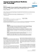
To evaluate the predictive value of tumour size between the groups, we used Receiver Operating Characteristic (ROC) curve analysis. As shown in Fig. 1,
the area under the ROC curve for tumour size between group I and group II was 0.691 (95% CI:
0.621–0.761; P < 0.001); the optimal cut-off value was
2.650 cm (sensitivity: 67%; specificity: 70%; Youden’s
index: 0.364).
Association of Individual Pathologic Characteristics with
Nodal Metastasis
Univariate analysis showed that tumour size greater than
2.650 cm (OR =4.62, 95% CI 2.59–8.24; P < 0.001),
tumour differentiation (I vs II + III, OR =6.22, 95% CI
2.58–15.03; P < 0.001), pleural invasion (absent vs
present, OR =1.93, 95% CI 1.05–3.54; P = 0.034) and
bronchus invasion (absent vs present, OR =3.64, 95% CI
1.78–7.44; P < 0.001) were the four significant risk
factors associated with the presence of metastatic lymph
nodes (Table 3).
Page 4 of 8
Multivariable analysis of pathologic characteristics
associated with nodal metastasis
Multivariate analysis of the four risk factors obtained on
univariate analysis showed that only the tumour size
(≤2.65 cm vs. >2.65 cm, OR =3.23, 95% CI 1.75–5.93;
P < 0.001), tumour differentiation (I vs II + III, OR
=3.64, 95% CI 1.44–9.16; P = 0.006) and bronchus invasion (absent vs. present, OR =2.54, 95% CI 1.18–5.46;
P = 0.017) were independent predictors associated with
the presence of metastatic lymph nodes. However,
pleural invasion (absent vs. present, OR =1.64, 95% CI
0.84–3.21; P = 0.146) was not a significant predictor of
lymph node metastasis (Table 4).
Multivariable logistic regression model derivation and
development
On multivariable analysis, only three covariates remained in
the final model. Using these three variables (Table 5), a scoring system was developed to discriminate between patients
with and without lymph node metastasis. The risk scores for
individual patients were calculated using the following formula: xβ = −2.947 + (1.368 × Differentiation (I vs. II + III,
I = 0, II + III = 1)) + (1.188 × Tumour Size (2.65 cm vs.
>2.65 cm, ≤2.65 cm = 0, >2.65 cm = 1)) + (0.876 × Bronchus
Invasion (absent =0, present =1)).
The probabilities of lymph node metastasis were
calculated using the following formula (ŷ = 1/
[1 + exp.(−xβ)]): ŷ = 1/[1 + exp. (2.947 - (1.368 × Differentiation (I vs. II + III, I = 0, II + III = 1)) - (1.188 × Tumour
Size (≤2.65 cm vs. >2.65 cm, ≤2.65 cm = 0, >2.65 cm = 1))
- (0.876 × Bronchus Invasion (absent =0, present =1))].
Fig. 1 The ROC (Receiver Operating Characteristic) curve of tumor size between group I and group II
Zhao et al. BMC Cancer (2017) 17:267
Page 5 of 8
Table 3 Univariate analysis of the risk factors for lymph node
metastases
Variables
OR (95% CI)
P value
0.75 (0.44–1.30)
0.304
1.0 (0.58–1.72)
0.997
0.60 (0.28–1.27)
0.179
Age
Gender
Pathology
Tumor location
1.00 (0.57–1.77)
0.98
Upper lobes vs Middle +Left lobes
1.45 (0.82–2.56)
0.199
Single lobes vs Mixed lobes
1.12 (0.39–3.24)
0.832
6.22 (2.58–15.03) <0.001*
I VS II + III
Tumor size
≤ 2.65 cm vs >2.65 cm
4.62 (2.59–8.24)
<0.001*
1.93 (1.05–3.54)
0.034*
3.64 (1.78–7.44)
<0.001*
1.04 (0.36–2.98)
0.939
2.14 (0.59–7.83)
0.249
Pleura invasion
Absent vs Present
Bronchus invasion
Absent vs Present
Multicentric invasion
Absent vs Present
Angiolymphatic invasion
Absent vs Present
P < 0.05
*
Model performance and selecting cut-off values to discriminate patients with lymph node metastasis
As shown in Fig. 2, the area under the ROC curve of the
selected model was 0.753 (95% CI 0.692–0.814, standard
error 0.031) and the optimal cut-off value was
0.7997 ≈ 0.80 (sensitivity: 71%, specificity: 71%, Youden’s
index: 0.417). In all patients, using a score threshold of
Table 4 Multivariate analysis of the risk factors for lymph node
metastases
β
OR (95% CI)
P value
1.291
3.64 (1.44–9.16)
0.006*
1.171
3.23 (1.75–5.93)
<0.001*
0.496
1.64 (0.84–3.21)
0.146
Absent vs Present
0.931
2.54 (1.18–5.46)
0.017*
Intercept
−3.013
Differentiation
Tumor size
Pleura invasion
Bronchus invasion
P < 0.05
3.93 (1.57–9.83)
0.003*
1.188
3.28 (1.79–6.01)
<0.001*
Absent vs Present
0.876
2.40 (1.13–5.13)
0.023*
Intercept
−2.947
≤ 2.65 cm vs >2.65 cm
P < 0.05
Differentiation
*
1.368
I VS II + III
*
Right lobes vs Left lobes
Absent vs Present
P value
Bronchus invasion
Squamous cell carcinoma VS
Adenocarcinoma
≤ 2.65 cm vs >2.65 cm
OR (95% CI)
Tumor size
male vs female
I VS II + III
β
Variables
Differentiation
≤ 60 vs >60
Variables
Table 5 Multivariate analysis of the risk factors for development
of model
≤0.80, 20 (12%) of 172 patients with lymph node metastasis were correctly identified, whereas 152 (88%) of 172
without lymph node metastasis were correctly identified.
Using a score threshold of >0.80, 49 (44%) of 112
patients with lymph node metastasis were correctly
identified, whereas 63 (56%) of 112 without lymph node
metastasis were correctly identified.
When all three covariates (tumour size, tumour differentiation, bronchus invasion) were equal to zero, we
found that the cut-off value was 0.42685 ≈ 0.43. In all
patients, using a score threshold of ≤0.43, 2 (3%) of 71
patients with lymph node metastasis were correctly
identified, whereas 69 (97%) of 71 without lymph node
metastasis were correctly identified. Using a score
threshold of >0.43, 67 (31%) of 213 patients with lymph
node metastasis were correctly identified, whereas 146
(69%) of 213 without lymph node metastasis were
correctly identified.
Using a score threshold between 0.43 and 0.80, 18 (18%)
of 101 patients with lymph node metastasis were correctly
identified, whereas 83 (82%) of 101 without lymph node
metastasis were correctly identified. So, we obtained three
score thresholds, ŷ ≤ 0.43, 0.43 < ŷ ≤ 0.80 and ŷ > 0.80.
Discussion
A complete lymph node dissection, removing all ipsilateral lymph nodes which can be seen at operation [16],
can provide more accurate pathologic staging and better
clinical outcomes for some patients. It is considered a
standard surgical treatment for patients diagnosed preoperatively with lymph node metastases. However,
complete lymph node dissection is not regarded as a
routine surgical procedure for patients intraoperatively
diagnosed as stage I NSCLC, as some studies have
demonstrated a lack of significant differences in
outcome between selective lymph node sampling and
complete lymph node dissection in patients with earlystage lung cancer [13, 17].
However each patient exhibits different clinical characteristics that affect the risk of lymph node metastasis in
early-stage lung cancer. In this study, we collected
Zhao et al. BMC Cancer (2017) 17:267
Page 6 of 8
Fig. 2 The ROC (Receiver Operating Characteristic) curve of the selected model
pathology data from 284 patients intraoperatively diagnosed as stage I NSCLC who underwent lobectomy with
complete lymph node dissection and investigated factors
that might be associated with metastasis to lymph nodes
(age, gender, pathology, tumour location, tumour differentiation, tumour size, pleural invasion, bronchus invasion,
multicentric invasion and angiolymphatic invasion).
First, we used univariate analysis to find associations between pathologic factors and lymph node metastasis. The
results showed that only the tumour size (>2.65 cm),
tumour differentiation, pleural invasion and bronchus
invasion were significant risk factors. The other factors
tested, including age, gender, pathologic type, tumour
location, multicentric invasion, angiolymphatic invasion
and neural invasion were excluded as risk factors associated with lymph node metastasis.
Furthermore, multivariate analysis of the four risk
factors identified on univariate analysis found that only
tumour size (>2.65 cm), tumour differentiation and
bronchus invasion were independent predictors of
lymph node metastasis. Pleural invasion was excluded as
an independent predictor in this analysis.
These three independent predictors were kept in the
final model. After developing the multivariable logistic regression model, we finally obtained three score thresholds,
ŷ ≤0.43, 0.43 < ŷ ≤ 0.80 and ŷ > 0.80 (Table 6). As shown
Table 6 Analysis of lymph Node Metastases (LNM)
Variables
ŷ ≤ 0.43
ŷ > 0.80
Negative LNM
Positive LNM (%)
Total
Negative LNM
Positive LNM (%)
Total
Negative LNM
Positive LNM (%)
Total
Num
69
2(3)
71
83
18(18)
101
63
49(44)
112
I
69
2(3)
71
11
2(15)
13
0
2(100)
2
II + III
−
−
−
72
16(18)
88
63
47(43)
110
0.43 ~ 0.80
Differentiation
Tumor size(cm)
≤2
57
0(0)
57
50
13(20)
64
4
4(50)
8
2 ~ 2.65
12
2(14)
14
22
4(15)
26
5
0(0)
5
> 2.65
−
−
−
11
1(8)
12
54
45(45)
99
Bronchus invasion
Absent
69
2(3)
71
83
17(17)
100
44
32(42)
76
Present
−
−
−
0
1(100)
1
19
17(47)
36
Zhao et al. BMC Cancer (2017) 17:267
in the table, we found that when ŷ was ≤0.43, patients with
lymph node metastasis accounted for 3% of all patients,
and when ŷ was ≤0.43 and tumour size was ≤2 cm, no
patients had lymph node metastasis. However, when ŷ was
≤0.43 and tumour size was >2 cm, the percentage of
patients identified with lymph node metastasis increased
to 14%. With 0.43 < ŷ ≤ 0.80, patients with lymph node
metastasis accounted for 18% of all patients. When ŷ was
>0.80, the patients with lymph node metastasis accounted
for 44% of all patients.
Thus we demonstrated that lymph node dissection is
not necessary for those patients intraoperatively diagnosed as stage I NSCLC whose ŷ value obtained from
the model is less than or equal to 0.43 and whose
tumour size is ≤2 cm. Complete lymph node dissection
or lymph node sampling would be appropriate if the ŷ
value from the model is less than or equal to 0.43 but
the tumour size is >2 cm or if ŷ is more than 0.43 and
less than or equal to 0.80. Complete lymph node dissection must be performed for patients whose ŷ value
obtained from the model is more than 0.80.
However, our study has some limitations. This study
was conducted at a single institution with retrospective
methods and demonstrated the necessity of further prospective study. Further prospective study with multicenter trial should be performed to comprehensively
evaluate this model for prediction of lymph node metastases in patients intraoperatively diagnosed as Stage I
non-small cell lung cancer.
Conclusions
After a comprehensive analysis of our results concerning
various clinical factors, we conclude that the incidence
of lymph node metastasis would be lowest when we obtained a ŷ value from the model less than or equal to
0.43 along with a tumour size ≤2 cm. For other patients
intraoperatively diagnosed as stage I NSCLC, the risk of
lymph node lymph node metastasis was greater, so that
and complete lymph node dissection or lymph node
sampling is necessary.
Additional file
Additional file 1: Support file containing the Age ranges, Pathology,
location, Differentiation, Tumor size 2.65 cm, Pleura invasion, Bronchus
invasion, Multicentric invasion, Angiolymphatic invasion, Neural invasion
and LNM (lymph node metastasis) described in categorical variables and
Tumorsize, xβ and ŷ described in continuous variables. (XLSX 32 kb)
Abbreviations
ACOSOG: American College of Surgeons Oncology Group; CT: Computed
tomography; NSCLC: Non-small-cell lung cancer; ROC: Receiver Operating
Characteristic; SD: Standard Deviation
Page 7 of 8
Acknowledgments
We thank Dr. Liang Chen and Dr. Quan Zhu for their constructive
suggestions and comments.
Funding
This work was supported by Natural Science Foundation of Jiangsu Province
(BK20151589) which provided funds for collection and analysis of clinical data.
Availability of data and materials
We presented raw data within Additional file 1.
Authors’ contributions
ZF and ZY drafted the manuscript. GP, HC, YY, LJ, SY, MY, XJ, JT, ZZ, SJ
participated in collecting clinical data and performed the statistical analysis.
WW conceived of the study, and participated in its design and coordination
and helped to draft the manuscript. All authors read and approved the final
manuscript.
Competing interests
The authors declare that they have no competing interests.
Consent for publication
Not applicable.
Ethics approval and consent to participate
This study was conducted in accordance with the amended Declaration of
Helsinki. The approval of the Ethical Committee of Nanjing Medical
University was obtained (project approval no. 2012-SRFA-161). The written informed consent from either the patients or their representatives was waived
due to the retrospective nature of this study in accordance with the American Medical Association.
Publisher’s Note
Springer Nature remains neutral with regard to jurisdictional claims in
published maps and institutional affiliations.
Received: 18 December 2016 Accepted: 7 April 2017
References
1. Reif MS, Socinski MA, Rivera MP. Evidence-based medicine in the treatment
of non-small-cell lung cancer. Clin Chest Med. 2000;21:107–20. ix
2. Gdeedo A, Van Schil P, Corthouts B, Van Mieghem F, Van Meerbeeck J, Van
Marck E. Prospective evaluation of computed tomography and
mediastinoscopy in mediastinal lymph node staging. Eur Respir J.
1997;10:1547–51.
3. Gupta NC, Graeber GM, Bishop HA. Comparative efficacy of positron
emission tomography with fluorodeoxyglucose in evaluation of small (<1
cm), intermediate (1 to 3 cm), and large (>3 cm) lymph node lesions. Chest.
2000;117:773–8.
4. Prenzel KL, Monig SP, Sinning JM, Baldus SE, Brochhagen HG, Schneider PM,
Holscher AH. Lymph node size and metastatic infiltration in non-small cell
lung cancer. Chest. 2003;123:463–7.
5. Sioris T, Jarvenpaa R, Kuukasjarvi P, Helin H, Saarelainen S, Tarkka M.
Comparison of computed tomography and systematic lymph node
dissection in determining TNM and stage in non-small cell lung cancer. Eur
J Cardiothorac Surg. 2003;23:403–8.
6. Steinert HC, Hauser M, Allemann F, Engel H, Berthold T, von Schulthess GK,
Weder W. Non-small cell lung cancer: nodal staging with FDG PET versus CT
with correlative lymph node mapping and sampling. Radiology.
1997;202:441–6.
7. Izbicki JR, Passlick B, Pantel K, Pichlmeier U, Hosch SB, Karg O, Thetter O.
Effectiveness of radical systematic mediastinal lymphadenectomy in patients
with resectable non-small cell lung cancer: results of a prospective
randomized trial. Ann Surg. 1998;227:138–44.
8. Hermens FH, Van Engelenburg TC, Visser FJ, Thunnissen FB, Termeer R,
Janssen JP. Diagnostic yield of transbronchial histology needle aspiration in
patients with mediastinal lymph node enlargement. Respiration.
2003;70:631–5.
9. Annema JT, Veselic M, Versteegh MI, Willems LN, Rabe KF. Mediastinal
restaging: EUS-FNA offers a new perspective. Lung Cancer. 2003;42:311–8.
Zhao et al. BMC Cancer (2017) 17:267
Page 8 of 8
10. Freixinet Gilart J, Garcia PG, de Castro FR, Suarez PR, Rodriguez NS, de
Ugarte AV. Extended cervical mediastinoscopy in the staging of
bronchogenic carcinoma. Ann Thorac Surg. 2000;70:1641–3.
11. Allen MS, Darling GE, Pechet TT, Mitchell JD, Herndon 2nd JE, Landreneau
RJ, Inculet RI, Jones DR, Meyers BF, Harpole DH, et al. Morbidity and
mortality of major pulmonary resections in patients with early-stage lung
cancer: initial results of the randomized, prospective ACOSOG Z0030 trial.
Ann Thorac Surg. 2006;81:1013–9. discussion 1019–1020
12. Kim S, Kim HK, Kang DY, Jeong JM, Choi YH. Intra-operative sentinel lymph
node identification using a novel receptor-binding agent (technetium-99m
neomannosyl human serum albumin, 99mTc-MSA) in stage I non-small cell
lung cancer. Eur J Cardiothorac Surg. 2010;37:1450–6.
13. Naruke T, Tsuchiya R, Kondo H, Nakayama H, Asamura H. Lymph node
sampling in lung cancer: how should it be done? Eur J Cardiothorac Surg.
1999;16(Suppl 1):S17–24.
14. Silverberg SG, Connolly JL, Dabbs D, Muro-Cacho CA, Page DL, Ray MB,
Wick MR. Recommendations for processing and reporting of lymph node
specimens submitted for evaluation of metastatic disease. Am J Clin Pathol.
2001;115:799–801.
15. Rami-Porta R, Bolejack V, Giroux DJ, Chansky K, Crowley J, Asamura H,
Goldstraw P. The IASLC lung cancer staging project: the new database to
inform the eighth edition of the TNM classification of lung cancer. J Thorac
Oncol. 2014;9:1618–24.
16. Martini N. Mediastinal lymph node dissection for lung cancer. The memorial
experience. Chest Surg Clin N Am. 1995;5:189–203.
17. Jeon HW, Moon MH, Kim KS, Kim YD, Wang YP, Park HJ, Park JK. Extent of
removal for mediastinal nodal stations for patients with clinical stage I nonsmall cell lung cancer: effect on outcome. Thorac Cardiovasc Surg.
2014;62:599–604.
Submit your next manuscript to BioMed Central
and we will help you at every step:
• We accept pre-submission inquiries
• Our selector tool helps you to find the most relevant journal
• We provide round the clock customer support
• Convenient online submission
• Thorough peer review
• Inclusion in PubMed and all major indexing services
• Maximum visibility for your research
Submit your manuscript at
www.biomedcentral.com/submit