Corepressive function of nuclear receptor coactivator 2 in androgen receptor of prostate cancer cells treated with antiandrogen
Bạn đang xem bản rút gọn của tài liệu. Xem và tải ngay bản đầy đủ của tài liệu tại đây (1.4 MB, 8 trang )
Takeda et al. BMC Cancer (2016) 16:332
DOI 10.1186/s12885-016-2378-y
RESEARCH ARTICLE
Open Access
Corepressive function of nuclear receptor
coactivator 2 in androgen receptor of
prostate cancer cells treated with
antiandrogen
Keisuke Takeda1, Noboru Hara1,2,3*, Tsutomu Nishiyama1, Masayuki Tasaki1, Fumio Ishizaki1 and Yoshihiko Tomita1
Abstract
Background: Recruitment of cofactors in the interaction of the androgen receptor (AR) and AR ligands plays a
critical role in determining androgenic/antiandrogenic effects of the AR ligand on signaling, but the functions of
key cofactors, including nuclear receptor coactivator (NCOA), remain poorly understood in prostate cancer cells
treated with AR ligands.
Methods: We examined prostate cancer cell lines LNCaP and VCaP expressing mutated and wild-type ARs,
respectively, to clarify the significance of NCOAs in the effect of antiandrogens. Hydroxyflutamide showed
antagonistic activity against VCaP and an agonistic effect on LNCaP. Bicalutamide served as an antagonist for both.
We analyzed mRNA transcription and protein expression of NCOAs in these cells pretreated with
dihydrotestosterone and thereafter treated with the mentioned antiandrogens. Transcriptional silencing of
candidate NCOAs and AR was performed using small interfering RNA (siRNA). Cell proliferation was evaluated with
MTT assay.
Results: LNCaP treated with bicalutamide showed an about four-fold increase in the expression of NCOA2 mRNA
compared to those pretreated with dihydrotestosterone alone (P <0.01). In VCaP pretreated with
dihydrotestosterone, transcriptions of NCOA2 and NCOA7 were slightly increased with bicalutamide (1.96- and 2.
42-fold, respectively) and hydroxyflutamide (1.33-fold in both). With Western blotting, the expression of NCOA2
protein also increased in LNCaP cells treated with bicalutamide compared with that in control cells pretreated with
dihydrotestosterone alone. Following silencing with siRNA for NCOA2, PSA levels in media with LNCaP receiving
bicalutamide were elevated compared with those in non-silencing controls (101.6 ± 4.2 vs. 87.8 ± 1.4 ng/mL,
respectively, P =0.0495). In LNCaP cells treated with dihydrotestosterone and bicalutamide, NCOA2-silencing was
associated with a higher proliferation activity compared with non-silencing control and AR-silencing.
Conclusion: NCOA2, which has been thought to be recruited as a coactivator, possibly plays a corepressive role in
AR of prostate cancer cells when treated with antiandrogens, suggesting its potential as a therapeutic target.
Keywords: Androgen receptor, Antiandrogen, Coactivator, Corepressor
* Correspondence: ;
1
Division of Urology, Department of Regenerative and Transplant Medicine,
Graduate School of Medical and Dental Sciences, Niigata University, Niigata,
Japan
2
Division of Molecular Oncology, Department of Signal Transduction
Research, Graduate School of Medical and Dental Sciences, Niigata
University, Niigata, Japan
Full list of author information is available at the end of the article
© 2016 The Author(s). Open Access This article is distributed under the terms of the Creative Commons Attribution 4.0
International License ( which permits unrestricted use, distribution, and
reproduction in any medium, provided you give appropriate credit to the original author(s) and the source, provide a link to
the Creative Commons license, and indicate if changes were made. The Creative Commons Public Domain Dedication waiver
( applies to the data made available in this article, unless otherwise stated.
Takeda et al. BMC Cancer (2016) 16:332
Background
NR3C4 (nuclear receptor subfamily 3, group C, member 4),
also well-known as the androgen receptor (AR), is a member of the nuclear hormone receptor superfamily of ligandregulated transcription factors [1, 2]. It has been wellestablished that the androgen-AR interaction is involved in
the proliferation of both benign and malignant prostate epithelial cells, and the role played by the androgen-AR interaction has been a therapeutic target [3, 4]. Androgen
deprivation therapy (ADT) has thus been the mainstay for
patients with metastatic prostate cancer and non-metastatic
high-risk disease to prevent recurrence after definitive local
therapy [5, 6].
Patients who have undergone ADT often show resistance to it, and develop castration-resistant prostate cancer (CRPC) [2, 7]. Despite being refractory to ADT,
experimental studies as well as clinical practice suggest
that CRPC has AR, which remains transcriptionally and
functionally active beyond late-stage disease; human kallikrein (KLK) 3/prostate-specific antigen (PSA), whose
production is regulated by androgen-dependent transcription, continues to elevate in the serum in men with
CRPC receiving ADT [2–4, 8]. Such contradictions cannot be explained just by AR mutations or amplification
[9]. Recently, the agonistic or antagonistic role of the AR
ligand has been shown to be determined by the recruitment profile of cofactors in the AR complex, and it has
also been suggested that the altered recruitment of cofactors in the androgen-AR interaction and the signaling
pathway thereof are involved in the development of
CRPC, and possibly have an impact on oncological outcomes [10–12]; however, relevant studies are limited.
Combined castration and peroral antiandrogens/AR-antagonists have been associated with better survival outcomes in men with prostate cancer compared with
castration alone, although some studies suggest that their
advantage may be limited in advanced disease [4, 5, 7].
Interestingly, antiandrogens represented by bicalutamide
and hydroxyflutamide can give an agonistic effect; castrated men treated with antiandrogens, who have disease
progression thereafter, occasionally show an decrease in
serum PSA and improvement in disease following the discontinuation of them. This phenomenon of disease remission after the withdrawal of antiandrogens has been
regarded as antiandrogen-withdrawal syndrome, and men
showing such PSA reduction are associated with better
oncological outcomes [13–15]. Correspondingly, a few exploratory studies reported that commonly used antiandrogens showed agonistic activity in cells with increased AR
levels [16]; the altered arrangement or recruitment of
coactivators and corepressors to the promoters of AR target genes may account for the mentioned antagonistagonist conversion. Additionally, enhanced expressions of
AR possibly intensify signaling from low levels of residual
Page 2 of 8
ligands, and change the normal response to antiandrogens, leading to resistance to them. However, roles played
by cofactors in AR complex in prostate cancer cells remain unclear; approaching them may clarify the mechanism of castration resistance as well as antiandrogen
withdrawal syndrome, potentially developing innovative
therapy.
In the present study, we examined the expression profile of AR cofactors in prostate cancer cells under various hormonal conditions in the presence/absence of
antiandrogens to clarify the significance of nuclear receptor coactivators (NCOAs) in the effect of antiandrogens, and to identify cofactors as targets for prostate
cancer treatment.
Methods
Cells, agents, and antibodies
The protocol of this research project was approved by a
suitably constituted Ethics Committee of Niigata University School of Medicine (#2050), and all experimental
protocols for cell experimentation did not require ethical
review and approval. Human prostate cancer cell lines
LNCaP and VCaP were purchased from the American
Type Culture Collection (Manassas, VA, USA). Hydroxyflutamide and bicalutamide, antagonists against AR,
were purchased from Toronto Research Chemicals Inc.
(Toronto, Canada) and Tocris Bioscience (Bristol, UK),
respectively. Testosterone and dihydrotestosterone
(DHT) were purchased from Steraloids (Wilton, NH,
USA). Anti-NCOA2 (ab10491, Lot: GR167494-1) was
obtained from Abcam plc. (CAMRIDGE, UK). Anti-beta
actin (A5441, Lot: 122M478V) was obtained from
SIGMA-ALDRICH Corp. (St. Louis, MO, USA).
Cell culture
Cells were cultured in Roswell Park Memorial Institute1640 (Gibco; Life Technologies, Carlsbad, CA, USA),
supplemented with 10 % heat-inactivated fetal bovine
serum (FBS), 1 % MEM nonessential amino acids, 1 %
sodium pyruvate solution 100 mM, 0.14 % NaHCO3, and
80 mg/L of kanamycin, at 37 °C in a humidified, 5 %
CO2 atmosphere. These cells grown to subconfluence
were switched to steroid hormone-depleted medium
without phenol-red, containing 10 % charcoal-dextran
stripped FBS (Biowest, Paris, France), and were then exposed to hydroxyflutamide or bicalutamide at 10−5 M
for 3 days. For the Tandem-R PSA test (Beckman
Coulter Inc., San Diego, CA, USA) in media, cells were
plated at a population of 1 × 10⁵ cells/mL in triplicate.
RNA extraction and quantification of gene expression by
quantitative-PCR
We analyzed mRNA transcription levels of cofactors in
LNCaP and VCaP cells pretreated with DHT of 10−9 M
Takeda et al. BMC Cancer (2016) 16:332
and thereafter treated with bicalutamide or hydroxyflutamide at 10−5 M. mRNA expressions of AR-related genes
were determined by quantitative PCR. Cells were plated
at a concentration of 5 × 10⁵ cells per 25-cm2 flasks
(5 mL of media) (Falcon Labware, Lincoln Park, NJ,
USA) for RNA isolation. Total RNA was isolated with
Ambion’s RNAqueous-4PCR Kit (Applied Biosystems;
Life Technologies) and cDNA was synthesized by
reverse-transcription according to the protocol of the
High-Capacity cDNA Reverse Transcription Kits (Applied Biosystems). The expression levels of genes and
the internal reference beta-actin were estimated using
quantitative PCR with the TaqMan system and ABI 7500
Sequence Detection System (Applied Biosystems). The detectors/probes were purchased from Applied Biosystems.
Each experiment was triplicated, and we used the deltadelta Ct method for analysis. An increase with ratio values
of 2.5-fold or higher in the transcription was considered
indicative of significant overexpression [17, 18].
Transcriptional silencing using small interfering RNA
(siRNA)
LNCaP cells (passage number: 19 times) cultured in steroid hormone-depleted media containing 10 % charcoaldextran stripped FBS were treated with 10−9 M DHT,
and were prepared for the transfection of si-RNAs. A
total of 5 × 10⁵ LNCaP cells were resuspended in 100 μL
of buffer R with 5 × 10⁻⁶ M siRNA for NCOA2 or control non-silencing siRNA, and were transfected in
100 μL of Neon tip with the Neon transfection system
(Life Technologies) using two pulses (1,100-V input
pulse voltage/20-ms input pulse width). NCOA2-transfected cells were plated at a concentration of 1 × 10⁵ cells/
mL per 25-cm2 flasks, and cultured in phenol-red free
media with 10 % charcoal-dextran stripped of FBS with
DHT of 10−9 for 3 days before the experiment. Functions
of cofactors with small interfering RNA (siRNA)-based silencing were evaluated by measuring the concentration of
PSA in cell culture media [19].
Protein extract and Western blot assays
The cell tissues were prepared in lysis solution (PBS containing 1.0 % Triton-X 100 and 20 mM HEPES), and the
supernatant was collected after being centrifuged at
25,000 × g for 30 min at 4 °C. Aliquots of proteins (24 μg
of each sample) were prepared using sample buffer, heated
for denaturing at 70 °C for 10 min, and separated on a
10 % Bis-Tris SDS-PAGE gel. Subsequently, they underwent electrophoresis and were transferred to PVDF membranes using iBlot Gel Transfer Stacks and the iBlot Dry
Blotting System (Novex; Life Technologies). Membranes
were preincubated for one hr at room temperature with
blocking buffer (5 % skim milk and 0.1 % Tween20 in
PBS), and incubated with the first antibody (NCOA2
Page 3 of 8
[ab10491], 1:500 dilution) overnight at 4 °C. Thereafter,
membranes were exposed to peroxidase-labeled second
antibodies for 1 h at room temperature. Protein expressions were visualized using the ECL Prime System (GE
Healthcare, Buckinghamshire, UK) and a cooled CCD
camera system (AE-9300z, ATTO CORP, Tokyo, Japan).
MTT assay
A total of 5 × 10⁵ LNCaP cells were resuspended in
10 μL of buffer R with 5 × 10⁻⁶ M siRNA for NCOA2,
AR, or non-silencing control, and were transfected in
10 μL of Neon tip with the Neon transfection system
(Life Technologies) using two pulses (1,100-V input
pulse voltage/20-ms input pulse width). Cells were
plated at a concentration of 5 × 10⁵ cells/mL per 96-well
assay plate, and were cultured in phenol-red free media
with 10 % charcoal-dextran stripped of FBS with DHT
of 10−9. Twenty μl of Celltiter 96 AQueous One Solution
Reagent (Promega, Fitchburg, WI, USA) were pipetted
into wells containing 100 μl of culture medium, incubated the plate at 37 °C in a humidified, 5 % CO2 atmosphere, and subsequently, the absorbance at 490 nm was
recorded using iMARK (BIO-RAD, Hercules, CA, USA).
Assays were performed every 24 h for 3 days.
Statistical analysis
The Kruskal-Wallis test was used to verify the significance in differences in expression level of mRNA and
cell proliferation analysis with MTT assay. The Mann–
Whitney U test was used to compare changes in paired
parameters before and after siRNA. The test was twosided and p < 0.05 was considered significant. All analyses were performed using SPSS version 15.0 J (SPSS
Inc., Chicago, IL, USA) on a Windows-based computer.
Results
Effects of bicalutamide or hydroxyflutamide treatment on
KLK3/PSA and AR transcription in prostate cancer cells
pretreated with DHT
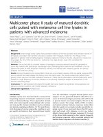
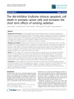
In VCaP cells cultured in DHT-added media, the transcription level of KLK3/PSA with quantitative PCR was
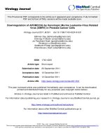
markedly reduced with bicalutamide treatment (0.05fold, Fig. 1); this was also the case with hydroxyflutamide
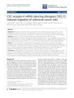
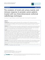
treatment (0.04-fold, Fig. 2). In LNCaP cells pretreated
with DHT, KLK3/PSA transcription was downregulated
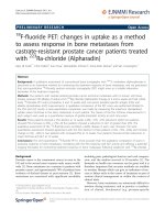
with bicalutamide treatment (0.28-fold, Fig. 3), while
hydroxyflutamide treatment clearly increased the transcription level of KLK3/PSA (2.90-fold, Fig. 4).
In VCaP cells cultured in DHT-added media, bicalutamide and hydroxyflutamide treatment significantly upregulated the transcription of AR (3.39- and 2.51-fold,
respectively) (Figs. 1 and 2). In LNCaP cells pretreated
with DHT, bicalutamide treatment slightly increased
AR transcription (1.24-fold, Fig. 3), whereas AR
Takeda et al. BMC Cancer (2016) 16:332
Page 4 of 8
Fig. 1 Alterations in transcription of androgen receptor (AR), NCOAs,
NCORs, and human kallikrein 3/prostate-specific antigen (KLK3) in
VCaP cells pretreated with dihydrotestosterone (DHT) and thereafter
cultured with the addition of bicalutamide (BC). Relative mRNA
expression levels were assessed in comparison with those in cells
treated with DHT alone
Fig. 3 Alterations in transcription of androgen receptor (AR), NCOAs,
NCORs, and human kallikrein 3/prostate-specific antigen (KLK3) in
LNCaP cells pretreated with dihydrotestosterone (DHT) and
thereafter cultured with the addition of bicalutamide (BC). Relative
mRNA expression levels were assessed in comparison with those in
cells treated with DHT alone. *P < 0.01 (Kruskal-Wallis test)
transcription decreased with hydroxyflutamide (0.44fold, Fig. 4).
and NCOR2 did not show an increase in the transcription level.
Impact of bicalutamide or hydroxyflutamide treatment on
the transcription of NCOA and nuclear receptor
corepressor (NCOR) families
Influence of knock-down of NCOA2 in LNCaP cells on the
production of KLK3/PSA
Transcriptions of NCOA2 and NCOA7 were also elevated with bicalutamide (1.96- and 2.42-fold, respectively, Fig. 1) and hydroxyflutamide (1.33-fold in both,
Fig. 2) in VCaP cells pretreated with DHT. LNCaP cells
pretreated with DHT receiving bicalutamide showed a
4-fold increase in the expression of NCOA2 mRNA
compared with those cultured with DHT pretreatment
alone (Fig. 3); transcription levels of other coactivators
such as NCOA1, NCOA3, and NCOA4 did not increase
with bicalutamide. Also, corepressors such as NCOR1
We also established knock-down models of NCOA2
in LNCaP cells with transcriptional silencing using
small interfering RNA (siRNA). Transcription levels
of NCOA2 were significantly reduced compared with
negative controls (Fig. 5). Thereafter, knock-down of
NCOA2 with siRNA showing most efficient silencing
was performed in LNCaP cells pretreated with DHT;
the KLK3/PSA concentration in media increased
compared with that in non-silencing controls (Fig. 6,
P =0.0495).
Fig. 2 Transcriptional alterations of androgen receptor (AR), NCOAs,
NCORs, and human kallikrein 3/prostate-specific antigen (KLK3) in
VCaP cells pretreated with dihydrotestosterone (DHT) and
subsequently cultured with the addition of hydroxyflutamide (HF).
Relative mRNA expression levels were evaluated by comparing those
in cells treated with DHT alone
Fig. 4 Transcriptional alterations of androgen receptor (AR), NCOAs,
NCORs, and human kallikrein 3/prostate-specific antigen (KLK3) in
LNCaP cells pretreated with dihydrotestosterone (DHT) and
subsequently cultured with the addition of hydroxyflutamide (HF).
Relative mRNA expression levels were evaluated in comparison with
those in cells treated with DHT alone
Takeda et al. BMC Cancer (2016) 16:332
Fig. 5 The efficacy of siRNA for silencing NCOA2 in transcriptions.
1.00 ± 0.04 (control) vs. 0.29 ± 0.01, P = 0.0495
Protein expressions of NCOA2 in LNCaP cells cultured
with DHT and bicalutamide
With Western blotting, LNCaP cells pretreated with
DHT showed an increased protein level of NCOA2 with
bicalutamide than those without (Fig. 7).
Alterations in cell proliferation of LNCaP with silencing of
NCOA2 or AR
There was no difference among proliferations in the
NCOA2-silencing, AR-silencing, and non-silencing cells
cultured with dihydrotestosterone alone (Fig. 8). Relative
absorbance with MTT assay in LNCaP cells cultured
with DHT plus bicalutamide was shown in Fig. 9. Cells
with NCOA2-silencing showed a higher proliferation activity compared with non-silencing control cells and
those with AR-silencing.
Fig. 6 Impact of knock-down of NCOA2 using siRNA on the production of prostate-specific antigen (PSA) in LNCaP cells cultured with
dihydrotestosterone (DHT) plus bicalutamide (BC) (left columns) and
those treated with DHT alone. *101.6 ± 4.2 vs. 87.8 ± 1.4 ng/mL,
respectively, P = 0.0495, **144 ± 4.4 vs. 145 ± 5.6 ng/mL,
respectively, n.s
Page 5 of 8
Fig. 7 Protein expression levels of NCOA2 with Western Blot in
LNCaP cells cultured with dihydrotestosterone (DHT) plus
bicalutamide (BC). The protein level of NCOA2 increased with
compared to that without BC
Discussion
LNCaP and VCaP cells have mutated and wild-type AR,
respectively [20, 21]. Hydroxyflutamide has an agonistic
effect on LNCaP cells, and bicalutamide serves as an antagonist against them [22, 23]. For VCaP cells, the effect
of these antiandrogens has not been determined. In the
current study, both bicalutamide and hydroxyflutamide
treatments reduced KLK3/PSA transcription in VCaP
cells (Figs. 1 and 2), while the former decreased and the
latter increased KLK3/PSA transcription in LNCaP
(Figs. 3 and 4), suggesting the different nature of AR in
the response to antiandrogens between the two cell
types. The transcriptional regulation in AR in response
to antiandrogens also differed between VCaP and
LNCaP cells (Figs. 1, 2, 3 and 4), but bicalutamide inhibited KLK3/PSA transcription in both VCaP and LNCaP
cells to a similar extent.
Fig. 8 Relative absorbance with MTT assay in LNCaP cells cultured
with dihydrotestosterone. There was no difference among
proliferations in the NCOA2-silencing, AR-silencing, and
non-silencing cells
Takeda et al. BMC Cancer (2016) 16:332
Fig. 9 Relative absorbance with MTT assay in LNCaP cells cultured
with dihydrotestosterone (DHT) plus bicalutamide (BC). NCOA2silencing cells showed an increased proliferation compared with nonsilencing control cells and those with silencing androgen receptor (AR).
LNCaP cells with silencing AR showed decreased proliferation
compared with those silencing NCOA2. *P < 0.01 (Kruskal-Wallis test)
The present study verified that the absence of DHT
negated the role of antiandrogens; transcriptions of AR,
NCOAs, and KLK3/PSA were not altered by antiandrogens in the androgen-deprived milieu, except for KLK3/
PSA being increased by hydroxyflutamide. In the absence of endogenous ligands, AR is segregated in the
cytoplasm, with its nuclear localization sequence (NLS)
masked by heat-shock proteins. Binding to ligands and
the separation of these chaperones cause AR to
dimerize, and lead to conformational changes and the
exposure of the NLS [24–27]. Thus, the nuclear translocation of AR and binding to androgen response elements (AREs) activate androgen-responsive genes. This
process is transcriptionally regulated and posttranscriptionally modified through various mechanisms
involving interactions with multiple coactivator and corepressor proteins [28–30]. AR activity is modulated by
the recruitment of multitudes of positive and negative
cofactors, being closely associated with the regulation of
protein stability, interaction with others, intracellular receptor localization, and alteration of the AR structure.
Thus, the elucidation of cofactor-related modifications
of AR may be a promising approach to develop novel
and efficient therapeutic options.
In our study, the transcription level of NCOA2 increased with bicalutamide treatment in LNCaP cells
(Figs. 3 and 7); transcription levels of other coactivators
were not altered. On the other hand, such upregulation
of NCOA2 was not marked in VCaP, and the difference
may possibly be due to the aforementioned issues in AR
and the response to antiandrogens being different between the 2 types of cell. Moreover, knock-down of
NCOA2 in LNCaP cells increased the KLK3/PSA concentration in media (Figs. 5 and 6), and cell proliferation
analysis with MTT assay further supported the mentioned
Page 6 of 8
novel finding (Figs. 8 and 9); NCOA2 is possibly associated with the downregulation of AR signaling in prostate
cancer cells treated with bicalutamide. In the presence of
bicalutamide, interestingly, knock-down of NCOA2 was
associated with a higher cell proliferation than knockdown of AR. These results suggest that NCOA2 plays an
inhibitory role in prostate cancer cells treated with bicalutamide. Steroid receptor coactivators represented by
NCOA2 and NCOA3 are key regulators, having multiple
effects, of transcription factors necessary for cancer cell
proliferation, survival, and metastasis [31]. Their overexpression and/or overactivation has been shown in a number
of human cancers with various genomic, transcriptional,
and posttranslational mechanisms, and are associated with
refractory disease leading to poor outcomes [31]. In human
prostate cancer, NCOAs have been reported to regulate cell
proliferation and invasion and be involved in castration resistance, coupled with AR transcriptional activity [32–34].
NCOA2 has therefore been shown to play a role as a coactivator in the androgen-AR interaction [35–38]; however,
there has been no study examining the function of NCOAs
in prostate cancer cells treated with antiandrogens. The
current results suggest that NCOA2 may possibly explain
the reduction of PSA on the withdrawal or conversion of
antiandrogens; it possibly also serves as a corepressor in the
presence of antiandrogens in prostate cancer cells pretreated with androgens. It is thus necessary to characterize
the functional and recruitment profile of each cofactor-AR
complex and signaling in prostate cancer in accordance
with antagonists as well as agonists.
The present study had several limitations. With the
current cell lines and hormonal milieu, cell proliferation
assays lack in reproducibility. Knock-down studies with
siRNA require further verification of the response, and
the application of different siRNAs is necessary to rule
out unrelated effects.
Conclusions
Although the recruitment of NCOA2 has been thought
to induce agonistic signaling in AR, the current study
showed that it possibly also serves as a corepressor in
the presence of antiandrogens in prostate cancer cells
cultured in a physiological hormonal milieu, suggesting
its potential as a therapeutic target for prostate cancer.
Characterization of the function and recruitment profile
of each cofactor related to AR signaling brought about
by various AR ligands may lead to advanced therapy for
men with prostate cancer.
Additional files
Additional file 1: Figure S1. Screening of the siRNA efficacy on target
mRNA expression levels. Catalog numbers were s20580, s20581, and
s20582 (Neon Transfection System; Life Technologies, Carlsbad, CA, USA)
Takeda et al. BMC Cancer (2016) 16:332
for siRNA NCOA2 #1 (siNCOA2 #1), #2 (siNCOA2 #2), and #3 (siNCOA2 #3),
respectively. Based on this result, siRNA NCOA2 #2 was selected, and the
relevant data were indicated in Fig. 5. (DOC 54 kb)
Additional file 2: Table S1. Ct values of gene expression assessed by
quantitative PCR in VCaP cells cultured with dihydrotestosterone-added
media. Delta-delta Ct value was calculated from the following formula:
delta(target gene Ct – internal control Ct) with specific cell culture media
- delta(target gene Ct – internal control Ct) with standard cell culture
media. (DOC 32 kb)
Additional file 3: Table S2. Ct values of quantitative PCR in VCaP cells
cultured with dihydrotestosterone- and bicalutamide-added media. (DOC
31 kb)
Additional file 4: Table S3. Ct values of quantitative PCR in VCaP cells
cultured with dihydrotestosterone- and hydroxyflutamide-added media.
(DOC 31 kb)
Additional file 5: Table S4. Ct values of quantitative PCR in LNCaP cells
cultured with dihydrotestosterone-added media. (DOC 31 kb)
Additional file 6: Table S5. Ct values of quantitative PCR in LNCaP cells
cultured with dihydrotestosterone- and bicalutamide-added media. (DOC
31 kb)
Additional file 7: Table S6. Ct values of quantitative PCR in LNCaP cells
cultured with dihydrotestosterone- and hydroxyflutamide-added media.
(DOC 31 kb)
Additional file 8: Figure S2. Full membranes of Western blotting
(corresponding to Fig. 7). The membrane in the left panel showed
NCOA2 staining part, and the right showed actin beta staining part.
Starting from the left lane, NCOA2 expressions in LNCaP with
bicalutamide and without bicalutamide, and positive control were
shown. Twenty-seven ug of Jurkat cell lysate (Catalog #611451, BD Biosciences, Franklin Lakes, NJ, USA) was used as positive control. Densitometry
data of Western blotting were attached below them. (DOC 1266 kb)
Abbreviations
ADT, androgen deprivation therapy; AR, androgen receptor; ARE, androgen
response element; CRPC, castration-resistant prostate cancer; DHT, dihydrotestosterone; FBS, fetal bovine serum; KLK, human kallikrein; NCOA, nuclear
receptor coactivator; NCOR, nuclear receptor corepressor; PSA, prostatespecific antigen; siRNA, small interfering RNA
Acknowledgements
The authors thank Dr. T. Hoshii for assistance in preliminary studies and
analyses.
Funding
It was supported in part by a Grant-in-Aid-for Scientific Research from the
Ministry of Education, Culture, Sports, Science and Technology, Japan
(#21791493 and #24592380).
Availability of data and materials
The datasets supporting the conclusions of this article are included within
the article and its additional files (Additional files 1, 2, 3, 4, 5, 6, 7 and 8).
Authors’ contributions
Conception and initiation of the study was performed by TN and KT. KT, MT,
and TN performed the experimental assays, and KT and NH conducted data
analyses. TN, FI and NH participated in the design of the study, and KT and
FI performed the statistical analysis. KT and NH wrote the manuscript, and YT
helped to draft it. NH supervised all through the study. All authors read and
approved the final version of the manuscript.
Competing interests
This work has not been funded by any commercial company. The authors
declare that they have no competing interests.
Consent for publication
Not applicable.
Page 7 of 8
Ethics approval and consent to participate
Not applicable.
Author details
Division of Urology, Department of Regenerative and Transplant Medicine,
Graduate School of Medical and Dental Sciences, Niigata University, Niigata,
Japan. 2Division of Molecular Oncology, Department of Signal Transduction
Research, Graduate School of Medical and Dental Sciences, Niigata
University, Niigata, Japan. 3Asahimachi 1, Niigata 951-8510, Japan.
1
Received: 7 August 2015 Accepted: 23 May 2016
References
1. Ryan CJ, Tindall DJ. Androgen receptor rediscovered: the new biology and
targeting the androgen receptor therapeutically. J Clin Oncol.
2011;29:3651–8.
2. George D, Moul JW. Emerging treatment options for patients with
castration-resistant prostate cancer. Prostate. 2012;72:338–49.
3. Massard C, Fizazi K. Targeting continued androgen receptor signaling in
prostate cancer. Clin Cancer Res. 2011;17:3876–83.
4. van Poppel H, Klotz L. Gonadotropin-releasing hormone: an update review
of the antagonists versus agonists. Int J Urol. 2012;19:594–601.
5. Michaelson MD, Cotter SE, Gargollo PC, Zietman AL, Dahl DM, Smith MR.
Management of complications of prostate cancer treatment. CA Cancer J
Clin. 2008;58:196–213.
6. Gilbert SM, Kuo YF, Shahinian VB. Prevalent and incident use of androgen
deprivation therapy among men with prostate cancer in the United States.
Urol Oncol. 2011;29:647–53.
7. Mostaghel EA, Montgomery B, Nelson PS. Castration-resistant prostate
cancer: targeting androgen metabolic pathways in recurrent disease. Urol
Oncol. 2009;27:251–7.
8. Osanto S, van Poppel H. Emerging novel therapies for advanced prostate
cancer. Ther Adv Urol. 2012;4:3–12.
9. Stein MN, Goodin S, Dipaola RS. Abiraterone in prostate cancer: a new
angle to an old problem. Clin Cancer Res. 2012;18:1848–54.
10. Zhang C, Wang L, Wu D, Chen H, Chen Z, Thomas-Ahner JM, et al.
Definition of a FoxA1 Cistrome that is crucial for G1 to S-phase cell-cycle
transit in castration-resistant prostate cancer. Cancer Res. 2011;71:6738–48.
11. Zhong WD, Qin GQ, Dai QS, Han ZD, Chen SM, Ling XH, et al. SOXs in
human prostate cancer: implication as progression and prognosis factors.
BMC Cancer. 2012;12:248.
12. Fujimoto N, Miyamoto H, Mizokami A, Harada S, Nomura M, Ueta Y, et al.
Prostate cancer cells increase androgen sensitivity by increase in nuclear
androgen receptor and androgen receptor coactivators; a possible
mechanism of hormone-resistance of prostate cancer cells. Cancer Invest.
2007;25:32–7.
13. Okihara K, Ukimura O, Kanemitsu N, Mizutani Y, Kawauchi A, Miki T, et al.
Clinical efficacy of alternative antiandrogen therapy in Japanese men with
relapsed prostate cancer after first-line hormonal therapy. Int J Urol.
2007;14:128–32.
14. Suzuki H, Okihara K, Miyake H, Fujisawa M, Miyoshi S, Matsumoto T, et al.
Alternative nonsteroidal antiandrogen therapy for advanced prostate cancer
that relapsed after initial maximum androgen blockade. J Urol.
2008;180:921–7.
15. Okegawa T, Nutahara K, Higashihara E. Alternative antiandrogen therapy in
patients with castration-resistant prostate cancer: a single-center experience.
Int J Urol. 2010;17:950–5.
16. Chen CD, Welsbie DS, Tran C, Baek SH, Chen R, Vessella R, et al. Molecular
determinants of resistance to antiandrogen therapy. Nat Med. 2004;10:33–9.
17. Kenneth JL, Thomas DS. Analysis of relative gene expression data using realtime quantitative PCR and the 2-ΔΔCT method. Methods. 2001;25:402–8.
18. Rodriguez C, Hughes-Davies L, Vallès H, Orsetti B, Cuny M, Ursule L, et al.
Amplification of the BRCA2 pathway gene EMSY in sporadic breast cancer is
related to negative outcome. Clin Cancer Res. 2004;17:5785–91.
19. Lubik AA, Gunter JH, Hendy SC, Locke JA, Adomat HH, Thompson V, et al.
Insulin increases de novo steroidogenesis in prostate cancer cells. Cancer
Res. 2011;71:5754–64.
20. Veldscholte J, Ris-Stalpers C, Kuiper GG, Jenster G, Berrevoets C, Claassen E,
et al. A mutation in the ligand binding domain of the androgen receptor of
Takeda et al. BMC Cancer (2016) 16:332
21.
22.
23.
24.
25.
26.
27.
28.
29.
30.
31.
32.
33.
34.
35.
36.
37.
38.
Page 8 of 8
human LNCaP cells affects steroid binding characteristics and response to
anti-androgens. Biochem Biophys Res Commun. 1990;173:534–40.
Korenchuk S, Lehr JE, Mclean L, Lee YG, Whitney S, Vessella R, et al. VCaP, a
cell-based model system of human prostate cancer. In Vivo. 2001;15:163–8.
Wang LG, Liu XM, Kreis W, Budman DR. Phosphorylation/dephosphorylation
of androgen receptor as a determinant of androgen agonistic or
antagonistic activity. Biochem Biophys Res Commun. 1999;259:21–8.
Wilding G, Chen M, Gelmann EP. Aberrant response in vitro of hormoneresponsive prostate cancer cells to antiandrogens. Prostate. 1989;14:103–15.
Prescott J, Coetzee GA. Molecular chaperones throughout the life cycle of
the androgen receptor. Cancer Lett. 2006;231:12–9.
Mangelsdorf DJ, Thummel C, Beato M, Herrlich P, Schutz G, Umesono K, et
al. The nuclear receptor superfamily: the second decade. Cell.
1995;83:835–9.
Kaku N, Matsuda K, Tsujimura A, Kawata M. Characterization of nuclear
import of the domain-specific androgen receptor in association with the
importin alpha/beta and Ran-guanosine 5′-triphosphate systems.
Endocrinology. 2008;149:3960–9.
Cutress ML, Whitaker HC, Mills IG, Stewart M, Neal DE. Structural basis for
the nuclear import of the human androgen receptor. J Cell Sci.
2008;121:957–68.
Gottlieb B, Pinsky L, Beitel LK, Trifiro M. Androgen insensitivity. Am J Med
Genet. 1999;89:210–7.
Shiota M, Song Y, Yokomizo A, Tada Y, Kuroiwa K, Eto M, et al. Human
heterochromatin protein 1 isoform HP1beta enhances androgen receptor
activity and is implicated in prostate cancer growth. Endocr Relat Cancer.
2010;17:455–67.
Shiota M, Yokomizo A, Tada Y, Inokuchi J, Tatsugami K, Kuroiwa K, et al.
Peroxisome proliferator-activated receptor gamma coactivator-1alpha
interacts with the androgen receptor (AR) and promotes prostate cancer
cell growth by activating the AR. Mol Endocrinol. 2010;24:114–27.
Xu J, Wu RC, O’Malley BW. Normal and cancer-related functions of the p160
steroid receptor co-activator (SRC) family. Nat Rev Cancer. 2009;9:615–30.
Otsuka T, Iguchi K, Fukami K, Ishii K, Usui S, Sugimura Y, et al. Androgen
receptor W741C and T877A mutations in AIDL cells, an androgenindependent subline of prostate cancer LNCaP cells. Tumor Biol.
2011;32:1097–102.
Zhou HJ, Yan J, Luo W, Ayala G, Lin SH, Erdem H, et al. SRC-3 is required for
prostate cancer cell proliferation and survival. Cancer Res. 2005;65:7976–83.
Yan J, Erdem H, Li R, Cai Y, Ayala G, Ittmann M, et al. Steroid receptor
coactivator-3/AIB1 promotes cell migration and invasiveness through focal
adhesion turnover and matrix metalloproteinase expression. Cancer Res.
2008;68:5460–8.
Taylor BS, Schultz N, Hieronymus H, Gopalan A, Xiao Y, Carver BS, et al.
Integrative genomic profiling of human prostate cancer. Cancer Cell.
2010;18:11–22.
Rosales T, Georget V, Malide D, Smirnov A, Xu J, Combs C, et al. Quantitative
detection of the ligand-dependent interaction between the androgen
receptor and the co-activator, Tif2, in live cells using two color, two photon
fluorescence cross-correlation spectroscopy. Eur Biophys J. 2007;36:153–61.
Shi XB, Xue L, Shi D, de Vere WRW. Influence of short polyglutamine tracts
and p160 coactivators on the transactivation of the androgen receptor.
Cancer Biother Radiopharm. 2011;26:191–201.
Kino T, Ichijo T, Chrousos GP. FLASH interacts with p160 coactivator
subtypes and differentially suppresses transcriptional activity of steroid
hormone receptors. J Steroid Biochem Mol Biol. 2004;92:357–63.
Submit your next manuscript to BioMed Central
and we will help you at every step:
• We accept pre-submission inquiries
• Our selector tool helps you to find the most relevant journal
• We provide round the clock customer support
• Convenient online submission
• Thorough peer review
• Inclusion in PubMed and all major indexing services
• Maximum visibility for your research
Submit your manuscript at
www.biomedcentral.com/submit