METCAM/MUC18 is a novel tumor and metastasis suppressor for the human ovarian cancer SKOV3 cells
Bạn đang xem bản rút gọn của tài liệu. Xem và tải ngay bản đầy đủ của tài liệu tại đây (2.78 MB, 15 trang )
Wu and Zeng BMC Cancer (2016) 16:136
DOI 10.1186/s12885-016-2181-9
RESEARCH ARTICLE
Open Access
METCAM/MUC18 is a novel tumor and
metastasis suppressor for the human
ovarian cancer SKOV3 cells
Guang-Jer Wu1,2,3* and Guo-fang Zeng1,4
Abstract
Background: Increased expression of METCAM/MUC18, a trans-membrane cell adhesion molecule in the Ig-like
gene superfamily, has been associated with the malignant progression of epithelial ovarian carcinomas. To investigate
if this is a fortuitous correlation or if METCAM/MUC18 actually plays a role in the progression of the cancer, we tested
effects of enforced expression of METCAM/MUC18 on in vitro behaviors, in vivo tumorigenesis, and in vivo malignant
progression of human ovarian cancer SK-OV-3 cells, which minimally expressed this protein.
Methods: For in vitro and in vivo tests, we transfected human METCAM/MUC18 cDNA gene into SK-OV-3 cells in a
mammalian expression vector pcDNA3.1+ and obtained G418-resistant (G418R) clones, which expressed various levels
of human METCAM/MUC18. To mimic physiological situations, we used pooled METCAM/MUC18-expressing and control
(vector) clones for testing effects of human METCAM/MUC18 over-expression on in vitro motility and invasiveness, and on
in vivo tumor formation and metastasis in female athymic nude mice. Effects of METCAM/MUC18 on the expression of
various downstream key factors related to tumorigenesis were also evaluated by Western blot analyses.
Results: The over-expression of METCAM/MUC18 inhibited in vitro motility and invasiveness of SK-OV-3 cells. SK-OV-3
cells of the control (vector) clone (3D), which did not express human METCAM/MUC18, supported the formation
of a solid tumor after SC injection of the cells at dorsal or ventral sites and also formation of solid tumor and ascites after
IP injection in the intraperitoneal cavity of nude mice. In contrast, SK-OV-3 cells from the METCAM/MUC18-expressing
clone (2D), which expressed a high level of METCAM/MUC18, did not support the formation of a solid tumor at
SC sites, or formation of ascites in the intraperitoneal cavity of nude mice. Expression levels of downstream key
factors, which may affect tumor proliferation and angiogenesis, were reduced in tumors induced by the
METCAM/MUC18-expressing clone (2D).
Conclusions: We conclude that increased human METCAM/MUC18 expression in ovarian cancer SK-OV-3 cells
suppressed tumorigenesis and ascites formation in nude mice, suggesting that human METCAM/MUC18 plays a
suppressor role in the progression of ovarian cancer, perhaps by reducing proliferation and angiogenesis.
Keywords: Human METCAM/MUC18 expression, Ovarian cancer SKOV3 cells, SC & IP injections, Tumorigenesis
and progression, Athymic nude mice
* Correspondence:
1
Department of Microbiology and Immunology, Emory University School of
Medicine, Atlanta, GA 30322, USA
2
Department of Bioscience Technology, Chung Yuan Christian University,
Chung Li 32023, Taiwan
Full list of author information is available at the end of the article
© 2016 Wu and Zeng. Open Access This article is distributed under the terms of the Creative Commons Attribution 4.0
International License ( which permits unrestricted use, distribution, and
reproduction in any medium, provided you give appropriate credit to the original author(s) and the source, provide a link to
the Creative Commons license, and indicate if changes were made. The Creative Commons Public Domain Dedication waiver
( applies to the data made available in this article, unless otherwise stated.
Wu and Zeng BMC Cancer (2016) 16:136
Background
Epithelial ovarian cancer (EOC) is the fifth leading cause
of female cancers in USA with a high fatality rate (about
65 %) [1]. The high lethality of the cancer is because the
early stage of the disease is mostly asymptomatic and
therefore remains undiagnosed until the cancer has
already disseminated throughout the peritoneal cavity
[2]. The early stage disease can be treated successfully,
however, effective therapy for the advanced-stage disease
is lacking because of the strong chemo-resistance of recurrent ovarian cancer [2]. The major challenges for
combating ovarian cancer are: (a) the ovarian cancer is
histologically and molecularly heterogeneous with at
least four major subtypes [3, 4], (b) there is a lack of reliable specific diagnostic markers for an effective early
diagnosis of each subtype, though molecular signatures
of the major subtypes are available [5], and (c) very little
is known of how ovarian tumor emerges and how it progresses to malignancy ([6] for a review).
In general, tumorigenesis is a complex process involving
changes of several biological characteristics [7], including
the aberrant expression of cell adhesion molecules [8].
Tumor progression is induced by a complex cross-talk between tumor cells and stromal cells in the surrounding tissues [8]. These interactions are, at least in part, mediated
by cell adhesion molecules (CAMs), which govern the social behaviors of cells by affecting the adhesion status of
cells and cross-talk and modulating intracellular signal
transduction pathways [8]. Thus the altered expression of
CAMs can change motility and invasiveness, affect survival and growth of tumor cells, and alter angiogenesis [8].
As such, CAMs may promote or suppress the metastatic
potential of tumor cells [9]. Aberrant expression of various
CAMs, such as mucins [10], integrins [11], CD44 [12],
L1CAM [13], E-cadherin [14], claudin-3 [15], EpCAM
[16], and METCAM/MUC18 [17, 18], has been associated
with the malignant progression of ovarian cancer.
We have been focusing our studies on the possible
role of METCAM/MUC18 in the progression of several
epithelial tumors [19]. Human METCAM/MUC18 (or
MCAM, Mel-CAM, S-endo1, or CD146), an integral
membrane cell adhesion molecule (CAM) in the Ig-like
gene superfamily, has an N-terminal extra-cellular domain of 558 amino acids, a transmembrane domain, and
a short intra-cellular cytoplasmic domain (64 amino acids)
at the C-terminus [19, 20]. The extra-cellular domain of the
protein comprises a signal peptide sequence and five
immunoglobulin-like domains and one X domain [19, 20].
The cytoplasmic domain contains five consensus sequences
potentially to be phosphorylated by PKA, PKC, and CK2
[19, 20]. Thus human METCAM/MUC18 is capable of performing typical functions of CAMs, such as governing the
social behaviors by affecting the adhesion status of cells and
modulating cell signaling. Therefore, an altered expression
Page 2 of 15
of METCAM/MUC18 may affect motility and invasiveness
of many tumor cells in vitro and tumorigenesis and metastasis in vivo [19].
Human METCAM/MUC18 is only expressed in several normal tissues, such as hair follicular cells, smooth
muscle cells, endothelial cells, cerebellum, normal mammary epithelial cells, basal cells of the lung, activated T
cells, and intermediate trophoblasts [19, 21]. Human
METCAM/MUC18 is also expressed in several epithelial
tumors, such as melanoma, prostate cancer, osteosarcoma, breast carcinoma, and intermediate trophoblast
tumors [19, 21]. Over-expression of METCAM/MUC18
promotes the tumorigenesis of prostate cancer [22] and
breast carcinoma [23, 24], but it has a minimal effect on
the tumorigenesis of melanoma [25]. Over-expression of
METCAM/MUC18 also initiates the metastasis of prostate cancer [26] and promotes the metastasis of melanoma
[25] and breast carcinoma [27].
On the contrary, the possibility that the overexpression of METCAM/MUC18 might play a tumor
suppressor role was first suggested by Shih et al. [28],
who found that METCAM/MUC18 expression suppressed tumorigenesis of a breast cancer cell line MCF-7
in SCID mice. However, this notion was contradicted by
recently published evidence, which supported the positive role of METCAM/MUC18 in the progression of
breast cancer cells [23, 24, 27], similar to its role in the
progression of melanoma and prostate cancer cells.
The role of METCAM/MUC18 in the progression of
ovarian cancer has not been well studied, except that the
METCAM/MUC18 expression has been recently reported
to correlate with the progression of ovarian cancer
[17, 18], and perhaps affects the behaviors of ovarian
cancer cells [29]. To directly test the role of METCAM/
MUC18 in the progression of epithelial ovarian cancer, we
first chose to use SK-OV-3 cells for testing the effect of
over-expression of METCAM/MUC18 on in vitro motility
and invasiveness, in vivo tumor formation in nude mice
after subcutaneous (SC) injection, and in vivo progression
in nude mice after intraperitoneal (IP) injection. We found
that the over-expression of METCAM/MUC18 inhibited
in vitro motility and invasiveness and suppressed in vivo
tumorigenesis and the malignant progression of the human ovarian cancer cell line SK-OV-3. We conclude that
METCAM/MUC18 is a novel tumor and metastasis suppressor for the progression of human ovarian cancer cells.
Methods
Cell lines and culture
SK-Mel-28, a human melanoma cell line from ATCC,
which was maintained in EMEM supplemented with
1 mM Na.pyruvate, extra nonessential amino acids and
vitamins, and 10 % fetal bovine serum (FBS), was used as
a positive control (100 %) for the expression of human
Wu and Zeng BMC Cancer (2016) 16:136
METCAM/MUC18. LNCaP, a human prostate cancer cell
line from ATCC, which was maintained in modified RPMI
1640 medium supplemented with 25 mM HEPES, 1 mM
Na.pyruvate, 1 mM glutamine, and 10 % FBS, was used as
a negative control (0 %) for the expression of human
METCAM/MUC18. Human ovarian cancer cell lines,
CAOV3, SK-OV-3, and NIHOVCAR3, were from ATCC.
CAOV3, which was established from human primary
ovarian adenocarcinoma, was maintained in DMEM
(4.5 g/L of glucose) and 10 % FBS. SK-OV-3, which was
established from malignant ascites of human ovarian
adenocarcinoma, was maintained in McCoy’s 5A medium
with 10 % FBS. NIHOVCAR3, which was established from
malignant ascites of human ovarian progressive adenocarcinoma, was maintained in modified RPMI medium-4.5 g/
L glucose-1 mM Na.pyruvate, 10 μg/ml insulin, and 20 %
FBS. IOSE from Dr. Nelly Auesperg, Vancouver, Canada,
which was a normal human ovarian surface epithelial cell
line immortalized by the SV40 virus large T antigen [30],
was maintained in M199/MCDB105 (1:1) medium with
15 % FBS and 50 μg/ml of gentamicin. BG-1 from Drs.
Erin Dickerson and Nathan Bowen at Georgia Institute of
Technology, Atlanta, GA, which was established from
poorly differentiated human primary ovarian adenocarcinoma [31], was maintained in DMEM/F12 with 10 % FBS.
HEY from Dr. Gordon Mills at M.D. Anderson Cancer
Center, Houston, TX, which was established from a mouse
xenograft of human primary ovarian adenocarcinoma
[32], was maintained in a modified RPMI 1640 medium
supplemented with 25 mM HEPES, 1 mM Na.pyruvate,
1 mM glutamine, 4.5 g/L glucose and 10 % FBS. All the
SK-OV-3 clones were maintained in the McCoy’s 5A
medium with 10 % FBS plus 0.5 mg/ml of G418. All media
were from Invitrogen/Life Technology/GIBCO/BRL. FBS
was from Cellgro/MediaTech. All the cell lines and
SK-OV-3 clones were maintained in a humidified 37 ° C
incubator with 5 % CO2.
Page 3 of 15
sequentially from 24-well to 12-well and 6-well culture
plates. Cell lysate of each clone grown in each well of
6-well plates was made by addition of 100 μl of Western
blot lysis buffer [22–24] and processed for Western
blot analysis [22–24]. Liquid-nitrogen-frozen stocks of
the METCAM/MUC18-expressing clones (METCAM/
MUC18 clones) and the control (vector) clones were
made from duplicated 6-well plates. The single METCAM/
MUC18-expressing clones were designated as METCAM/
MUC18 clone 2D-1 to 2D-12 (or abbreviated as METCAM
clone 2D-1 to 2D-12). After single colonies were picked,
the remaining colonies in the plates were treated with
trypsin and pooled together, and seeded to duplicate T25 flasks. After growth, cells from the pooled clones in
one flask were frozen and designated either as METCAM/
MUC18 clone 2D (or abbreviated as METCAM clone 2D)
or control (vector) clone 3D, and those in another flask
were made Western blot lysate, designated as cell lysate of
METCAM clone 2D or control (vector) clone 3D.
Cell motility assay
The in vitro cell motility assay was carried out [23–26].
2 × 10⁵ cells of the METCAM clone 2D or the control
(vector) clone 3D of SK-OV-3 cells in 0.4 ml of growth
medium containing 0.1 %-BSA were seeded to each of
the top insert with 8.0 μm pore size of the polycarbonate
membrane (Fisher #08-771-12 or Falcon 35-3182) that
fits into the bottom wells of a companion 12-well plate
of the Boyden type Transwell system (Fisher #08-771-22
or Falcon 35-3503). Each bottom-well was added 1.1 ml
of regular growth medium containing 10 % FBS. After
6 h, cells migrating to the bottom wells were treated
with trypsin, concentrated by centrifugation, and counted
with a hemocytometer [23–26]. The mean value and the
standard deviation of three measurements of cell numbers
migrated to bottom wells were calculated and presented.
Cell invasiveness assay
Lipofection of SK-OV-3 cells and selection for human
METCAM/MUC18-expressing clones
1 × 10⁶ of SKOV3 cells per well were seeded (about 60 %
confluence) in 6-well plates 1 day before lipofection.
Lipofection was carried out with a mixture in 2 ml of
Opti-MEM containing 12 μg of DEMRIE-C, or 6 μg of
FuGene HD (Cat.no.04-709-691-001, Roche), and 2 μg
each of the plasmid pcDNA3.1+ with or without the human METCAM/MUC18 cDNA gene for 6 h at 37 C. At
the end of lipofection, 0.2 ml FBS was added to make
the final serum concentration to 10 %. After cultured for
two more days, the transfected cells were split into two
plates containing the growth medium plus 0.5 mg/ml
of G418 (active component 71.3 %). G418-resistant
(G418R)-clones emerged in 2 weeks. Twelve clones from
each lipofection were picked, transferred and expanded
The in vitro cell invasiveness assay was carried out [23–26].
All procedures were similar to the cell motility assay except
each top well (with a pore size of 12 μm) was coated with
150 μg of diluted Matrigel (growth factors-reduced and
phenol-red free grade, BD Biosciences Cat # 354237 or
Collaborative Research Cat. #40234C). After 6 h, cells
migrating to the bottom wells were determined. The
mean value and the standard deviation of three measurements of cell numbers migrated to bottom wells were
calculated and presented.
Determination of tumorigenesis of SK-OV-3 clones/cells at
the subcutaneous (SC) sites of athymic nude mice
All animal studies complying with the Institutional, national and international guidelines were approved by the
Emory University’s animal ethics committee, Institutional
Wu and Zeng BMC Cancer (2016) 16:136
Animal Care and Use Committee (IACUC), with an approval ID of 275-2008 (from 2/16/2009 to 2/16/2011).
Emory’s Animal Welfare Assurance Number is A3180-01.
Ten 33 days-old female athymic nude mice from Harlan
Sprague Dawley Inc. (Indianapolis, Indiana, USA) were
used for SC injection of cells from each clone. A single cell
suspension was made from monolayer cultures of SK-OV-3
clones/cells after trypsin treatment, washed, re-suspended
in PBS (5 × 10⁶ cells/ml), cooled in ice, centrifuged, resuspended in 0.05 ml of cold McCoy’s 5A medium
without FBS, and mixed with an equal volume of
Matrigel (16 mg/ml, Cultrex, Trevigen) to make a final
concentration of 5 × 10⁷ cells per ml and Matrigel at
8 mg/ml [22–25]. 5 × 10⁶ cells of the METCAM clone
2D (p24) and the control (vector) clone 3D (p24) of
SK-OV-3 cells in 0.1 ml were subcutaneously injected
with a gauge #28G1/2 needle into the right dorsal flank
or the right ventral side. After injection, the size of
tumor was weekly measured with a caliper till 40 days.
Tumor volumes were calculated by using the formula
V = π/6 (d1 × d2)3/2 (mm)3 [22–25]. At the endpoint,
mice were euthanatized, tumor from each mouse was
excised, weighed, and a portion was made cell lysate for
Western blot analysis. The rest of the tumor was fixed
in phosphate-buffered 10 % formaldehyde (Fisher), paraffinized, and sectioned for histology and immunohistochemistry staining.
Determination of tumorigenesis and progression of SK-OV-3
clones/cells in the intra-peritoneal cavity of female athymic
nude mice
All animal studies complying with the Institutional, national and international guidelines and were approved by
the Emory University’s animal ethics committee, Institutional Animal Care and Use Committee (IACUC), with an
approval ID of 275-2008 (from 2/16/2009 to 2/16/2011).
Emory’s Animal Welfare Assurance Number is A3180-01.
Five 34 days-old female athymic nude mice from Harlan
Sprague Dawley Inc. were used for IP injection of cells
from each clone [22–26]. A single cell suspension was
made from monolayer cultures of SK-OV-3 clones/cells
after trypsin treatment, washed, re-suspended in PBS
(3 × 10⁷ cells /ml), cooled in ice, centrifuged, and resuspended in 2 ml of cold PBS, and mixed with 1 ml of
cold Matrigel (16 mg/ml, Cultrex, Trevigen) to make a
final concentration of 1 × 10⁷ cells per ml and Matrigel
at 5.55 mg/ml [22–25]. 5 × 10⁶ cells of the METCAM
clone 2D (p19) and the control (vector) clone 3D (p19)
of SK-OV-3 cells in 0.5 ml containing Matrigel were
injected into intra-peritoneal cavity. The formation of
solid tumors and ascites in the abdomen of each mouse
was weekly monitored till the end of the experiments
(10 weeks). After euthanasia, ascites were carefully
withdrawn from abdominal cavities with pipets and
Page 4 of 15
total volumes of ascites were recorded. Ascites were
centrifuged at 700 rpm for 10 min to separate the pelleted cells from the supernatant and collected in new
tubes. The volumes of pelleted cells were also recorded
and lysates made. Solid tumors in the abdominal walls
and cavity were collected, weighed, and recorded. A
portion of solid tumors was made cell lysate for Western blot analysis. The rest of the tumor was fixed in
formaldehyde (Fisher), paraffinized, and sectioned for
histology and immunohistochemistry staining.
Western blot analysis
Lysates from cells grown in monolayers and from tumors were prepared as described [22–26]. Protein concentration of each lysate was determined and verified
as described [22–26]. The expression of METCAM/
MUC18 in the lysates from various cells lines/clones
(5 μg proteins of each lysate) was determined by Western blot (WB) analysis [22–26] by using a chicken antihuman METCAM/MUC18 IgY as the primary antibody
(1/300 dilutions) [22–26]. An AP-conjugated rabbit
anti-chicken IGY (AP162A) from Chemicon (1/2000 dilutions) was used as the secondary antibody. Primary
antibodies for detection of Bcl2 (N-19, SC-492), Bax
(N-20, SC-493), and VEGF (A-20, SC-152) were rabbit
polyclonal antibodies from Santa Cruz Biotech. The rabbit
anti-human LDH-A polyclonal antibody was previously
made in our group [33]. Those for detection of phosphoAKT (Ser473) (D9E, #4060), pan-AKT (C67E7, #4691),
and VEGFR2 (53B11, #24790) were rabbit monoclonal
antibodies from Cell Signaling Technology. The primary
antibody for detection of PCNA (PC-10, SC-56, Santa
Cruz Biotech) was a mouse monoclonal antibody. The
1/2000 dilution of the corresponding AP-conjugated
secondary antibody, goat anti-rabbit antibody (AP132A),
or rabbit anti-mouse antibody (AP160A) from Chemicon,
was used. As the loading controls, the same WB membrane was reacted with three primary antibodies (1/200
dilutions) against three house-keeping genes, such as
actin, β-tubulin, and GAPDH, which were goat polyclonal
antibody (C-11, SC-1615), rabbit polyclonal antibody (H235, SC-9104), and goat polyclonal antibody (SC-20358),
respectively, from Santa Cruz Biotech. The 1/2000 dilution
of AP-conjugated rabbit anti-goat (AP106A) or goat antirabbit (AP132A) antibody from Chemicon was used as the
secondary antibodies. Substrates BCIP/NBT (S3771,
Promega) were used for color development. The image
of the specific protein band corresponding to METCAM/
MUC18, each key downstream parameter, or each of the
three house-keeping genes on the same membrane, was
scanned by an Epson Scanner model 1260 and its intensity
was quantitatively determined by a NIH software program
Image J version 1.31.
Wu and Zeng BMC Cancer (2016) 16:136
Histology and immunohistochemistry (IHC) of the tumor
tissue sections
Paraffin-embedded tissue sections (5 μm) were deparaffinized, rehydrated with graded alcohol and PBS,
and used for histological staining (H&E) and IHC analyses
[22–26]. A tissue section of SC tumors derived from the
human prostate cancer LNCaP-expressing clone (LNS239)
was used as a positive external control for IHC staining
[22]. 1/200 to 1/300 dilution of the chicken antihuMETCAM/MUC18 IGY antibody was used as the
primary antibody and 1/250 dilution of the biotinylated
rabbit anti-chicken IGY antibodies (G2891, Promega)
as the secondary antibody [22–26]. A streptavidin-conjugated horseradish peroxidase complex (Dako LSAAB-2
system) and diaminobenzidine were used for color development. Hematoxylin was used as the counter staining. Negative controls had the primary antibody
replaced by non-fat milk or control chicken IGY.
Statistical analysis of data
All the data were statistically analyzed by the Student’s t
test by using the 1 tailed distribution type1, 2, or 3 method.
Page 5 of 15
Two corresponding sets of data were considered significantly different if the P value was < 0.05.
Results
Expression of METCAM/MUC18 in various human ovarian
cancer cell lines
We initiated the investigation by determining expression
levels of METCAM/MUC18 in several ovarian cancer
cell lines. Figure 1a shows that the expression level of
METCAM/MUC18 in one immortalized normal ovarian
epithelial cell line (IOSE) was about 10 % and that in five
ovarian cancer cell lines, BG-1, HEY, CAOV-3, SK-OV-3
and NIHOVCAR3, ranged from zero to 50 %, assuming
that a positive control, human melanoma cell line SKMel-28, expressed 100 % of METCAM/MUC18. This
provided an important information for us to choose two
cell lines, BG-1 (established from a poorly differentiated
adenocarcinoma) and SK-OV-3 (established from an
adenocarcinoma metastasis as malignant ascites), which
expressed very low levels of METCAM/MUC18 (zero
and 1 %, respectively), for in vitro and in vivo studies. In
this report, we have provided the results of the following
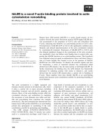
Fig. 1 Expression of METCAM in various human ovarian cancer cell lines (a) and in G418R- clones derived from SK-OV-3 (b). a The expression of
METCAM/MUC18 in the lysates from various cells lines was determined by Western blot (WB) analysis as described in “Methods”. Cell lysate from
a human melanoma cell line, SK-Mel-28, was used as a positive control (lane 1) and those from human ovarian cancer cell lines, BG-1 (lane 3)
and SK-OV-3 (lane 6) as negative controls. METCAM/MUC18 expression levels in cell lysates from one immortalized human ovarian epithelial
cells (IOSE) and in five human ovarian cancer cell lines are shown in lanes 2 to 7. The number under each lane indicates the relative level of
METCAM/MUC18 of each cell line, assuming that in SK-Mel-28 is 100 %. Only the house-keeping genes, actin and GAPDH, are shown here as
the loading controls. b Human METCAM/MUC18 expression in lysates prepared from various clones/cells was determined by Western blot
analysis as described in “Methods”. METCAM/MUC18 expression level in cell lysates from a human melanoma cell line, SK-Mel-28, was used as
a positive control (lane 1) and from the parental human ovarian cancer cell line, SK-OV-3, as a negative control (lane 2). METCAM/MUC18
expression in cell lysates from one single SK-OV-3 clone (METCAM Clone 2D-9) and two pooled SK-OV-3 clones (METCAM Clone 2D and
Control (Vector) Clone 3D) are shown in lanes 3–5. Both the METCAM Clone 2D-9 and the METCAM Clone 2D were derived from SK-OV-3 cells
transfected with the human METCAM/MUC18 cDNA gene. The Control (Vector) Clone 3D was from SK-OV-3 cells transfected with the empty
vector. The number under each lane indicates the relative level of METCAM/MUC18 of each cell line, assuming that in SK-Mel-28 was 100 %.
β-tubulin is shown as the loading control
Wu and Zeng BMC Cancer (2016) 16:136
studies by using the human ovarian cancer cell line, SKOV-3. The results of similar studies by using the BG-1
cell line will be reported elsewhere.
METCAM/MUC18 expression in G418R-clones derived
from SK-OV-3 cells
Since the SK-OV-3 cell line does not express METCAM/
MUC18, to determine if METCAM/MUC18 expression
affects the in vitro and in vivo cellular behaviors of the
cells, it would be desirable to ectopically make SK-OV-3
express the protein by transfecting the cells with the
human METCAM/MUC18 cDNA. To facilitate the expression of the transfected gene, the cDNA is inserted
in a mammalian expressible plasmid vector, pcDNA3.1+,
in which the inserted gene is driven by a strong CMV promoter to facilitate the high expression of the inserted gene
in mammalian cells. Since the pcDNA3.1+ also contains
the cDNA encoding for neomycin (or G418)-resistant
gene, which is driven by the SV40 promoter, the transfected cells should also express the neomycin-resistant
gene and be resistant to the killing of neomycin (G418).
As such, the majority of the cells, which were not successfully transfected with the plasmid, should be killed in the
growth medium containing G418. In contrast, a minority
of the cells, which were successfully transfected with the
plasmid, should be resistant to the killing of G418 and
enriched in the presence of G418; most of them should
also express METCAM/MUC18, albeit at different levels
in different clones. To obtain high expressing clones after
transfecting SK-OV-3 cells with the human METCAM/
MUC18 cDNA, the G418-resistant (G418R)-clones were
selected and the expression level of METCAM/MUC18 in
each clone was determined by Western blot analysis. The
control cells, which were transfected with the empty vector that did not contain the human METCAM/MUC18
cDNA, should not express METCAM/MUC18 similar to
the parental SK-OV-3 cells, even though they were G418R.
We found that DEMRIE-C was an excellent transfecting
reagent, since 2/3 were high-expressing clones. However,
the transfecting reagent of FuGene HD (Roche) was not,
since no high-expressing clones were obtained and 2/3
clones were low-expressing clones and 1/3 mediumexpressing clones. Figure 1b shows that the expression of
METCAM/MUC18 in three typical G418R clones when
DEMRIE-C was used as the transfecting reagent. When
compared to the positive control cell line, human melanoma SK-Mel-28 cells (assuming expression of 100 % of
METCAM/MUC18) (lane 1), the METCAM clone 2D-9
(lane 3) and the METCAM clone 2D (lane 4) of SKOV3
cells showed much higher expression of METCAM/
MUC18 (137 and 51 %, respectively) than that of clone of
the control (vector) clone 3D (lane 5), which expressed
0 % of METCAM/MUC18, similar to the parental SK-OV3 cells (lane 2).
Page 6 of 15
Effects of METCAM/MUC18 expression on the cell motility
and invasiveness in vitro
Figure 2a shows the effect of METCAM/MUC18 overexpression on the motility of SK-OV-3 cells. As shown
in Fig. 2a, the motility of the METCAM clone 2D, which
expressed a high level of METCAM/MUC18, was 1.65fold lower than that of the control (vector) clone 3D,
which expressed 0 % of METCAM/MUC18. Figure 2b
shows the effect of METCAM/MUC18 over-expression
on the invasiveness of SK-OV-3 cells. As shown in Fig. 2b,
the invasiveness of the METCAM clone 2D was 1.57-fold
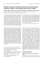
Fig. 2 Effects of huMETCAM/MUC18 expression on the in vitro
motility (a) and invasiveness (b) of SK-OV-3 clones/cells. a For the
motility test, the METCAM clone 2D and the Control (Vector) clone
3D of SK-OV-3 cells were used. Six hours after seeding to the top
wells, cells migrating to the bottom wells were determined as
described in “Methods”. Means and standard deviations of triplicate
values of the motility tests are indicated. P value, which was determined
by analyzing two sets of data with the Student’s t test by using
the one-tailed distribution-type 2 method, was 0.014, indicating
that the result was statistically different. b For invasiveness test, the
METCAM clone 2D and the Control (Vector) clone 3D of SK-OV-3
cells were used. Six hours after seeding cells to the top wells, cells
migrating to the bottom wells were determined as described in
“Methods”. Means and standard deviations of triplicate values of
the invasiveness tests are indicated. P value, which was determined
by analyzing two sets of data with the Student’s t test by using
the one-tailed distribution-type 2 method, was 0.0015, indicating
that the result was statistically different
Wu and Zeng BMC Cancer (2016) 16:136
lower than that of the control (vector) clone 3D. Taken
together, we conclude that increased METCAM/MUC18
expression decreased both motility and invasiveness of
SK-OV-3 cells.
METCAM/MUC18 expression inhibits in vivo
tumorigenicity of SK-OV-3 cells in nude mice
The effect of METCAM/MUC18 over-expression on in
vivo tumorigenicity of SKOV3 cells was determined in
female nude mice after SC injection at either dorsal or
ventral side. As shown in Figs. 3a and b, the tumor proliferation of the METCAM clone 2D was much lower
than that of the control (vector) clone at both sites, indicating that over-expression of METCAM/MUC18 decreased tumorigenicity of SK-OV-3 cells in nude mice.
Consistent with the results in Figs. 3a and b, Fig. 3c shows
that final tumor weights of the METCAM clone 2D were
also lower than those of the control (vector) clone 3D at
both sites, indicating that over-expression of METCAM/
MUC18 decreased the final tumor weights of SK-OV-3
cells in nude mice. Interestingly, as also shown in Fig. 3,
tumorigenicity of the control clone 3D on the dorsal side
was significantly better than that on the ventral side, in
contrast tumorigenicity of the METCAM clone 2D on the
ventral side was significantly better than that on the dorsal
site. Taken together, we conclude that over-expression of
METCAM/MUC18 suppressed in vivo tumorigenesis of
SK-OV-3 cells in nude mice.
Expression of METCAM/MUC18 in subcutaneous tumors
derived from SK-OV-3 clones
Figure 4a shows results of Western blot analysis that
METCAM/MUC18 was not expressed in tumors derived
from the control (vector) clone 3D, but was expressed in
tumors derived from the METCAM clone 2D. Since the
apparent electrophoretic mobility of the proteins from
tumors in the gel (lanes 5–16) was similar to that from
the tissue culture cells before injection (lanes 3–4), we
concluded that the tumors were from the injected clones/
cells. The IHC results in Fig. 4b showed that the tumor
sections from the METCAM clone 2D (panels e and f)
were stained much stronger than those from the control
(vector) clone 3D (panels g and h), consistent with the
Western blot results in Fig. 4a.
It is intriguing to find that the tumors derived from
the METCAM clone 2D were barely visible with the
naked eye, but visible under microscope in the tumor
sections (Fig. b, panels a and b in H&E stain and panels
e and f in IHC), which appeared to be confined to small
regions, whereas tumors derived from the control (vector)
3D were not confined (Fig. 4b, panels c and d in H&E
stain and panels g and h in IHC).
Page 7 of 15
METCAM/MUC18 expression inhibits tumorigenicity and
ascites formation of SK-OV-3 cells in the abdominal cavity
of nude mice
To further determine the effect of METCAM/MUC18
over-expression on in vivo tumorigenicity of SK-OV-3
cells in the orthotopic site (IP cavity), SK-OV-3 cells
from the METCAM clone 2D and the control (vector)
3D were IP injected into female nude mice. As shown in
Fig. 5a, the mice in the control group, which were
injected with the control (vector) clone 3D, developed
swollen abdominal cavity, but not the mice in the test
group, which were injected with the METCAM clone
2D. After dissection of the abdominal cavities, we found
that tumors and ascites were formed in four of five mice
in the control group, whereas no tumors and ascites
were found in the test group (Figs. 5b–d). Consistent
with the observation, the final weights of abdominal tumors and volumes of ascites were measured, and were
significantly heavier in the group injected with the control (vector) clone 3D than those injected with the METCAM clone 2D, as shown in Figs. 5b–d. We concluded
that over-expression of METCAM/MUC18 suppressed
the tumorigenicity and ascites formation of SK-OV-3
cells in IP cavities in nude mice.
Expression of METCAM/MUC18 in abdominal tumors and
ascites derived from SK-OV-3 clones
The METCAM/MUC18 expression in the IP tumors
and ascites formed by the vector control 3D clone in
mice was also determined by Western blot analysis. The
results showed that METCAM/MUC18 was minimally
detectable in the ascites and tumors similar to the parental SK-OV-3 cells (data not shown), suggesting that
those tumors were from the injected SK-OV-3 clones.
Preliminary mechanisms of METCAM/MUC18-mediated
suppression of the progression of SK-OV-3 cells
Mechanisms of METCAM/MUC18-mediated suppression of the progression of human ovarian cancer cells
have not been studied. By deducing knowledge learned
from METCAM/MUC18-induced tumorigenesis of other
tumor cell lines, such as, melanoma, cancers in breast
and prostate and nasopharyngeal carcinoma, METCAM/
MUC18 may affect tumorigenesis by cross-talk with many
downstream signaling pathways that regulate proliferation,
survival pathway, apoptosis, metabolism, and angiogenesis
of tumor cells [7, 22–25]. To investigate if METCAM/
MUC18-mediated tumor suppression also affected expression of its downstream effectors, such as indexes of
apoptosis/anti-apoptosis, proliferation, survival, aerobic
glycolysis, and angiogenesis, we determined the expression
of levels of Bcl2, Bax, PCNA, LDH-A, VEGF, pan-AKT,
phospho-AKT(Ser 473), and the ratio of phospho-AKT/
AKT in tumor lysates. Figure 6a shows the Western blot
Wu and Zeng BMC Cancer (2016) 16:136
Fig. 3 (See legend on next page.)
Page 8 of 15
Wu and Zeng BMC Cancer (2016) 16:136
Page 9 of 15
(See figure on previous page.)
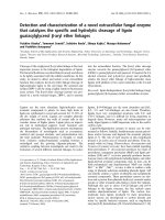
Fig. 3 Effects of huMETCAM/MUC18 expression on the in vivo tumorigenesis of SK-OV-3 clones/cells at the SC injection sites. a Tumorigenicity of
the METCAM clone 2D and the Control (Vector) clone 3D of SK-OV-3 was determined by subcutaneous injection of 5 × 10⁶ cells of cells from each
clone at the dorsal and ventral sides in female athymic nude mice. Tumor proliferation by the two clones is shown by plotting mean tumor volumes/
weights versus time after injection. P values were determined by analyzing all the data with the student’s t test by using 1-tailed distribution-type 1
method. P values between tumor volumes through the time course of the METCAM clone 2D and that of the control (vector) clone 3D were 0.0142 at
the dorsal site and 0.025 for the ventral site of injection, respectively. P value between the dorsal and the ventral sites of the METCAM clone 2D was
0.024 (**) and that between the two sites of the control (vector) clone 3D was 0.016 (*). b The panels a and b show the mice bearing tumors from the
METCAM clone 2D and the control (vector) clone 3D, respectively, at the dorsal sites (DSC). The panels c and d show the mice bearing tumors from
the METCAM clone 2D and the control (vector) clone 3D, respectively, at the ventral sites (VSC). c The mean final tumor weights of the two clones
injected at both dorsal and ventral sites in athymic nude mice were compared at the endpoint. Both the mean final tumor weights from five mice of
the control (vector) clone 3D were statistically significantly heavier than the mean tumor weight from those of the METCAM clone 2D, since
the P values, which were analyzed by the Student’s t test (one-tailed distribution-type 1 method) between the tumors from the METCAM
clone 2D and the control (vector) clone 3D at the dorsal and ventral sites were 0.0008 and 0.0022, respectively. The P values of the final tumor
weights analyzed by the Student’s t test (one-tailed distribution-type 1 method) between the dorsal and ventral sites were 0.047 for the METCAM clone
2D and 0.05 for the control (vector) clone 3D, respectively
results of the expression levels of Bcl2, Bax, PCNA, LDHA, VEGF, pan-AKT, and phospho-AKT (Ser473) in tumor
lysates. Figure 6b shows that the ratios of Bax/Bcl2 were
not statistically different between tumors derived from the
METCAM clone 2D and those from the control (vector)
clone 3D, indicating that over-expression of METCAM/
MUC18 did not affect apoptosis or anti-apoptosis of SKOV-3 cancer cells during in vivo tumorigenesis. Figures 6a
and c show that tumor lysates from the METCAM clone
2D had a lower level of PCNA than the control (vector)
clone 3D, indicating that over-expression of METCAM/
MUC18 decreased proliferation of SK-OV-3 cancer cells
during in vivo tumorigenesis. Figures 6a and d show that
tumor lysates from the METCAM clone 2D had a lower
level of LDH-A than the control (vector) clone 3D, indicating that over-expression of METCAM/MUC18
decreased proliferation of SK-OV-3 cancer cells by decreasing aerobic glycolysis during in vivo tumorigenesis.
Figures 6a and e show that tumor lysates from the
METCAM clone 2D had a lower level of VEGF than
the control (vector) clone 3D, indicating that overexpression of METCAM/MUC18 decreased proliferation
of SK-OV-3 cancer cells by decreasing angiogenesis during
in vivo tumorigenesis. Figures 6a and f show that the level
of pan-AKT was lower in tumors from the METCAM
clone 2D than those from the control (vector) clone 3D,
indicating that over expression of METCAM/MUC18 decreased the expression of pan-AKT. Figures 6a and g show
that phospho-AKT (Ser473) was lower in tumors from the
METCAM clone 2D than those from the control (vector)
clone 3D, indicating that over expression of METCAM/
MUC18 decreased the expression of phospho-AKT
(Ser473), which in turn affects motility and cell growth.
Figures 6a and h show that ratios of phospho-AKT (Ser
473)/AKT in tumors of the METCAM clone 2D was not
statistically significantly different from those in tumors of
the control (vector) clone 3D, indicating that METCAM
over-expression did not affect the survival pathway of
SK-OV-3 cancer cells during in vivo tumorigenesis.
Taken together, we suggest that over expression of METCAM/MUC18 may suppress tumorigenesis and malignant
progression of ovarian cancer cells in nude mice by decreasing their abilities in proliferation, aerobic glycolysis,
and angiogenesis, and by decreasing motility and invasiveness, but not altering the apoptosis/anti-apoptosis and
survival pathways.
Discussion
In this study, we initiated the investigation by determining
expression levels of METCAM/MUC18 in several ovarian
cancer cell lines. We found that METCAM/MUC18 was
expressed at a level of 31–50 % in two out of three cell
lines established from primary adenocarcinomas (HEY
and CAOV3), but poorly expressed (1–11 %) in two cell
lines established from malignant ascites (SKOV3 and
NIHOVCAR3). It appeared that METCAM/MUC18 was
expressed poorer in malignant cell lines than in primary
adenocarcinomas, suggesting that METCAM/MUC18
may play a negative role in the progression of ovarian cancer. To further support this hypothesis, we provided in
vitro evidence to show that a high expression level of
METCAM/MUC18 inhibited the migration and invasion
of SKOV3 cancer cells. We also provided in vivo evidence
in animal tests to show that METCAM/MUC18 expression inhibited the tumorigenicity at the subcutaneous sites
as well as the tumorigenicity and ascites formation in the
intra-peritoneal cavity of an athymic nude mouse model.
Since the METCAM/MUC18 expressed in the tumors
and ascites cells were similar to that in the injected
clones/cells, the protein was not modified to manifest
these processes. Taken together, we conclude that METCAM/MUC18 serves as a tumor suppressor as well as a
metastasis suppressor for the human ovarian cancer cells
SK-OV-3. METCAM/MUC18 may suppress tumorigenesis and malignant progression of ovarian cancer cells in
nude mice by decreasing their abilities in proliferation,
aerobic glycolysis, and angiogenesis, and by decreasing
Wu and Zeng BMC Cancer (2016) 16:136
Fig. 4 (See legend on next page.)
Page 10 of 15
Wu and Zeng BMC Cancer (2016) 16:136
Page 11 of 15
(See figure on previous page.)
Fig. 4 HuMETCAM/MUC18 expression in the subcutaneous tumors of SK-OV-3 clones/cells. a The expression of huMETCAM/MUC18 in the lysates
from the tumors was determined by Western blot analysis as described in “Methods”. Lysates from SK-Mel-28 cells (lane 1) and from LNCaP cells
(lane 2) were used as the positive control and the negative control, respectively. The huMETCAM/MUC18 expression levels in the tissue cultured
cells of two SK-OV-3 clones, METCAM clone 2D and the control (vector) clones 3D, are shown (lanes 3–4) in comparison with those in the tumor
lysates (lanes 5–16). The huMETCAM/MUC18 expression levels in the combined lysate from the two dorsal tumors (DSC) of the METCAM clone
2D (#421-422), the lysate of each of the five dorsal tumors (DSC) from the control (vector) clone 3D (#428-432), the combined lysate from two
ventral tumors (VSC) from the METCAM clone 2D (#423-424), and the lysate of each of the five ventral tumors (VSC) from the control (vector)
clone 3D (#433-437) are shown. As loading controls, the same membranes were reacted with antibodies against three house-keeping genes, but
only actin and GAPDH are shown. b Histology and immunohistochemistry (IHC) of SC tumors of the two SK-OV-3 clones. Panels a–d show histology of
the tumor sections from the two SK-OV-3 clones injected at dorsal (DSC) or ventral (VSC) subcutaneous sites. Panels e to l show the IHC of these tumor
sections. A tissue section of SC tumors derived from the human prostate cancer LNCaP-expressing clone (LNS239) was used as a positive
external control for IHC staining (data not shown). IHC of all tumor sections were carried out as previously described in “Methods”. Panels
e to h show the anti-huMETCAM/MUC18 antibody staining of the cells in the tumor sections. Both tumor sections (DSC and VSC) from
the METCAM clone 2D showed strong brown color staining in IHC when the antibody was added (Panels e & f), however, the two tumor
sections (DSC and VSC) from the control (vector) clone 3D showed a weak background staining (Panels g & h). Panels i to l show the corresponding
negative controls which show no staining in the adjacent sections when no antibody or when the control chicken IgY was added
their abilities in EMT, but not altering the apoptosis/antiapoptosis and survival pathways.
This conclusion contradicts the results of a positive
correlation of clinical prognosis with the increased expression of METCAM/MUC18 in malignant ovarian
cancer specimens [17, 18, 29]. This suggests that the
positive correlation in this case is fortuitous and that we
should not assume a positive role of METCAM/MUC18
in the progression of ovarian cancer without the support
of tests in an animal model. Our results also contradict
the previously established notion that METCAM/
MUC18 serves as a tumor promoter in both prostate
cancer cells [22] and breast cancer cells [23, 24], and as
a metastasis promoter in human melanoma cells [25],
Fig. 5 Effect of huMETCAM/MUC18 expression on the in vivo tumorigenesis of SK-OV-3 clones/cells at the IP injection sites. Tumorigenicity of the
METCAM clone 2D and the control (vector) clone 3D of SK-OV-3 was also determined by IP injection of 5 × 10⁶ cells of the two clones in athymic
nude mice. a Shows that the mice injected with the control (vector) clone 3D bore tumors and developed ascites in the intra-peritoneal cavity
(#365-369), whereas the mice injected with the METCAM clone 2D did not bear any tumors and develop ascites in the intra-peritoneal cavity
(#360-363). The bar shows the 1 cm mark. b Shows the final mean tumor weights of the two clones in the abdominal cavity, and c the final
volumes of total ascites fluid and d the pelleted volume of ascites cells of both clones. P values were determined by analyzing all the data with
the Student’s t test by using the 2-tailed distribution-type 1 method in (b) and the 1-tailed distribution-type 1 method in (c) and (d)
Wu and Zeng BMC Cancer (2016) 16:136
Fig. 6 (See legend on next page.)
Page 12 of 15
Wu and Zeng BMC Cancer (2016) 16:136
Page 13 of 15
(See figure on previous page.)
Fig. 6 The effect of METCAM/MUC18 expression on levels of various key parameters expressed in the tumor lysates, which may affect the tumor
growth. Tumor lysates were used in the western blot analysis by using various antibodies, as described in “Methods”. a The summary of Western
blot results of levels of various key parameters are shown, and their quantitative results are shown in (b) Bax/BCl2 ratios, c PCNA, d LDH-A, e VEGF,
f pan-AKT, g Phospho-AKT(Ser473), and h Phospho-AKT(Ser473)/pan-AKT ratios. P values were determined by using the Student’s t test (1-tailed
distribution-type 2/3 method) to analyze the data between the tumor lysates from the METCAM clone 2D clone and those from the Control
(Vector) Clone 3D
prostate cancer [26], and breast cancer [27]. The conclusion, nevertheless, appears to be consistent with the first
notion suggested by one group that METCAM/MUC18
is a tumor suppressor in human breast cancer cell line
MCF-7 [28]; albeit the notion was later proven to
contradict to the evidence from two different groups
[23, 24, 27]. Regardless, the role of METCAM/MUC18
as a tumor suppressor was not only conclusively demonstrated in a human ovarian cancer cell line, SK-OV-3 (as
shown here), but also in another human ovarian cancer
cell line BG-1 [Wu, unpublished results], as well as in a
mouse melanoma cell line, K1735-9 [34] and one NPC
cell line, NPC-TW01 ([35, 36], & Wu, unpublished results). METCAM/MUC18 has also been demonstrated
as a metastasis suppressor in the two human ovarian
cancer cell lines, SK-OV-3 (as also shown here) and BG1 [Wu, unpublished results], and one mouse melanoma
cell line, K1735-9 [34]. Thus sufficient evidence is provided to support the novel suppressor role of METCAM/MCU18 in the progression of these human
cancers.
E-cadherin, a cell adhesion molecule, has been demonstrated as a tumor suppressor role in many tumors derived from epithelium; however, E-cadherin has not been
found to play a tumor or metastasis promoter role in
any tumor [8]. Thus the most intriguing, unique biological
function of METCAM/MUC18 in tumorigenesis and metastasis is that it seems to play a dual role in the progression of some tumor cell lines. It can be a tumor/
metastasis promoter in prostate cancer cell lines [22, 26],
breast cancer cell lines [23, 24, 27], and most melanoma
cell lines [19, 25, 34]. It can also be a tumor/metastasis
suppressor in the progression of other tumor cell lines in
animal studies, such as, two ovarian cancer cell lines
(in this report and Wu, unpublished results), one
mouse melanoma subline ([34] and Wu, unpublished
results), nasopharyngeal carcinoma ([35, 36] and Wu,
unpublished results), and perhaps hemangioma [37].
It is not clear why METCAM/MUC18 plays a dual
role in tumorigenicity and metastasis. One point is
clear, which is that METCAM/MUC18 plays an opposite role in different cancer types or in different
clones/sublines of the same cancer type [38]. Thus it
is logical to propose that the effect of METCAM/
MUC18 on the progression of epithelial cancers is
modulated by different intrinsic factors in different
tumor cells/types. The dual role of METCAM/
MUC18 is very likely due to the presence of different
interacting partners intrinsic to each cancer cell type
and different clone, or perhaps due to different heterophilic ligands, which unfortunately have not been
identified [19, 34, 38]. Interactions of METCAM/
MUC18 with different sets of intrinsic partners may
result in the promotion or suppression of tumorigenicity
and metastasis via increasing or decreasing aerobic glycolysis, proliferation, angiogenesis, other growth-promoting
pathways, as well as altering tumor cell motility, invasiveness, and vascular metastasis, as suggested in this report. In
the future, the identification of these partners and/or
ligands is essential to understand further detailed
mechanisms.
Interestingly, many molecules have recently been
shown to play a dual role in the progression of cancer.
The most well-known examples are TGF-β, which is
context dependent and acts as a tumor suppressor in the
early stage of tumorigenesis, but as a progression promoter in the late stage [7], and VEGF, which also plays a
dual role in tumor progression [39].
One point worth noting is that the tumors induced by
the METCAM clone 2D were confined to small regions,
as shown in the results of H&E and IHC, whereas the
tumors induced by the control (vector) clone 3D developed serious tumors, suggesting that tumors from the
2D clone appeared to be dormant; thus METCAM/
MUC18 may function similarly to other tumor suppressors in other tumor cells [40].
Another point also worth noting is that tumorigenicity
of the control (vector) clone 3D in the dorsal site appeared to be significantly better than that in the ventral
site (P value = 0.016), whereas tumorigenicity of the 2D
clone in the ventral site was significantly better than that
in the dorsal site (P value = 0.024). We don’t know why
different SC sites have different effects on tumorigenicity. This also requires further investigation.
Conclusion
In summary, we have provided the first conclusive evidence to suggest that human METCAM/MUC18 is a
novel suppressor in the progression of human ovarian
cancer. The notion is supported by the evidence that the
over-expression of human METCAM/MUC18 inhibited
in vitro motility and invasion and in vivo tumor
Wu and Zeng BMC Cancer (2016) 16:136
formation of a human ovarian cancer cell line, SK-OV-3,
at SC sites as well as in the IP cavities of an athymic
nude mouse model. It also inhibited in vivo ascites formation of SKOV3 cells in the mouse IP cavities. The
tumor/metastasis suppressor role of human METCAM/
MUC18 in the progression of human ovarian cancer
cells is opposite to its role in breast cancer, prostate cancer, and most melanoma cell lines. This novel role of
METCAM/MUC18 is not unique in this human ovarian
cancer cell line, but is also found in another human
ovarian cancer cell line, BG-1, one mouse melanoma
subline, and one nasopharyngeal cancer cell line. The
dual role played by METCAM/MUC18 in the progression of different cancers may be dependent upon the
unique intrinsic constituents and cell surface heterophilic ligands in different cancer cell types, which require
future investigation. How METCAM/MUC18 affects
tumor dormancy should also be an interesting aspect for
future investigation, since tumor dormancy may be due
to intrinsic growth inhibition, immunological suppression, and/or angiogenic suppression [40].
Abbreviations
CAM: cell adhesion molecule; FBS: fetal bovine serum; G418R: G418-resistant;
huMETCAM/MUC18: human METCAM/MUC18; IHC: immunohistochemistry;
METCAM: metastasis cell adhesion molecule; IP: intraperitoneal;
SC: subcutaneous.
Competing interests
The authors declare that they have no competing interests.
Authors’ contributions
GJW conceived of the idea and study, participated in its design and
coordination, carried out in vivo animal studies, performed the statistical
analysis, and revised the manuscript many times suitable for publication. GFZ
carried out western blots analyses, migration and invasion studies, and
colony formation study, participated in in vivo animal studies, performed the
statistical analysis, and drafted the manuscript. Both authors read and
approved the final manuscript.
Acknowledgements
We thank Mrs. Mei-Whey H. Wu for critical reading the manuscript and Mr.
Jonathan Wu for editing and improving the style of written English. We also
thank the financial supports from Emory University School of Medicine (USA),
Chung Yuan Christian University, and grants from NSC (NSC-101-2320-B-033001 and −003), Taiwan (GJW), and from the government of the People’s
Republic of China (GFZ).
Page 14 of 15
3.
4.
5.
6.
7.
8.
9.
10.
11.
12.
13.
14.
15.
16.
17.
18.
19.
20.
21.
Author details
1
Department of Microbiology and Immunology, Emory University School of
Medicine, Atlanta, GA 30322, USA. 2Department of Bioscience Technology,
Chung Yuan Christian University, Chung Li 32023, Taiwan. 3Center for
Biomedical Technology, Chung Yuan Christian University, Chung Li 32023,
Taiwan. 4Present Address: Department of Hepatobiliary Surgery, Institute of
Plastic Surgery, and Laboratory of Regenerative Medicine, Affiliated Hospital
of Guangdong Medical College, Zhanjiang 542001, China.
22.
23.
24.
Received: 16 September 2015 Accepted: 15 February 2016
25.
References
1. Siegel R, Ma J, Zou Z, Jemal A. Cancer statistics 2014. CA Cancer J Clin.
2014;64(1):9–29.
2. Clarke-Pearson DL. Screening for ovarian cancer. N Engl J Med.
2009;361:170–7.
26.
McCluggage WG, Wilkinson N. Metastatic neoplasms involving the ovary: a
review with an emphasis on morphological and immunohistochemical
features. Histopathology. 2005;47:231–47.
Wei W, Dizon D, Vathipadiekal V, Birrer MJ. Ovarian cancer: genomic
analysis. Ann Oncol. 2013;24(supplement 10):x7–15.
Menon U, Griffin M, Gentry-Maharaj A. Ovarian cancer screening-current
status, future directions. Gynecol Oncol. 2014;132:490–5.
Jacob F, Nixdorf S, Hacker NF, Heinzelmann-Schwarz VA. Reliable in vitro
studies require appropriate ovarian cancer cell lines. J Ovarian Res.
2014;7:60 (10 pages).
Harahan D, Weinberg RA. Hallmarks of cancer: the next generation. Cell.
2011;144:646–74.
Cavallaro U, Christofori G. Cell adhesion and signaling by cadherins and
Ig-CAMs in cancer. Nat Rev Cancer. 2005;4:118–32.
Bellone S, Siegel ER, Cocco E, Cargnelutti M, Silasi DA, Azodi M, et al. Overexpression of epithelial cell adhesion molecule in primary, metastatic, and
recurrent/chemotherapy-resistant epithelial ovarian cancer: implications for
epithelial cell adhesion molecule-specific immunotherapy. Int J Gynecol
Cancer. 2009;19(5):860–6.
Rump A, Morikawa Y, Tanaka M, Minami S, Umesaki N, Takeuchi M, et al.
Binding of ovarian cancer antigen CA125/MUC16 to mesothelin mediates
cell adhesion. J Biol Chem. 2004;279:9190–8.
Strobel T, Cannistra SA. β1-integrins partly mediate binding of ovarian
cancer cells to peritoneal mesothelium in vitro. Gynecol Oncol. 2004;73:362–7.
Bourguignon LYW, Gilad E, Rothman K, Peyrollier K. Hyaluronan-CD44
interaction with IQGAP1 promotes Cdc42 and ERK signaling, leading to
actin binding, Elk-1/estrogen receptor transcription activation, and ovarian
cancer progression. J Biol Chem. 2005;280:11961–72.
Euer NI, Kaul S, Deissler H, Mobus VJ, Zeillinger R, Weidle UH. Identification
of L1CAM, Jagged2, and neuromedin U as ovarian cancer-associated
antigens. Oncol Rep. 2005;13:375–87.
Imai T, Horiuchi A, Shiozawa T, Osada R, Kikuchi N, Ohira S. Elevated
expression of E-cadherin and alpha-, beta-, and gamma-catenins in
metastatic lesions compared with primary epithelial ovarian carcinomas.
Human Pathol. 2004;35:1469–76.
D’Souza T, Agarwal R, Morin PJ. Phosphorylation of claudin-3 at threonine
192 by cAMP-dependent protein kinase regulates tight junction barrier
function in ovarian cancer cells. J Biol Chem. 2005;280:26233–40.
Pietzner K, Woopen H, Richter R, Joens T, Braicu EI, Dimitrova D, et al.
Expression of EpCAM in paired tumor samples of patients with primary and
recurrent serous ovarian cancer. Int J Gynecol Cancer. 2013;23(5):797–802.
Aldovini D, Demichelis F, Doglioni C, Di Vizio D, Galligioni E, Brugnara S, et
al. M-CAM expression as marker of poor prognosis in epithelial ovarian
cancer. Int J Cancer. 2006;119(8):1920–6.
Wu GJ, Dickerson EB. Frequent and increased expression of human
METCAM/MUC18 in cancer tissues and metastatic lesions associates with
the clinical progression of human ovarian carcinoma. Taiwan J Obstet
Gynecol. 2014;53:509–17.
Wu GJ. METCAM/MUC18 expression and cancer metastasis. Curr Genomics.
2005;6:333–49.
Lehmann JM, Reithmuller G, Johnson JP. MUC18, a marker of tumor
progression in human melanoma. Proc Natl Acad Sci U S A.
1989;86:9891–5.
Shih IM. The role of CD146 (Mel-CAM) in biology and pathology. J Pathol.
1999;189:4–11.
Wu GJ, Wu MWH, Liu Y. Enforced expression of human METCAM/MUC18
increases the tumorigenesis of human prostate cancer cells in nude mice.
J Urol. 2011;185:1504–12.
Zeng G, Cai S, Wu GJ. Up-regulation of METCAM/MUC18 promotes motility,
invasiveness and tumorigenesis of human breast cancer cells. BMC Cancer.
2011;11:113. doi:10.1186/1471-2407-11-113.
Zeng G, Cai S, Liu Y, Wu GJ. METCAM/MUC18 augments promotes
migration, invasion and tumorigenicity of human breast cancer SK-BR-3
Cells. Gene. 2012;492:229–38.
Wu GJ, Fu P, Wang S-W, Wu MWH. Enforced expression of MCAM/MUC18
increased in vitro motility and invasiveness and in vivo metastasis of two
mouse melanoma K1735 Sublines in a syngeneic mouse model. Mol Cancer
Res. 2008;6(11):1666–77.
Wu GJ, Qiong P, Fu P, Wang S-W, Chiang CF, Dillehay DL, et al. Ectopical
expression of human MUC18 increases metastasis of human prostate cancer
cells. Gene. 2004;327:201–13.
Wu and Zeng BMC Cancer (2016) 16:136
Page 15 of 15
27. Zeng Q, Li W, Lu D, Wu Z, Duan H, Luo Y, et al. CD146, an epithelialmesenchymal transition inducer, is associated with triple-negative breast
cancer. Proc Natl Acad Sci U S A. 2012;109(4):1127–32.
28. Shih IM, Hsu M-Y, Palazzo JP, Herlyn M. The cell-cell adhesion receptor
MEL-CAM acts as a tumor suppressor in breast carcinoma. Am J Pathol.
1997;151:745–51.
29. Wu Z, Wu Z, Li J, Yang X, Wang Y, Yu Y, et al. MCAM is a novel metastasis
marker and regulates spreading, apoptosis and invasion of ovarian cancer
cells. Tumor Biol. 2012;33:1619–28.
30. Auersperg N, Maines-Banders SL, Dyck HG, Kruk PA. Characterization of
cultured human ovarian surface epithelial cells (IOSE): phenotypic plasticity
and premalignant changes. Lab Invest. 1994;71:510–8.
31. Geisinger KR, Kute TE, Pettenati MJ, Welander CE, Dennard Y, Collins LA,
et al. Characterization of a human ovarian carcinoma cell line, BG-1, with
estrogen and progesterone receptors. Cancer. 1989;63:280–8.
32. Buick RN, Pullano R, Trent JM. Comparative properties of five human ovarian
adenocarcinoma cell lines. Cancer Res. 1985;45:3668–76.
33. Wu GJ, Lu SY, Lowe LL, Kinkade Jr JM. Identification of lactate
dehydrogenase-M polypeptide translated in vitro from human and mouse
tumor cell poly(A)-containing mRNA. Int J Biochem. 1985;17:355–63.
34. Wu GJ. Chapter 11: Dual roles of the melanoma CAM (MelCAM/METCAM) in
malignant progression of melanoma. In: Murph M, editor. Research on
Melanoma: a glimpse into current directions and future trends. Rijeka:
InTech-Open Access Publisher, University Campus STeP Ri; 2011. p. 229–42.
ISBN 978-953-307-293-7.
35. Lin JC, Chiang CF, Wang SW, Wang WY, Kwan PC, Wu GJ. Significance and
expression of human METCAM/MUC18 in nasopharyngeal carcinoma (NPC)
and metastatic lesions. Asian Pac J Cancer Prev. 2014;15(1):245–52.
36. Liu YC. The putative role of human METCAM/MUC18 in modulating the
development and progression of nasopharyngeal carcinoma. Chung Li: M.S.
thesis under the supervision of Dr. Guang-Jer Wu, Department of Bioscience
Technology, Chung Yuan Christian University; 2014. .
tw/thesis.
37. Li Q, Yu Y, Bischoff J, Milliken JB, Olsen BR. Differential expression of CD146
in tissues and endothelial cells derived from infantile haemangioma and
normal human skin. J Path. 2003;201:296–302.
38. Wu GJ. Dual roles of METCAM in the in the progression of different cancers.
J Oncol. 2012;2012:853797. doi:10.1155/2012/853797.
39. Vecchiarelli-Federico LM, David Cervi D, Haeri M, Li Y, Nagy A, Ben-David Y,
et al. Vascular Endothelial Growth Factor-A Positive and Negative Regulator
of Tumor Growth. Cancer Res. 2010;70:863–7.
40. Aguirre-Chiso JA. Models, mechanisms and clinical evidence for cancer
dormancy. Nat Rev Cancer. 2007;7:834–46.
Submit your next manuscript to BioMed Central
and we will help you at every step:
• We accept pre-submission inquiries
• Our selector tool helps you to find the most relevant journal
• We provide round the clock customer support
• Convenient online submission
• Thorough peer review
• Inclusion in PubMed and all major indexing services
• Maximum visibility for your research
Submit your manuscript at
www.biomedcentral.com/submit