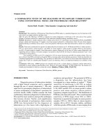
Real-time polymerase chain reaction analysis of MDM2 and CDK4 expression using total RNA from core-needle biopsies is useful for diagnosing adipocytic tumors
Bạn đang xem bản rút gọn của tài liệu. Xem và tải ngay bản đầy đủ của tài liệu tại đây (1.2 MB, 9 trang )
Sasaki et al. BMC Cancer 2014, 14:468
/>
RESEARCH ARTICLE
Open Access
Real-time polymerase chain reaction analysis of
MDM2 and CDK4 expression using total RNA from
core-needle biopsies is useful for diagnosing
adipocytic tumors
Taro Sasaki1*, Akira Ogose1, Hiroyuki Kawashima1, Tetsuo Hotta1, Hiroshi Hatano3, Takashi Ariizumi3,
Hajime Umezu2, Riuko Ohashi2, Tsuyoshi Tohyama4, Naohito Tanabe5 and Naoto Endo1
Abstract
Background: Diagnosing adipocytic tumors can be challenging because it is often difficult to morphologically
distinguish between benign, intermediate and malignant adipocytic tumors, and other sarcomas that are
histologically similar. Recently, a number of tumor-specific chromosome translocations and associated fusion genes
have been identified in adipocytic tumors and atypical lipomatous tumors/well-differentiated liposarcomas
(ALT/WDL), which have a supernumerary ring and/or giant chromosome marker with amplified sequences of the
MDM2 and CDK4 genes. The purpose of this study was to investigate whether quantitative real-time polymerase
chain reaction (PCR) could be used to amplify MDM2 and CDK4 from total RNA samples obtained from core-needle
biopsy sections for the diagnosis of ALT/WDL.
Methods: A series of lipoma (n = 124) and ALT/WDL (n = 44) cases were analyzed for cytogenetic analysis and
lipoma fusion genes, as well as for MDM2 and CDK4 expression by real-time PCR. Moreover, the expression of
MDM2 and CDK4 in whole tissue sections was compared with that in core-needle biopsy sections of the same
tumor in order to determine whether real-time PCR could be used to distinguish ALT/WDL from lipoma at the
preoperative stage.
Results: In whole tissue sections, the medians for MDM2 and CDK4 expression in ALT/WDL were higher than those
in the lipomas (P < 0.05). Moreover, karyotype subdivisions with rings and/or giant chromosomes had higher MDM2
and CDK4 expression levels compared to karyotypes with 12q13-15 rearrangements, other abnormal karyotypes,
and normal karyotypes (P < 0.05). On the other hand, MDM2 and CDK4 expression levels in core-needle biopsy
sections were similar to those in whole-tissue sections (MDM2: P = 0.6, CDK4: P = 0.8, Wilcoxon signed-rank test).
Conclusion: Quantitative real-time PCR of total RNA can be used to evaluate the MDM2 and CDK4 expression levels
in core-needle biopsies and may be useful for distinguishing ALT/WDL from adipocytic tumors. Thus, total RNA from
core-needle biopsy sections may have potential as a routine diagnostic tool for other tumors where gene overexpression
is a feature of the tumor.
Keywords: Liposarcoma, Atypical lipomatous tumor, Adipocytic tumors, MDM2, CDK4, Real-time PCR
* Correspondence:
1
Division of Orthopedic Surgery, Niigata University Graduate School of
Medical and Dental Sciences, 757-1, Asahimachi-dori, Niigata City, Niigata
951-8510, Japan
Full list of author information is available at the end of the article
© 2014 Sasaki et al.; licensee BioMed Central Ltd. This is an Open Access article distributed under the terms of the Creative
Commons Attribution License ( which permits unrestricted use, distribution, and
reproduction in any medium, provided the original work is properly credited. The Creative Commons Public Domain
Dedication waiver ( applies to the data made available in this article,
unless otherwise stated.
Sasaki et al. BMC Cancer 2014, 14:468
/>
Background
Adipocytic tumors represent the largest group of soft tissue tumors [1]. The diagnosis of adipocytic tumors is primarily based on clinical features and histologic patterns
[2]. However, the distinction between lipomas and atypical
lipomatous tumors/well-differentiated liposarcomas (ALT/
WDL) may be difficult to distinguish morphologically.
Cytogenetic studies of adipocytic tumors have revealed a clear association between chromosomal findings and clinicohistopathological features [3,4]. Clonal
chromosome aberrations have been found in nearly 60%
of all lipomas [4], of which two-thirds are rearrangements involving the 12q13-15 chromosomal region. A
variety of rearrangements, mainly involving the 6p and
13q regions, are observed in the remaining lipoma cases
[5-7]. In tumors with aberrations involving 12q13-15 region, the high mobility group protein gene (HMGA2,
also known as HMGIC) on chromosome 12 is rearranged. These aberrations may also result in the creation of chimeric genes, in which the HMGA2 gene is
fused to multiple genes. The most frequent gene aberration in lipomas is HMGA2/LPP [8].
ALT/WDL and dedifferentiated liposarcomas (DDL)
most often have a supernumerary ring and giant marker
chromosomes composed of amplified sequences from
the 12q13-15 region [9,10], including the murine doubleminute type 2 gene (MDM2) and the cyclin-dependent
kinase 4 gene (CDK4) [11-13]. Amplification of the 12q1315 region has not been observed in lipoma, and the
MDM2 and CDK4 proteins are known to be overexpressed
in ALT/WDL but not in lipoma [14]. Immunohistochemistry for MDM2 and CDK4 plays a helpful role in the differential diagnosis of adipocytic tumors. Aleixo et al. [15]
reported that MDM2 has very high sensitivity (100%) in
the identification of ALT/WDL among lipomas, but has
low specificity (58.8%), whereas CDK4 has low sensitivity
(68.4%), but high specificity (88.2%). Immunohistochemistry may be used to demonstrate MDM2 and CDK4 amplification, but the sections sometimes show several staining
patterns such as diffuse, moderate, and focal positivity.
Categorization of these staining patterns has been developed differently by different researchers, making it difficult
to compare studies effectively.
The use of minimally invasive biopsies to diagnose soft
tissue tumors has become increasingly common. On the
other hand, ALT/WDL can be difficult to distinguish morphologically from benign lipomatous lesions, especially
with limited material in which the diagnostic features of
scattered atypical cells are not present because of heterogeneity of the neoplasm. However, distinguishing benign
lipomatous tumors from ALT/WDL is important at primary biopsy.
In this study, we used whole tissue sections from surgically resected specimens to retrospectively analyze
Page 2 of 9
cytogenetic findings by quantifying MDM2 and CDK4
expression levels in lipomas and ALT/WDL with realtime polymerase chain reaction (PCR) from total RNA.
We evaluated the clinical utility of measuring MDM2
and CDK4 expression levels to establish a diagnosis of
adipocytic tumors, with the aim of making a distinction
between lipoma and ALT/WDL. Moreover, we compared the results of MDM2 and CDK4 expression in
whole tissue sections with those in core-needle biopsy
sections in order to investigate whether real-time PCR
for MDM2 and CDK4 could be used to distinguish between ALT/WDL and lipoma prior to surgery.
Methods
Specimens
Tumor samples were obtained from patients that underwent surgical resection at Niigata University Hospital
between August 2001 and December 2012. In total, 124
cases of lipoma and 44 cases of ALT/WDL were studied
(Additional file 1: Table S1). In all cases, the diagnosis of
lipoma or ALT/WDL was established according to the
World Health Organization (WHO) Classification of
Tumors [2] by using hematoxylin and eosin-stained tissue
sections from the surgical resection specimens. Two experienced pathologists independently reviewed the cases in
which it was difficult to distinguish between lipoma and
ALT/WDL. There were 159 primary and 9 recurrent tumors. The patient cohort consisted of 96 men and 72
women between 24 and 86 years of age (mean 59.0 years;
range 24–86 years).
The samples were taken from both core-needle biopsy
sections and whole tissue sections of the adipose tissue
tumors. Some of the samples represent paired whole tissue sections and core-needle biopsy sections from the
same tumor. Core-needle biopsy sections were sampled
prior to or after surgical resection using a 16G Tru-Cut
trocar with at least two passes or until an adequate sample was obtained.
Cytogenetic analysis
The tumor specimens that were analyzed were obtained
immediately after surgical excision. Portions of the tumor
were treated with collagenase and cultured at 37°C for
4 days. The chromosome slides were prepared from shortterm-cultured tumor cells using the standard trypsin
Giemsa banding technique. Karyotypes were described on
the basis of the short system of the International System
for Human Cytogenetic Nomenclature (ISCN) [16]. The
karyotypes were classified as either normal or abnormal.
The abnormal karyotypes were further subdivided according to the presence of a rearrangement in 12q13-15, rearrangement or loss of chromosome 13q, rearrangement of
6p21-23, and the presence of a supernumerary ring and/or
giant marker chromosome, as well as other aberrations
Sasaki et al. BMC Cancer 2014, 14:468
/>
[4-6]. Some tumors had more than one of these aberrations
and were thus included in more than one subgroup.
Reverse transcription PCR
Total RNA was prepared using Isogen reagent (Nippon
Gene; Tokyo, Japan) from core-needle biopsy sections according to the manufacturer’s recommendations. Synthesis
of cDNA was performed using a PrimeScript™ RT reagent
kit (TaKaRa Bio; Tokyo, Japan), and PCR was performed
with rTaq DNA Polymerase (Toyobo; Osaka, Japan). Glyceraldehyde 3-phosphate dehydrogenase (GAPDH: forward;
5′TGAAGGTCGGAGTCAACGGATTTGGT 3′, reverse;
5′CATGTGGGCCATGAGGTCCACCAC 3′) was used as
the internal control for uniform RNA loading. The primers
that were used to detect HMGA2 transcripts are listed in
Additional file 1: Table S2 as HMGA2/LPP, HMGA2/
RDC1, and HMGA2/NFIB [17]. The PCR conditions used
were as follows: the reaction mixture was heated for 3 min
at 94°C, followed by 30 cycles of 30 s denaturation at 94°C,
30 s annealing at 55 °C, and a 30 s extension at 72°C using
a PTC-200 Peltier Thermal Cycler (MJ Research; Waltham,
MA, USA). PCR products were analyzed by electrophoresis
on a 1.5% agarose gel containing ethidium bromide, and
were photographed under ultraviolet light.
Quantitative real-time PCR
RNA samples were taken from both core-needle biopsy
sections and whole-tissue sections. Total RNA and synthesis of cDNA were prepared as described above. Quantitative real-time PCR was performed using SYBR Premix Ex
Taq II in a Thermal Cycler Dice Real Time System TP800
(TaKaRa Bio; Otsu, Japan). The primers of target genes
used for this analysis were MDM2 and CDK4, and the primer sequences are listed in Additional file 1: Table S3.
GAPDH was selected as the reference gene (forward; 5′
GCACCGTCAAGGCTGAGAAC 3′, reverse; 5′ TGGT
GAAGACGCCAGTGGA3′). The gene copy numbers of
MDM2 and CDK4 were calculated by using a standard
curve that was constructed using the NDDLS-1 cell line
[18]. The level of expression for the target gene was calculated as the ratio of the copy number of the target gene
(MDM2 or CDK4) to that of the reference gene (GAPDH).
Total RNA from normal human adipose tissue was purchased from BioChain (Newark, CA, USA), and used as
a calibrator. Finally, the relative level of expression was
calculated as follows: [copy number of the target gene
(MDM2 or CDK4)/copy number of the reference gene
(GAPDH)]/copy number of the target gene (MDM2 or
CDK4) in normal adipose tissue.
Statistical analysis
Results from quantitative real-time PCR are reported as
the median of MDM2 and CDK4 relative expression
levels. The Mann–Whitney U test was used to compare
Page 3 of 9
differences in MDM2 and CDK4 median relative expression levels between lipoma and ALT/WDL. The SteelDwass test was used for comparison of differences in each
of the subdividing karyotypes. MDM2 and CDK4 relative
expression levels in the core-needle biopsy sections were
compared to those in the whole-tissue sections by the
Wilcoxon signed-rank test and Spearman rank correlation
coefficient. P values < 0.05 were considered to be statistically significant.
Consent
The study complies with the Declaration of Helsinki and
was approved by the Institutional Review Board of Niigata
University Hospital. Written informed consent was obtained from each patient before the specimens were taken
in accordance with the local ethics committee (Niigata
University Hospital).
Results
Cytogenetic findings
Cytogenetic analysis was performed on 104/168 cases (66
lipoma cases and 38 ALT/WDL cases). Table 1 shows the
results from the clinical and cytogenetic analyses of the
lipomas, which indicate that an abnormal karyotype was
present in 56 of the lipoma cases (85%). By subdividing
the karyotypes into previously identified cytogenetic subgroups, it was discovered that 21 lipomas had a 12q13-15
rearrangement (38%), 6 had a 13q rearrangement or loss
of chromosome 13 (11%), 3 had a 6p21-23 rearrangement
(5%), 4 had one or more ring chromosomes (7%), and 25
had other rearrangements (45%). In addition, 10 cases of
lipoma (15%) had a normal karyotype.
Analysis of ALT/WDL (Table 2) demonstrated that 36
ALT/WDL (95%) cases had an abnormal karyotype while
the remaining 2 cases (5%) had a normal karyotype. Subdividing the karyotypes showed that most of the abnormal
karyotypes had ring and/or giant chromosomes; 15 ALT/
WDLs had one or more rings and/or giant chromosomes
(42%), 5 had a 12q13-15 rearrangement (14%), 5 had a
13q rearrangement or loss of chromosome 13 (14%), 3
had a 6p21-23 rearrangement (8%), and 10 had other rearrangements (28%).
HMGA2 fusion genes
Reverse transcription PCR was used to evaluate 128/168
samples (96 lipoma samples and 32 ALT/WDL samples)
(Table 3). The HMGA2/LPP gene fusion transcript was
detected in 10 samples (8%) while the HMGA2/RDC1 fusion transcript was only detected in 3 samples (2%). No
sample expressed the HMGA2/NFIB fusion gene. Most of
these cases were categorized as lipomas, except for one
HMGA2/LPP case, which was diagnosed as ALT/WDL.
Cytogenetic analysis of the 6 cases that tested positive
for HMGA2/LPP revealed that 5 of them had a t(3;12)
Sasaki et al. BMC Cancer 2014, 14:468
/>
Page 4 of 9
Table 1 Clinical and cytogenetic findings in lipomas
Sex
Age (years)
Location
Total
Karyotype
M
F
20-40
40-60
>60
U
L
T
H
= 66
Normal
6
4
2
6
2
1
1
3
5
10 (15%)
Abnormal
36
20
5
23
28
13
17
17
9
56 (85%)
Ring/Giant chromosome
1
3
0
1
3
1
2
1
0
4 (7%)
12q13-15 rearrangement
13
8
0
8
13
6
7
7
1
21 (38%)
13q rearrangement
5
1
0
2
4
0
1
1
4
6 (11%)
6p21-23 rearrangement
2
1
1
0
2
1
0
1
1
3 (5%)
Other
17
8
4
13
8
5
8
7
5
25 (45%)
Abbreviations: M male, F female, U upper extremity, L lower extremity, T trunk, H head and neck. Note that some cases showed more than one
karyotypic aberration.
(q27-28;q13-15) translocation that fused the HMGA2
and LPP genes.
MDM2 and CDK4 expression in whole tissue sections
The gene expression levels of MDM2 and CDK4 were
studied in 149/168 whole tissue sections (108 lipoma samples and 41 samples from the 38 cases of ALT/WDL). The
medians for MDM2 relative expression levels were 2.0
(range, 0.2–54.1) for lipoma and 3.4 (range, 0.4–52.5) for
ALT/WDL. The medians for CDK4 relative expression
levels were 1.0 (range, 0.1–19.9) for lipoma and 2.9 (range,
0.4–22.4) for ALT/WDL (Figure 1). Both MDM2 and
CDK4 relative expression levels in ALT/WDL were higher
than those in lipoma (P < 0.05, Mann–Whitney U test).
In each of the subdividing karyotypes, the medians for
relative MDM2 expression were 5.1 (range, 3.1–52.5) for
the 16 samples with a ring and/or giant chromosomes (3
lipoma samples and 13 ALT/WDL samples), 2.3 (range,
1.0–5.0) for the 23 samples with 12q13-15 rearrangements (19 lipoma samples and 4 ALT/WDL samples),
2.6 (range, 0.4–22.4) for the 34 samples with other rearrangements (21 lipoma samples and 13 ALT/WDL samples), and 1.5 (range, 0.2–12.0) for the 9 samples with a
normal karyotype. The medians for CDK4 expression
were 8.4 (range, 0.9–22.4) for the 16 samples with ring
and/or giant chromosomes, 1.1 (range, 0.3–4.5) for the
23 samples with 12q13-15 rearrangements, 1.1 (range,
0.2–16.0) for the 34 samples with other rearrangements,
and 1.0 (range, 0.1– 2.1) for the 9 samples with a normal
karyotype (Figure 2). Relative MDM2 and CDK4 expression levels in lipoma and ALT/WDL cases with a ring
and/or giant chromosome were higher than those with
12q13-15 rearrangements and other abnormal karyotypes
(P < 0.05, Steel-Dwass test). However, expression levels of
cases with a ring and/or giant chromosome were not
significantly higher than those with normal karyotypes
(P < 0.1, Steel-Dwass test), because of the small number
of samples with normal karyotypes.
MDM2 and CDK4 expression in core-needle biopsy
sections
The relative gene expression levels of MDM2 and CDK4
were studied in 38/168 samples (28 lipoma samples and
10 ALT/WDL samples) from core-needle biopsy sections. The medians for relative MDM2 expression were
1.3 (range, 0.1–28.2) for lipoma and 3.9 (range, 0.4–
21.6) for ALT/WDL. The medians for relative CDK4 expression were 0.9 (range, 0.3–8.0) for lipoma and 1.4
(range, 0.3–12.8) for ALT/WDL (Figure 3). Both MDM2
and CDK4 expression levels in core-needle biopsy sections
showed no significant difference between lipoma and ALT/
WDL (MDM2: P < 0.1, CDK4: P < 0.1, Mann–Whitney U
Table 2 Clinical and cytogenetic findings in atypical lipomatous tumors/well-differentiated liposarcomas
Sex
Age (years)
Location
Total
Karyotype
M
F
20-40
40-60
>60
U
L
T
H
= 38
Normal
1
1
0
1
1
0
2
0
0
2 (5%)
Abnormal
21
15
2
16
18
6
20
8
2
36 (95%)
Ring/Giant chromosome
7
8
1
5
9
1
9
5
0
15 (42%)
12q13-15 rearrangement
4
1
0
2
3
1
3
0
1
5 (14%)
13q rearrangement
4
1
0
3
2
3
1
1
0
5 (14%)
6p21-23 rearrangement
1
2
0
2
1
1
1
0
1
3 (8%)
Other
5
5
1
3
6
1
7
2
0
10 (28%)
Abbreviations: M male, F female, U upper extremity, L lower extremity, T trunk, H head and neck. Note that some cases showed more than one
karyotypic aberration.
Sasaki et al. BMC Cancer 2014, 14:468
/>
Page 5 of 9
Table 3 Reverse transcription PCR of HMGA2 fusion genes
HMGA2-LPP(+)
Lipoma
ALT/WDL
9 (9%)
1 (3%)
HMGA2-RDC1(+)
3 (3%)
0 (0%)
HMGA2-NFIB(+)
0 (0%)
0 (0%)
Fusion gene(−)
85 (88%)
31 (97%)
test). MDM2 and CDK4 expression levels in the coreneedle biopsy sections were comparable to those in
the whole-tissue sections (MDM2: P = 0.6, CDK4: P =
0.8, Wilcoxon signed-rank test) (MDM2: ρ = 0.827, P =
0.000001, CDK4: ρ = 0.746, P = 0.000001, Spearman rank
correlation coefficient) (Figure 4).
Discussion
In the WHO classification, ALT/WDL is considered an
intermediate (locally aggressive) malignancy. It accounts
for approximately 40–45% of all liposarcomas and mostly
occurs in the deep soft tissue of the extremities, especially
in the thigh, retroperitoneum, and paratesticular areas.
ALT/WDL mostly occurs in middle-aged and older individuals. Histologically, the tumor is composed either
entirely or partially of mature adipocytic proliferation
showing significant variation in cell size and, at least focal,
nuclear atypia in both adipocytes and stromal cells. In
some situations, ALT/WDL may be indistinguishable from
benign adipocytic tumors at the histological level, and
evaluation of inadequate samples can lead to misdiagnosis.
Lipomatous tumors are cytogenetically heterogeneous.
Of the more than 200 cases with karyotypic abnormalities
that have been described to date, most cytogenetic aberrations have been found to correlate with morphological
subtype. In the present study, 36 out of the 38 (95%) ALT/
WDL cases had an abnormal karyotype, whereby the ring
and/or giant marker chromosome was identified in 15 of
them (42 %). Fletcher et al. [3] reported that 29 of 37
(78 %) ALT cases (including 5 dedifferentiated cases) had
a ring chromosome. In ordinary lipoma, however, the
presence of a supernumerary ring chromosome is a rare
finding [3,7,11]. It is interesting that tumors diagnosed
as ordinary lipomas occasionally display rings and/giant
chromosomes, which were found in 3% [3], 6% [5], and
2% [6] of ordinary lipoma samples in three different studies. The patients with ring chromosomes often have deepseated lipomas that are, on average, larger and older than
the other lipomas [1,5]. Furthermore, Bartuma et al. reported that it is interesting that in the 5 local recurrences
among the 272 cases, 2 of the 5 cases that contained ring
chromosomes were recurrent compared to 3/257 lipomas
without ring chromosomes [5].
Ordinary lipoma is the most common soft tissue tumor
and may appear at any site. It occurs mainly between 40
and 60 years of age and is more frequent in obese individuals [1]. Ordinary lipomas usually present as painless,
slowly growing soft tissue masses, and can arise within subcutaneous tissue or within deep soft tissue or even on the
surfaces of bone. The 12q13-15 region is the most common gene alteration involved in such aberrations, followed
by the 6p21-23 and 13q rearrangements [5,6,8,19]. This
chromosomal region has been found to recombine with a
large number of bands through translocations. The most
frequent translocation is t(3;12)(q27-28;q13-15), which
fuses the HMGA2 and LPP genes. This particular translocation is seen in more than 20% of tumors with 12q13-15
aberrations.
Figure 1 Amplification of target genes from whole tissue sections by real-time PCR (A: MDM2, B: CDK4). Abbreviations: L, lipoma;
ALT/WDL, atypical lipomatous tumors/well-differentiated liposarcomas.
Sasaki et al. BMC Cancer 2014, 14:468
/>
Page 6 of 9
Figure 2 MDM2 and CDK4 amplification in subdividing karyotypes of whole tissue sections (A: MDM2, B: CDK4).
In this study, an abnormal karyotype was found in
many more cases (85%), and rearrangements in the
12q13-15 region were found in lower frequency than
previously described. In addition, the HMGA2/LPP gene
fusion transcript was detected by reverse transcription
PCR in 10 samples (8%). Hatano et al. [17] reported that
the HMGA2/LPP gene fusion transcript was present in 23
of 102 cases (22.5%). Some of the discrepancies between
our results and theirs may be due to the fact that there
was a higher proportion of older patients in our study.
There was one case of HMGA2/LPP diagnosed as ALT/
WDL, which was a deep-seated adipocytic tumor in the
ankle. Histopathologically, there were variations in adipocytic cell size and extensive septa, but upon further review,
few hyperchromatic stromal cells were observed (Figure 5).
Furthermore, this case had a 12q13-15 rearrangement,
which was confirmed by cytogenetic analysis, and MDM2
and CDK4 amplification was not detected by quantitative
real-time PCR. It is possible that this case was a lipoma
cytogenetically.
ALT/WDL is characterized by the presence of a supernumerary ring and/or a giant marker chromosome that
contains an amplification of the 12q13-15 region, including
the MDM2 and CDK4 genes [11-13,20]. This 12q13-15
Figure 3 Amplification of target genes from core-needle biopsy sections by real-time PCR (A: MDM2, B: CDK4). Abbreviations: L, lipoma;
ALT/WDL, atypical lipomatous tumors/well-differentiated liposarcomas.
Sasaki et al. BMC Cancer 2014, 14:468
/>
Page 7 of 9
Figure 4 Comparison of real-time PCR results from core-needle biopsy sections and whole tissue sections (A: MDM2, B: CDK4).
Abbreviations: L lipoma, ALT/WDL atypical lipomatous tumors/well-differentiated liposarcomas.
amplification is not observed in benign adipocytic tumors, and therefore, its detection can be used as an ancillary diagnostic technique for the diagnosis of ALT/
WDL [21,22]. Fluorescence in situ hybridization (FISH)
analysis is a potential tool for showing MDM2 and
Figure 5 Variation in adipocytic size and extensive collagenous
stroma were observed.
CDK4 gene amplification. Weaver et al. [23] demonstrated
that detection of MDM2 amplification by FISH is a more
sensitive and specific adjunctive test compared to MDM2
immunohistochemistry when aiming to differentiate ALT/
WDL from various benign lipomatous tumors, especially
if there are limited tissue samples.
In this study, MDM2 and CDK4 expression levels, as determined by real-time PCR, were higher in ALT/WDL
than in lipoma samples in whole tissue sections (P < 0.05)
(Figure 1). Moreover, the expression levels from adipocytic
tumors with rings and/or giant marker chromosomes were
significantly higher compared to those from other aberrations (P < 0.05) (Figure 2). However, there were some
lipomas with MDM2 and CDK4 amplification, cases L27
(MDM2 54.1, CDK4 17.5) and L30 (MDM2 43.8, CDK4
19.9), as shown in Figure 1. L27 was a deep-seated intramuscular lipoma in the thigh and did not recur during
one year (Figure 6). Whereas L30 was a superficial intramuscular lipoma in the thigh, ring chromosomes were
identified in cytogenetic analysis. There was no recurrence
in L30 during 3 years after surgery (Figure 7). In a histopathological review, L27 and L30 had a few hyperchromatic stromal cells within fibrous septa. Therefore, it is
Sasaki et al. BMC Cancer 2014, 14:468
/>
Figure 6 Sample L27 showed an infiltrative pattern with
mature adipocytes and a few hyperchromatic stromal cells
within fibrous septa.
possible that L27 and L30 were actually cases of ALT/
WDL. On the other hand, Nakayama et al. reported that
MDM2 amplification was frequently found in deep-seated
intra- or inter-muscular lipomas [24].
Using total RNA samples, we could detect fusion genes
by reverse transcription PCR as well as MDM2 and CDK4
expression levels by real-time PCR. This genetic profile is
particularly useful for the differential diagnosis of ALT/
WDL and lipoma.
In addition, while both MDM2 and CDK4 expression
levels in core-needle biopsy sections were not significantly
difference between lipoma and ALT/WDL (MDM2: P < 0.1,
CDK4: P < 0.1, Mann–Whitney U test) (Figure 3), MDM2
and CDK4 expression levels in core-needle biopsy sections
were compared to those in whole-tissue sections (MDM2:
P = 0.6, CDK4: P = 0.8, Wiloxon signed-rank test) (MDM2:
ρ = 0.827, P = 0.000001, CDK4: ρ = 0.746, P = 0.000001,
Page 8 of 9
Spearman rank correlation coefficient), which revealed
no marked difference (Figure 4).
Because fast and useful methods that are applicable to
core-needle biopsy are necessary in routine diagnosis,
quantitative real-time PCR appears to be a reliable method
for evaluating MDM2 and CDK4 gene expression in adipocytic tumors. Furthermore, using total RNA, and not
DNA samples, the fusion genes of various sarcomas could
be identified, such as HMGA2-LPP and TLS-CHOP, while
detecting MDM2 and CDK4 overexpression by quantitative real-time PCR.
In the design of this study, there were two limitations
of diagnosing adipocytic tumors by real-time PCR using
total RNA. First, because of cytogenetic heterogeneity of
adipocytic tumors, it is theoretically possible that realtime PCR using RNA may lead to both false-negatives
and false-positives. Second, while the median levels of
MDM2 and CDK4 expression were higher in ALT/WDL,
the overlapping range of values for each tumor type is a
limitation to the diagnostic usefulness of this test.
Conclusions
The ease of use and reliability of real-time PCR when analyzing total RNA from core-needle biopsy sections makes
it a potential routine diagnostic tool for liposarcoma. Furthermore, it may have potential use when diagnosing other
cancers in which gene overexpression is a feature.
Additional file
Additional file 1: Table S1. Summary of the performed methods.
Table S2. Primer sequences of the fusion genes. Table S3. Primers used
to amplify target genes.
Abbreviations
MDM2: Murine double-minute type 2; CDK4: Cyclin-dependent kinase 4;
L: Lipoma; ALT/WDL: Atypical lipomatous tumors/well-differentiated
liposarcomas; PCR: Polymerase chain reaction; DDL: Dedifferentiated
liposarcomas; ISCN: International system for human cytogenetic
nomenclature; MFH: Malignant fibrous histiocytoma; MPNST: Malignant
peripheral nerve sheath tumor.
Competing interests
The authors declare that they have no competing interests.
Authors’ contributions
TS participated in the design of the study, conducted and evaluated the
in vitro assay, performed the statistical analysis, and drafted the manuscript.
AO contributed to the design of the study and helped to draft the
manuscript. HK, TH, and HH participated in the design and coordination of
the study. TA contributed to the design of the study and evaluated the
in vitro assay. HU and RO conducted the pathological examination. NT
contributed to the statistical analysis. TT conducted the cytogenetic analysis.
NE participated in the design, evaluated the in vitro assay, and helped to
draft the manuscript. All authors approved the final manuscript.
Figure 7 Sample L30 was a superficial intramuscular lipoma
composed of mature adipocytes.
Acknowledgements
The authors would like to thank Yoshiaki Tanaka and Keiko Tanaka for their
technical assistance (Division of Orthopedic Surgery, Department of
Regenerative and Transplant Medicine, Niigata University Graduate School of
Medical and Dental Sciences).
Sasaki et al. BMC Cancer 2014, 14:468
/>
Author details
1
Division of Orthopedic Surgery, Niigata University Graduate School of
Medical and Dental Sciences, 757-1, Asahimachi-dori, Niigata City, Niigata
951-8510, Japan. 2Division of Pathology, Niigata University Medical and
Dental Hospital, Niigata, Japan. 3Departments of Orthopedic Surgery, Niigata
Cancer Center Hospital, Niigata, Japan. 4Center of Molecular Biology and
Cytogenetics, SRL, Inc, Tokyo, Japan. 5Department of Health and Nutrition,
Faculty of Human Life Studies, University of Niigata Prefecture, Niigata, Japan.
Received: 29 January 2014 Accepted: 19 June 2014
Published: 26 June 2014
References
1. Weiss SW, Goldblum JR: Enzinger & Weiss’s soft tissue tumors. 5th edition.
Philadelphia: Mosby Elsevier; 2008.
2. Fletcher CDM, Unni KK: Pathology and genetics: Tumors of soft tissue and
bone. World Health Organization Classification of Tumors. Lyon: IARC Press;
2002.
3. Fletcher CDM, Akerman M, Dal Cin P, De Wever I, Mandahl N, Mertens F,
Mitelman F, Rosai J, Rydholm A, Sciot R, Tallini G, Van Den Berghe H,
Van de Ven W, Vanni R, Willen: Correlation between clinicopathological
features and karyotype in lipomatous tumors: a report of 178 cases
from the chromosomes and morphology (CHAMP) collaborative study
group. Am J Pathol 1996, 148:623–630.
4. Nishio J: Contributions of cytogenetics and molecular cytogenetics to
the diagnosis of adipocytic tumors. J Biomed Biotechnol 2011, 2011:1–9.
5. Bartuma H, Hallor KH, Panagopoulos I, Collin A, Rydholm A, Gustafson P,
Bauer HCF, Brosjo O, Domanski HA, Mandahl N, Mertens F: Assessment of
the clinical and molecular impact of different cytogenetic subgroups in
a series of 272 lipomas with abnormal karyotype. Genes Chromosome
Cancer 2007, 46:594–606.
6. Willen H, Akerman M, Cin PD, Wever ID, Fletcher CDM, Mandahl N, Mertens F,
Mitelman F, Rosai J, Rydholm A, Sciot R, Tallini G, Berghe HVD, Vanni R:
Comparison of chromosomal patterns with clinical features in 165 lipomas:
a report of the CHAMP study group. Cancer Genet Cytogenet 1998, 102:46–49.
7. Sreekantaiah C, Leong SPL, Karakousis CP, McGee DL, Rappaport WD, Villar HV,
Neal D, Fleming S, Wankel A, Herrington PN, Carmona R, Sandberg AA:
Cytogenetic profile in 109 lipomas. Cancer Res 1991, 51:422–433.
8. Sandberg AA: Updates on the cytogenetics and molecular genetics of
bone and soft tissue tumors: lipoma. Cancer Genet Cytogenet 2004,
150:93–115.
9. Dal CP, Kools P, Sciot R, Wever ID, Van Damme B, Van de Ven W,
Van Den Berghe H: Cytogenetic and fluorescence in situ hybridization
investigation of ring chromosomes characterizing a specific pathologic
subgroup of adipose tissue tumors. Cancer Genet Cytogenet 1993,
68:85–90.
10. Sandberg AA: Updates on the cytogenetics and molecular genetics of
bone and soft tissue tumors: liposarcoma. Cancer Genet Cytogenet 2004,
155:1–24.
11. Pedeutour F, Forus A, Coindre JM, Berner JM, Nicolo G, Michiels JF, Terrier P,
Ranchere-Vince D, Collin F, Myklebost O, Turc-Carel C: Structure of the
supernumerary ring and giant rod chromosomes in adipose tissue
tumors. Genes Chromosomes Cancer 1999, 24:30–41.
12. Dei Tos AP: Liposarcoma: new entities and evolving concepts. Ann Diagn
Pathol 2000, 4:252–266.
13. Dei Tos AP, Doglioni C, Piccinin S, Sciot R, Furlanetto A, Boiocchi M, Dal Cin P,
Maestro R, Fletcher CDM, Tallini G: Coordinated expression and
amplification of the MDM2, CDK4, and HMGI-C genes in atypical
lipomatous tumours. J Pathol 2000, 190:531–536.
14. Pilotti S, Della Torre G, Mezzelani A, Tamborini E, Azzarelli A, Sozzi G,
Pierotti MA: The expression of MDM2/CDK4 gene product in the
differential diagnosis of well differentiated liposarcoma and large
deep-seated lipoma. Br J Cancer 2000, 82:1271–1275.
15. Aleixo PB, Hartmann AA, Menezes IC, Meurer RT, Oliveira AM: Can MDM2
and CDK4 make the diagnosis of well differentiated/dedifferentiated
liposarcoma? an immunohistochemical study on 129 soft tissue
tumours. J Clin Pathol 2009, 62:1127–1135.
16. Mitelman F: ISCN(1995). In An International System for Human Cytogenetic
Nomenclature. Basel: Karger; 1995.
Page 9 of 9
17. Hatano H, Morita T, Ogose A, Hotta T, Kobayashi H, Segawa H, Uchiyama T,
Takenouchi T, Sato T: Clinicopathological features of lipomas with gene
fusions involving HMGA2. Anticancer Res 2008, 28:535–538.
18. Ariizumi T, Ogose A, Kawashima H, Hotta T, Li G, Xu Y, Hirose T, Endo N:
Establishment and characterization of a novel dedifferentiated
liposarcoma cell line, NDDLS-1. Pathol Int 2011, 61:461–468.
19. Petit MMR, Mols R, Schoenmakers EFPM, Mandahl N, Van de Ven WJM: LPP,
the preferred fusion partner gene of HMGIC in lipomas, is a novel
member of the LIM protein gene family. Genomics 1996, 36:118–129.
20. Hostein I, Pelmus M, Aurias A, Psdeutour F, Mathoulin-Pelissier S, Coindre JM:
Evaluation of MDM2 and CDK4 amplification by real-time PCR on
paraffin wax-embedded material: a potential tool for the diagnosis of
atypical lipomatous tumors/well-differentiated liposarcomas. J Pathol 2004,
202:95–102.
21. Sirvent N, Coindre JM, Maire G, Hostein I, Keslair F, Guillou L, Ranchere-Vince D,
Terrier P: Detection of MDM2-CDK4 amplification by fluorescence in situ
hybridization in 200 paraffin-embedded tumor samples: utility in
diagnosing adipocytic lesions and comparison with immunohistochemistry
and real-time PCR. Am J Surg Pathol 2007, 31:1476–1489.
22. Weaver J, Downs-Kelly E, Goldblum JR, Joyce MJ, Turner SL, Lazar AJF,
Lopez-Terada D, Tubbs RR, Rubin BP: Fluorescence in situ hybridization for
MDM2 gene amplification as a diagnostic tool in lipomatous neoplasms.
Mod Pathol 2008, 21:943–949.
23. Shimada S, Ishizawa T, Ishizawa K, Matsumura T, Hasegawa T, Hirose T: The
value of MDM2 and CDK4 amplification levels using real-time polymerase
chain reaction for the differential diagnosis of liposarcoma and their
histologic mimickers. Hum Pathol 2006, 37:1123–1129.
24. Nakayama T, Toguchida J, Wadayama B, Kanoe H, Kotoura Y, Sasaki M:
MDM2 gene amplification in bone and soft-tissue tumors:association
with tumor progression in differentiated adipose-tissue tumors.
Int J Cancer 1995, 64:342–346.
doi:10.1186/1471-2407-14-468
Cite this article as: Sasaki et al.: Real-time polymerase chain reaction
analysis of MDM2 and CDK4 expression using total RNA from
core-needle biopsies is useful for diagnosing adipocytic tumors. BMC
Cancer 2014 14:468.
Submit your next manuscript to BioMed Central
and take full advantage of:
• Convenient online submission
• Thorough peer review
• No space constraints or color figure charges
• Immediate publication on acceptance
• Inclusion in PubMed, CAS, Scopus and Google Scholar
• Research which is freely available for redistribution
Submit your manuscript at
www.biomedcentral.com/submit