Bài tập tổng hợp: B1-8, B2-8, B3-8
Bạn đang xem bản rút gọn của tài liệu. Xem và tải ngay bản đầy đủ của tài liệu tại đây (897.95 KB, 6 trang )
<span class='text_page_counter'>(1)</span><div class='page_container' data-page=1>
*Corresponding author.
1,3*
<sub>Erwanto, Y., </sub>
1<sub>Abidin, M.Z., </sub>
2,3<sub> Sismindari and </sub>
2,3<sub>Rohman, A.</sub>
<i>1<sub>Division of Animal Products Technology, Faculty of Animal Science, Gadjah Mada </sub></i>
<i>University, Jl. Fauna No. 3, Bulaksumur, Yogyakarta 55281, Indonesia</i>
<i>2<sub>Department of Pharmaceutical Chemistry, Faculty of Pharmacy, Gadjah Mada </sub></i>
<i>University, Jl. Kaliurang Km 4,5, Sekip, Yogyakarta 55281, Indonesia</i>
<i>3<sub>Halal Products Research Centre, Gadjah Mada University, Jl. Kaliurang Km 4, </sub></i>
<i>Sekip, Yogyakarta 55281, Indonesia</i>
<b>Pig species identification in meatballs using polymerase chain </b>
<b>reaction- restriction fragment length polymorphism for</b>
<i><b> Halal authentication</b></i>
<b>Abstract:</b> The detailed information on the chemical and nutritional content is essential for consumers in choosing
meat-derived food products. For moslem communities, it is prohibited to consume pork-contained or other pig
derivatives foods. Unfortunately, meat adulteration by means of mixing beef and chicken with pork or other pig
derivatives frequently occurs in the market. This habits cause difficult identification of beef and chicken that
are free from pork and other derivatives products. Genomic DNA of pig, bovine, and chicken were isolated and
subjected to PCR amplification targeting the mitochondrial cytochrome b gene. Pig species differentiation was
determined by digestion of 359 bp amplified product obtained with <i>BseDI</i> restriction enzymes, which generated
pig species electrophoresis pattern. PCR-Restriction Fragment Length Polymorphism (RFLP) revealed the
presence of pork in meatball product which can be distinguished among bovine, chicken, and pig samples.
Pig mitochondrial cytochrome DNA gene was cleaved into 228 bp and 131 bp fragments but the bovine, and
chicken cytochrome b gene were not digested by <i>BseDI</i> enzyme. PCR-RFLP technique using <i>BseDI</i> restriction
enzymes is reliable for the detection of pork in meatball for the <i>Halal</i> authentication.
<b>Key words: Pig species, Identification, PCR-RFLP, </b><i><b>Halal</b></i><b> authentication</b>
<b>Introduction</b>
Indonesian traditional meatballs or known as
“bakso” is one of the comminuted meat products and
gains the popularity among all classes of Indonesian
society. The products are served in hot soup with
other stuffs such as tofu, noodle, cabbage and chili
or tomato sauce. Meat used to make <i>bakso</i> originally
comes from beef, but nowadays some others such as
chicken, fish, and pork are also commonly used in
some meatball products (Purnomo and Rahardiyan,
2008). The wide variety of meatball products availabe
on the market in Indonesia seems favourable but leads
to several fears for Muslim community, because the
presence of pork in meatball products are prohibited
to be consumed (Rohman <i>et al</i>., 2011). This is an
important challenge for the people in charge of the
official control of food which have an obligation to
to verify the species of meat ingridients that are not
always easily identifiable.
The strategies used to detect the adulterated
products have traditionally relied on wet chemistry
to determine the amount of a marker compound or
compounds in a test material followed by a comparison
of the value(s) obtained with those previously
documented for authentic material of the same type.
This approach is often time-consuming and therefore
expensive; therefore, some analytical methods
offering fast and reliable results are continuously
developed by some researchers (Downey, 1998). One
of them is DNA-based methods.
Many various methods based on DNA techniques
have developed such as multiplex PCR assay
(Matsunaga <i>et al</i>., 1999) and PCR-based finger
printing (Saez <i>et al</i>., 2004). Colgan <i>et al.</i> (2001)
analyzed meat bone meal using real time PCR to
investigate the meat source origin and to verify the
quantity of meat in DNA mixture complex.
Lopes-andreo <i>et al.</i> (2005) also studied meat species
</div>
<span class='text_page_counter'>(2)</span><div class='page_container' data-page=2>
<b>Materials and Methods</b>
<i>Sample preparation and DNA extraction </i>
Authentic muscle samples of beef, pork and
chicken were obtained from the traditional market
in Yogyakarta, Indonesia. Meatball was prepared
in laboratory scale with separate equipment to
prevent cross contamination. Meatball samples were
prepared by mixing pork with beef or chicken at a
final concentration of pork at 0; 1.0; 2.5; 5.0; 10.0
and 25.0 % (w/w).
DNA was extracted from meatball samples using
the <i>High Pure PCR Template </i>protocol for animal
tissue provided with the <i>High Pure PCR Template</i>
<i>Kit</i> (Roche, Germany). Approximately 50-100 mg of
meatballs was blended using a commercial blender
and placed in a 1,5 ml microcentrifuge tube. A-100 µl
of <i>tissue buffer </i>and 40 µl <i>Proteinase </i>K were added
and mixed by vortexing. The mixture was incubated
at 55°C in a water bath overnight to disperse the
sample until the tissue was completely lysed. The
samples were then added with 200 µl binding buffer
and incubated at 70°C for 10 min. The mixture was
mixed b vortexing for seconds, added with 100 µl
isopropanol, mixed vigorously and placed high filter
tubes. The samples was subsequently poured in the
collection tube, placed in table top centrifuge, and
spun at 8,000 g for 1 min. The flow-through and
<i>collection tube</i> were discarded and the <i>High Filter </i>
<i>Tube </i>was placed in a new 2 ml <i>collection tube</i>. A-500
µl of <i>wash buffer</i> was added and spun at 8,000 g for
1 min. The flow-through and collection tube were
discarded and the <i>High Filter Tube</i> was placed in
another 2 ml <i>collection tube</i>. The high filter tube
was dried by centrifugation for 10 seconds, and the
supernatant flow-through was discarded. The <i>High </i>
<i>Filter Tube </i>was placed in a clean 1.5 ml micro
centrifuge tube. A-200 µl of <i>pre-warmed elution </i>
<i>buffer </i>was added and spun at 8,000 g for 1 min to
elute. The DNA solution was stored at 4 °C.
<i>PCR amplification of a conserved Cytochrome 2b of </i>
<i>Mitochondrial gene fragment </i>
The set of primers used for amplification
consisted of Cyt b-FW and Cyt b-REV oligonucleotides
as follows: CYT b FW 5’-CCA TCC AAC ATC TCA
GCA TGA TGA AA-3’, CYTb REV 5’-GCC CCT
CAG AAT GAT ATT TGT CCT CA-3’. Amplification
of the mt cyt b gene was performed in a final volume
of 25 µl containing 250 ng of extracted DNA,
mega-mix royal (optimized mega-mixture of <i>Taq</i> polymerase,
anti-<i>Taq </i>polymerase monoclonal antibodies in 2 X
reaction buffer (6 mM MgCl<sub>2</sub> with 400 µM dNTPs,
stabilizer and blue loading dye) (Microzone Ltd,
West Sussex, UK), and 20 pmol of each primer.
Amplification was performed with a thermal cycler
according to the following PCR step-cycle program:
pre-denaturation of 94°C for 2 min to completely
denature the DNA template, followed by 35 cycles of
denaturation at 95°C for 36 s, annealing at 51°C for
73 s, and extension at 72°C for 84 s. Final extension at
72°C for 3 min followed the final cycle for complete
synthesis of elongated DNA molecules. Two
microlitres of PCR products were electrophoresed at
constant voltage (50V) on 2% agarose gel (Promega,
Madison, USA) for about an hour in 1x TBE buffer,
pH 8.0 and stained by ethidium bromide. A-100 bp
DNA ladder (Promega, Madison, USA) was used
as size reference. The gel photo was taken using the
Syngene gel documentation system.
<i>Restriction fragment length polymorphism</i>
Two units/µl of RE <i>BseDI</i> (Fermentas) were
applied to 10 µl of amplified DNA in a final volume
of 20 µl digestion mixture [containing 1x reaction
buffer (10 mM Tris-HCl, 100 mM KCl, 1 mM EDTA,
0,2 mg/ml BSA, 1 mM DTT and 50% glycerol)] and
were incubated at 55°C for 3 h for optimal result. A-5
µl of the digested samples were electrophoresed at
constant voltage (50 V) on 2% agarose gel (Promega,
Madison, USA) for about an hour in 1x TBE buffer,
pH 8.0 and stained by ethidium bromide. A-100 bp
(Promega, Madison, USA) was used as size reference.
The gel photo was taken using the Syngene gel
documentation system.
<b>Results and Discussion</b>
PCR based amplification was carried out based
on the sequence of the mitochondrial cytochrome
b of the products. For restriction fragment length
polymorphism was carried out by digesting the PCR
products using BseD I enzymes. Genomic DNA
isolation from the meatball can be extracted with this
kit, but it is ascribed to the fact that thermal strongly
accelerates DNA degradation from the meatball
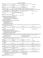
samples (Figure 1). The data was in comfort with
the finding of Arslan <i>et al.</i> (2006) and Tanabe <i>et </i>
<i>al</i>. (2007) who reported that heating of the samples
by various treatment did not significantly affect the
DNA and it was able to detect. Matsunaga <i>et al</i>.
(1999) has also studied of DNA isolation in meat
which was processed with high temperature around
100 and 120°C for 30 min of various meat flesh such
as cattle, goat, chicken, sheep, horse and pig, while
Tanabe <i>et al. </i> (2007) provided similar data of pork
at various cooked. According to Martinez and Yman
</div>
<span class='text_page_counter'>(3)</span><div class='page_container' data-page=3>
which mainly affected the quality DNA can cause the
DNA degradation into small size fragment.
<b>Figure 1.</b> Total genomic DNA extracted from beef-pork
meatball and chicken-pork meatball. (A) M: marker 100
bp DNA ladder (Invitrogen), 1: pork (100%), 2: (beef 75%
: pork 25%) 3: (Beef 90% : Pork 10%), 4: (Beef 95% :
Pork 5%)5: (Beef 97% : Pork 3%), 6: (Beef 99% : Pork
1%), 7: (Beef 100 %). (B): M: marker 100 bp DNA ladder
(Invitrogen), 1: pork (100%), 2: (chicken 75% : pork 25%)
3: (chicken 90% : Pork 10%), 4: (Chicken 95% : Pork
5%)5: (Chicken 97% : Pork 3%), 6: (Chicken 99% : Pork
1%), 7: (Beef 100 %).
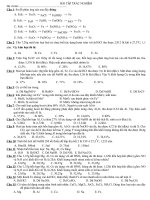
<b>Figure 2. </b>PCR products of cytochrome b gene fragments
359 bp long of samples from different meatballs
product separated by 2% high-resolution agarose gel
electrophoresis. PCR amplification using cyt b universal
</div>
<span class='text_page_counter'>(4)</span><div class='page_container' data-page=4>
Genomic DNA was applied as a template for the
PCR amplification using universal primers. Gene of
cytochrome <i>b</i> was selected for the PCR amplification
and resulted a DNA fragment of approximately 359 bp
(Figure 2). This result indicated that isolated DNA of
mixture meatball was enough for PCR amplification.
The same result of PCR amplification has also been
reported previously (Kocher <i>et al., </i>1989; Aida <i>et </i>
<i>al</i>., 2005; Erwanto <i>et al</i>., 2011). The selection of
target gene and primers affecting sensitivity and
specification of method for detection. PCR method
was very sensitive when primer target represent a
gene multicopy of like gene mitochondrial. This
research used the area mitochondrial DNA of the
cytochrome <i>b</i> as target for detection of porcine.
The PCR reaction allowed fragments of the
expected length to be obtained in all meatball samples
either beef or chicken mixed with pork, although with
various efficiencies. The mitochondrial cytochrome
b gene was selected in this study as template for DNA
amplification, because it has an acceptable length and
an adequate grade of mutation and there are numerous
sequences available in the DNA bank databases
(Kocher <i>et al</i>., 1989). The mitochondrial primers Cyt
b-FW and Cyt b-REV was able to amplify a conserved
359 bp region of the cytochrome b gene of all animal
studied, namely chicken, beef and pork.
Sequence DNA of cytochrome b gene of cattle,
goat, chicken and pig obtained from database of
NCBI was further employed for sequence alignment
using software of CLC sequencer. The similarity of
the mitochondrial cytochrome b gene among beef,
mutton, chicken and pork was 86.64%. As a result
of the preliminary CLC sequencer software analysis
for the detection of specific restriction sites on pig
sequence, a site recognized by <i>BseDI</i> enzyme was
cleaved into two fragments, namely 131 bp and 228
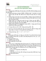
bp (Figure 3). Based on RFLP pattern using CLC
sequencer, <i>Bse</i>DI was applicable to differentiate or
identify among four species.
The digestion of PCR products resulted the
different fragment sizes, it was 131 and 228 bp at PCR
product of porcine. Basically, PCR product of mutton
could also be digested, but DNA length size was very
short (approximately 5-20 bp), consequently, it could
not be seen at 2% agarose gel (Figure 4).A clear band
with a length between 100 and 150 bp was observed
and thus referable to the 131 bp fragment, as shown
in Figure 4 (lane 1). In the same lane, a thicker band
can be traced back to the 228 bp fragment.
The data obtained suggests that compared with
<i>BsaJI</i> endonuclease profiles, the DNA restriction
patterns obtained after digestion of the amplicons
with <i>BseDI</i> enzymes consisted of same patterns.
<b>Figur</b>
<b>e 3.</b>
Sequences of nucleotide
<i>cytochr</i>
<i>ome b</i>
of
<i>Sus scr</i>
<i>ofa</i>
(pig) restriction site by
<i>Bse</i>
DI using CLC
Sequencing Software
and universal primer position in the fragment
of
<i>cytochr</i>
<i>ome b </i>
</div>
<span class='text_page_counter'>(5)</span><div class='page_container' data-page=5>
The difference between <i>BsaJI</i> and <i>BseDI</i> restriction
enzyme is the incubation time for the digestion. <i>BseDI</i>
needed 3 h for digestion, while <i>BsaJI</i> enzyme needed
more than 12 h (Aida <i>et al</i>., 2005).
PCR amplification of cytochrome b gene followed
by digestion by BseDI restriction enzymes was a
powerful technique for the identification of pork or
other pig derivative products contamination, due to
its simplicity and sensitivity. The cytochrome b gene
alignment using CLC sequencer software showed
that pig intra species have the same restriction sites
and their homology was 98.2%.
<b>Conclusions</b>
Our results allow us to conclude that PCR-RFLP
of the mitochondrial Cytochrome b gene is a suitable
alternative technique that can be applied to the
detection of pig species present in the commercialized
food products such as meatballs.
<b>Acknowledgement</b>
This research was financially supported by
grants from Riset Unggulan Strategis Nasional
LPPM Universitas Gadjah Mada (Grant number
LPPM-UGM/1309/2009). The authors also deeply
thanks to Dr. Widodo for the critical reading of this
manuscript.
<b>References</b>
Aida, A. A., Che Man, Y. B., Wong, C. M. V. L., Raha,
A. R. and Son, R. 2005. Analysis of raw meats and
fats of pigs using polymerase chain reaction for Halal
authentication. Meat Science (69): 47–52.
Arslan A., Ilhak, O. I. and Calicioglu, M. 2006. Effect of
method of cooking on identification of heat processed
beef using polymerase chain reaction (PCR) technique.
Meat Science (72): 326–330.
Colgan S., O’Brien, L., Maher, M., Shilton, N., McDonnell,
K. and Ward, S. 2001, Development of a DNA-based
assay for spesies identification in meat and bone meal.
Food Research International(34): 409-414.
Downey, G. 1998. Food and food ingredient authentication
by mid-infrared spectroscopy and chemometrics.
Trends in Analytical Chemistry (17): 418-424.
Erwanto, Y., Abidin, M. Z., Rohman, A. and Sismindari.
2011. PCR-RFLP Using <i>BseDI</i> enzyme for Pork
Authentication In Sausage and Nugget Products.
Media Peternakan (Journal of Animal Science and
Technology), (34): 14-18.
Kocher, T. D., Thomas, W. K.; Meyer, A.; Edwards, S. V.;
Paabo, S.; Villablanca, F. X. and Wilson, A. C. 1989.
Dynamics of mitochondrial DNA evolution in animals:
<b>Figure 4. </b> Restriction fragment produced by <i>BseDI</i>
restriction enzyme on 359 bp amplicons of cytochrome
b gene from different meatball products separated by
2% high-resolution agarose gel electrophoresis. PCR
amplification using cyt b universal primer. (A) M: marker
</div>
<span class='text_page_counter'>(6)</span><div class='page_container' data-page=6>
amplification and sequencing with conserved primers.
Proc. Natl. Acad. Sci. U.S.A. (86): 6196-6200.
Lopez-Andreo, I., Lugo, L., Garrido-Pertierra, A., Prieto,
M. I. and Puyet, A. 2005. Identification and quantitation
of spesies in complex DNA mixture by real-time
polymerase chain reaction. Analytical Biochemistry
(339): 73-82.
Matsunaga, T., Chikuni, K., Tanabe, R., Muroya, S.,
Shibata, K., Yamada, J., and Shinmura, Y. 1999. A
quick and simple method for the identifcation of
meat species and meat products by PCR assay. Meat
Science(51): 143-148.
Martinez and Yman, M. I. 1998. Species identification
in meat product by RAPD analysis. Food Research
International(31): 459-466.
Purnomo, H. and Rahardiyan, D. 2008. Review Article:
Indonesian Traditional Meatball. International Food
ResearchJournal (15): 101-108.
Rohman, A.,Sismindari, Erwanto, Y. and Che Man, Y. B.
2011. Analysis of pork adulteration in beef meatball
using Fourier transform infrared (FTIR) spectroscopy.
Meat Science (88): 91 – 95.
Saez, R, Sanz, Y. and Toldra, F. 2004. PCR-based
fingerprinting technique for rapid detection of animal
species in meat product. Meat Science (66): 659-665.
Tanabe, S., Eiji M., Akemi, M. and Kazuhiro, M.
</div>
<!--links-->