- Trang chủ >>
- Đề thi >>
- Đề thi lớp 7
Bài tập tổng hợp
Bạn đang xem bản rút gọn của tài liệu. Xem và tải ngay bản đầy đủ của tài liệu tại đây (231.27 KB, 9 trang )
<span class='text_page_counter'>(1)</span><div class='page_container' data-page=1>
<i>Full Length Research Paper </i>
<b>Molecular detection of invA and spv virulence genes in </b>
<i><b>Salmonella enteritidis isolated from human and animals </b></i>
<b>in Iran </b>
<b>Kumarss Amini</b>
<b>1</b><b>, Taghi Zahraei Salehi</b>
<b>1</b><b>*, Gholamreza Nikbakht</b>
<b>2</b><b>, Reza Ranjbar</b>
<b>3</b><b>, Javid Amini</b>
<b>4</b><b>and Shahrnaz Banou Ashrafganjooei</b>
<b>5 </b>1
Department of Microbiology, Faculty of Specialized Veterinary Science, Islamic Azad University, Science and Research
Branch, Tehran, Iran.
2
Department of Microbiology, Faculty of Veterinary Medicine, University of Tehran, Tehran, Iran.
3
Molecular Biology Research Center, Baqiyatallah University of Medical Sciences, Tehran, Iran.
4
Department of Microbiology, Islamic Azad University of Kerman, Iran.
5
Department of Microbiology, Kerman University of Medical Science, Kerman, Iran.
Accepted 23 August, 2010
<b>It is important to study the genotypic diversity of </b><i><b>Salmonella</b></i><b> plasmid genes which are responsible for its </b>
<b>virulence. In the present study multiplex polymerase chain reaction (multiplex PCR) assay was carried </b>
<b>out for detection of </b><i><b>Salmonella enteritidis</b></i><b> and presence of </b><i><b>invA</b></i><b> and </b><i><b>spv </b></i><b>genes. In the first stage of the </b>
<b>study, 1001 poultry samples were collected from a slaughterhouse in Kerman province (southern Iran). </b>
<b>Biochemical and serological tests were then performed for identification of </b><i><b>Salmonella</b></i><b> serovars and </b>
<b>6.79% (68/1001) were positive for </b><i><b>Salmonella</b></i><b>. Multiplex PCR with three set primers was then applied to </b>
<b>confirm serovar</b> <b>enteritidis 51.4% (35/68). Simple-PCR was then applied to detect </b><i><b>spvA </b></i><b>(</b><i><b>Salmonella</b></i>
<b>plasmid virulence), and </b><i><b>spvB </b></i><b>genes. Finally, multiplex PCR assay was carried out to simultaneously </b>
<b>detect and identify </b><i><b>invA</b></i><b> and </b><i><b>spvC</b></i> <b>genes. The presence of</b><i><b>spvA</b></i><b>, </b><i><b>spvB</b></i><b> and </b><i><b>spvC</b></i><b>in</b><i><b> S. enteritidis </b></i><b>was </b>
<b>88.6% for each gene. In the second stage of the study, thirty-three bovine (n = 13) and human (n = 20) </b><i><b>S. </b></i>
<i><b>enteritidis</b></i><b> strains were isolated from the culture collection in the Department of Microbiology, Faculty of </b>
<b>Veterinary Medicine, University of Tehran, Iran. The analyses of the samples revealed that </b><i><b>spvA, spvB, </b></i>
<b>and </b><i><b>spvC </b></i><b>genes were present in 90% of </b><i><b>S. enteritidis</b></i><b> from human sources as compared to 100% in </b>
<b>bovine sources. The study represents the first report in Iran about the genotypic diversity of </b><i><b>spvA</b></i><b>, </b><i><b>spvB</b></i>
<b>and </b><i><b>spvC</b></i><b>genes of </b><i><b>S. enteritidis. </b></i>
<b>Key words:</b><i>Salmonella enteritidis,</i> multiplex PCR, virulence genes.
<b>INTRODUCTION </b>
Salmonellosis is associated with medium to severe
morbidity and even mortality in farm animals representing
a major economic productivity loss in the food and animal
industries (Malkawi et al., 2004). <i>Salmonella enterica</i>
subspecies <i>enterica</i> serovar enteritidis is a major cause
of food-borne illness in animals and human disease
worldwide (Agron et al., 2001). An important pathogen,
<i>Salmonella</i> shows different disease syndromes and host
specificities according to their antigenic profiles (Lim et
al., 2003; Ranjbar et al., 2010). Poultry products have
been recognized as a major source of human illness
caused by this pathogen (Amavisit et al., 2001). During
*Corresponding author. E-mail: Tel:
982166427517. Fax: 982166427517.
the last decades, the isolation of <i>Salmonella</i> worldwide
has been on the increase (Madadgar et al., 2008). Several
gastroenteritis outbreaks have been reported in Iran due to
the consumption of contaminated food products (Fardsanei
et al., 2009). In fact, the incidence of food-borne cases of
infection caused by <i>Salmonella enteritidis has increased </i>
dramatically in the country during the past years. In a study
of 480 broiler chicken farms around Tehran, 67% were
reported to be contaminated with <i>S. enteritidis</i>
(Bozorgmehri et al., 1992). The contamination rate in 171
commercial poultry farms around the country was
reported to be 45% (Akbarian et al., 2007). Moreover, in
a study of 146 clinical human fecal samples, 31 cases
(21.23%) were confirmed as <i>Salmonella </i>spp<i>,</i> out of which
11 strains (35.48%) were S. enteritidis (Fardsanei et al.,
2009).
</div>
<span class='text_page_counter'>(2)</span><div class='page_container' data-page=2>
enteritidis, and its complex life cycle, many researchers
emphasize the necessity and importance of finding a
more rapid and effective detection method as a basis of
control (Agron et al., 2001; Lim et al., 2003). Presently,
<i>Salmonella</i> is detected by standard bacteriological,
biochemical and serological tests. These tests are
generally time-consuming, tedious and costly as they
require hundreds of antisera as well as well-trained
technicians (Echeita et al., 2002; Nori et al., 2010).
Several rapid and sensitive methods have been
developed for identification of <i>Salmonella</i> serotypes from
clinical samples (Zahraei et al., 2007). These methods,
however, still lack the necessary sensitivity and specificity
(Widjojoatmodjo et al., 1992; Aabo et al., 1993).
<i>In vitro</i> amplification of DNA by the polymerase chain
reaction (PCR) method is a powerful tool in
microbiological diagnostics (Malorny et al., 2003).
Multiplex PCR provides us with a specific method and
superior ability to detect <i>S</i>. <i>enterica</i> and the serovar <i>S</i>.
<i>enteritidis</i> and/ or <i>Salmonella</i> <i>typhimurium</i> in the
presence of other bacteria simultaneously (Yan et al.,
2010; Malkawi et al. 2004). In this method several genes
are used to detect <i>Salmonella</i> genus or serovars
including: Virulent chromosomal genes such as <i>invA</i>
(Malorny et al., 2003; Zahraei et al., 2006), <i>iroB</i> (Soumet
et al., 1998), <i>invE</i> (Feder et al., 2001) and <i>slyA</i> (Del
Cerro et al., 2003); fimbriae genes such as <i>fimy</i> (Yeh et
al., 2002) and <i>sefA</i> (Pan et al., 2002); unique sequence
such as Sdf I (Agron et al., 2001) and ST (Malkawi et al.,
2004) and finally plasmid genes such as <i>spv</i> (Soumet et
al., 1998). The <i>invA</i> gene of <i>Salmonella</i> contains
sequences unique to this genus and has been proved to
be a suitable PCR target with potential diagnostic
application (Jamshidi et al., 2008). This gene is
recognized as an international standard for detection of
<i>Salmonella</i> genus (Malorny et al., 2003). The <i>sefA</i> on the
other hand, is a good candidate for specific detection of
<i>S.enteritidis</i> (Pan et al., 2002; Woodward et al., 1996).
Strains of <i>S.</i> <i>enterica</i> serovar enteritidis often carry
serovar-associated plasmids which encode a virulence
operon consisting of five genes <i>spvR</i> , <i>spvA , spvB, spvC </i>
and<i> spvD</i> (Asten et al., 2005; Aabo et al., 1999). The <i>spv </i>
genes play a role in the virulence of the host strain (Chu
et al., 1999; Saule et al., 1997). It is possible that
virulence plasmid is sequentially or independently formed
by recombination and hybridization (Hong et al., 2008;
Del Cerro et al., 2003). The integration of resistance
genes and additional replicons into a <i>Salmonella</i>
virulence plasmid constitutes a new and interesting
example of plasmid evaluation posing a serious threat to
public health. These genes can be horizontally
transferred and mobilized by an F or F-like conjugative
plasmid between the <i>Salmonella</i> strains and species
(Hong et al., 2008; Chu et al., 2006). One main function
of the <i>spv</i> operon is to potentiate the systemic spread of
the pathogen (Heithoff et al., 2008). This potential is
associated with multidrug-resistance with <i>spv </i>operon
which has been demonstrated in <i>Salmonella</i> strains (Chu
et al., 2006). Some studies have provided evidence that
the virulence plasmid plays a significant role in human
disease (Guiney et al., 1994; Chu et al., 1996). Detection
of these <i>spv</i> genes allows us to decide whether the
pathogenesis of the isolates from positive clinical
samples is attributable to chromosome or plasmid born
virulence factor (Trafny et al., 2006).
The present study has three aims. Firstly, it aims at
determining whether <i>invA </i>(invasion gene of the genus
<i>Salmonella</i>) is specific for identification of <i>Salmonella</i>
genus. It also intends to determine if genes <i>sefA </i>(fimbrial
antigen of <i>S. enteritidis</i>) and <i>spv</i> (S1-S4 primers) are
specific for detection of <i>S</i>. <i>enteritidis</i> serovars. Secondly,
the study tends to assess the occurrence of <i>Salmonella</i>
spp. and <i>S. enteritidis</i> in a chicken slaughterhouse in
Kerman, Iran by multiplex PCR. This assay will then be
compared with conventional culture and biochemical
methods. The third and more important aim of the
present study is detection and determining of the
distribution of <i>spvA</i>, <i>spvB </i> and<i> spvC </i> genes in <i>S.</i>
<i>enteritidis</i> isolates from poultry, bovine and human
sources. This is the first report of the prevalence of these
genes in Iran.
<b>MATERIALS AND METHODS </b>
<b>Samples and bacterial strains </b>
A total of 1001 poultry samples including feces, liver, spleen, caecal
content, and bile (feces dominant) were collected from two poultry
slaughterhouses located in Kerman, Iran. Two days a week for a
period of nine months starting from January (2009) to September
(2009), 15 samples were randomly obtained from 15 animals.
In addition, 33 isolates of <i>S. enteritidis</i> lyophilized from human (n
= 20) and bovine (n = 13) sources were obtained from the culture
collection in the Department of Microbiology, Faculty of Veterinary
Medicine, University of Tehran, Iran. The positive control <i>S. </i>
<i>enteritidis</i> isolate and negative control <i>Escherichia coli </i>ATCC 35218
or <i>Klebsiella pneumoniae</i> were also obtained from the culture
collection in the Department of Microbiology, Faculty of Veterinary
Medicine, University of Tehran, Iran.
<b>Microbiological methods </b>
The samples, collected twice a week (15 samples in the beginning
and 15 samples in the middle of the week), were immediately
placed into sterile polyethylene bags. To determine the presence of
</div>
<span class='text_page_counter'>(3)</span><div class='page_container' data-page=3>
<b>Table 1.</b> Nucleotide sequence and primers used for identification of <i>S</i>. <i>enteritidis</i> by multiplex PCR (Pan et al., 2002).
<b>Primer</b> <b>Target gene</b> <b>Sequence</b> <b>Amplified fragment size (bp)</b>
ST11
ST14
Randoma
Sequence
5'-GCCAACCATTGCTAAATTGGCGCA
5'-GGTAGAAATTCCCAGCGGGTACTGG
429
SEFA2
SEFA4
<i>sefA</i>b 5'-GCCGTACACGAGCTTATAGA
5'-ACCTACAGGGGCACAATAAC
310
S1
S4
<i>spv</i>c 5'-GCCGTACACGAGCTTATAGA
5'-ACCTACAGGGGCACAATAAC
250
a. Randomly cloned sequence specific for the genus <i>Salmonella,</i> b. <i>S. enteritidis</i> fimbrial antigen gene (specific for the <i>S. enteritidis</i>)
and c. <i>Salmonella</i> plasmid virulent gene.
<b>Table 2. Nucleotide sequence used as primers in the multiplex PCR </b><i>invA</i>+<i>spvC </i>genes and simple-PCR <i>spvA</i>, <i>spvB</i> genes in <i>S. enteritidis</i>.
<b>Name of primer </b> <b>Gene </b> <b>Sequence </b> <b>Length (bp) </b> <b>Reference </b>
Multiplex invA and spvC
<i>inv</i>A+<i> spv</i>C
f 5’-ACAGTGCTCGTTTACGACCTGAAT-3’
r 5’-AGACGACTGGTACTGATCTAT-3’
f 5’-GTCCTTGCTCGTTTACGACCTGAAT-3’
r 5’-TCTCTTCTGCATTTCGTCA-3’
244
571
Chiu et al. (1996)
Chiu et al. (1996)
Simple- spvA -f/B
<i>spvA</i> f 5’-GTCAGACCCGTAAACAGT-3’
r 5’- GCACGCAGAGTACCCGCA-3’ 604
Del Cerro et al. (2003)
Simple- <i>s</i>pvB -f/B
<i>spvB</i> f 5’-ACGCCTCAGCGATCCGCA-3’
r 5’-GTACAACATCTCCGAGTA- 3’ 1063
Del Cerro et al. (2003)
Selenite-Cystein broth respectively for overnight enrichment at
<i>Salmonella</i> agar (Hi-Media, India). Suspicious colonies were
identified with biochemical tests (IMVIC, TSI, SIM, Urease,
Phenylalanine deaminase, and Ornithine decarboxylase). The
isolates identified as <i>Salmonella</i> were serotyped by slide
agglutination tests. Serogroup D1 was serotyped with specific O and
H antisera (<i>S.enteritidis</i>) and the other serogroups were serotyped
with only O antisera (Difco Detroit, MI, USA).
<b>Bacterial growth </b>
Lyophilized or recently isolated strains, after one-night at 37°C
incubation in 2 mL brain-heart infusion broth (BHI, Difco, Detroit, MI,
USA), were transferred to Luria-Bertani (LB) agar (Difco, France)
for one- night at 37°C to isolate single colony.
<b>DNA preparation </b>
Three colonies of each isolate on agar plate were picked and
suspended in 200 µl of distilled H2O. After vortexing, the
suspension was boiled for 10 min, and 50 µl of the supernate was
37°C. The enrichment samples for primary diagnosis were then
applied on to Xylose-Lysine-Sodium-Deoxycholate agar (Merck,
using a stomacher for 2 min, followed by incubation for 24 h at
collected after spinning for 10 min at 14,000 rpm in a
microcentrifuge (Madadgar et al., 2008).
<b>DNA primers </b>
In the first panel of multiplex PCR assay for identification of <i>S. </i>
<i>enteritidis</i> three set OF primers were selected: ST11-ST14 (429
bp), SEFA2-SEFA4 (310 bp), and S1-S4 (250 bp). In the second
panel of multiplex PCR assay, two set primers were selected: for
<i>invA </i>gene (244 bp), which is specific to <i>Salmonella </i>genus, and for
<i>spvC</i>gene (571 bp) in <i>S. enteritidis</i> (Chiu et al., 1996). Moreover,
simple-PCR with a pair of primer for <i>spvA</i> gene (604 bp) and a pair
of primer for <i>spvB</i> gene (1063 bp) in<i> S. enteritidis</i> were selected
(Del Cerro et al., 2003). The primers sequences and their
corresponding genes are shown in Tables 1 and 2.
<b>DNA amplification </b>
Multiplex PCR was performed in a reaction of 25 µl containing
reaction buffer (50 mM KCl, 1.5 mM MgCl2, 10 mM Tris-HCl pH =
8.3) (CinaGen, Iran), 2 µl of DNA sample, 200 µM dNTPs, 1 U <i>Taq</i>
polymerase (CinaGen, Iran) and 1 µm of each primer (CinaGen,
Iran). The multiplex PCR amplification program for <i>S</i>. <i>enteritidis</i>
</div>
<span class='text_page_counter'>(4)</span><div class='page_container' data-page=4>
<b>Table 3.</b> The results of serotyping evaluation and identification of S. enteritidis by multiplex PCR in poultry samples
<b>Serogroup </b>
<i><b>Salmonella</b><b>enteritidis</b></i><b> with three </b>
<b>bonds: ST11-ST14 (429bp), </b>
<b>SEFA2-SEFA4 (310 bp), S1-S4 (250 bp) </b>
<i><b>Salmonella</b><b>enteritidis</b></i><b> with two </b>
<b>bonds: ST11-ST14 (429 bp), </b>
<b>SEFA2-SEFA4 (310 bp) </b>
<i><b>Salmonella</b></i><b> spp. with two </b>
<b>bonds: ST11-ST14(429 bp), </b>
<b>S1-S4 (250 bp) </b>
<i><b>Salmonella</b></i><b> spp. with </b>
<b>one bond: ST11-ST14 </b>
<b>(429 bp) </b>
<b>Total </b>
<b>(serogroup) </b>
D1 (<i>S</i>. <i>enteritidis</i>)
31/68 (45.6%)
4/68 (5.9%)
-
-
35/68
(51.4%)
C1 and C4 (<i>Salmonella</i> spp.)
-
-
1/68 (0.68%)
18/68 (26.4%)
19/68
(27.9%)
A1 (<i>Salmonella</i> spp.)
-
-
-
7/68 (10.2%)
7/68 (10.2%)
E (<i>Salmonella</i> spp.)
-
-
-
7/68 (10.2%)
7/68 (10.2%)
Total ( Multiplex PCR )
32/68 (47%)
1/68 (0.68%)
4/68 (5.9%)
31/68 (45.6%)
68 (100%)
The PCR product was electrophoresed in 1.2% agarosis
gel (Fermentas) and afterward stained with ethidium
bromide and visualized by UV light illumination (Bio- rad,
Molecular Imager, Gel DocTM, XR Imaging system, USA).
<b>RESULTS </b>
<b>Panel 1 </b>
<b>Detection of S. enteritidis by culture and </b>
<b>serotyping </b>
Sixty-eight out of the 1001 poultry samples
(6.79%) were culture positive for Salmonella spp.
Serotyping evaluation showed that thirty-five out
of sixty-eight samples (51.4%) were serogroup D1
(S. enteritidis) with O and H antisera. Moreover,
nineteen out of sixty-eight samples (27.9%) were
serogroups C1 and C4. Seven out of sixty eight
samples (10.2%) were serogroup E. Also, seven
out of sixty eight samples (10.2%) were serogroup
A1. Finally, no serogroup B was observed in the
study (Table 3)
<b>Identification of </b><i><b>S. enteritidis</b></i><b> by multiplex PCR</b>
Multiplex PCR assay was applied to confirm
<i>S. enteritidis</i> in sixty-eight poultry samples which
were confirmed to be <i>Salmonella</i> positive through
culture and serotyping method. Results showed that
thirty-one out of sixty-eight samples (45.6%) were
positive for <i>S. enteritidis</i> with three bands
(ST11-ST14, SEFA2-SEFA4, and S1-S4) amplifying the
expected 429 310 and 250 bp fragments
res-pectively. Four out of sixty-eight samples (5.9%)
were positive <i>S. enteritidis</i> with two bands
(ST11-ST14 and SEFA2-SEFA4). One sample (0.68%)
was <i>Salmonella </i>spp. with two bands (ST11-ST14
and S1-S4). Finally, thirty-two out of sixty-eight
samples (47%) were <i>Salmonalla</i> spp. with only
one band ST11-ST14 (Table 3 and Figure 1).
<b>Panel 2 </b>
<b>Detection of </b><i><b>spvA, spvB </b></i><b>and</b><i><b> invA+spvC </b></i><b>genes </b>
<b>in</b><i><b> S.</b><b>enteritidis </b></i>
Simple-PCR to detect virulence gene <i>spvA </i>and
<i>spvB </i>with one pair primer and multiplex PCR to
detect both<i> invA</i> and <i>spvC </i>genes in the samples
yielded the following results:
<b>Poultry isolated</b><i><b> S. enteritidis</b></i>
The study showed that <i>spvA</i>,<i> </i>and<i> spvB</i>, genes
were present in 88.6% (31/35) of the samples
respectively (Figure 2). In 88.6% of the samples
(31/35) <i>spvC</i> and <i>invA</i> were present. In the same
samples (without <i>spvC</i>) <i>invA </i>genes were present
in 11.4% of the samples (4/35), (Table 4 and
Figure 3).
<b>Human isolated </b><i><b>S. enteritidis </b></i>
The study showed that <i>spvA</i>, and<i> spvB</i>, genes
were present in 90% of the samples (18/20).
Similarly, <i>spvC</i> and <i>invA</i> were present in 90% of
the samples (18/20). In the same samples
(without <i>spvC</i>) <i>invA </i>genes were present in 10%
(2/20), (Table 4).
<b>Bovine isolated</b><i><b> S. enteritidis </b></i>
As Table 2 shows, positive band appears for
<i>spvA</i>, <i>spvB</i> and <i>invA</i> + <i>spvC </i> genes in 100%
(13/13) of the all isolates (Figure 3).
<b>DISCUSSION </b>
</div>
<span class='text_page_counter'>(5)</span><div class='page_container' data-page=5>
429bp
310bp
250bp
100bp
<b> NC M PC 1 2 3 4 5 M </b>
<b>M PC 1 2 3 4 5 6 7 8 9 10 11 12 13 14 15 16 NC 17 </b>
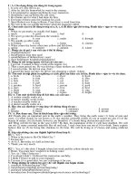
<b>Figure 1.</b> Multiplex PCR with three pairs of primers for detected <i>S. enteritidis</i> isolated (poultry source); M:
marker 100 bp; PC: positive control; NC: negative control (<i>E. coli</i>); lane 17: Product without the DNA
template; lane 1, 2, 4, 15: <i>Salmonella</i> spp. and other lane for positive <i>S. enteritidis</i>.
604bp
<b> M PC 1 2 3 4 5 6 7 8 9 10 11 12 NC </b>
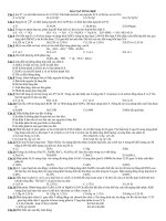
<b>Figure 2.</b> Simple- PCR with one pair of primer for spvA gene (604 bp) in <i>S.</i> <i>enteritidis</i> (poultry
source) ; M: 100 bp marker; lane PC: positive control; lane NC: negative control (PCR product
without the DNA); lanes 5, 6, 8, 10: negative spvA gene; other lanes: positive spvA gene.
<b>Table 4.</b> Distribution <i>of spvA</i>, <i>spvB, invA</i> + <i>spvC</i> genes in <i>S.enteritidis. </i>
<b>Serotype </b>
<b>Serogroup </b>
<b>Source </b>
<b>Total </b>
<i><b>spvA</b></i><b> (+) (%) </b>
<i><b>spvB</b></i><b> (+) (%) </b>
<i><b>invA+spvC </b></i>
<i><b>spv</b></i><b>C (-) </b>
<i><b>inv</b></i><b>A (+) (%) </b>
<i><b>spv</b></i><b>C (+) </b>
<i><b>inv</b></i><b>A (+) (%) </b>
<i>S. enteritidis </i>
D1
Poultry
35
88.6 (31/35)
88.6 (31/35)
11.4 (4/35)
88.6 (31/35)
<i>S.enteritidis </i>
D1
Human
20
90 (18/20)
90 (18/20)
10 (2/20)
90 (18/20)
<i>S.enteritidis </i>
D1
Bovine
13
100 (13/13)
100 (13/13)
0 (0/13)
100 (13/13)
</div>
<span class='text_page_counter'>(6)</span><div class='page_container' data-page=6>
<b>M PC 1 2 3 4 5 6 7 8 9 10 11 12 13 14 15 16 NC 17 </b>
571bp
244bp
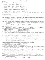
<b>Figure 3.</b> Multiplex PCR with two pairs of primers for <i>inv</i>A (244bp), <i>spv</i>C (571 bp) virulence genes in
<i>S. enteritidis</i> (bovine, and poultry source); lane M: 100 bp marker; lane PC: positive control; lane
NC: negative control (<i>Klebsiella pneumoniae</i>); lane 17: PCR product without the DNA template;
lanes 15, 16: <i>S. enteritidis</i> (positive <i>inv</i>A and negative<i> spv</i>C genes); other lanes: <i>S. enteritidis</i>
(positive <i>invA</i> and<i> spvC </i>genes).
in intensive livestock production presents explicit public
health risks in addition to food industry losses. Multiplex
PCR provides a rapid means to monitor specific
microorganisms in a variety of samples. This assay is an
epidemiologically useful tool to distinguish serovar
enteritidis. In previously reported studies where S1-S4
primers were chosen to detect the <i>spv</i> gene of a virulent
plasmid of <i>S. enteritidis</i> through multiplex PCR assay;
this gene was not confirmed in all isolates (Woodward et
al., 1999). This is also confirmed by the results obtained
in the present study (Figure 1).
Wood et al. (1994) found that this gene is only present
in 30% of <i>S</i>. <i>enteritidis</i> strains isolated from poultry. The
present study yielded different and unexpected results.
The <i>spv</i> genes were not detected in 5.9% of isolates of
the <i>S.</i> <i>enteritidis</i> as compared with 7.4% of the isolates
reported by Pan et al. (2002) and 70% of the isolates
reported by Wood et al. (1994). The study suggests that
<i>spv gene for S. enteritidis is on the increase in recent years.</i>
Previous studies suggest that selection of S1-S4
primers for the multiplex PCR as marker for the presence
of serovar enteritidis is not a suitable candidate as they
were not detected in some <i>S.enteritidis</i> serovars (Pan et
al., 2002; Wood et al., 1994). In order for the presence of
strains of <i>S</i>. <i>enteritidis</i> to be confirmed, a new pair of
primers is needed. The set of primer suggested in a
number of studies is <i>S</i>. <i>enteritidis</i> fimbrial antigen (SEFA)
(Pan et al., 2002). Agron et al. (2001) found <i>sef</i> gene in
other <i>Salmonella</i> serovars non-enteritidis. Consequently,
they suggested that <i>sef</i> gene is not specific for detection
of <i>Salmonella</i> serovar enteritidis and proposed a novel <i>S</i>.
<i>enterica</i> serovar enteritidis locus that serves as a marker
for DNA-based identification of <i>S</i>. <i>enteritidis</i>. While these
researchers suggest that <i>Salmonella</i> difference fragment
(Sdf I - 333 bp) primer is a highly specific marker for
<i>Salmonella</i> serovar enteritidis and yield’s clear results in
laboratory testing, this fragment (Sdf I) cannot detect
clearly infectious <i>S</i>. <i>enteritidis </i> isolates (Agron et al.,
2001). On the other hand, results obtained from other
researchers suggest that <i>sef</i> gene is a robust marker for
detection of <i>Salmonella</i> serovar enteritidis (Soumet et al.,
1999; Pan et al., 2002; Malkawi et al., 2004). This gene
was detected in all of the isolated <i>S</i>. <i>enteritidis</i> in the
present study (Figure 1).
Invasion gene operon, <i>invA</i> was detected in all
<i>Salmonella</i> spp. isolates in our study. This gene is
essential for full virulence in <i>Salmonella</i> and is thought to
trigger the internalization required for invasion of deeper
tissue (khan et al., 1999). There are studies reporting the
detection of this gene in all <i>Salmonella</i> spp. isolates
(Zahreai et al., 2006; Nashwa et al., 2009; Trafny et al.,
2006; Jamshidi et al., 2008). Oliveira et al. (2003)
reported that PCR assay using the invA primers specific
for <i>Salmonella </i>spp. considerably decreases the number
of false-negative results which commonly occur in
diagnostic laboratories. Amplification of <i>invA</i> is now
recognized as an international standard procedure for
detection of <i>Salmonella</i> genus (Malorny et al., 2003).
This increases the value of the present research because
of virulence properties and clinical importance of <i>invA</i>
gene. According to the results of this study, PCR method
based on<i> invA</i> gene is useful for rapid identification of
<i>Salmonella</i> serovares.
</div>
<span class='text_page_counter'>(7)</span><div class='page_container' data-page=7>
samples, 52 samples (18%) were positive for <i>Salmonella</i>
spp. by culture method (Cortez et al., 2006). From these,
5.6 and 2.4% were positive for <i>S</i>. <i>enteritidis</i> and <i>S</i>.
<i>typhimurium</i>, respectively by multiplex PCR assay.
Another study shows that from among a total of 93
samples collected from poultry carcass, 19/93 (20%)
were detected as <i>S. enteritidis</i> (Malkawi et al., 2004). Yet
in another study in U.S.A, prevalence of <i>Salmonella</i> in
poultry was reported as 25 to 29% (Harrison et al., 2001).
Agron et al. (2001) using three pair primers Sdf I, Sdf II,
and Sdf III by S.S.H (suppression subtractive
hybridization), which is a PCR-based technique, identified
81 <i>S. enterica</i> isolates with various serovars. They
detected and amplified Sdf II and Sdf III in a few <i>S.</i>
<i>enterica</i> serovars. They could detect and amplify Sdf I in
only one pathogenic <i>Salmonella</i> serovar <i>enteritidi</i>(Agron
et al., 2001). In a 1996 report from Italy, the prevalence of
food-borne <i>Salmonella </i>was reported 81% from which, 50
samples were <i>S. enteritidis</i> and only 3 samples were <i>S. </i>
<i>typhimurium </i>(Scuderi et al., 1996).
According to the results of the present study, 6.8% of
the samples were <i>Salmonella</i> serovars. This may
suggest a drop in the incidence of <i>Salmonella </i> as
compared with the previous years. There is also the
possibility of cross-contamination of products, differences
in sample origin, detection methods, sampling procedure,
and level of processing in the previous studies (Brayan
and Doyle., 1995).
From among 68 poultry source <i>Salmonella </i> spp.
isolates, no <i>S. typhimurium</i> serovar was detected after
serotyping and multiplex PCR. This is similar to a study
by Soltan et al. (2010) who did not detect <i>S</i>. <i>typhimurium</i>
in 22 <i>Salmonella</i> spp. isolates taken out of 1950 fecal
samples from diarrheic children in Tehran, Iran. This
does not suggest that <i>S</i>. <i>typhimurium</i> is an insignificant
pathogen. The pathogen is in fact detected in a number
of epidemics worldwide. In Iran, Yousefi Mashoof et al.
(2005) detected <i>S</i>. <i>typhimurium</i> in 20% of 100
<i>Salmonella </i> spp. isolates from human source. The
detected <i>S</i>. <i>typhimurium</i> from poultry source by Jafari et
al. (2005) was 8.5% in Ahwaz, Iran. In their studies in
Iran, Zahraei et al. (2006) detected this pathogen in 66%
of a total of 33 <i>Salmonella</i> spp. detected from 400 bovine
fecal samples. Soltan et al. (2008) detected 9.1% <i>S</i>.
<i>typhimurum</i> in 195 raw beef samples in Tehran.
Namimatso et al. (2005) in a report about the prevalence
of <i>S</i>. <i>typhimurium</i> from porcine source in Japan detected
this pathogen in 35.8% of a total of 106 porcine isolates.
Hughs et al. (2007) detected this pathogen in 90.6% of
32 <i>S.</i> <i>enterica</i> isolates from wild birds in northern
England.
Operon <i>spvR, spvA, spvB, spvC, spvD</i> (7.8 kb) in
virulence plasmid (<i>S</i>. <i>enteritidis</i>, 60 kb) which are present
in a few serovars of subspecies of <i>S. enterica</i> are
responsible for the systemic infection and
multidrug-resistance in both human and animals (Boyed et al.,
1998; Rotger et al., 1999; Chiu et al., 2006; Gebreyes et al.,
2009). They are also responsible for the induction of
intracellular bacterial proliferation and apoptosis of
infected macrophages (Kurita et al., 2003; Heithoff et al.,
2008). The carriage of <i>spv</i> gene may increase the
propensity of <i>Salmonella</i> straines to be of major clinical
relevance (Gebreyes et al., 2009). Heithof et al. (2008)
showed that<i> Salmonella</i> serovar <i>typhimurium</i> isolates
driven from human gastroenteritis patients often lose the
<i>spv </i>gene and, accordingly, lack the capacity to cause
systemic disease in mice. In the present study, presence
of <i>spvA</i>, <i>spvB, </i>and <i>spvC</i> genes in <i>S. enteritidis</i> from
human source was 90%.<i> Spv</i>A, <i>spvB, </i>and<i> spvC</i>genes
were present in 100% of the bovine source isolates. In
the case of <i>S. enteritidis</i> in poultry source, presence of
<i>spvA</i>, <i>spvB, </i> and <i>spvC</i> was 88.6%. Regarding the
presence of virulent plasmid genes in <i>S. enteritidis, </i>
another study has reported lack of<i> spvC</i> gene in 8/110
(7.2%) of the samples from human, pig, and poultry
sources (Castilla et al., 2006). Chiu et al. (1996)
analyzing 38 isolates from <i>Salmonella </i>serovars with two
primers <i>invA and spvC</i> reported the presence of <i>invA</i>in
all strains100% (38/38)<i>. </i>The same study reports that in
only 21strains21/38 (55.26%) there were<i> spvC</i> together
with <i>invA</i>. A study reported in 2000 has shown that from
among a total of 17 isolates of <i>S. enteritidis </i>and <i>S. </i>
<i>typhimurium,</i> all samples possess <i>invA</i> and <i>spvC</i>
together (Jenikova et al., 2000). Ling et al. (2009)
analyzed 152 isolates of <i>S. enteritidis</i> from human feces
in which only 4 isolates (8%) lacked <i>spvC</i>and possessed
<i>invA</i>. Del Cerro et al. (2003) reported that of 56 <i>S. </i>
<i>enteritidis</i> samples from human feces only 2 isolate
lacked <i>spvA, spvB, </i>and<i> spvC. </i>This last study is also in
line with the present study. Finally, the only report in Iran
about detection of <i>spvR</i> gene has confirmed that the
gene was present in 100% (16/16) of the <i>Salmonella</i>
serovars samples (Nikhbakht et al., 2004). The present
study shows a remarkably high distribution for <i>spvA, </i>
<i>spvB,</i> and<i> spvC </i>in poultry, human, and bovine sources
(88 to 100%).
There are some discrepancies about distribution of
virulence plasmid of various <i>Salmonella</i> spp. serovars
between samples from human and animal origins. In
some studies results show a higher distribution for the
virulence plasmid from animal-origin isolates than that of
human-origin (Del Cerro et al., 2003). The findings of the
present study show a higher distribution of spvA, spvB, and
<i>spvC genes in bovine sources and lower distribution in </i>
poultry sources compared with human-origin sources.
Drastic genetic variations in <i>Salmonella </i>could be derived
from transfer of this organism between human-origin and
animal-origin strains (Chiu et al., 2006). Whether this can
transfer virulence plasmid from animal-origin strains to
human-origin strains or vice versa remains to be
investi-gated. Strains of <i>Salmonella </i> bacterium (Particularly
typhimurium and enteritidis serovars) which carry
virulence plasmid can cause systemic disease, while
plasmidless strains can cause local or asymptomatic
disease (Heitoff et al.,2008).
</div>
<span class='text_page_counter'>(8)</span><div class='page_container' data-page=8>
prevalence of <i>S</i>. <i>enteritidis</i> in Iran is more than that of <i>S</i>.
<i>typhimurium</i> while the trend was opposite in the earlier
studies (Zahraei et al., 2008; Yoosefi Mashoof et al.,
2005). This study also shows that poultry carcasses have
potential for salmonellosis<i>.</i> Food hygiene necessitates
more control on <i>Salmonella</i> as this contributes to safe
food production and can help improve public health.This
is why a more reliable and rapid method for the detection
of <i>Salmonella</i> is needed in epidemiological studies and
monitoring of <i>S</i>. <i>enteritidis</i> in Iran. This study confirms
that compared with bacteriological culture method,
multiplex PCR is remarkably faster saving precious time
to control <i>S. enteritidis</i> which is the predominant serotype
in Iran. Multiplex PCR provides us with reliable, rapid,
specific, and reproducible results about the status of the
sample and detection of certain microorganisms in
large-scale epidemiological studies involving several
laboratories (Ebner et al., 2001; Chiu et al., 2006).
Multiplex PCR assay carried out in this study suggest
that set primers ST11-ST14 and invA are specific for
detection of <i>Salmonella </i>spp. Also set primers
SEFA2-SEFA4 are specific for detection of <i>S</i>. <i>enteritidis</i> serovar.
However, the study shows that presence of <i>spv</i> gene
(with set primers S1-S4) is not specific for detection of <i>S</i>.
<i>enteritidis</i> as it is seen in other <i>Salmonella</i> non-enteritidis
serovars. This is contrary to an earlier report by Pan et al.
(2002) who considered presence of set primers S1-S4
specific for detection of <i>S. enteritidis</i>.
In conclusion, epidemiological survey, identification of
<i>S</i>. <i>enteritidis</i>, and screening of <i>spv </i>gene through
PCR-based procedures can have major benefit in public health
specifically for rapid diagnosis, etiology, epidemiological
investigations, ideal vaccine, development of treatment,
and prophylactic strategies for sallmonelosis in Iran. This
is the first study on the distribution of genotypes of <i>spv</i>A,
<i>spv</i>B, <i>inv</i>A+<i>spv</i>C genes in isolates from poultry, human
and bovine sources in the country.
<b>ACKNOWLEDGEMENTS </b>
This work was supported by a grant from Faculty of
Specialized Veterinary Science, Islamic Azad University,
Science and Research Branch, Tehran, Iran. The authors
wish to thank Professor I. Sohrabi Haqdoost, Mr. M.
Abedi, Mr. A. Mokhtary, and Mr. A. H. Jangjou. Special
thanks are given to Mr. Mohammad Reza Masrour for his
considerable support during the preparation of the final of
draft of the paper.
<b>REFERENCES </b>
Aabo S, Rasmussen F, Rossen, Sorensen LPD, Olsen, JE (1993).
<i>Salmonella</i> Identification by the polymerase chain reaction. Molecular
and Cellular Probs., 7: 171-178.
Aabo S, Brown D J, Olsen E J (2000). Virulence characterization of a
strain of <i>Salmonellaenterica </i>subspecies houten with chromosomal
integrated <i>Salmonella</i> plasmid virulence (<i>spv</i>) genes. Res. Microbiol.,
151: 183-189.
Agron GP, Walker RL, Kind H, Sawyer SJ, Hayes DC, Wollard J,
Andersen GL (2001). Identification by subtractive hybridization of
sequences specific for <i>Salmonella</i> serovar Enteritidis. Appl. Environ.
Microbiol., 67(11): 4984-4991.
Amavisit P, Browing GF, Lightfood D, Anderson CS (2001). Rapid PCR
detection of <i>Salmonella </i>in horse faecal samples. Vet. Microbiol., 79:
63-74.
Asten AJ, Dijk JE (2005). Distribution of “Classic” virulence factors
<i>Salmonella </i>serovars. femsImmunol. Med. Microbiol., 44: 251- 259.
Boyed EF, Hartl DL (1998). <i>Salmonella</i> virulence plasmid Modular
Acquisition of the <i>spv </i> virulence Region by an F-Plasmid in
<i>Salmonellaenterica</i> subspecies I and Insertion into the chromosome
of subspecies II, III a, IV and VII Isolate. Gen. Soc. Am., 149:
1183-1190.
Bozorgmehri (1992). An introduction of contamination rate of
<i>Salmonella </i>Enteritidis in broiler chicken from of Tehran. Int. J. Fac.
Vet. Med. Univ. Tehran, 47(1): 70-76.
Brayan FL, Doyle MP (1995). Health risk and consequences of
<i>Salmonella</i> and <i>Campylobacter</i> Jeujeni in raw poultry J. Food
Protect., 58: 326-344.
Castilla KS, Ferreira CSA, Moreno AM, Nunes IA, Ferreira AJP (2006).
Distribution of virulence genes <i>sef</i>C<i>, pef</i>Aand <i>spv</i>C in <i>Salmonella </i>
Enteritidis phage type 4 strines isolated in Brazil. Br. J. Microbiol.,
37(2): 1-7.
Chiu CH, Ou JT (1996). Rapid identification of <i>Salmonella </i>serovars in
faeces by specific detection of virulence genes <i>inv</i>A, and <i>spv</i>C, by an
enrichment broth culture-Multiplex PCR combination assay. J. Clin.
Microbiol., 96: 2619-2622.
Chiu CH, Su LH, Chu CH, Wang MH, Yeh CM, Weill FX, Chu C (2006).
Detection of Multidrug-Resistant <i>Salmonella</i> <i>enterica </i> serovar
Typhimurium phage types DT102, DT 104 and U302 by Multiplex
PCR. J. Clin. Microbiol., 10: 2354-235.
Chu C, Hong SF, Tsai C, Lin WS, Liu TP, Ou J (1999). Comparative
physical and genetic maps of the virulence plasmids of <i>Salmonella </i>
<i>enterica </i>serovar Typhimurium, Enteritidis, Choleraesuis and Dublin.
Infect. Immun., 99: 2611- 2614.
Chu C, Chiu CH (2006). Evolutions of the Virulence Plasmid of
non-typhoid <i>Salmonella </i>and its association with antimicrobial resistance.
Microb. Infect., 8: 1931-1935.
Cortez AL, Carvalho AC, Ikuno AA, Burger KP, Idal-Marthins AM
(2006). Identification of <i>Salmonella serovars </i>Isolate from chicken
abattoirs by Multiplex PCR. Res. Vet. Sci., 81: 340-344.
Del Cerro A, Soto SM, Mendoza MC (2003). Virulence and
antimicrobial-resistance gene Profiles determined by PCR-Based
Procedures for <i>Salmonella</i> isolated from samples of animal origin.
Food Microbiol., 20: 431-436.
Ebner PD, Mathew AG (2001). Three molecular methods to identify
<i>Salmonella </i> <i>enterica</i> serotype Typhimurium DT104: PCR
fingerprinting multiplex PCR and rapid PFGE .FEMS Microbiol. Lett.,
205: 25-29.
Echeita MA, Herrera S, Garaizar J, Usera MA (2002). Multiplex
PCR-based detection an identification of the most common <i>Salmonella </i>
second-Phase flagellar antigen. Res. Microbiol., 153: 107-113.
Fardsanaei F, Soltan Dallal M, Eeshraqi SS, Zahraei T, Ranjbar R,
Akbari A, Abdosamadi Z, Amin Harati F, Nikmanesh FB (2010).
Study of <i>Salmonella</i> Eneritidis prevalence in patient referred to TUMS
hospitals.10TH Iranian congers of Microbiology, 42: 37-38.
Feder I, Nietfeld JC, Gallan J, Yeary T, Sargeant JM, Oberst R, Tamplin
ML, Luchansky JB (2001). Comparison of cultivation and PCR
hybridization for detection of <i>Salmonella</i> in porcine faecal and water
sample. J. Clin. Microbiol., 39: 2477-2484.
Gebreyes WA, Thakur S, Dorr P, Tadesse DA, Post K, Wolf L (2009).
Occurrence of <i>spvA </i>virulence Gene and clinical significance for
Multidrug-Resistant <i>Salmonella </i>strains. J. Clin. Microbiol., 10:
777-780.
Guiney D, Fang F, Krause, Libby S (1994). Plasmid mediated virulence
genes in non-typhoid <i>Salmonella</i> serovares. FEEMS Microbiol. Lett.,
124: 1-9.
Harrison AW, Griffith CJ, Tennant D, Peters AC (2001). Incidence of
</div>
<span class='text_page_counter'>(9)</span><div class='page_container' data-page=9>
33: 450-454.
Heithoff DM, Shimp WR, lau PW, Badie G, Enioutina EY, Daynes,
Barbara RA, Byrne A, House J, Mahan MJ (2008). Human
<i>Salmonella </i>Clinical Isolates Distinct from Those of Animal origin.
Appl. Environ. Microbiol., 10: 1757-1766.
Hong SF, Chiu H, Chu C, Feng C, Ou TJ (2008). Complete nucleotide
sequence of a virulence plasmid of <i>Salmonella enterica</i> serovar
Dublin and it phylogenetic relationship to the virulence plasmids of
serovars Choleraesuis, Enteritidis,Typhmurium. FEMS Microbial.,
282: 39-43.
Hughes AL, Shopland S, Wigley Paul, Bardon H, Leatherbarrow LA,
William N, Bennett M, Pinna E, Lawson B (2008). Characterisation of
<i>Salmonella </i>enteric serovar Typhimurium isolates from wild bird in
northern England from 2005-2006. BMC Vet. Res., 4: 4.
Jafari A, Ghorbanpour M, Jaidrei A (2005). A survey of the <i>Salmonella </i>
Infection in backyard chickens in Iran. Int. J. Ahvaz Vet. Microbiol.,
(35): 145.
Jamshidi A, Bassami MR, Afshari-Nic S (2008). Identification of
<i>Salmonella </i>serovars Typhimurium by a multiplex PCR-Based assay
from Poultry carcasses in Mashhad-Iran. Int. J. Vet. Res., 3(1):
43-48.
Jenikova G, Pazlarova J, Demnerova K (2000). Detection of <i>Salmonella</i>
in food sample by the combination of immunomagnetic separation
and PCR assay. Int. Microbiol., 3: 225-229.
Karimi A, Ranjbar R, Ahmadi Z, Safiri Z (2007). Rapid Detection of
Different Serovars of <i>Salmonella enterica</i> by Multiplex PCR. Iranian
J. Publ. Health, 36(2): 38-42.
Khan AA, Nawaz MS, Khan S, Sernigelia CE (1999). Detection of
Multidrug Resistant <i>Salmonella </i>Typhimurium DT104 by Multiplex
polymerase chain reaction. FEEMS Microliol. Lett., 182: 355-360.
Kurita AI, Gotoh H, Eguchi M, Okada N, Matsuura S, Matsui H, Danbara
H, Kikuchi Y (2003). Intracellular expression of the <i>Salmonella</i>
plasmid virulence protein, SPVB,causes apoptotic cell death in
eukaryotic Cells. Microb. Patho., 35: 43-48.
Lim YH, Hirose K, Izumiya H, Arakawa, Shi HT, Terajima J, Itoh K,
Tamura K, Kim S, Watanabe H (2003). Multiplex Polymerase chain
Reaction Assay for selective of <i>Salmonella enterica</i> serovar
Typhimurium. Jpn. J. Infect. Dis., 56: 151-155.
Ling JML (2009). Rapid detection of food-borne pathogens in clinical
specimens, food and environmental samples Hong Kong Med. J.,
15(1): 26-29.
Madadgar O, Zahraei Salehi T, Tadjbaksh H, Mahzounieh M, Feizabadi
M (2008). Genomic and Phenotypic evaluation of <i>Salmonella </i>
Typhimurium and <i>Salmonella </i>Enteritidis in Iran. Com. Clin. Pathol.,
17: 2298-235.
Malkawi HI, Gharaibeh R (2004). Rapid and simultaneous Identification
of two <i>Salmonella enterica</i> serotypes Enteritidis and Typhimurium
from chicken and meat product by multiplex PCR. Biotechnol., 3(1):
44-48.
Namimatsu T, Asai T, Osumi T, Imai Y, Soto S (2006). Prevalence of
the virulence plasmid in <i>Salmonella </i>Typhimurium isolates from pigs.
J. Vet. Med. Sci., 68(2): 187-188.
Nashwa HM, Mahmoud AH, Sami, Adawy S (2009). Application of
Multiplex Polymerase Chain Reaction (M-PCR) for Identification an
Characterization <i>Salmonella </i>enteritidisand <i>Salmonella typhimurium</i>,
5(12): 2343-2348.
Nikbakht G, Tadjbakhsh H (2004). Study of plasmid virulence genes
(<i>spvR</i>) in <i>Salmonella</i> <i>enterica</i> serovars isolated in Iran. J. Fac. Vet
.Med. Univ. Tehran, 59 (2):137-140.
Nori EEM, Thong KL (2010). Differentiation of <i>Salmonella</i> enterica
based on PCR detection of selected somatic and flagellar antigen.
Afr. J. Microbiol. Res., 4(9): 871-879.
Oliveira SD, Rodenbusch CR, Ce MC, Rocha SLS, Canal CWl (2003).
Evaluation of selective and non-selective enrichment PCR. Lett. Appl.
Microbiol., 36(4): 217-221.
Pan TM, liu YJ (2002). Identification of <i>Salmonella </i>Enteritidis isolate by
polymeras chain reaction and multiplex polymerase chain reaction J.
Microbiol. Immuno. Infect., 35: 147-151.
Ranjbar R, Giammanco GM, Aleo A, Plano MR, Naghoni A, Owlia P,
Mammina C. (2010). Charecterization of the first extended –
spectrum beta lactamese – producing nonthyphoidal Salmonella
staines isolated in Tehran, Iran. Foodborne Pathog. Dis., 7(1): 91-95.
Rotger R, Casadesus J (1999). The virulence plasmids of <i>Salmonella.</i>
Internal Microbiol., 2: 177-184.
Saul V R, Schaeffer F, kowarz L, Norel F (1997). Relationships between
H-NS, SPVR and growth phase in the control of spvR, the regulatory
gene of the Salmonella plasmid virulence operon. Mol. Gen. Genet.,
25b: 333-347.
Scuderi G, Fantasia M, Filetici E, Anastasio MP (1996). Food-borne
outbreaks causes by <i>Salmonella</i> in Italy, 1991-1996. Epidemiol.
Infectol., 116: 257-265.
Shakerian A, Sarofzade A, Rahimi E, Kargar Y (2010). <i>Salmonella</i>
serotype as a potential pathogen in poultry and cow carcasses
slaughtered in Isfahan-Iran. 10TH Iranian congers of Microbiology. 42:
333.
Soltan DM, Fardsanaei F, Eesraqi SS, Zahraei T, Ranjbar R, Akbari A,
Abdosamadi Z, Amin harati F, Nikmanesf B (2009). The antimicrobial
resistance pattern among <i>Salmonella</i> Enteritidis isolate from 1950
diiarhoic children in Iran J. Med. Univ. Tehran, 67(12): 35-48.
Soltan Dallal M, Vahedi S, Zeraati H, Kalantar E (2008). Incidence of
<i>Salmonella </i>serovars and its antimicrobial pattern in barbecued meat
and ground beef burgers in Tehran. Iranian J. Microbiol., 1: 37-41.
Soumet C, Ermel G, Rose N, Rose V, Drouin P, Salvat G, Colin P
(1999). Evaluation of a Multiplex PCR assay for for simultaneous
identification of <i>Salmonella </i> serovar <i>Salmonella </i> Enteritidis and
<i>Salmonella </i>Typhimuriumfrom environmental swab of poultry houses.
Lett. Appl. Microbiol., 25: 113-117.
Trafny EA, Kozlowska k, Szpakowska (2006). A novel Multiplex PCR
assey for the detection of <i>Salmonella</i> enteric serovar Enteritidis in
human faeces. Lett. Appl. Microbiol., 43: 673-679.
Widjojoatmodjo, N M, Fluit A C, Torensma R, Verdonk G P and Verhoef
J (1992). The magnetic immune polymerase chain reaction assay for
direct detection of <i>Salmonella</i> in faecal sample. J. Clin. Microbiol., 30:
3195-3199.
Wood MW, Mahon J, Lax AJ (1994). Development of a probe and PCR
primers specific to virulence plasmid of <i>Salmonella </i>Enteritidis<i>.</i> Mol.
Cell Probes, 8: 473-479.
Woodward MJ, Kirwan SE (1996). Detection of <i>Salmonella </i>Enteriitidisin
eggs by thepolymerase chain reaction. Vet . Record, 138: 411-413.
Yan, Yusof MY, Sekaran SD (2010). Multiplex polymerase chain
reaction (M-PCR) assay for the detection of <i>Enterobactericeae</i> in
clinical samples. Afr. J. Microbiol. Res., 4(11): 1186-1186-1191.
Yeh KS, Chen TH, Liao CW, Chang CVS, Lo HC (2002). PCR
amplification of the <i>Salmonella</i> Typhimurium <i>fimY </i>gene sequence to
detect the <i>Salmonella</i> species. Int. J. Food Microbiol., 78: 227 234.
Yousefi M, Goodarzi A, Azadi S (2006). Identification and non typhid
<i>Salmonella</i> typing by SDS-PAGE method. Int. J. Fac. Med. Uni. Iran-
Hamadan, 12(3): 48.
Zahraei T, Mahzoonae MR, Ashrafi A (2006). Ampllification of <i>invA</i>
gene of <i>Salmonella</i> by polymerase chain reaction (PCR) as a specific
method for detection of <i>Salmonella</i>. J. Fac. Vet. Med. Univ. Tehran.,
61(2): 195-199.
</div>
<!--links-->