Microwire arrays for studying the influence of temperature gradients on the cellular behavior
Bạn đang xem bản rút gọn của tài liệu. Xem và tải ngay bản đầy đủ của tài liệu tại đây (6.14 MB, 144 trang )
Fakultät für Elektrotechnik und Informationstechnik
Professur für Neuroelektronik
Microwire arrays for studying the
influence of temperature gradients
on the cellular behavior
Truong Ka My Dang
Vollständiger Abdruck der von der Fakultät für Elektrotechnik und
Informationstechnik der Technischen Universität München zur Erlangung des
akademischen Grades eines
Doktor-Ingenieurs (Dr.-Ing.)
genehmigten Dissertation.
Vorsitzende: Prof. Dr.-Ing.Christian Jirauschek
Prüfer der Dissertation:
1. Prof. Dr. Bernhard Wolfrum
2. Prof. Dr. Oliver Hayden
Die Dissertation wurde am 13.08.2018 bei der Technischen Universität
München eingereicht und durch die Fakultät für Elektrotechnik und
Informationstechnik am 03.12.2018 angenommen.
A BSTRACT
Temperature plays an important role in regulating the biological function
of cellular development. Over the past few decades, the development
of microfabrication technology has enabled highly localized temperature
control at the scale of a few microns. This allows the study of thermal
influences on biological microsystems. In this context, the present thesis
investigates the changes of cellular behavior depending on temperature
gradients using microwire arrays. In addition, the thesis also introduces
a sacrificial layer approach to fabricate microchannels for studying neuronal guidance. The integration of the microwire array into microchannels presented in this thesis proposes a novel method for governing neuronal networks under the influences of temperature gradients.
The first part of this thesis presents the application of microwire arrays
to study the effects of localized temperature gradients on signal propagation in cardiac cell networks. In order to investigate the effects of temperature on cellular networks, a monolayer of cardiomyocytes is cultured
onto the chip. The localized temperature is induced by using a power
supply connected to microwire arrays. A calcium imaging method is
performed to evaluate changes in signal propagation under local heat
stimulation. Calcium signals are recorded using a high speed and low
light camera. Also in this part, the velocity of the calcium signal propagation in cellular networks is locally increased upon heat stimulation.
Additionally, this part presents a relocation of the pacemaker cell and a
deformation of the calcium wavefront caused by an increase of the local
temperature.
In the second part, the growth of neurons is investigated under the influence of temperature gradients. In this study, the PC12 neuron-like cells
are cultured on the microwire array chip. The effects of temperature gradients on neurons are examined, focusing on the speed and direction of
neurite outgrowth. The growth of neurites under local heat stimulation
is recorded using a time-lapse imaging method. The images are then analyzed to estimate the development of neurons within temperature gradients. The result indicates that the growth speed of neurons increases with
heat stimulation. Moreover, the evidence that temperature gradients affect the growth direction of neurites is observed in a long-term period
experiment (24 h after heating).
The last part of the thesis presents a promising fabrication approach,
based on standard clean room fabrication and sacrificial layer etching, for
combining microwire arrays with a set of perpendicular axon-guiding microchannels. This approach enables the positioning of neurites, as well
as control over their polarity. In particular, an array of asymmetrical microchannels with wide inlets and narrow outlets is fabricated to allow
neurites to grow in one direction. The experiment with neurites from
PC12 cells demonstrates that neurites can grow from the entrance to the
exit of the microchannels. Furthermore, the fabrication of microchannels,
integrated with microwire arrays, allows the study of temperature gradients effects on the formation of functional neuronal networks.
Overall, this thesis focuses on investigating thermal impacts on cellular behavior using the localized heating generated by microwire arrays.
This work demonstrates the important role of microscopic heating in regulating signal propagation, as well as the growing properties of cellular networks. Specifically, the temperature highly confined in a small
area governs the calcium signal propagation of cardiomyocytes and the
growth of neurites towards the heat source. Other than that, concerning
the technical aspects, the fabrication of thin microwire enables us to control high temperature in a microscopic area. Furthermore, the fabrication
of sacrificial microchannels contributes benefits for neuronal guidance,
ii
such as reducing a number of fabrication steps and improving the adhesion between microchannels and the chip’s surface.
iii
iv
C ONTENTS
1
Introduction
1
2
Fundamentals & Theory
5
2.1
The cell . . . . . . . . . . . . . . . . . . . . . . . . . . . . . .
6
2.1.1
The cell structure . . . . . . . . . . . . . . . . . . . .
6
2.1.2
Membrane potential . . . . . . . . . . . . . . . . . .
8
2.1.3
Action potentials and signal propagation . . . . . .
9
2.1.4
Signal propagation in cardiac cells . . . . . . . . . .
13
2.1.5
TRPV channels as temperature sensors . . . . . . .
15
Cell Models . . . . . . . . . . . . . . . . . . . . . . . . . . .
16
2.2.1
HL-1 cells: A cardiomyocyte cell line
. . . . . . . .
16
2.2.2
PC12 cells as a model for neurons . . . . . . . . . .
16
Devices and fabrication techniques . . . . . . . . . . . . . .
17
2.3.1
Microwire array . . . . . . . . . . . . . . . . . . . . .
17
2.3.2
Microfluidic channels for cell culture . . . . . . . . .
18
2.3.3
Fabrication technologies for microfluidic channels .
22
2.2
2.3
2.4
2.5
3
Heat stimulation
. . . . . . . . . . . . . . . . . . . . . . . .
25
2.4.1
Resistive heating . . . . . . . . . . . . . . . . . . . .
25
2.4.2
Heat transfer . . . . . . . . . . . . . . . . . . . . . .
27
Optical detection . . . . . . . . . . . . . . . . . . . . . . . .
33
2.5.1
Calcium imaging . . . . . . . . . . . . . . . . . . . .
33
2.5.2
Fluorescence lifetime imaging (FLIM) . . . . . . . .
35
Heat stimulation for modulating signal propagation in HL-1 cell
networks
37
C ONTENTS
3.1
Preamble . . . . . . . . . . . . . . . . . . . . . . . . . . . . .
38
3.2
Introduction . . . . . . . . . . . . . . . . . . . . . . . . . . .
38
3.3
Materials and methods . . . . . . . . . . . . . . . . . . . . .
39
3.3.1
Fabrication of microwire array chips . . . . . . . . .
39
3.3.2
Culture of HL-1 cells on the microwire array chip .
40
3.4
3.3.3
Thermal stimulation and Ca
imaging . . . . . . .
41
3.3.4
Cross-correlation analysis . . . . . . . . . . . . . . .
41
Results and discussions . . . . . . . . . . . . . . . . . . . .
43
Effect of localized heat stimulation on Ca2+ signal
propagation . . . . . . . . . . . . . . . . . . . . . . .
43
Effect of localized heat stimulation on the pacemaker
position . . . . . . . . . . . . . . . . . . . . . . . . .
50
Conclusions and outlook . . . . . . . . . . . . . . . . . . . .
51
3.4.1
3.4.2
3.5
4
Heat activation and guidance of neurite outgrowth
55
4.1
Preamble . . . . . . . . . . . . . . . . . . . . . . . . . . . . .
56
4.2
Introduction . . . . . . . . . . . . . . . . . . . . . . . . . . .
56
4.3
Materials and methods . . . . . . . . . . . . . . . . . . . . .
57
4.3.1
PC12 cell culture on chips . . . . . . . . . . . . . . .
57
4.3.2
Heat stimulation Setup . . . . . . . . . . . . . . . . .
58
4.3.3
Thermal stimulation and imaging . . . . . . . . . .
58
4.3.4
Image processing using the optical flow method . .
60
Results and discussion . . . . . . . . . . . . . . . . . . . . .
60
4.4.1
Effect of temperature gradients on cellular growth .
60
4.4.2
Influence of temperature gradients on the direction
of cellular growth . . . . . . . . . . . . . . . . . . . .
65
Temperature distribution on microwire . . . . . . .
69
Conclusions and outlook . . . . . . . . . . . . . . . . . . . .
70
4.4
4.4.3
4.5
5
vi
2+
Fabrication of microchannel structures for neuronal guidance towards heat activation
73
5.1
Preamble . . . . . . . . . . . . . . . . . . . . . . . . . . . . .
74
5.2
Introduction . . . . . . . . . . . . . . . . . . . . . . . . . . .
74
5.3
Materials and Methods . . . . . . . . . . . . . . . . . . . . .
75
C ONTENTS
5.3.1
5.3.2
5.3.3
5.3.4
Fabrication of PDMS microchannels . . . . . . . . .
Fabrication of aligned SU-8 microchannel . . . . . .
Cell Culture . . . . . . . . . . . . . . . . . . . . . . .
Fluorescence imaging of the neurite length in microchannels . . . . . . . . . . . . . . . . . . . . . . .
Results and discussion . . . . . . . . . . . . . . . . . . . . .
5.4.1 PDMS microchannels and its drawbacks . . . . . .
5.4.2 Microwire array and SU-8 microchannel structures
5.4.3 Neurite outgrowth into SU-8 microchannels . . . .
Conclusion and Outlook . . . . . . . . . . . . . . . . . . . .
75
76
79
Conclusions and outlook
6.1 Summary of the Thesis . . . . . . . . . . . . . . . . . . . . .
6.2 Outlook . . . . . . . . . . . . . . . . . . . . . . . . . . . . . .
89
90
91
5.4
5.5
6
80
80
80
82
85
87
Appendix
95
A Geometry and parameters for temperature simulation
95
B Protocol for bonding PDMS to a polyimide surface
97
C Protocol for HL-1 cell culture
99
D Protocol for PC12 cell culture
103
E Protocol for preparing dyes
E.1 Fluo-4 . . . . . . . . . . . . . . . . . . . . . . . . . . . . . . .
E.2 Calcein-AM/Ethidium Homodimer(EthD) . . . . . . . . .
E.3 CellTracker Green . . . . . . . . . . . . . . . . . . . . . . . .
107
107
107
107
Author’s list of publications
127
Conference presentations
129
Acknowledgment
131
vii
C ONTENTS
viii
C HAPTER 1
I NTRODUCTION
Temperature is an important physical parameter affecting every life process. In mammals, temperature is well known as an essential vital sign
that indicates sickness or health. Additionally, temperature plays a crucial role in regulating a variety of mechanisms in biological systems. It
has been demonstrated that cells decrease their growth rate under low
temperature conditions [1, 2]. Conversely, the proliferation rate of cells
rises in response to an increase of ambient temperature [3]. In line with
this, the previous study of Kang et al. [4] shows that temperature has a
comprehensive influence on intracellular calcium dynamics, which manipulates most biochemical processes such as neural transmission, muscle contraction, and heart beat. In fact, many studies have demonstrated
that temperature has a profound effect on living systems such as birth
length [5], life span [6] and aging-associated diseases [7].
Together with temperature itself, temperature gradients also play a significant role in regulating the living systems. Spatial and temporal variation of temperature induces changes in the nature of chemical reactions
and alters the configuration of atoms that build up nucleic acids, proteins, lipids and other biomolecules [8]. A spatial temperature gradient
has been demonstrated to be a guidance cue for the migration of organisms. Many studies indicate that thermotaxis can cause an organism to
move up or down according to a gradient temperature [9–11]. An exper-
1. I NTRODUCTION
iment with Caenorhabditis elegans, a transparent nematode (roundworm),
has shown that Caenorhabditis elegans migrates to a certain temperature
when subjected to a spatial temperature gradient [12]. While the temperature sensitivity of organisms is well known, it has been not well understood how individual cells respond to different temperatures. The study
of temperature sensitivity at a cellular level has been a challenging engineering problem, possibly due to the lack of microscopic systems which
can produce temperature at microscales.
Over the past few decades, microtechnology has been developed as a
new approach for studies in biophysics and physical chemistry [13–15].
Microtechnology employs fabrication methods (e.g. photolithography)
to create microsystems on chips, such as lab-on-a-chip or micro-totalanalysis-systems. As a result of the development of microelectromechanical systems (MEMS) and microfluidic devices, scientists are now able
to investigate and manipulate biological systems at a cellular level [16–
19]. MEMS offer the ability to precisely manipulate cells on microscales,
which makes it possible to study cellular interactions with physical and
chemical microenvironments surrounding them [17, 20]. These interactions offer deep insights into the fundamental behaviors of biological microsystems, such as pacemaker cells, calcium concentrations, neuronal
outgrowth, cellular viability, and cellular locomotion.
This thesis aims to study cellular networks regulating and responding
to local heating generated by microwire array chips. Particularly, the influence of localized temperature gradients on calcium signal propagation
in cardiomyocytes will be investigated. A rise of local temperature will
cause an increase in the propagation speed of these signals. This results
in a deformation of the calcium wave in the cardiac networks. Additionally, the local temperature will be used to stimulate cells in the heated
zone. This stimulation causes the cells to become more active. This results in the relocation of pacemaker cells to the heated area. Moreover,
the effects of temperature gradients on neuron-like cells will be examined in this work. Temperature gradients could accelerate the speed of
neurite outgrowth and be a guidance cue for neurites. To access cellular
2
behavior in response to localized heating, a fabrication approach which
combines microheaters and microfluidic channels for neurite guidance
will be introduced in this thesis.
The local heating with high spatial accuracy induced from microwire
array chips provides an effective approach to study heat effects at cellular levels. Studying the influence of heat on living cells at a microscale
creates a pathway to understanding how cells are sensed by the adjacent cells and communicate with each other in response to temperature
changes.
The thesis contains six chapters. In the introduction, the motivation for
the research is presented. The second chapter takes up the fundamentals
and methods. This chapter describes the basic concepts of biology and
physics which are used in the following experiments and studies. The
third chapter presents the study of local heat stimulation for modulating
signal propagation in cardiomyocyte-like cell (HL-1 cells) networks. The
study of the effect of heat activation on the guidance of neurite outgrowth
will be conducted in the fourth chapter. The next chapter describes the
fabrication of microchannels perpendicular to microwire arrays for studying the effects of thermal gradients on neurite outgrowth and neuronal
guidance. The last chapter presents the conclusions and outlook, which
suggests possible applications of microwire arrays in studies of neuronal
regeneration, cell migration, and dopamine releases.
3
1. I NTRODUCTION
4
C HAPTER 2
F UNDAMENTALS & T HEORY
2. F UNDAMENTALS & T HEORY
2.1 T HE CELL
2.1.1 T HE CELL STRUCTURE
The cell is the basic unit of biological activity. The name “cell” was
suggested by Robert Hooke in 1665. The meaning is derived from the
Latin cella which means storeroom or chamber [21]. There are two different types of cells, prokaryotic cells and eukaryotic cells. The structure
of prokaryotic cells is very simple, whereas the structure of eukaryotic
cells is more complex. The cells from plants and animals are all eukaryotic cells. Since the cells used in this work are cardiomyocyte-like cells
and neuron-like cells, only the structure of eukaryotic cells is described
in detail.
The size of eukaryotic cells is between 1 and 100 µm. The structure of
eukaryotes is shown in Figure 2.1. Surrounding the cell is the plasma
membrane, which protects and separates the cell from the external environment. The cell membrane contains many biomolecules such as proteins. These proteins form ion channels which are embedded in the membrane to allow substances to cross through the cell membrane. Inside the
cell, many subcellular components work together to maintain the cell’s
activity. The nucleus contains genetic information, which controls the activity of the cell by regulating gene expression. Gene expression is the
process used to synthesize a functional protein from a gene [22]. Within
the nucleus is the nucleolus where ribosomes are synthesized. Ribosomes
are found “free” in the cytoplasm or attached to the rough endoplasmic
reticulum to produce proteins.
Another type of endoplasmic reticulum is the smooth endoplasmic
reticulum, which plays an important role in the metabolism and the synthesis of lipids. In muscle cells, the sarcoplasmic reticulum is a special
type of the smooth endoplasmic reticulum that stores and releases calcium for moderating the excitation and contraction process. Proteins produced in the endoplasmic reticulum are transported to the membrane
or lysosomes by vesicles via the Golgi apparatus. Inside the lysosome,
6
2.1. T HE
CELL
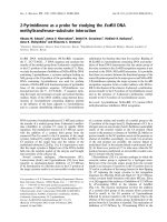
F IGURE 2.1: Structure of the eukaryotic cell contains many structural subunits:
1) Nucleolus, 2) Nucleus, 3) Ribosome, 4) Vesicle, 5) Rough endoplasmic reticulum, 6) Golgi apparatus, 7) Cytoskeleton, 8) Smooth endoplasmic reticulum, 9)
Mitochodria, 10) Vacuoles, 11) Cytoplasm, 12) Lysosomes, 13) Centriole.
biomolecules, including macromolecules such as proteins, nucleic acids,
or small molecules, are disassembled by hydrolytic enzymes. This process is similar to the activity of digestion [22].
Besides the degradation process of molecules, lysosomes also participate in recycling processes inside the cell. To maintain the shape and
the size of the cell, the cytoskeleton is present in all cells. The cytoskeleton is a dynamic structure which is involved in the rapid growth of the
cell. Three main kinds of cytoskeletal filaments are microfilaments, microtubules, and intermediate filaments [22]. Microfilaments are also called
actin filaments, which are made of actin proteins that act as tracks for the
movement of myosin molecules.
In muscle cells, the binding of myosin on actin generates forces which
perform the contraction in cardiomyocyte. Microtubules serve as tracks
for organelles to move along within the cell. Interactions between microtubules and microflilaments play a crucial role in processes of neurite
outgrowth and neurite guidance [23]. Intermediate filaments together
with microfilaments maintain the cell shape and form cell-cell connections. The centriole consists of two bundles of microtubules which are
involved in cell polarity. To supply energy for the cell, mitochondria generate the energy and transform it into adenoine triphosphate (ATP). For
this reason, they are sometimes considered as the “powerhouse” of the
cell.
7
2. F UNDAMENTALS & T HEORY
2.1.2 M EMBRANE POTENTIAL
As described in subsection 2.1.1, numerous types of ion channels are
embedded in the cell membranes. These ion channels serve as conduits
that regulate the flow of ions through the cell membrane. The influx and
efflux of ions through the cell membrane result in a difference in electrical
potential between the intracellular and extracellular environment. This
potential difference is called membrane potential.
Signals are transmitted through the cell by opening or closing the ion
channels in the membrane, causing a local change of membrane potential.
The movement of ions results in an asymmetric distribution, causing ion
gradients to cross through the cell membrane. Cells control and use the
ion gradients in different ways to help them communicate with their surrounding environment. In electrogenic cells such as neurons and muscle
cells, the membrane potential triggers action potentials to transmit signals.
When the influx and the efflux of ions are in balance, the cell is at rest
and this state is called the resting membrane potential. The equilibrium
distribution of ions through the membrane is maintained by the movement of ions. Three important ions which contribute to the membrane
potential are sodium (Na+ ), potassium (K+ ) and chloride (Cl – ). These
ions are distributed inside and outside of the cell. Their concentrations
are given in Table 2.1 [24]. Since the membrane is more permeable for
K+ than for other ions, the outflow of K+ is the dominant process needed
to maintain the resting potential. The voltage at which the net ion flow
through the membrane is zero is called the equilibrium potential. The
TABLE 2.1: Approximate concentrations of intracellular and extrallular ions in
mammalian cells (Note: these are concentrations of free ions).
Ion
Na+
K+
Cl –
8
Intracellular
concentration (mM)
15
140
10
Extracellular
concentration (mM)
140
4.5
120
2.1. T HE
CELL
equilibrium potential of an ion can be calculated using the Nernst equation. This equation calculates the equilibrium potential for an ion based
on the charge of the ion and its concentration gradient across the membrane. For example, the equilibrium potential for K+ can be calculated as
follows:
RT [K+ ]out
Eeq,K+ =
ln
,
(2.1)
zFa
[K+ ]in
where Eeq,K+ is the equilibrium potential for K+ (V), R is the universal gas
constant (J mol−1 K−1 ), T is the absolute temperature (K), z is the number
of elementary charges of the ion, Fa is the Faraday constant, [K+ ]out is
the extracellular concentration of ion K+ , and [K+ ]in is the intracellular
concentration of ion K+ .
The above equation is calculated only for K+ . In real cells, the resting
membrane potential is estimated for the ions which give the strongest current through the membrane. In fact, there are contributions from ions Cl –
and Na+ ; therefore three ions K+ , Cl – and Na+ must be considered. The
resting membrane potential for these three ions is given by the GoldmanHodgkin-Katz equation:
EM =
RT PK [K+ ]out + PNa [Na+ ]out + PCl [Cl− ]in
ln
,
Fa
PK [K+ ]in + PNa [Na+ ]in + PCl [Cl− ]out
(2.2)
where PK , PNa and PCl are the relative permeabilities of the ion K+ , Na+
and Cl – respectively. Normally, permeability values are reported as relative permeability with PK . In most cells at resting membrane potential,
the permeability of K+ is higher than Na+ and Cl – . The charge z is not
explicitly present as it was considered within the terms of the individual
K+ . A sketch of various ion channels embedded in the plasma membrane
is shown in Figure 2.2.
2.1.3 ACTION POTENTIALS AND SIGNAL PROPAGATION
As described in subsection 2.1.2, the electrical potential difference is
caused by the different concentrations of ions through the membrane.
Rapid changes in the membrane potential result in changes in the volt9
2. F UNDAMENTALS & T HEORY
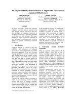
F IGURE 2.2: Schematic of the ion channels are embedded in the plasma membrane. Image reprinted with permission from [25].
age through the membrane, which leads to the propagation of action potentials. The action potential can be divided into three main phases. A
typical action potential is shown in Figure 2.3. The initial phase is a rapid
change in membrane potential from resting state to the equilibrium potential of the ions. This phase is called depolarization. In this phase, a
stimulus causes the channels to open, leading to an influx of Na+ ions,
which in turn produces an increase in the membrane potential from negative to positive values. For example, in neuronal cells, the membrane
potential changes from −70 mV at rest to 40 mV in the depolarization
phase [26].
The second phase is called repolarization, which is a return to resting
membrane potential. In this phase, potassium channels are opened, leading to an outward flow of K+ ions, which causes the membrane to return
to the resting state. The third phase is a short period of time after repolarization, which is called the refractory period. In this phase, sodium
channels are inactivated and some potassium ion channels are still open
to let the K+ ions flow out of the membrane. After this phase, Na+ /K+
pumps eventually bring the membrane back to the resting state.
10
2.1. T HE
CELL
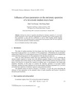
F IGURE 2.3: A schematic shows various phases of a typical action potential.
C ARDIAC ACTION POTENTIALS Cardiac action potentials differ from action potentials found in other electrogenic cells, which are initiated by
nervous activity. The cardiac action potential is generated by specific
cells within cardiac cells (pacemaker cells). A typical action potential of
cardiac cells is shown in Figure 2.4. As shown from the schematic, there
are five phases of the cardiac action potential. Before the action potential occurs, the membrane potential maintains at a constant level which
is called resting membrane potential (phase 4). At this phase, the membrane potential results from the balance of the influx of ions (e.g. sodium
and calcium) and the efflux of ions (e.g. potassium). Following a stimulus
of the membrane, the fast sodium channels open, causing an increase in
sodium influx, leading to the depolarization of the cell membrane (phase
0). As the sodium channels rapidly close, the potassium channels open
and close rapidly, allowing a small flow of potassium ions out of the cell
and leading to the membrane potential slightly more negative (phase 1).
The abrupt depolarization triggers the opening of calcium and potassium
channels, which allows the calcium influx and the potassium efflux. The
opening of calcium channels and potassium channels results in a balance
of the calcium influx and the potassium efflux. This results in a plateau
11
2. F UNDAMENTALS & T HEORY
F IGURE 2.4: Schematic of an action potential from a cardiomyocyte. 0 Depolarisation. 1 First repolarization. 2 Plateau. 3 Second repolarization. 4 Resting
membrane potential.
of the action potential (phase 2). As the calcium channels close, the potassium channels still open causing the potassium efflux, which leads to a
full repolarization of the cell (phase 3). Finally, the potasium channels
close and the membrane potential reaches the resting state of membrane
potential (phase 4).
PACEMAKER CELLS Some cells can generate action potentials without
any stimulus, and these are called pacemaker cells. In cardiac cells, these
cells reside in the sinoatrial node of the heart, regulating the beating
rate. At the resting state of an action potential, the pacemaker cells generate a gradual increase of the membrane potential by allowing an influx of sodium ions. When the membrane potential reaches the threshold
−40 mV, it activates an opening of the calcium channels. The influx of
calcium ions initiates depolarization of the cell. Subsequently, potassium
channels open, allowing an efflux of potassium ions and establishing the
repolarization state of the cell. Once the cell is repolarized, leak channels
(e.g sodium channels) cause an instability of the resting potential and
result in a firing of the next action potential [27]. The unstable resting
potentials of pacemaker cells enable spontaneous depolarizations which
are relatively fast and repeat themselves regularly. The fast depolarizations of pacemaker cells control contractile cardiomyocytes by means of a
mechanism called overdrive suppression, which states that the cells with
the most rapid frequency of depolarization control the overall beating of
12
2.1. T HE
CELL
F IGURE 2.5: Schematic of a gap junction. Adapted from [31].
cardiac cells [27]. The pacemaker cells are connected to neighboring cells
via gap junctions (see below), which allow them to locally depolarize adjacent cells. This function allows pacemaker cells to control contractions
in cardiomyocytes [28].
2.1.4 S IGNAL PROPAGATION IN CARDIAC CELLS
G AP JUNCTIONS Gap junctions are channels shared with the plasma
membrane of neighboring cells (see Figure 2.5). They are formed by proteins called connexons; each connexon consists of six proteins called connexins. Molecules, ions, and electrical impulses can pass through these
channels, allowing cells to communicate with each other. In this way,
when the cells are depolarized above a certain threshold, this depolarization triggers the propagation of action potentials in the next cells through
the gap junctions. The propagation of these signals is very fast. The propagation speed of cardiomyocyte-like cells (HL-1 cells) is in the range of
7 −30 mm s−1 [29, 30].
E XCITATION - CONTRACTION COUPLING OF CARDIAC CELLS In cardiac
cells, the excitation-contraction coupling is a key process of turning electrical excitations into mechanical contractions. This process is initiated by
13
2. F UNDAMENTALS & T HEORY
F IGURE 2.6: Schematic of excitation-contraction coupling of cardiac cells. Image reprinted with permission from [32].
local membrane depolarization, causing calcium-induced calcium release
from the sarcoplasmic reticulum (SR). The released calcium activates myofilaments that eventually initiate the contraction as explained below.
Membrane depolarization during the action potential leads to an opening of voltage-gated L-type calcium channels (Figure 2.6). The opening
of L-type calcium channels, located in the T-tubule membrane, causes
an influx of calcium, which results in the increase of calcium concentration. The inward flow of calcium ions binds to ryanodine receptors (RyR)
located on the SR, triggering intracellular calcium release from the sarcoplasmic reticulum (SR). The increase in the free intracellular calcium
allows calcium ions to bind to troponin C, a protein of the thin filament.
This binding causes tropomyosin to shift, exposing the myosin binding
site on the actin. Once the myosin binds to the actin, a force is generated.
At the end of the cycle, to relax the contraction, calcium is released from
troponin-C and then either pumped out of the cell by the sodium-calcium
exchanger or pumped into the SR by the sarcoplasmic reticulum calcium
ATPase (SERCA) pump [33].
14
2.1. T HE
CELL
This is the explanation of the process of contraction in an individual
cardiomyocyte. In the standard experiment of this thesis, these cells are
grown as a confluent layer to form gap junctions. Therefore, signals can
propagate across the cell layer via gap junctions (see above).
2.1.5 TRPV CHANNELS AS TEMPERATURE SENSORS
Temperature sensing allows organisms to adapt to changes of environmental conditions. Over the past years, it is known that the temperature
is sensed by means of ion channels located in cellular membrane [34, 35].
The ion channels are sensitive to heat called thermal-transient receptor
potential (TRPV) channels. The temperature dependence of these TRPV
channels is estimated based on Q10 value. Q10 is a change in the rate of
reaction when the temperature rises by 10 ◦C. The Q10 value is calculated
as follows:
Q10 = (
R2 T 10
) 2 −T1 ,
R1
(2.3)
where R1 and R2 are the rate of the reaction at temperature T1 and T2 ,
respectively.
The thermal TRPV channels are characterized for having greater thermal sensitivity with Q10 values much higher than 2 (6 < Q10 < 30)
[36, 37]. There are four different types of thermal-sensing ion channels
in the TRPV family called TRPV1, TRPV2, TRPV3, and TRPV4. The
structure of these TRPV channels is similar to the TRP (Transient Receptor Potential) ion channels superfamily, which consists of proteins with
six transmembrane domains. These groups of proteins are assembled to
form cation-permeable ion channels. The rise of temperature causes these
ion channels to open, leading to the influx of ions through open channels.
TRPV1 ion channels are calcium-permeable receptors activated by capsaicin with temperature threshold of 43 ◦C. The activation of TRPV1 is
relatively fast with the time response in a range of a few milliseconds. If
the temperature is kept constant for a while, the sensitivity of the channels is decreased. The TRPV2 ion channels are activated by noxious tem15