Nghiên cứu hoạt tính gây độc một số dòng tế bào ung thư của các hợp chất phân lập từ ba loài san hô mềm sinularia nanolobata, sinularia leptoclados, sinularia conferta thu thập ở vùng biển trung bộ việt nam TT TA
Bạn đang xem bản rút gọn của tài liệu. Xem và tải ngay bản đầy đủ của tài liệu tại đây (1.57 MB, 26 trang )
MINISTRY OF EDUCATION
AND TRAINING
VIETNAM ACADEMY OF
SCIENCE AND TECHNOLOGY
GRADUATE UNIVERSITY OF SCIENCE AND TECHNOLOGY
-----------------------------
Ninh Thi Ngoc
STUDY ON CYTOTOXIC ACTIVITY OF SOME CANCER
CELL LINES OF ISOLATED COMPOUNDS FROM THREE
SOFT CORALS SINULARIA NANOLOBATA, SINULARIA
LEPTOCLADOS, SINULARIA CONFERTA COLLECTED
FROM THE SEA AREA OF CENTRAL VIETNAM
Major: Biochemistry
Code: 9.42.01.16
SUMMARY OF BIOLOGY DOTORAL THESIS
Ha Noi - 2021
This thesis was completed at: Graduate university of Science and
Technology - Vietnam Academy of Science and Technology
- Institute of Biotechnology
- Institute of Marine Biochemistry
Advisor 1: PhD. Nguyen Hoai Nam
Advisor 2: PhD. Tran My Linh
Reviewer 1: Assoc. Prof. PhD. Nguyen Dinh Thang
Reviewer 2: Assoc. Prof. PhD. Tran Thu Huong
Reviewer 3: PhD. Bui Thi Thuy Luyen
This thesis will be defended at Graduate University of Science and
Technology - Vietnam Academy of Science and Technology at hour
date month 2021.
The thesis can be found in:
- The Library of Graduate University of Science and Technology, Vietnam
Academy of Science and Technology.
- National Library of Vietnam.
- Institute of Biotechnology
1
INTRODUCTION
1. The urgency of the thesis
In recent years, many countries have exploited bioactive substances
from marine organisms to serve researches to find drugs to treat dangerous
diseases such as cancer, hepatitis, and diseases infections and viruses. Up to
this point, a number of pharmaceuticals derived from marine organisms
have reached the users' hands, such as Cytarabine, Vidarabine, Eribulin,
Trabectedin ... To achieve this achievement, research institutes around the
world has screened the biological activity of millions of compounds from
marine species, and invested financial resources and time in pre-clinical and
clinical research phases for potential compounds.
With the advantage of owning a long coastline of over 3.260 km along
with many islands and bays, Vietnam has great potential for exploiting a
diverse marine resources, rich in both species composition and storage
amount. However, up to now, there are not many studies searching for
valuable active ingredients from Vietnamese marine organisms and limited
in the in vivo activity test step and research on drug-cell interaction
mechanisms cancer. The studies are currently in the early stages compared
with other countries in the region and far behind the advanced countries.
The reason is that there are still some difficulties such as: surveying and
collecting samples of marine organisms requires modern equipment,
compounds isolated from marine organisms often have very small
concentrations, complex structures impurities, some compounds that are
easy to decompose right in the analysis process. Therefore, an urgent
requirement for our country is to develop research to step by step systemize
the chemical composition and biological activity of marine species.
Genus Sinularia is one of the genus of soft corals that is interested in
research by many scientists around the world. Up to now, there have been
many studies on the chemical composition and biological activities of many
2
compounds isolated from soft corals belonging to this genus. However,
studies on many soft corals of the genus Sinularia such as S. nanolobata, S.
leptoclados, S. conferta in Vietnam are few and almost no systematic and
methodical studies on these species mentioned above. Stemming from the
above fact, I chose the thesis topic "Study on cytotoxic activity of some
cancer cell lines of isolated compounds from three soft corals Sinularia
nanolobata, Sinularia leptoclados, Sinularia conferta collected from the
sea area of Central Vietnam" was chosen.
2. The objectives of the thesis
- Determine the chemical composition of three soft coral species S.
nanolobata, S. conferta, S. leptoclados collected in the central sea of
Vietnam.
- Detecting active substances with cytotoxic activity in soft corals
studied, orienting their application for biomedical and pharmacological
studies.
3. The main contents of the thesis
- Determine the scientific names of three soft corals collected from the
sea area of Central Vietnam by molecular markers.
- Isolation and Determination of chemical structures of isolated
compounds from three soft corals S. nanolobata, S. conferta, S. leptoclados.
- Evaluation of cytotoxic activity in vitro of isolated compounds.
- Evaluation of cytotoxicity mechanism of some typical compounds.
3
CHAPTER 1: OVERVIEW
This chapter provides an overview domestic and international studies on
the extraction of natural compounds from marine organisms in cancer
treatment, research on cytotoxic activity, general characteristics of the soft
corals and genus Sinularia, about the chemical composition and biological
activity of soft corals genus Sinularia
1.1. Test to evaluate cytotoxic activity
1.2. Natural compounds from marine organisms in cancer treatment
1.3. Introduction to soft corals
1.3.1. Characteristics of soft corals
1.3.2. Overview of soft corals of the genus Sinularia
1.3.3. Application of molecular indicators in the classification of soft
corals
1.3.4. Study on the biological activity of isolated compounds from soft
corals of the genus Sinularia
Statistics of published studies show that compounds isolated from soft
corals genus Sinularia mainly include sesquiterpen, diterpen and steroid
compounds. Many of these compounds exhibit interesting biological
activity such as cytotoxic, anti-inflammatory, antibacterial, antiviral,
neuroprotective and antioxidant activities.
CHAPTER 2: SUBJECTS AND METHODS
2.1. Subjects
Sample of S. nanolobata Verseveldt, 1977 was collected at Lang Co,
Hue, Vietnam (4/2015).
Sample of S. leptoclados Ehrenberg, 1834 was collected at Con Co,
Quang Tri, Vietnam (5/2016).
Sample of S. conferta Dana, 1846 was collected at Con Co, Quang Tri,
Vietnam (5/2015).
4
2.2. Methods
2.2.1. Determination of scientific name of soft coral samples by
molecular markers (msh1 and 28S RNA)
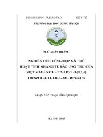
2.2.1.2. Amplification and sequencing of marker DNA fragments
M
SN SLE SCO M
SN SLE SCO
A
M 1 2
B
3 4
5
6 7
M 8
Figure 2.5. PCR product
electrophoresis image cloning
indicator gene segments from
studied soft coral samples. (A)
28S rRNA gene segment using
28SF and 28SR primers. (B)
msh1 gene segment using
MSHF and MSHR primers.
M: GeneRulerTM 1kb DNA
ladder
9 10 11 12 13 14 M 15 16 17 18 19 20 21
Hình 2.6. Electrophoresis of PCR-colony products from some colonies after
transformation of vector pTZ57R/T at 28S rRNA of soft coral samples SN (well
No. 1-7), SLE (well No. 8-14), SCO (well No. 15-21). M: GeneRulerTM 1kb
DNA ladder
M 22 23 24 25 26 27 28 29 30 31 32 33 34 M
35 36 37 38 39 40 41
42
Hình 2.7. Electrophoresis of PCR-colony products from some colonies after
transformation of vector pTZ57R/T with msh1 gene attached to soft coral samples
SN (well No. 22-28), SLE (well No. 29-35), SCO (well No. 36-42). M:
GeneRulerTM 1kb DNA ladder
5
2.2.2. Methods for isolation of secondary metabolites
2.2.2.1. Isolation of compounds from S. nanolobata
Hình 2.8. Isolation of compounds from S. nanolobata
2.2.2.2. Isolation of compounds from S. leptoclados
Hình 2.9. Isolation of compounds from S. leptoclados
6
2.2.2.3. Isolation of compounds from S. conferta
Hình 2.10. Isolation of compounds from S. conferta
2.2.3. Methods for determination of chemical structure of compounds
2.2.4. Methods of assessment of activity and mechanism of cytotoxicity
CHAPTER 3: RESULTS
3.1. Determination of species by molecular makers
3.1.1. Sequencing 28S rARN and msh1 gene fragments of 3 soft corals
- Nucleotide sequence msh1 gene segment of SN sample with length 639
bp, including 190 A, 113 C, 121 G, 215 T
- Nucleotide sequence msh1 gene segment of SLE sample with length
639 bp, including 189 A, 113 C, 119 G, 218 T
- Nucleotide sequence msh1 gene segment of SCO sample with length
639 bp, including 189 A, 113 C, 122 G, 215 T
7
- Nucleotide sequence of 28S rRNA gene segment of SN sample with
length 695 bp, including 139 A, 194 C, 228 G, 134 T
- Nucleotide sequence of 28S rRNA gene segment of SLE sample with
length 709 bp, including 145 A, 197 C, 231 G, 136 T
- Nucleotide sequence of 28S rRNA gene segment of SCO sample with
length 709 bp, including 144 A, 199 C, 228 G, 138 T
3.1.2. Sequence comparison of 3 soft corals by BLAST program
The results of analyzing the msh1 and 28S rRNA gene sequences of the
study samples with the reference sequences on the NCBI international gene
bank showed that the sequences of the indicator genes of the study samples
have similarities. high with the corresponding sequences on the gene bank,
namely:
- Sequence of msh1 gene fragment of SN sample had 100% similarity
with the corresponding sequence (code FJ621451.1) of the sample S.
nanolobata vourcher RMNH coel. 38441 on the NCBI gene bank.
- Sequence of msh1 gene fragment of SLE sample has 100% similarity
with the corresponding sequence (code KC542857.1) of the sample S.
leptoclados vourcher ZMTAU: CO35308 on the NCBI gene bank.
- Sequence of msh1 gene fragment of SCO sample has 100% similarity
with corresponding sequence (code FJ621389.1) of sample S. conferta
vourcher NTM C13972 on NCBI gene bank
- Sequence of 28S rRNA gene fragment of SN sample has 99.8%
similarity with corresponding sequence (code KF915519.1) of Sinularia sp.
RMNH voucher: Coel.41326 on the NCBI gene bank.
- Sequence of 28S rRNA gene fragment of SLE sample has 100%
similarity with the corresponding sequence (code KC542837.1) of the
sample S. leptoclados voucher ZMTAU: CO34095 on the NCBI gene bank.
8
- Sequence of 28S rRNA gene fragment of SCO sample has 99.6%
similarity with corresponding sequence (code MF817932.1) of Sinularia sp.
on the NCBI gene bank.
Table 3.1. The results of species identification of the soft coral samples
studied based on analysis of the similarity (%) of the sequence of the
marker DNA segments (genes 28S rARN and msh1) of the study samples
with the reference sequences on gene bank.
msh1 maker
28S rRNA maker
Name
Species
SN
SLE
SCO
Sinularia
nanolobata
Sinularia
leptoclados
Sinularia
conferta
Ident
Species
Ident
100%
Sinularia sp.
≥ 99.8%
100%
Sinularia
leptoclados
100%
100%
Sinularia sp.
≥ 99.6%
The general
conclusion is based
on 2 DNA markers
Sinularia
nanolobata
Sinularia
leptoclados
Sinularia conferta
3.2. Determination of chemical structure of the compounds
3.2.1. Determination of chemical structure of compounds from S.
nanolobata
28
21
22
18
11
19
9
13
H
HO
10
3
5
4
17
16
14
1
26
24
23
29
12
2
20
25
27
OH
15
8
H
6
7
H
Figure 3.2. Chemical structure of isolated compounds from S. nanolobata
3.2.2. Determination of chemical structure of compounds from S.
leptoclados
9
Figure 3.3. Chemical structure of isolated compounds from S. leptoclados
3.2.3. Determination of chemical structure of compounds from S. conferta
28
22
21
18
12
19
13
11
20
24
23
26
25
27
17
16
14
9
1
8
10
3
HO
5
6
OH
Figure 3.4. Chemical structure of isolated compounds from S. conferta
3.3. Evaluation of cytotoxic activity of compounds isolated from soft
corals
3.3.1. Evaluation of cytotoxic activity of compounds isolated from S.
nanolobata
10
Table 3.2. Results of cytotoxic activity of SN compounds
Compounds
KB
SKLU-1
HepG2
SN 4
-
-
-
SN 6
-
-
SN 30
1.26±
0.20
2.07±
0.33
Elipticine*
64.35±
7.00
1.95±
0.28
IC50 (µM)
MCFLNCaP
7
SKMel-2
-
-
-
-
-
-
2.36±
0.24
2.07±
0.28
1.46±
0.20
HL-60
89.02±
9.93
33.53±
4.25
1.91±
0.37
SW480
71.02±
4.00
2.24±
0.16
*: Positive control; "-": No activity
The results of evaluation of 10 SN compounds showed that: The new
compound SN 6 showed an average cytotoxic activity on the line of 3 cells
HL-60, HepG2 and SW480 (IC50 from 33.53-71.02 µM). Compound SN 4
exhibits toxic activity on the HL-60 cell line. The remaining compounds did
not show activity.
3.3.2. Evaluation of cytotoxic activity of compounds isolated from S.
leptoclados
Table 3.3. Results of cytotoxic activity of SLE compounds
Compou
-nds
IC50 (µM)
SKLU-1
51.80±
31.22
KB
HepG2
MCF-7
SLE 10
34.95±
4.21
21.13±
0.70
38.92±
6.26
SLE 20
-
-
-
19.03±
2.92
32.86±
3.46
21.79±
2.20
39.54±
4.90
17.29±
1.91
36.97±
2.24
21.21±
1.47
49.13±
4.74
SLE 30
-
-
-
-
Ellipticine*
1.79±
0.28
1.38±
0.28
1.34±
0.16
1.26±
0.28
SLE 27
SLE 28
-
62.07±
10.88
SW480
28.65±
1.53
SKMel-2
59.35±
4.23
92.96±8
.29
83.84±
3.72
-
13.45±
1.81
20.53±
2.26
82.80
± 3.65
1.91±
0.12
14.4±
1.88
26.6±
1.59
HL-60
2.03±
0.16
29.01±
3.21
33.87±
3.82
72.32
± 1.30
1.91±
0.20
LNCaP
64.12±
1.71
89.80±
3.34
17.13±
1.81
40.55±
3.63
1.95±
0.20
*: Positive control; "-": No activity
The results of 15 SLE compounds showed that: 3 compounds SLE 10,
SLE 27 and SLE 28 (IC50 in the range of 1.78 to 78.33 µM) showed
cytotoxic activity on 8 cancer cell lines test letter. Compounds SLE 20 and
11
SLE 30 showed activity in 2-3 tested cancer cell lines. The remaining
compounds did not show activity.
3.3.3. Evaluation of cytotoxic activity of compounds isolated from S.
conferta
Table 3.4. Results of cytotoxic activity of SCO compounds
Compound
SCO 27
SCO 35
SCO 37
Etoposide *
Camptothecin*
*: Positive control;
A-549
3.64±0.18
78.73±2.11
27.12±1.69
IC50 (µM)
Hela
19.34±0.42
30.5±0.77
24.64±1.28
27.99±2.01
PANC-1
1.78±0.69
9.35±0.37
20.51±2.72
1.17±0.42
12.65±1.01
The results of evaluation of 12 SCO compounds showed that
compounds SCO 27, SCO 35 and SCO 37 exhibited significant cytotoxic
activity in all 3 experimental cancer cell lines. The remaining compounds
did not show activity
3.3.4. Study on cytotoxicity mechanism of compounds SLE 27 and SCO27
3.3.4.1. Study on cytotoxicity mechanism of compounds SCO 27 in lung
cancer cell line A549
a. Evaluation of the effect of SCO 27 on morphological change in cancer
cells
Figure 3.5. Cell morphology A549 under the influence of SCO 27 at
different concentrations and positive control (camptothecin 5 µM). Arrows
indicate cells in a state of apoptosis
12
b. Evaluate the ability of SCO 27 to stimulate production of the enzyme caspase 3
Figure 3.6. The ability to stimulate production of caspase 3 of SCO 27;
sco-10 µM; sco-5 µM and sco-2,5 µM: The analyzed samples were
supplemented with SCO 27 at the respective concentrations. Control:
Analytical sample with no added SCO 27 compound. Camptothecin:
analytical sample supplemented with camptothecin
c. Determination of the ability of SCO 27 to induce apoptosis in lung
cancer cell line A549
A
C
B
D
Figure 3.7. Effects
of SCO 27 on
apoptosis of cell line
A549 at 24 h at
different
concentrations of 5
µM (C), 10 µM (D),
negative control (A)
and positive control
(B)
using
the
Method of dyeing
Annexin V/PI
13
Table 3.4. Apoptosis rate under the influence of SCO 27 on lung cancer
cell line A549
Compounds
The rate of
survival
cells (%)
Control
SCO 27 - 5 µM
SCO 27 - 10 µM
Camptothecin-5µM
89.08
88.50
83.33
79.17
The rate of
early
apoptosis
cells (%)
3.89
4.90
4.76
15.49
The rate of
late
apoptosis
cells (%)
5.18
5.72
8.93
1.57
The rate
of necrotic
cells (%)
1.85
0.88
2.99
3.77
3.3.4.2. Study on cytotoxicity mechanism of compound SLE 27 in breast
cancer cell line MCF-7
a. Evaluation of the effect of SLE 27 on morphological change in cancer
cells
Figure 3.8. Effects of compound SLE 27 at concentrations of 10, 30 and
100 µM on morphology of breast cancer cell line MCF-7. Arrows indicate
cells in a state of apoptosis. 10X and 20X magnification. Control: negative
control - MCF-7 cells not supplemented with SLE 27 compound.
b. Determination of induction apoptosis of breast cancer cell line MCF-7 of
compound SLE 27
Table 3.5. Apoptosis rate under the influence of SLE 27 on the breast
cancer cell line MCF-7
14
Compounds
The rate of
survival
cells (%)
The rate of
early
apoptosis
cells (%)
The rate of
late apoptosis
cells (%)
The rate of
necrotic
cells (%)
Control
SLE 27-10 µM
SLE 27-30 µM
SLE 27-100 µM
97.08
97.39
86.08
39.10
0.67
1.15
1.72
3.71
1.93
1.28
7.16
23.02
0.32
019
5.04
34.16
c. Evaluation of the effect of compound SLE 27 on breast cancer cell cycle
MCF-7
Figure 3.10.
Effect
of
compound
SLE27
(concentration
10; 30; 100
µM) on cell
cycle MCF-7
at 48 h, using
the
Flow
cytometer
Novocyte
system
Table 3.6. The rate (%) of MCF-7 cells in G0/G1, S, G2/M phases and
apoptosis (sub-G1) after 48 hours of treatment with SLE 27 compound
Compounds
Control
SLE 27 - 10 µM
SLE 27 - 30 µM
SLE 27- 100 µM
The rate of cells in all phases of the cell division
cycle (%)
sub-G1
G0/G1
S
G2/M
phase
phase
phase
phase
0.54
41.21
41.22
14.81
0.23
36.57
33.41
17.78
0.76
40.59
27.19
15.86
10.24
66.08
15.96
4.42
15
CHAPTER 4: DISCUSSIONS
4.1. Determination of species of soft coral based on DNA makers
After cloning and sequencing of marker gene segments from 3 soft coral
samples, the DNA sequences of Sinularia leptoclados (MW077896,
MW077906); Sinularia conferta (MW077897, MW077907) and Sinularia
nanolobata (MW077898, MW077908) are registered on the NCBI.
Sinularia-abruptaMF817864
Sinularia-slieringsiMH516803
Sinularia-penghuensisJX991181
Sinularia-molestaJX991172
30 Sinularia-abrubtaFJ621374
Sinularia-abruptaJX991168
37
Sinularia-leptocladosKC542857
66 SLE
46 Sinularia-compactaFJ621384
Sinularia-bisulcaFJ621378
99
Sinularia-acutaFJ621375
46
Sinularia-verseveldtiKC542859
Sinularia-robustaFJ621473
Sinularia-diffusaFJ621399
Sinularia-sp.KF915757
64
Sinularia-abhishiktaeFJ621373
Sinularia-tumulosaFJ621482
50
Sinularia-siaesensisFJ621478
Sinularia-polydactylaKU230374
90 Sinularia-confertaFJ621389
99
43
SCO
Sinularia-peculiarisJX023274
64
Sinularia-ornataJX991173
Sinularia-nanolobataFJ621451
65
90 SN
Sinularia-brassicaKF915724
48
47
0.01
Figure 4.1. The results of classification analysis by the NJ (NeighborJoining) method on MEGA6 of soft coral samples based on msh1 gene
sequence polymorphism of the studied samples and related soft corals
samples on NCBI. The tributary numbers are the Bootdtrap values that
represent the reliability of the genetic branching of the sequences and
groupings.
Analysis of genotypes of the msh1 gene segment based on the nucleotide
sequence was performed on 3 study sequences and 23 reference sequences
on NCBI. The results in Figure 4.1 show that the SLE sample has a high
genetic similarity with the species that has been sequenced in the world
gene bank, S. leptoclados KC542857 (bootstrap 66%). The SCO sample
16
has a high genetic affinity with S. conferta FJ621389 (bootstrap 90%). SN
pattern has a high genetic similarity with S. nanolobata species FJ62621451
(bootstrap 90%).
Similarly, genotyping analysis based on 28S rARN gene segment
nucleotide sequence was performed on 3 study sequences and 21 reference
sequences on NCBI. The results in Figure 4.2 show that the SLE sample
has a high genetic similarity with the species that have been sequenced on
the world gene bank, namely S. leptoclados KC542857, S. leptoclados
MF817912, S. densa KC542829, S. abrupta KC542822, S. australiensis
KC542825, Sinularia sp. MF817932 (bootstrap 100%). SCO samples have
high genetic relationships with species that have been sequenced on the
world gene bank such as S. bisulca KC542826, S. slieringsi MK333594, S.
penghuensis KC542842, S. robusta KC542843, S. verseveldti KC542845, S.
eilatesis KC542832, S. corpulentissima KC542827, S. maxima KC542839
(bootstrap 100%). The SN pattern has a highly similar genetic relationship
with the species Sinularia sp. KC915519 and S. polydactila KF915515
(bootstrap 98%).
Thus, the synthesis of the results comparing the sequences of msh1 and
28S marker genes of 3 soft corals are presented in Table 3.1. Sample SN
was identified scientifically as S. nanolobata, SLE sample was identified
scientifically as S. leptoclados, SCO sample was identified scientifically as
S. conferta. This result also coincided with the results identified by
analyzing the initial morphological characteristics of Prof. Dr. Do Cong
Thung, Institute of Marine Environmental Resources. This is an initial
research result that will partly help in the search for specific molecular
markers to support the fast and accurate scientific name determination of
marine species.
17
4.2. Determination of chemical structure of compounds from S.
nanolobata
From the soft coral S. nanolobata, 10 compounds were isolated. These
include: 4 new compounds named: 24(S),28-epoxyergost-5-ene-3β,4α-diol
(SN 6), 3β,4α-dihydroxyergosta-5,24(28)-diene (SN 8), nanolobatol B (SN
20),
nanolobatol
A
(SN
30)
and
6
known
compounds:
16α-
Hydroxysarcosterol (SN 3), sarcophytosterol (SN 4), sinularianin B (SN
10), sinularianin D (SN 11), Cholesta-5,24(28)-dien-3β-ol-7-one (SN 16),
dissesterol (SN 17). The compounds isolated were structurally in the sterol
and sesquiterpen classes
4.3. Determination of chemical structure of compounds from S.
leptoclados
From the soft coral S. leptoclados has isolated and determined the
structure of 15 compounds: all 15 compounds belong to the sterol class, of
which 2 new compounds are named: Leptosteroid (SLE 10), 5β,6βepoxygorgosterol (SLE 21) and 13 known compounds: sarcophytosterol
(SLE 13), Ergosta-24(28)-en-3β-ol (SLE 19), Ergosta-5,22,24(28)-trien-3βol (SLE 20), 3β,4α-Dihydroxyergosta-5,24(28)-die (SLE 22), Ergosta5,24(28)-dien-3β-ol-7-one (SLE 23), 7-oxogorgosterol (SLE 25), Ergosta5-en-3β-ol-7-one (SLE 26), Ergosta-5,24(28)-dien-3β,7β-diol (SLE 27),
Ergosta-5-en-3β,7β-diol (SLE 28), 7β-hydroxygorgosterol (SLE 29),
Ergosta-5,24(28)-dien-3β,7α-diol (SLE 30), Ergosta-5-en-3β,7α-diol (SLE
31), 7α-hydroxygorgosterol (SLE 32).
4.4. Determination of chemical structure of compounds from S.
conferta
From the soft coral S. conferta has isolated and determined the structure
of 12 compounds: all 12 compounds belong to the sterol class, of which 5
new compounds are named: 7α-Methoxygorgosterol (SCO 29), 7αMethoxy-ergosta-5-ene-3β-ol (SCO 30), 3β-Hydroxyergosta-4-ene-6-one
18
(SCO 44), 3β-Hydroxyergosta-4,24(28)-diene-6-one
(SCO 32), 24-
methylenecholestane-3β,5α,6β-triol-3-monoacetate (SCO 42) and 7 known
compounds: 3β-Hydroxy-24-methylenecholest-5-en-7-one (SCO 26), 24methylenecholestane-3β,5α,6β-triol-6-monoacetate
(SCO
27),
7-
methoxyergosta-5,24(28)-diene-3 -ol (SCO 31), 24-methylenecholestane3β,5α,6β-triol (SCO 35), Ergosta-3β,5α,6β-triol (SCO 37), 3β,7αDihydroxyergosta-5,24(28)-dien (SCO 34.1) và (24S)-ergost-5-en-3β,7αdiol (SCO 34.3)
4.5. Evaluation of cytotoxic activity of isolated compounds
4.5.4.1. Study on cytotoxicity mechanism of compounds SCO27 in lung
cancer cell line A549
a. Evaluation of the effect of SCO 27 on morphological change in cancer
cells
The results of morphological analysis of A549 lung cancer cells (Figure
3.5) showed that cells in samples treated with SCO 27 showed apoptosis in
some cells with characteristic morphology changes cells such as: condensed
cells, fragmented nuclei. The number of apoptosis cells increases with
increasing concentration of SCO 27 reagent from 2.5 to 10 µM. The
positive control (camptothecin) was stable, the cells after treatment also had
a homogeneous, bright, round and bright dye nucleus. Thus, compound
SCO 27 has the effect of changing cell morphology, this is the basis for
conducting further studies to determine the ability of this compound to
cause apoptosis.
b. Evaluate the ability of SCO 27 to stimulate production of the enzyme
caspase 3
The results of Figure 3.6 show that SCO 27 is capable of inducing A549
cells to produce caspase 3. This activity is only evident at a concentration of
10 µM with an increase of 1.74 times compared to the negative control. The
camptothecin positive control was stable in the experiment with a caspase 3
19
activity at a concentration of 2.86 times that of a negative control (P <0.05).
Thus, the SCO 27 compound has a lot of potential for further anti-cancer
oriented studies. To date, no documents have been published about the
ability to activate the enzyme caspase 3 of the compound SCO 27.
c. Determination of the ability of SCO 27 to induce apoptosis in lung
cancer cell line A549
Under the action of SCO 27 compound, the rate of early apoptosis, late
apoptosis and necrosis increased with increasing reagent concentration. At a
concentration of 10 µM SCO 27, the proportion of cells in the stage of late
apoptosis and necrotic cells both increased compared to the control
(control). In the control cells % late apoptosis was only 5.18%, however,
this cell population increased to 8.93% under the influence of SCO 27 with
concentration of 10 µM. When the sample concentration decreased to 5
µM, the rate of early apoptosis was 4.90% and late apoptosis was 5.72%,
the rate of necrotic cells was 0.88%. Thus, compound SCO 27 exhibits the
ability to cause cell death through apoptosis.
4.5.4.2. Study on cytotoxicity mechanism of compound SLE 27 in breast
cancer cell line MCF-7
a. Evaluation of the effect of SLE 27 on morphological change in cancer
cells
Morphological analysis of MCF-7 cells under reverse microscopy
showed a marked morphology change after 48 h treatment with SLE 27
compound at concentrations of 10, 30 and 100 µM. Cell samples showed
distinct apoptosis features such as reduced density, cell shrinkage, and
chromosomal concentration (Figure 3.8). From there, it can be concluded,
SLE 27 compound has the effect of altering breast cancer cell morphology
MCF-7. This is the basis for conducting further studies on the cytotoxic
mechanism of this compound.
20
b. Determination of induction apoptosis of breast cancer cell line MCF-7 of
compound SLE 27
Flow cytometry analysis showed that the proportions of cells after
treatment with SLE 27 in early apoptosis, late apoptosis and necrosis were
increased compared to the control (Control). At a concentration of 100 µM,
only 39.10 % of living cells, while this rate in the control sample is 97.08
%. The percentage of apoptosis significantly increased at a concentration of
100 µM increased to 26.73 % (23.02 % of late apoptosis and 3.71 % of
early apoptosis) (Figure 3.9). When the sample concentration decreased to
30 µM, the rate of early apoptosis was 1.72% and late apoptosis was 7.16
%, the rate of necrotic cells was 5.04 %. Thus, SLE 27 compound has
shown the ability to cause cell death through apoptosis.
c. Evaluation of the effect of compound SLE 27 on breast cancer cell cycle
MCF-7
The analysis results in Figure 3.10 show that the percentage of cells in
the G0/G1 phase increases when the concentration of SLE 27 compound
increases from a concentration of 30 µM to 100 µM. Specifically, at a
concentration of 10 µM, the rate of phase cells G0/G1 is 36.57 %, at a
concentration of 30 µM, the rate of phase cells of G0/G1 is 40.59 %, at a
concentration of 100 µM, the rate of the rate of G0/G1 phase cells increased
sharply to 66.08 %. At all 3 concentrations of SLE 27 compound, the
percentage of cells in phase G0/G1 accounted for the highest proportion.
Meanwhile, the proportion of cells in phase S and G/M phase is less and
decreases gradually when the concentration of SLE 27 compound increases
from 10 - 100 µM. This indicates that the cell does not undergo DNA
replication (phase S) and mitosis (Phase M). Thus, compound SLE 27 has
the effect of arresting breast cancer cell cycle MCF-7 in phase G0/G1. This
is a new and unprecedented effect on this activity of the SLE 27 compound.
21
CONCLUSIONS
1. Identification of soft coral samples
The scientific names of 3 studied soft coral samples S. nanolobata, S.
conferta, S. leptoclados collected from the sea area of Central Vietnam
were determined by morphological characteristics and two molecular
markers (msh1 and 28S rRNA) sequence analysis
2. Study on chemical composition:
Using chromatographic and modern spectroscopy methods, 37
compounds were isolated and structured from three soft corals S.
nanolobata, S. conferta, S. leptoclados. Among them, there are 11 new
compounds, as following:
- From S. nanolobata, 10 compounds (SN) were isolated and structured,
including 4 new compounds and 6 known compounds. These compounds
belong to the sesquiterpen and steroid group
- From S. leptoclados, 15 compounds belonging to the steroid group
were isolated and structured, including 2 new compounds and 13 known
compounds.
- From S. conferta, 12 compounds belonging to the steroid group were
isolated and structured, including 5 new compounds and 7 known
compounds.
3. Evaluation of cancer cytotoxic activity:
The evaluation of cytotoxic activity of all isolated compounds showed
that: Compounds SLE 10, SLE 27, SLE 28 showed strong cytotoxic
activity on 8 cell lines (KB, LNCaP, SK-LU-, SK-Mel-2, HepG2, MCF-7,
HL-60, SW480), SLE 20 and SN 6 compounds showed moderate activity
on 3 cell lines (HL-60, SW-480, LNCaP/HepG2,HL-60, SW-480), SLE 30
showed activity in 2 cell lines (HL-60, SK-Mel-2) and SN 4 showed
activity in one cell line (HL-60), SCO 27, SCO 35 and SCO 37 compounds
showed good activity in 3 cell lines (A-549, PANC1, Hela)
22
4. Analysis of cancer cytotoxicity mechanism
- SCO 27 compound exhibited cytotoxicity on A549 lung cancer cells
by activation of caspase 3 enzyme production, leading to apoptosis . The
apoptosis were expressed by changes in cell morphology depend on
increasing concentrationof tested compound.
- Similarly to SCO27, SLE 27 compound showed cytotoxicity by
induction of apoptosis on breast cancer cells MCF-7. In addition, this
compound affected to the cell cycle in the G0/G1 phase, thereby preventing
cancer cell division.
RECOMENDATION
The obtained results showed that SCO 27 and SLE 27 are two potential
compounds for development of drugs in breast and lung cancer treatment.
Therefore, further studies could be focused to clarify the cytotoxicity
mechanism of these compounds in in vivo condition.
NEW FINDING OF THE THESIS
1. Firstly, the scientific names of three studied soft corals species were
determined as Sinularia nanolobata, Sinularia conferta, Sinularia
leptoclados by combinating of morphological charteristics and two
molecular markers (msh1 and 28S rRNA) sequence analysis
2. Secondly, 11 new substances were isolated and structurally identified
from these soft coral species of Vietnam sea: (24 (S), 28-epoxyergost-5ene-3β, 4α-diol; 3β, 4α-dihydroxyergosta-5,24 ( 28) –diene; Nanolobatol A;
Nanolobatol
B;
Leptosteroid;
5β,
6β-epoxygorgosterol;
7α-
Methoxygorgosterol; 7α-Methoxy-ergosta-5-ene-3β-ol, 3β-Hydroxyergosta-
23
4-ene-6-one;
3β
-Hydroxyergosta-4,24
(28)
-diene-6-one;
24-
methylenecholestane-3β, 5α, 6β-triol-3-monoacetate).
3. Finally, the research firstly demonstrated the cytotoxic activity of 11
new compounds and the cytotoxic mechanism of two potential compounds
(SCO 27 and SLE 27) was based on stimulating apoptosis process in
cancer cell lines.