Calixpenams: synthesis, characterization, and biological evaluation of penicillins V and X clustered by calixarene scaffold
Bạn đang xem bản rút gọn của tài liệu. Xem và tải ngay bản đầy đủ của tài liệu tại đây (161.43 KB, 9 trang )
Turk J Chem
(2014) 38: 288 296
ă ITAK
c TUB
Turkish Journal of Chemistry
/>
doi:10.3906/kim-1307-32
Research Article
Calixpenams: synthesis, characterization, and biological evaluation of penicillins
V and X clustered by calixarene scaffold
Fazel NASUHI PUR1,2 , Karim AKBARI DILMAGHANI1,∗
Department of Chemistry, Faculty of Science, Urmia University, Urmia, Iran
2
Health Technology Incubator Center, Urmia University of Medical Science, Urmia, Iran
1
Received: 14.07.2013
•
Accepted: 14.09.2013
•
Published Online: 14.03.2014
•
Printed: 11.04.2014
Abstract: Four 6-aminopenicillanic acid moieties were grafted at either rim of calix[4]arene, giving 2 novel generations
of penicillin, which were named calixpenam. Antibiotic tests showed that they have amplified activity with respect to
the corresponding penams against 3 gram-positive nonpenicillinase-producing strains of Streptococcus.
Key words: Calixpenam, Calixarene, 6-aminopenicillanic acid, nonpenicillinase, Streptococcus strains
1. Introduction
The resistance of infective bacteria to present antibiotics demands research assigned to the discovery of new drugs
in the antibacterial drug field. Penicillin was the first antibiotic, but Staphylococcus aureus and Streptococcus
pneumoniae have resisted it.1,2
Streptococcus pneumonia is an important infectious agent, representing a significant cause of pneumonia
and the other corresponding diseases. Seven million cases of otitis media, 500,000 of pneumonia, 50,000 of
bacteremia, and 3000 of meningitis are attributed to S. pneumonia each year in the USA alone. 3
The discovery of penicillin in 1928 by Alexander Fleming initiated the use of antibiotics to fight human
diseases. The development of penicillins (penams) was due to the discovery and identification of the penicillin
nucleus, 6-aminopenicillanic acid (6-APA) with a 4-membered lactam ring (β -lactam) and thiazolidine ring,
which was isolated from culture of Penicillium chrysogenum. All penicillins are β -lactam (6-APA core) antibiotics and are used against several bacterial infections. 4 However, semisynthetic penams have been synthetized
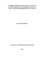
by acylating of the 6-APA amino group with various acid derivatives (Figure 1). 5
O
O
N
H2N
H S
(6-APA)
OH
O
O
O
OH
N
N
H
H S
(Penicillin G)
HO
O
O
O
N
N
H
O
OH
H S
(Penicillin X)
O
O
N
O
OH
N
H H S
(Penicillin V)
Figure 1. Structure of natural penams.
Penicillins are effective against diseases caused by gram-positive bacteria (streptococcus, pneumococcus)
and other infectious agents. They are not effective against the majority of gram-negative microorganisms (E.
∗ Correspondence:
288
NASUHI PUR and AKBARI DILMAGHANI/Turk J Chem
coli ). 5 These drugs act as antibiotics by suppressing the final steps of the synthesis of the bacterial cell wall. 6
It is accepted that the pharmacological activities of penicillin are associated with the conformations of the
thiazolidine ring and of the acylating agent. 7
Phenoxymethyl penicillin or penicillin V (Figure 1) is a natural acid-resistant penam and is used for oral
consumption. It is effective against gram-positive (streptococcus, pneumococcus) and other microorganisms and
is available from culture of the fungus P. chrysogenum. 5
p -Hydroxybenzyl penicillin or penicillin X (Figure 1), like penicillin G, is susceptible to penicillinase,
and can be produced in culture by strains of Penicillium notatum or chrysogenum as a natural penam. Like
penams G and V, it is active against gram-positive and in some cases is even more effective than penicillin G
and the other penicillins. 8
It is thought that antibiotic resistance is unavoidable, but medicinal chemistry can slow it down through
development of new antibiotics. There should be many strategies in order to develop new drugs.
These reasons prompted us to synthesize novel generations of penams by using a firm molecular platform
for the demonstration of the penicillin cluster. This idea could result in novel molecular structures with enhanced
effects and antibiotic activities in comparison to single penicillin units. It is attributed to their high density
antibiotic surface and synergistic effect of cluster arms.
Calixarenes have many structural characteristics that are preferable for the design and development of
new drugs. Recently, due to calix[4]arene’s limited toxicity, they have been used in the biological field as building
blocks or molecular scaffolds. 9−34 For medical applications, the toxicity of molecules is evidently a key factor;
to date, the calixarenes have shown neither toxicity nor immune responses. 9−36
We noticed that there are only 2 reports 37,38 in the literature regarding the application of calixarenes in
the field of β -lactam drugs. In them, calixarene is not used as a drug structure, but as a drug dispenser.
Here we wish to report the synthesis, characterization, and antibacterial activities of calix[4]arene derivatives, possessing four 6-APA units at either rim of the scaffold in all-syn orientation. The synthetic strategy
involves grafting of the 6-APA moieties via the formation of an amide bond between the calixarene platform
and the 6-APA arm.
2. Results and discussion
Compounds 2 and 3 were initially chosen as the core structures with a cone conformation for grafting of the
four 6-APA on one rim of the platform. Compound 2a was prepared according to Gutsche et al.’s method, 39,40
including Mannich dimethylaminomethylation of calix[4]arene, and quaternization of amines followed by eliminative nitrilation and acidic hydrolysis of nitrile groups to the corresponding tetraacid-calix[4]arene. Compound
3a was synthetized by the procedure of McKervey et al. 41,42 involving the transformation of calix[4]arene into
the corresponding ethyl ester and basic hydrolysis of ester groups.
The synthesis of calixpenams 4 (CP X) and 5 (CP V) is depicted in Scheme 1. We chose soft
conditions in the coupling reaction in order to avoid probable β -lactam degradation. It is clear that acid
chlorides as acylating agent are not preferred for this reaction because of problems due to their sensitivity
to water purification, low yield of the acylation reaction when using them, and problems in providing a lowtemperature acylation reaction (∼ −20 ◦ C). Thus, we chose a controlled peptide-bound formation process that
would involve the use of 2,2’-dibenzothiazole disulfide (DBTDS) as a carboxylic acid activator in the presence
of triphenylphosphine (TPP) as reducer and triethylamine (TEA) as catalyst. 43 The method for calixpenams
289
NASUHI PUR and AKBARI DILMAGHANI/Turk J Chem
synthesis has several advantages over the acid chloride method: easy handling, very mild reaction conditions,
high yield, no need for further purification of the acylating agent (thioester), and ambient temperature for the
reaction.
OH
HO
O
O
R
O
R
R
HO
O
OH OHHO HO
HO
N
2b R=MBT
S
H
1 (6-APA)
O O
O O
R
R
O
O
O
O
O
OH
O
N
S
N
H
H
4 (CP X)
[tetramer of pen. X]
OH OH HO HO
NH2
5 (CP V)
[tetramer of pen. V]
TEA/DCM
3 days at r.t.
workup by H2O
O
O
O
O
O
O
NH
O
H
H
N
3b R=MBT
O
O
NH
3a R=OH
i
O
O
N
H
H
R
R
NH
NH
O
S
O
O
H
H
N
i
S
O
O
O
O
N
N
S
R
2a R=OH
O
O
O
N
S
O
O
NH
S H
S
N
O
HN
O
H
OS
N
O
OH
OH
OH
O
HO
S
S
S
i)
S
N
N
/TPP/TEA/acetone
overnight at r.t.
and then 12h reflux
S
S
N
2-Mercaptobenzothiazole (MBT)
(2,2'-dibenzothiazole disulfide)
Scheme 1. Synthetic pathway to calixpenams.
In the first step, the product is an active thioester (Scheme 2) that is insensitive to aqueous media and
is very stable for isolation as the crystalline form.
Ph
Ph
P
N(C2H5)3
Ph
N
(TPP)
S
(TEA)
O
S
S
O
N
PPh3
R
H
O
O
R
P
O
Ph
Ph
Ph
2a or 3a
S
S
S
N
(DBTDS)
N
O
+
R
S
S
Thioester (2b or 3b)
Ph
P
N
Ph
Ph
(TPPO)
S
S
(MBT)
Scheme 2. The mechanism of thioesterification.
As shown in Scheme 2, the thioesterification is a redox condensation. Initially, the S–S bond of DBTDS
is broken up by TPP (reduction step), which is followed by its oxidation into triphenylphosphine oxide (TPPO)
(oxidation step). Polarity increased from the reactants to the transition state during the reaction process. Thus,
290
NASUHI PUR and AKBARI DILMAGHANI/Turk J Chem
polar solvent could stabilize the transition state and reduce the activation energy, which would accelerate the
reaction effectively (positive effect); on the other hand, due to reaction of protonic solvents with the anions
(MBT¯), and decline of its nucleophilic property (negative effect), dipolar aprotic solvents such as acetone are
suitable for this reaction.
The reaction occurs only in the presence of base (TEA). It is attributed to an increase in the nucleophilic
activity of DBTDS in basic condition, which is a positive factor for the reaction.
In the aminolysis reaction for the synthesis of calixpenams, 6-APA is added to a water-immiscible inert
organic solvent such as dichloromethane (DCM), followed by addition of the base, and then the activated
thioester (2b or 3b) is added to the reaction mixture and stirred until completion of the reaction to give the
corresponding calixpenams. Due to the low amount of impurities, triethylamine is a better choice of base
compared to other tertiary amines.
In the aminolysis reaction, TEA also serves to dissolve 6-APA in the form of triethyl ammonium salt and
will catalyze the reaction. The final products were obtained in the form of the corresponding triethylammonium
salt with good yield, followed by simple extraction with water, while 2-mercaptobenzothiazole (MBT), obtained
as a by-product, remained in the organic phase (DCM). The aqueous extracts were acidified to obtain the acid
form of the product. Finally, to increase the solubility of the final products in chloroform (recording of NMR
spectra) and water (in-vitro antimicrobial susceptibility testing), their potassium salt forms were prepared. The
products’ structures were characterized by IR, NMR, and ESI-MS spectra and elemental analysis.
IR analysis showed the presence of an intense band at 1690 cm −1 for CP X and at 1696 cm −1 for CP
V attributed to the amide carbonyl group, and 2 other close bands at the 1760 cm −1 zone were attributed to
the lactam carbonyl group.
According to de Mendoza et al., 44,45 compounds 4 (CP X) and 5 (CP V) are in a cone conformation,
as assessed by the Ar-CH 2 -Ar resonance signals at 30.7 and 33.9 ppm in the 13 C NMR spectra, respectively. It
was also confirmed through the presence of an AB system at 4.26–3.38 ppm for CP X and at 4.42 −3.32 ppm
(J AB = 13.7 Hz) for CP V in the 1 H NMR spectra.
In order to evaluate the potentially enhanced antibiotic activities of calixpenams (4 and 5), we compared
them with the penicillins X and V (6 and 7, respectively) as reference compounds. In fact, they can be
considered as 1/4 of the corresponding cluster compounds 4 (CP X) and 5 (CP V), respectively.
The in vitro antimicrobial susceptibility testing (AST) [e.g., minimum inhibition concentration (MIC)
determination] of compounds 4 −7 was determined by broth microdilution (BMD) in accordance with the
Clinical and Laboratory Standards Institute (CLSI) guidelines. 46 The results of these tests are shown in Table
1. As shown in Table 2, clusters 4 (CP X) and 5 (CP V) showed more antibiotic activities than the reference
monomers 6 and 7 (5- to 6-fold increases were observed). The numbers in Table 2 indicate the MIC ratio
of calixpenam and its corresponding monomer and they describe the increase in antibacterial effects from the
monomeric penicillin to calixpenam. The values are only slightly more for CP X than for CP V. This is
attributed to the larger contact surface of CP X with the bacterial membrane than CP V, due to the size of
the wider upper rim of calixarene compared to the lower rim.
2.1. Conclusion
In summary, the present work describes the first examples of calixpenams with efficient antibiotic activities.
These compounds could be considered as novel antibiotic structures with high density antibiotic surfaces.
291
NASUHI PUR and AKBARI DILMAGHANI/Turk J Chem
Table 1.
Minimal inhibitory concentration (MIC), in µ g/mL, obtained by broth microdilution (BMD) method,
according to CLSI guidelines.
MIC (µg/mL) values
Strain S. pyogenes
Compd.
ATCC 19615
4 (CP X)
0.002
5 (CP V)
0.003
6 (Pen. X)
0.012
7 (Pen. V)
0.016
S. agalactiae
ATCC 12386
0.004
0.006
0.025
0.032
S. pneumoniae
ATCC 49619
0.022
0.024
0.125
0.125
Table 2. MIC ratios between calixpenams and their corresponding monomers for Streptococcus strains.
MIC (µg/mL) values
Strain
Compd.
MICP en. X / MICCP X
MICP en. V / MICCP V
S. pyogenes
ATCC 19615
6.00
5.33
S. agalactiae
ATCC 12386
6.25
5.33
S. pneumoniae
ATCC 49619
5.68
5.20
The results of the present study demonstrate a noteworthy increase in antibacterial properties from the
monomeric penicillins (6 and 7) to their corresponding tetrameric cyclic isomers (4 and 5). This is attributed
to tethering and arraying of four 6-APA arms at either rim of the calixarene cores (CP X and CP V), which
causes a synergistic effect in interactions with the bacterial cell wall for creating effective antibacterial activity.
3. Experimental
3.1. General
The melting points of all compounds were recorded on a Philip Harris C4954718 apparatus without calibration.
IR spectra were determined on a Thermo Nicolet 610 Nexus FT-IR spectrometer with KBr disks. Ultraviolet
spectra were recorded on a Shimadzu UV-2401/PC spectrometer.
1
H NMR (400 MHz) and
13
C NMR (100
MHz) measurements were recorded on a Bruker AM-400 spectrometer in CDCl 3 using TMS as the internal
reference. Elemental analyses were obtained on a PerkinElmer 240c analyzer. Mass spectra were recorded on a
JEOL-JMS 600 (FAB MS) instrument. Thin layer chromatography (TLC) analyses were carried out on silica gel
plates. All chemicals were purchased from Merck (Tehran, Iran) and used as received by standard procedures.
3.2. Thioesterification: procedure for the synthesis of compounds 2b and 3b
2,2’-Dibenzothiazole disulfide (3.32 g, 10 mmol) and triphenylphosphine (2.63 g, 10 mmol) were suspended in
acetone (30 mL), and then stirred for 30 min at room temperature. After addition of tetraacid 2a or 3a (500
mg, 0.76 mmol), triethylamine (1.65 mL, 12 mmol) was gradually added dropwise into the mixture over 15
min. Then the mixture was stirred overnight at room temperature and finally was refluxed 12 h. After the
mixture was cooled, the formed precipitate was filtered, washed with cold acetone, dried, and recrystallized
from CH 2 Cl 2 /acetone to give pale fine powder of the target thioester 2b or 3b, respectively.
292
NASUHI PUR and AKBARI DILMAGHANI/Turk J Chem
5,11,17,23-Tetrakis(2-mercaptobenzothiazolyl carbonylmethyl)calix[4]arene-25,26,27,28-tetrol (2b)
Yield (760 mg, 80%), mp: 162− 164
◦
C. IR (KBr, ν , cm −1 ) : 3265 (O −H), 2951, 1733 (C=O), 1600, 1460.
The expanded structure of MBT moiety is shown in Figure 2.
1
H NMR (400 MHz, CDCl 3 )δ 10.12 (s, 4H, OH),
8.42 (d, J = 7.7 Hz, 4H, MBT), 8.05 (d, J = 7.6 Hz, 4H, MBT), 7.59–7.50 (m, 8H, H-5 & H-6 of MBT), 6.96
(s, 8H, Ar-H of calix), 4.20 (bd, 4H, ArCH 2 Ar, H ax ), 3.50 (bd, 4H, ArCH 2 Ar, H eq ), 3.40 (s, 8H, CH 2 CO 2 );
13
C NMR (100 MHz, CDCl 3 )δ 183.8 (C-2 of MBT), 148.9 (C=O), 147.2 (ArC− O), 140.4 (C-9 of MBT), 129.3
(C-8 of MBT), 128.3 (C (o) of Ar calix), 128.2 (C (m) of Ar calix), 127.1 (ArC* −CH 2 ) , 126.6 (C-5 of MBT),
123.1 (C-6 of MBT), 120.4 (C-7 of MBT), 110.7 (C-4 of MBT), 37.4 (CH 2 CO), 30.2 (ArCH 2 Ar). Anal. Calcd
for C 64 H 44 N 4 O 8 S 8 : C, 61.32; H, 3.54; N, 4.47; S, 20.46. Found: C, 61.38; H, 3.49; N, 4.42; S, 20.52. FAB
MS m/z: 1252.03 (M
+
+
).
25,26,27,28-Tetrakis(2-mercaptobenzothiazolyl carbonylmethoxy)calix[4]arene (3b)
Yield (685 mg, 72%), mp: 156−157
◦
C. IR (KBr, ν , cm −1 ): 2955, 1734 (C=O), 1601, 1459. The expanded
structure of MBT moiety is shown in Figure 2.
1
H NMR (400 MHz, CDCl 3 )δ 8.22 (d, J = 7.6 Hz, 4H, MBT),
8.08 (d, J = 7.7 Hz, 4H, MBT), 7.64–7.55 (m, 8H, H-5 & H-6 of MBT), 7.17 (d, J = 7.3 Hz, 8H, Ar-H m of calix),
6.72 (t, J = 7.3 Hz, 4H, Ar-H p of calix), 4.97 (d, J = 14 Hz, 4H, ArCH 2 Ar, H ax ) , 4.68 (s, 8H, ArO− CH 2 ),
3.26 (d, J = 14 Hz, 4H, ArCH 2 Ar, H eq );
13
C NMR (100 MHz, CDCl 3 )δ 187.2 (C-2 of MBT), 153.7 (ArC− O),
150.0 (C=O), 141.1 (C-9 of MBT), 133.1 (C (o) of Ar calix), 129.3 (C-8 of MBT), 127.1 (C-5 of MBT), 126.4
(C (m) of Ar calix), 124.4 (C-6 of MBT), 121.9 (C-7 of MBT), 120.8 (C (p) of Ar calix), 112.3 (C-4 of MBT), 72.4
(ArOCH 2 ), 32.4 (ArCH 2 Ar). Anal. Calcd for C 64 H 44 N 4 O 8 S 8 : C, 61.32; H, 3.54; N, 4.47; S, 20.46. Found:
C, 61.27; H, 3.58; N, 4.49; S, 20.59. FAB
+
MS m/z: 1252.05 (M
6
5
7
1
8 S
2
+
).
R
S
N
4 9 3
Figure 2. The numbering system for NMR spectra of MBT.
3.3. Aminolysis: procedure for the synthesis of compounds 4 and 5
A suspension of 6-APA (865 mg, 4 mmol) in dichloromethane (30 mL) was cooled to 5–10
◦
C with stirring
triethylamine (1.40 mL, 10 mmol) and then 2b or 3b (500 mg, 0.40 mmol) was added. The mixture was stirred
for 3 days at room temperature and then extracted twice with water (2 × 10 mL). The combined extracts were
adjusted to pH 3 by the addition of 3 M HCl (5 mL). The mixture was cooled to 0–5
◦
C and the resulting
precipitate was separated by filtration, washed successively with cold water (15 mL), cold ethanol (15 mL),
diethyl ether (2 × 15 mL), and dried for 4 h at 40 ◦ C to obtain 4 or 5, respectively, as white powder.
5,11,17,23-Tetrakis(6-aminopenicillanic acid carbonylmethyl)calix[4]arene-25,26,27,28-tetrol (4)
Yield (335 mg, 58%), mp: 201− 203
◦
C. IR (KBr, ν , cm −1 ) : 3375 (O −H), 2955, 1760 (C=O), 1758 (C=O),
1690 (C=O). The expanded structure of 6-APA is shown in Figure 3.
1
H NMR (400 MHz, CDCl 3 , in the form
293
NASUHI PUR and AKBARI DILMAGHANI/Turk J Chem
of potassium salt) δ 10.19 (d, J = 6.3 Hz, 4H, N −H), 9.59 (s, 4H, OH), 6.91 (s, 8H, Ar-H) 5.53 (d, J = 3.7 Hz,
4H, H-5 of APA), 5.46 (m, 4H, H-6 APA), 4.32 (s, 4H, H-2 of APA), 4.26 (bd, 4H, ArCH 2 Ar H ax ) , 3.48 (s,
8H, CH 2 CO 2 ) , 3.38 (bd, 4H, ArCH 2 Ar H eq ), 1.51 (s, 12H, CH 3 ), 1.48 (s, 12H, CH 3 );
13
C NMR (100 MHz,
CDCl 3 , in the form of potassium salt) δ 173.9 (COO), 173.5 (C-7 of APA), 172.6 (CONH), 148.2 (ArC− O),
129.5 (C (o) of Ar), 129.2 (C (m) of Ar), 128.1 (ArC*− CH2), 73.7 (C-3 of APA), 66.9 (C-5 of APA), 64.3 (C-2
of APA), 58.1 (C-6 of APA), 38.4 (*CH 2 CO), 30.7 (ArCH 2 Ar), 30.3 and 26.3 (C of Me). Anal. Calcd for
C 68 H 72 N 8 O 20 S 4 : C, 56.34; H, 5.01; N, 7.73; S, 8.85. Found: C, 56.47; H, 5.07; N, 7.66; S, 8.78. FAB
m/z: 1448.32 (M
+
+
MS
).
25,26,27,28-Tetrakis(6-aminopenicillanic acid carbonylmethoxy)calix[4]arene (5)
Yield (285 mg, 49%), mp: 211− 212
◦
C. IR (KBr, ν , cm −1 ) : 3392 (O −H), 2924, 1763 (C=O), 1759 (C=O),
1696 (C=O). The expanded structure of 6-APA is shown in Figure 3.
1
H NMR (400 MHz, CDCl 3 , in the form
of potassium salt) δ 10.22 (d, J = 6.7 Hz, 4H, N− H), 7.10 (d, J = 7.3 Hz, 8H, Ar-H m ) , 6.60 (t, J = 7.3 Hz,
4H, Ar-H p ), 5.60 (d, J = 4 Hz, 4H, H-5 of APA), 5.56 (m, 4H, H-6 of APA), 4.58 (s, 4H, H-2 of APA), 4.52 (s,
8H, ArO − CH 2 ), 4.42 (d, J = 13.7 Hz, 4H, ArCH 2 Ar H ax ), 3.32 (d, J = 13.7 Hz, 4H, ArCH 2 Ar H eq ) , 1.55
(s, 12H, CH 3 ), 1.51 (s, 12H, CH 3 );
13
C NMR (100 MHz, CDCl 3 , in the form of potassium salt) δ 173.0 (C-7
of APA), 171.2 (COO), 168.9 (CONH), 155.0 (ArC− O), 136.1 (C (o) of Ar), 128.3 (C (m) of Ar), 123.4 (C (p)
of Ar), 75.4 (ArO−CH 2 ), 70.5 (C-3 of APA), 67.6 (C-5 of APA), 64.8 (C-2 of APA), 58.1 (C-6 of APA), 33.9
(ArCH 2 Ar), 31.6 and 26.7 (C of Me). Anal. Calcd for C 68 H 72 N 8 O 20 S 4 : C, 56.34; H, 5.01; N, 7.73; S, 8.85.
Found: C, 56.42; H, 4.97; N, 7.78; S, 8.81. FAB
+
MS m/z: 1448.41 (M
+
).
H H6 H5 4
S
Me
N
3
R
N 2 Me
O7 1
OH
H
O
Figure 3. The numbering system for NMR spectra of 6-APA.
3.4. Preparation of final products for antimicrobial susceptibility testing and NMR spectra
recording
Distilled water (20 mL) was added to compound 4 or 5 (150 mg). The cooled and stirred mixture was
titrated in an ice-bath with 0.25 N KOH to pH 7.2. The mixture was concentrated under reduced pressure at
room temperature and lyophilized (freeze-dried) to yield the potassium salt of 4 or 5 as amorphous powder.
Recrystallization from acetone/water afforded pure salt.
3.5. Bacterial strains
In the present study, microbiological tests were carried out with compounds 4−7 against 3 gram-positive
nonpenicillinase producing strains of Streptococcus including S. pyogenes ATCC 19615, S. agalactiae ATCC
12386, and S. pneumonia ATCC 49619.
294
NASUHI PUR and AKBARI DILMAGHANI/Turk J Chem
3.6. Antimicrobial susceptibility testing (AST)
For determination of minimum inhibition concentration (MIC), suspensions were prepared by suspending
1 Streptococcus strain from Mueller–Hinton plates in 5 mL of Mueller–Hinton broth (MHB) (BD, 275730)
complemented with 5% lysed sheep blood. After 24 h of growth, suspensions were diluted in distilled water
to obtain a final inoculum of 5 × 10 5 − 5× 10 6 cfu/mL. Purity of strains was checked throughout the study
by examining the colony morphology and Gram staining. Two-fold serial dilutions of drugs were prepared in
Mueller–Hinton broth in 96-well U shape microtiter plates (Greiner, 650161), starting from a stock solution of
10 −2 M. An equal volume of bacterial inoculum was added to each well on the microtiter plate containing 0.05
mL of the serial compound dilutions. After incubation for 24 h at 35 ◦ C, MIC was determined with an ELISA
reader (read at 540 nm; Multiskan EX, Thermo Electron Corporation, France) as the lowest concentration of
compound whose absorbance was comparable with the negative control wells (broth only or broth with drug,
without inoculum). Results are expressed as mean values of 4 independent determinations.
Acknowledgment
We gratefully acknowledge the University of Urmia and Urmia University of Medical Science for providing
fellowships for the present work. The authors thank Dr Mohammad Badali (Daana Pharmaceutical Co. Tabriz,
Iran) for providing 6-APA, Penicillin X, and Penicillin V.
References
1. Leeb, M. Nature 2004, 431, 892–893.
2. Hansmann, D.; Bullen, M. M. Lancet 1967, 2, 264–265.
3. Thornsberry, C.; Jones, M. E.; Hickey, M. L.; Mauriz. Y.; Kahn, J.; Sahm, D. F. J. Antimicrob. Chemother. 1999,
44, 749–759.
4. Batchelor, F. R.; Doyle, F. P.; Nayler, J. H. C.; Rolinson, G. N. Nature 2003, 183, 257–258.
5. Vardanyan, R. S.; Hruby, V. J. Synthesis of Essential Drugs; Elsevier: Amsterdam, the Netherlands, 2006; p
431–433.
6. Morin, R. B.; Gorman, M. β -Lactam Antibiotics, Chemistry and Biology; Academic Press: New York, NY, USA,
1981.
7. Cohen, N. C. J. Med. Chem. 1983, 26, 259–264.
8. Eagle, H. J. Bacteriol. 1946, 52, 81–88.
9. Blaskovich, M. A.; Lin, Q.; Delarue, F. L.; Sun, J.; Park, H. S.; Coppola, D.; Hamilton, A. D.; Sebti, S. M. Nat.
Biotechnol. 2000, 18, 1065–1070.
10. Zhou, H.; Wang, D.; Baldini, L.; Ennis, E.; Jain, R.; Carie, A.; Sebti S. M.; Hamilton, A. D. Org. Biomol. Chem.
2006, 4, 2376–2386.
11. Cai, J.; Rosenzweig, B. A.; Hamilton, A. D. Chem. Eur. J. 2009, 15, 328–332.
12. Tsou, L. K.; Dutschman, G. E.; Gullen, E. A.; Telpoukhovskaia, M.; Cheng, Y.-C.; Hamilton, A. D. Bioorg. Med.
Chem. Lett. 2010, 20, 2137–2139.
13. Dilmaghani, K. A.; Pur, F. N. Turk. J. Chem. 2011, 35, 455–462.
14. Geraci, C.; Consoli, G. M. L.; Galante, E.; Bousquet, E.; Pappalardo, M.; Spadaro, A. Bioconjugate Chem. 2008,
19, 751758.
15. Karakuás, O. O.; Deligă
oz, H. Turk. J. Chem. 2011, 35, 87–98.
295
NASUHI PUR and AKBARI DILMAGHANI/Turk J Chem
ˇ
16. Bezouˇska, K.; Snajdrov´
a, R.; Kˇrenek, K.; Vanˇcurov´
a, M.; K´
adek, A.; Ad´
amek, D.; Lhot´
ak, P.; Kavan, D.; Hofbauerov´
a, K.; Man, P.; et al. Bioorg. Med. Chem. 2010, 18, 1434-1440.
17. Chen, X.; Dings, R. P. M.; Nesmelova, I.; Debbert, S.; Haseman, J. R.; Maxwell, J.; Hoye, T. A.; Mayo, K. H. J.
Med. Chem. 2006, 49, 7754–7765.
18. Sansone, F.; Dudic, M.; Donofrio, G.; Rivetti, C.; Baldini, L.; Casnati, A.; Cellai, S.; Ungaro, R. J. Am. Chem.
Soc. 2006, 128, 14528–14536.
19. Dudic, M.; Colombo, A.; Sansone, F.; Casnati, A.; Donofrio, G.; Ungaro, R. Tetrahedron 2004, 60, 11613–11618.
20. Bagnacani, V.; Sansone, F.; Donofrio, G.; Baldini, L.; Casnati, A.; Ungaro, R. Org. Lett. 2008, 10, 3953–3956.
21. Andre, S.; Sansone, F.; Kaltner, H.; Casnati, A.; Kopitz, J.; Gabius, H.-J.; Ungaro, R. ChemBioChem 2008, 9,
1649–1661.
22. Mourer, M.; Dibama, H. M.; Fontanay, S.; Grare, M.; Duval, R. E.; Finance, C.; Regnouf-de-Vains J.-B. Bioorg.
Med. Chem. 2009, 17, 5496–5509.
23. Mourer, M.; Psychogios, N.; Laumond, G.; Aubertin, A. M.; Regnouf-de-Vains J. − B. Bioorg. Med. Chem. 2010,
18, 36–45.
24. Dibama, H. M.; Clarot, I.; Fontanay, S.; Salem, A. D.; Mourer, M.; Finance, C.; Duval, R. E.; Regnouf-de-Vains
J. − B. Bioorg. Med. Chem. Lett. 2009, 19, 2679–2682.
25. Zamani, A. A.; Parinejad, M.; Jamali, F.; Yaftian, M. R.; Matt, D. Turk. J. Chem. 2012, 36, 907–916.
26. Rodik, R. V. Klymchenko, A. S.; Jain, N.; Miroshnichenko, S. I.; Richert, L.; Kalchenko V. I.; Mely Y. Chem. Eur.
J. 2011, 17, 5526–5538.
27. Paquet, V.; Zumbuehl, A.; Carreira, E. M. Bioconjugate Chem. 2006, 17, 1460–1463.
28. Marra, A.; Moni, L.; Pazzi, D.; Corallini, A.; Bridi, D.; Dondoni, A. Org. Biomol. Chem. 2008, 6, 1396–1409.
29. Frish, L.; Sansone, F.; Casnati, A.; Ungaro, R.; Cohen, Y. J. Org. Chem. 2000, 65, 5026–5030.
30. Arosio, D.; Fontanella, M.; Baldini, L.; Mauri, L.; Bernardi, A.; Casnati, A.; Sansone, F.; Ungaro, R. J. Am. Chem.
Soc. 2005, 127, 3660–3661.
31. Consoli, G. M. L.; Cunsolo, F.; Geraci, C.; Sgarlata, V. Org. Lett. 2004, 6, 4163–4166.
32. Gordo, S.; Martos, V.; Santos, E.; Men´endez, M.; Bo, C.; Giralt, E.; de Mendoza, J. Proc. Natl. Acad. Sci. USA
2008, 105, 16426–16431.
33. Cecioni, S.; Lalor, R.; Blanchard, B.; Praly, J.-P.; Imberty, A.; Matthews, S. E.; Vidal, S. Chem. Eur. J. 2009, 15,
1323213240.
ă S
34. S
á ahin, O.;
á ahin, M.; Ko¸cak, N.; Yılmaz, M. Turk. J. Chem. 2013, 37, 832–839.
35. Shinkai, S.; Araki, K.; Manabe, O. J. Chem. Soc. Chem. Commun. 1988, 3, 187–189.
36. Perret, F.; Lazar, A. N.; Coleman, A. W. Chem. Commun. 2006, 23, 2425–2438.
37. Salem, A. B.; Sautrey, G.; Fontanay. S.; Duval, R. E.; Regnouf-de-Vains, J.-B. Bioorg. Med. Chem. 2011, 19,
7534–7540.
38. Salem, A. B.; Regnouf-de-Vains, J.-B. Tetrahedron Lett. 2001, 42, 7031–7036.
39. Gutsche, C. D.; Nam, K. C. J. Am. Chem. Soc. 1988, 110, 6153–6162.
40. Sharma, S. K.; Kanamathareddy, S.; Gutsche, C. D. Synthesis 1997, 1268–1272.
41. Arnaud-Neu, F.; Collins, E. M.; Deasy, M.; Ferguson, G.; Harris, S. J.; Kaitner, B.; Lough, A. J.; McKervey, M.
A.; Marques, E.; Ruhl, B. L.; et al. J. Am. Chem. Soc. 1989, 111, 8681–8691.
42. Arnaud-Neu, F.; Barrett, G.; Cremin, S.; Deasy, M.; Ferguson, G.; Harris, S. J.; Lough, A. J.; Guerra, L.; McKervey,
M. A.; Schwing-Weill, M. J.; et al. J. Chem. Soc. Perkin Trans. 2 1992, 1119–1125.
43. Gao, S.; Gao, C.; Sun, C.; Zhao, X. Front. Chem. Eng. China 2008, 2, 80–84.
44. Jaime, C.; de Mendoza, J.; Prados, P.; Nieto, P. M.; Sanchez, C. J. Org. Chem. 1991, 56, 3372–3376.
45. Magrans, J. O.; de Mendoza, J.; Pons, M.; Prados, P. J. Org. Chem. 1997, 62, 4518–4520.
46. Clinical Laboratory Standards Institute. Methods for dilution antimicrobial susceptibility tests for bacteria that
grow aerobically; approved standard M7-A6. PA, USA: NCCLS Wayne, 2003.
296