Viêm mạc treo xơ hóa
Bạn đang xem bản rút gọn của tài liệu. Xem và tải ngay bản đầy đủ của tài liệu tại đây (2.86 MB, 18 trang )
Viêm mạc treo xơ
hóa
Đỗ Hồng Trọng
case
Name of lesion
Sclerosing mesenteritis : Viêm mạc treo xơ hóa
Mesenteric panniculitis : inflammation over fibrosis
Retractile mesenteritis: : predominance of fibrosis and retraction
Misty mesentery �
Mesenteric lipodystrophy: Fat necrosis
Male/female:2,3/1
44.2% (n = 85) chronic while 35.4% (n = 68) presented acutely.
Sharma P, Yadav S, Needham CM, Feuerstadt P. Sclerosing mesenteritis: a systematic review of 192 cases. Clin J Gastroenterol. 2017;10(2):103-111. doi:10.1007/s12328-017-0716-5
Symtom and Laboratory parameter
-
Normal
-
Abdominal pain, fever, weight loss, anorexia and abdominal tenderness
-
Bowel obstruction or ischemia. Obstructive uropathy, renal failure
44.2% (n = 85) chronic while 35.4% (n = 68) presented acutely.
CRP and leukocystosis: elevated in 86.5% (n = 45) và (n = 33, 17.2%), respectively
Sharma P, Yadav S, Needham CM, Feuerstadt P. Sclerosing mesenteritis: a systematic review of 192 cases. Clin J Gastroenterol. 2017;10(2):103-111. doi:10.1007/s12328-017-0716-5
Treatment and complication
There were 41.7% (n = 80) surgically resected, 34.9% (n = 67) underwent medical treatment and 17.2% (n = 33) were
managed conservatively.
When additional medical therapy was given, steroids (n = 18, 46.1%), azathioprine (n = 8, 20.5%), colchicine (n = 5,
12.8%) and cyclophosphamide (n = 3,7.7%) were the most frequently used medications.
20.8% (n = 40) developed complications with the most common being bowel obstruction/ileus/ischemia (n =
10,23.8%),obstructive uropathy/renal failure(n = 10,23.8%), steroid related complications (n = 6, 14.3%),sepsis/respiratory
failure(n = 6,14.3%) andvenous thromboembolism (n = 4, 10.0%). Other complications which included pericardial
effusion (n = 2, 5.0%) and pulmonary edema (n = 1, 2.5%) were seen less frequently.
Sharma P, Yadav S, Needham CM, Feuerstadt P. Sclerosing mesenteritis: a systematic review of 192 cases. Clin J Gastroenterol. 2017;10(2):103-111. doi:10.1007/s12328-017-0716-5
etiology and mechanism of disease
•
theory of abnormal post-surgical healing and ischemia to the mesentery
Sharma P, Yadav S, Needham CM, Feuerstadt P. Sclerosing mesenteritis: a systematic review of 192 cases. Clin J Gastroenterol. 2017;10(2):103-111. doi:10.1007/s12328-017-0716-5
Imaging
Hyperattenuating fat (approximately -40 to -60 HU)
Fat-ring sign or Fatty halo
Tumoral pseudocapsule
Soft-tissue nodules
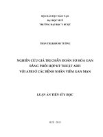
Differental diagnosis
Sclerosing mesenteritis
Metastasis of carcinoid tumor
Mesenteric stranding
+
76%
Calcification
20%
70%
Vessel displacement
40%
+
T2WI
low
Low to high
Radiat Med. 2006;24(3):220-223. doi:10.1007/s11604-005-1405-8
Radiat Med. 2006;24(3):220-223. doi:10.1007/s11604-005-1405-8
Radiat Med. 2006;24(3):220-223. doi:10.1007/s11604-005-1405-8
Radiat Med. 2006;24(3):220-223. doi:10.1007/s11604-005-1405-8
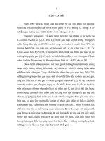
Trước tiêm
Radiat Med. 2006;24(3):220-223. doi:10.1007/s11604-005-1405-8
Sau tiêm và dựng coronal
Radiat Med. 2006;24(3):220-223. doi:10.1007/s11604-005-1405-8
Radiat Med. 2006;24(3):220-223. doi:10.1007/s11604-005-1405-8
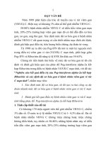
Sinh thiết
Radiat Med. 2006;24(3):220-223. doi:10.1007/s11604-005-1405-8
Trích dẫn
Matsuki M, Inada Y, Nakai G, et al. CT and MR features of sclerosing mesenteritis mimicking a
mesenteric metastasis from the carcinoid tumor. Radiat Med. 2006;24(3):220-223.
doi:10.1007/s11604-005-1405-8
Sharma P, Yadav S, Needham CM, Feuerstadt P. Sclerosing mesenteritis: a systematic review of
192 cases. Clin J Gastroenterol. 2017;10(2):103-111. doi:10.1007/s12328-017-0716-5