Broad range chemical profiling of natural deep eutectic solvent extracts using a high performance thin layer chromatography–based method
Bạn đang xem bản rút gọn của tài liệu. Xem và tải ngay bản đầy đủ của tài liệu tại đây (2.02 MB, 10 trang )
Journal of Chromatography A, 1532 (2018) 198–207
Contents lists available at ScienceDirect
Journal of Chromatography A
journal homepage: www.elsevier.com/locate/chroma
Broad range chemical profiling of natural deep eutectic solvent
extracts using a high performance thin layer chromatography–based
method
Xiaojie Liu a , Samantha Ahlgren a,b , Henrie A.A.J. Korthout c , Luis F. Salomé-Abarca a ,
Lina M. Bayona a , Robert Verpoorte a , Young Hae Choi a,d,∗
a
Natural Products Laboratory, Institute of Biology, Leiden University, 2333 BE Leiden, The Netherlands
Division of Pharmacognosy, Faculty of Pharmacy, Uppsala University, 751 05 Uppsala, Sweden
c
Fytagoras BV, 2333 BE Leiden, The Netherlands
d
College of Pharmacy, Kyung Hee University, 02447 Seoul, Republic of Korea
b
a r t i c l e
i n f o
Article history:
Received 16 October 2017
Received in revised form 3 December 2017
Accepted 4 December 2017
Available online 6 December 2017
Keywords:
Natural deep eutectic solvents
Chemical profiling
Ginkgo
Ginseng
High-performance thin-layer
chromatography
a b s t r a c t
Natural deep eutectic solvents (NADES) made mainly with abundant primary metabolites are being
increasingly applied in green chemistry. The advantages of NADES as green solvents have led to their
use in novel green products for the food, cosmetics and pharma markets. However, one of the main difficulties encountered in the development of novel products and their quality control arises from their low
vapour pressure and high viscosity. These features create the need for the development of new analytical
methods suited to this type of sample. In this study, such a method was developed and applied to analyse
the efficiency of a diverse set of NADES for the extraction of compounds of interest from two model
plants, Ginkgo biloba and Panax ginseng. The method uses high-performance thin-layer chromatography
(HPTLC) coupled with multivariate data analysis (MVDA). It was successfully applied to the comparative quali- and quantitative analysis of very chemically diverse metabolites (e.g., phenolics, terpenoids,
phenolic acids and saponins) that are present in the extracts obtained from the plants using six different
NADES. The composition of each NADES was a combination of two or three compounds mixed in defined
molar ratios; malic acid-choline chloride (1:1), malic acid-glucose (1:1), choline chloride-glucose (5:2),
malic acid-proline (1:1), glucose-fructose-sucrose (1:1:1) and glycerol-proline-sucrose (9:4:1). Of these
mixtures, malic acid-choline chloride (1:1) and glycerol-proline-sucrose (1:1:1) for G. biloba leaves, and
malic acid-choline chloride (1:1) and malic acid-glucose (1:1) for P. ginseng leaves and stems showed
the highest yields of the target compounds. Interestingly, none of the NADES extracted ginkgolic acids
as much as the conventional organic solvents. As these compounds are considered to be toxic, the fact
that these NADES produce virtually ginkgolic acid-free extracts is extremely useful. The effect of adding
different volumes of water to the most efficient NADES was also evaluated and the results revealed that
there is a great influence exerted by the water content, with maximum yields of ginkgolides, phenolics
and ginsenosides being obtained with approximately 20% water (w/w).
© 2017 The Authors. Published by Elsevier B.V. This is an open access article under the CC BY license
( />
1. Introduction
Natural products (NPs) are undoubtedly the most plentiful
source of new bioactive compounds and play an important role
in our daily lives, being used for their medicinal, nutritional
and cosmetic properties, and in industrial applications. However,
∗ Corresponding author at: Natural Products Laboratory, Leiden University, 2333
BE Leiden, The Netherlands and College of Pharmacy, Kyung Hee University, 02447
Seoul, Republic of Korea.
E-mail address: (Y.H. Choi).
extraction from their natural sources is generally a complex process, consisting of several steps that involve the use of large
volumes of organic solvents. Unfortunately, most of these solvents
are banned in the use of products for human consumption due to
their toxicity or are extremely restricted. In general, these solvents
are also highly volatile, posing as hazards to the environment. For
these reasons, the choice of suitable solvents is very limited [1].
The green extraction of NPs can be achieved by using innovative extraction techniques and/or sustainable alternatives to
conventional solvents. Great improvements have been accomplished with the use of non-conventional techniques such as
ultrasound-assisted extraction, microwave-assisted extraction,
/>0021-9673/© 2017 The Authors. Published by Elsevier B.V. This is an open access article under the CC BY license ( />
X. Liu et al. / J. Chromatogr. A 1532 (2018) 198–207
membrane-mediated extraction, and micro-extraction [2]. Some of
these techniques even allow for solvent-free extraction, including
microwave hydro-diffusion and gravity, enzyme-assisted extraction [3] and thermal desorption systems. However, for large-scale
extraction, the use of solvents is practically unavoidable hence
it is necessary to find alternatives to the conventional organic
solvents and/or mineral acids. Amongst the green options that
have appeared so far, the most popular are super- or subcritical fluids and ionic liquids. Supercritical fluid extraction (SFE), in
particular with supercritical carbon dioxide (SC-CO2 ), has been
used extensively for years to extract many natural bioactive compounds or eliminate toxic compounds from a variety of materials
[4–6]. However, despite its environmental benefits, the efficiency
of CO2 for the extraction of polar/hydrophilic compounds and
macromolecules is limited and many bioactive or nutritional compounds, such as phenolic glycosides or alkaloids, are not extractable
due to their poor solubility in CO2 . Other alternatives in the
green list include conventional solvents such as water, certain
agro-solvents (e.g., ethanol and glycerol) and surfactant aqueous
solutions [7,8].
Another type of novel alternative solvents that have gained considerable attention within the past few years are ionic liquids (ILs).
ILs are salts that consist of certain combinations of cations and
anions with melting points that are below 100 ◦ C. These liquids
possess many attractive physicochemical properties such as low
volatility, some electrolytic conductivity and tunable viscosity and
miscibility, making them promising substitutes for organic solvents
in numerous processes like biocatalytic processes, extractions,
catalysis, and electrochemistry [9–11]. However, their toxicity,
poor biodegradability and the high cost of synthesising their major
components cause major hindrance for their widespread application as extraction solvents.
Another option, deep eutectic solvents (DES), are mixtures of
specific organic compounds (not necessarily ionic) that have a
much lower melting point than either of the individual components and, similarly to ILs, are liquids at ambient temperatures
[12]. DES exhibit similar physicochemical properties as commonly
used ILs (e.g., high density and viscosity) but have the advantages
of being significantly cheaper, easier to prepare and less impactful
on the environment. In addition to this, they can be tailored to be
target-specific. These unique properties allow for a wide range of
applications of DES in fields such as extraction, catalysis, materials
chemistry, organic synthesis, metal processing, and electrochemistry [13,14], as well as in the extraction of DNA and as a Media for
enzymatic reactions [15,16].
A number of obstacles remain in the application of DES, particularly in those made with synthetic components in which toxicity
is an issue. Searching for alternatives, Choi and his co-workers
have explored more than 100 combinations of DES using only common metabolites that are abundant in plants [17]. They named
these combinations; ‘natural deep eutectic solvents’ (NADES) [17].
These liquids are bio-based DES which are composed of two or
more compounds that are generally functional primary metabolites, i.e., organic acids, sugars, (poly)alcohols, amines and amino
acids [17,18]. Apart from sharing the favorable characteristics of
ILs and DES as described previously, NADES have additional advantages of being composed of naturally-occurring compounds, being
much more sustainable and posing practically no environmental
hazards. This highlights the great potential of NADES for its application in various areas, namely in the NP field as an extraction solvent.
Currently, the most studied application of NADES as solvents for
NPs is the extraction of different phenolic compounds from plant
material such as Carthamus tinctorius flowers [19], Sophora japonica
flowers [20], Cajanus cajan leaves [21], Catharanthus roseus flowers [22], grape peel [23], and anthocyanins from wine lees [24].
They have also been applied for the extraction of ginsenosides
199
from Panax ginseng [25] and food contaminants [26]. Recently, the
first commercial NADES plant extracts were launched as cosmetic
ingredients and in the coming years a number of novel NADESbased products can be expected in the food, cosmetic and pharma
markets.
Given the increasing amount of NADES applications, the
methodological problems related to the analysis of NADES extracts
must be addressed. These problems are related mainly to the negligible volatility of NADES that makes the recovery of compounds
from NADES extracts or the elimination of interfering NADES components very difficult. So far, most analyses of NADES extracts
have been focused on phenolics that pose few methodological challenges given their highly-absorbing chromophores that allow direct
UV-spectrophotometric analysis with few to no sample clean-up
procedures. Different chromatographic methods have been used to
identify and quantify individual components, including the determination of the anthocyanin content in flower petal extracts from
C. roseus [19] and flavonoids in flower extracts from S. japonica via
HPLC-DAD, using anti-solvent strategies for recovery [20]. Alongside chromophore compounds, volatiles have been relatively easily
analysed by GC following simple liquid-liquid extraction from a
choline chloride and 4-chlorophenol (1:2) NADES [27]. However,
none of these methods can be applied to the full range of very chemically diverse compounds that are present in NADES extracts from
biological materials.
Within the past five decades, thin layer chromatography (TLC)
has advanced greatly from a simple planar chromatographic technique for major qualitative profiling into a multi-target quantitative
analytical tool with many applications in the field of medicinal
plants. The main disadvantages of TLC, such as its low resolution and poor quantitative performance, have been considerably
improved by the optimisation of the conventional TLC set-up.
Nowadays, the basic steps of TLC-based methods, i.e., sample
application, chromatographic development and detection, can be
automated and computer-controlled. Other undesirable characteristics, such as low efficiency, have been improved with the use
of high-performance thin-layer chromatography (HPTLC) plates,
which use stationary phases with smaller particle sizes (5–6 M)
and high-resolution sorbents with improved silica particles and
chemically-modified phases. These modern systems feature high
reproducibility in retention, detection and quantitative analysis. For further statistical evaluation, electronic images of the
chromatograms can be generated and used for multivariate data
analysis. As for the identification of compounds, notable progress
has been made in the direct online coupling of HPTLC with mass
spectrometry (MS) [28–31] which introduces new opportunities
for the application of planar chromatography in metabolomics.
Combining all of these improvements with the advantages of
multi-sample analysis in a single run and the option for selective
detection of a wide range of chemically diverse compounds makes
HPTLC a useful tool for the qualitative and quantitative analysis of
NADES extracts from natural products. Furthermore, the application of multivariate image analysis to HPTLC chromatograms allows
for the extraction of incalculably more information than that made
available by simple visual inspection [31].
The aim of the current study was to develop a suitable analytical
protocol for the HPTLC analysis of NADES plant extracts and then
to implement it to investigate the application of diverse NADES
(including a variation in water content) with the extraction of three
chemically different groups of active compounds in Ginkgo biloba
(i.e., ginkgolides, phenolics and ginkgolic acids) [33] and of ginsenosides from Panax ginseng [34–37]. The influence of adjustments to
the water content on the extraction ability and efficacy of the most
efficient NADES was also studied due to its known impact on their
physicochemical properties.
200
X. Liu et al. / J. Chromatogr. A 1532 (2018) 198–207
2. Materials and methods
2.1. Plant material
Ginkgo biloba leaves were collected in Seoul, Republic of Korea
in 2012. Panax ginseng leaves and stems were kindly provided by
Fytagoras (Leiden, the Netherlands). Samples used for this study
were identified by one of the authors, Dr. Y. H. Choi and a voucher
sample was deposited in the Natural Products Laboratory, Institute
of Biology, Leiden University. The dry plant material was powdered
in a blender with liquid nitrogen.
2.2. Chemicals and reagents
Bilobalide, ginkgolides (A, B and C), ginkgolic acids (C13:0, C15:1
and C17:1) and ginsenosides (Rb1, Rb2, Rb3, Re, Rg1, Rg2 and
Rg3) were purchased from Biopurify Phytochemicals (Chengdu,
China). Rutin, chlorogenic acid and quercetin were purchased from
Sigma (St. Louis, MO, USA). Methanol, ethanol, chloroform, acetone, ethyl acetate and toluene of analytical grade were purchased
from Sigma. Acetic anhydride, acetic acid and formic acid were
obtained from Sigma. Milli-Q water was used. Polyethylene glycol 400 was purchased from Alfa Aesar (Kandel, Germany). All
of the NADES components, i.e., choline chloride (≥98.0%), glycerol (≥99.5%), L-proline (≥99.0%), d-fructose (≥99.0%), d-glucose
(≥99.5%), malic acid (≥99.0%), and sucrose (≥99.5%) were obtained
from Sigma. Sodium acetate and 2-aminoethyl diphenylborinate
were purchased from Sigma. Solid phase extraction (SPE) cartridges
(OASIS HLB 3cc) were purchased from Waters (Milford, MA, USA).
Silica gel 60 F254 HPTLC plates were purchased from Merck (Darmstadt, Germany).
2.3. NADES preparation
The NADES employed in this study were combinations of fixed
molar ratios as follows; malic acid-choline chloride (1:1, N1), malic
acid-glucose (1:1, N2), choline chloride-glucose (5:2, N3), malic
acid-proline (1:1, N4), glucose-fructose-sucrose (1:1:1, N5) and
glycerol-proline-sucrose (9:4:1, N6) (Table S1). The first five NADES
were prepared by stirring mixtures of their components at 50 ◦ C
until a clear liquid was formed [19], whereas glycerol-prolinesucrose (9:4:1) was prepared using the freeze-drying method [25].
All of the prepared NADES were mixed with 10% (w/w) water. The
two most efficient NADES for the extraction of each plant were
found to be malic acid-choline chloride (1:1) and glycerol-prolinesucrose (9:4:1) for G. biloba, and malic acid-choline chloride (1:1)
and malic acid-glucose (1:1) for P. ginseng. These NADES were
selected to further study the effect of adding varying amounts of
water (0%, 10%, 20%, 30% and 40%, w/w) on their extraction properties.
2.4. Preparation of reference compound solutions for HPTLC
analysis
All reference solutions were prepared separately in methanol
in the following concentrations: bilobalide and ginkgolides B
and C (250 g/mL); ginkgolide A and C13:0, C15:1 and C17:1
ginkgolic acids (125 g/mL); rutin (1.8 g/mL); chlorogenic acid
and quercetin (4 g/mL); ginsenosides Rb1, Rb2, Rb3, Re, Rg1, Rg2,
and Rg3 (30 g/mL).
2.5. Preparation of extracts and sample solutions for HPTLC
analysis
Powdered plant material (200 mg) was mixed with 4 mL
methanol or NADES in a 15-mL centrifugation tube. After vortex-
ing for 1 min, the mixture was placed into a water bath at 40 ◦ C for
1 h and then ultrasonicated at room temperature for 30 min. The
mixture was then centrifuged at 13,000 rpm for 20 min. An aliquot
of 1 mL of the supernatant was used for the HPTLC analysis following pre-treatment. All extracts were prepared by triplicate. Extracts
prepared with methanol were used to compare the yield of the
diverse NADES extracts.
2.6. Recovery of samples from NADES
The removal of NADES from the mixture was conducted with
solid-phase extraction (SPE) using HLB cartridges [25]. Briefly, a
cartridge was placed in a vacuum manifold and equilibrated with
5 mL of ethanol, followed by 5 mL of water. After loading the extract
solution (1 mL), the cartridge was subsequently rinsed with 6 mL
of water twice and then eluted with 6 mL of ethanol. The ethanol
eluate was dried and re-dissolved in 1 mL of methanol for HPTLC
analysis.
2.7. General HPTLC analysis
Reference and sample solutions (100 L) were spotted by triplicate on HPTLC Si 60 F254, 20 × 10 cm (Merck) plates as 7 mm
bands, under a stream of nitrogen, using the CAMAG Automatic
TLC sampler (ATS 4) (CAMAG, Muttenz, Switzerland) with a 100 L
Hamilton syringe. The CAMAG auto-sample system is controlled by
WINCATS software. All plates were prepared similarly, spotting the
methanol extracts in the first three lanes, the reference solutions
in the next three and then the NADES extracts.
2.8. Analysis of NADES extracts with 10% water (w/w)
The layout of the tracks on the HPTLC plates varied in each case.
For the ginkgolides in G. biloba, a total of 13 tracks were used,
including triplicates of the methanol extract, four references and
six different NADES extracts. Bands were applied at a distance of
10 mm from the bottom of the plate and 16 mm from the left and
the right edges. For the determination of phenolics and ginkgolic
acids in G. biloba, the number of tracks per plate was 12, i.e., triplicates of methanol extract, three references and six different NADES
extracts. Bands were applied at a distance of 10 mm from the bottom of the plate and 20 mm from the left and the right edges. For
ginsenosides in P. ginseng leaves and stems, the number of tracks
per plate was 17, i.e., duplicates of methanol extract of leaves and
stems respectively, one reference and six different NADES extracts
of leaves and stems respectively. Bands were applied at a distance
of 10 mm from the bottom of the plate and 18 mm from the left and
the right edges.
2.9. The use of NADES with different water contents
The extracts obtained from G. biloba material with the most efficient NADES (N1 and N6, see above) with different added water
contents were then compared with each other and against the
methanol extracts. In the case of ginkgolides, this resulted in 17
tracks per plate with 16 used for the phenolics and ginkgolic acids,
i.e., triplicates of the methanol extract, three references, and the
two NADES with five different water contents. Bands were applied
at a distance of 10 mm from the bottom of the plate and 16 mm from
the left and the right edges. With the NADES extraction of ginsenosides from P. ginseng leaves and stems, the effect of added water
contents was studied using the most efficient NADES in this case,
i.e., N1 and N2. There were 14 tracks on each plate including triplicates of the methanol extract, one reference, and the N1 and N2
extracts with five different water contents. Bands were applied at a
distance of 10 mm from the bottom of the plate and 18 mm from the
X. Liu et al. / J. Chromatogr. A 1532 (2018) 198–207
left and the right edges. The HPTLC conditions used for the analysis
were those described for G. biloba and P. ginseng in the application notes of CAMAG laboratory (F-16A, F-16B, F-16C) [38] and the
Chinese Pharmacopeia [39], respectively (Table S2). Derivatisation
reagents were applied with an auto-spraying instrument (Derivatizer, CAMAG).
2.10. Determination of ginkgolides in Ginkgo biloba leaves
Prior to sample spotting, the plates were immersed in an ethanolic solution of sodium acetate (8 g NaOAc in 200 mL of 80% aqueous
ethanol) for 2 s, allowed to dry in the hood for 5 min at room
temperature and then activated at 90 ◦ C for 30 min as indicated
in the application notes of CAMAG laboratory (F-16A) [38]. Volumes of 25 L of methanol extracts and reference solutions, and
80 L of NADES extract solutions were spotted onto the HPTLC
plate as described. The plate was developed in a saturated chamber
with a mobile phase of toluene-ethyl acetate-acetone-methanol
(20:10:10:1.2, v/v/v/v). After drying the plates to remove the
mobile phase, they were evenly sprayed with acetic anhydride and
heated at 180 ◦ C for 10 min. Densitometric scanning was performed
at UV 366 nm. Bilobalide and ginkgolides A, B and C were used as
reference compounds.
2.11. Determination of phenolics in Ginkgo biloba leaves
A volume of 25 L of each reference solution, methanol extract,
and NADES extracts was applied onto the HPTLC silica plate and
developed with a mobile phase consisting of ethyl acetate-acetic
acid-formic acid-water (100:11:11:27, v/v/v/v) in a chamber saturated for 20 min before use, according to the application note of
CAMAG laboratory (F-16B) [38]. For the derivatisation, the plate
was heated at 100 ◦ C for 3 min on a TLC Plate Heater (CAMAG),
then dipped first in Natural Products reagent (1% 2-aminoethyl
diphenylborinate in methanol), dried with cold air, and then subsequently dipped in PEG reagent (5% polyethylene glycol 400 in
dichloromethane). Densitometric scanning was performed at UV
366 nm after derivatisation. Rutin, chlorogenic acid and quercetin
were used as reference compounds.
2.12. Determination of ginkgolic acids in Ginkgo biloba leaves
A volume of 25 L each of reference solutions of ginkgolic acids
(C13:0, C15:1 and C17:1), the methanol extract and the NADES
extracts was spotted separately onto the plate and developed
with a mobile phase consisting of toluene-ethyl acetate-acetic acid
(8:2:0.2, v/v/v) in a saturated chamber as indicated in the F-16-C
of the application notes of CAMAG laboratory [38]. The plate was
dried with a stream of cold air and scanned at UV 366 nm.
2.13. Determination of ginsenosides in Panax ginseng leaves and
stems
The plate was developed with chloroform-ethyl acetatemethanol-water (15:40:22:10) in a chamber saturated for 20 min
[39]. Ginsenosides were detected by spraying the plate with freshly
prepared anisaldehyde-sulfuric acid reagent and heating on a plate
heater at 105 ◦ C for 5 min. Images were captured under white light
after derivatisation. Ginsenosides Rb1, Rb2, Rb3, Re, Rg1, Rg2 and
Rg3 were used as standards.
2.14. Image processing and multivariate analysis
The HPTLC chromatograms were processed using rTLC software
[32] which converts the HPTLC images into a numerical data matrix
which is then integrated. This process generated digital data of 50
201
sequential 0.02 bins over the full retention factors (Rf ) range for
each track. An Rf of 0.02 was selected as the bin size because it represents the individual band in a single bin but avoids the Rf drift
which may result from batch-to-batch variation factors. To reduce
the variations between the replicates that were performed at different times, the intensity recorded for each Rf value was normalised
with respect to the methanol extract (reference extract sample) in
each plate. These 0.02 Rf bins were subjected to multivariate data
analysis. Principal component analysis (PCA) and orthogonal partial least square (OPLS) were performed with the SIMCA-P software
(v. 14.1, Umetrics, Umeå, Sweden). The unit variance (UV) scaling
method was used both for PCA analysis and OPLS modelling.
3. Results and discussion
This study was performed using six types of NADES to extract
four groups of chemically diverse bioactive compounds (phenolics,
terpenoids and phenolic acids from G. biloba leaves, and ginsenosides from P. ginseng leaves and stems). The NADES have been
grouped into five typical types according to their components,
i.e., NADES composed of acids and bases (N1), acids and sugars (N2), bases and sugars (N3), amino acid and acids (N4), and
sugar mixtures (N5). In addition to these typical NADES, glycerolproline-sucrose (9:4:1, N6) was selected because in previous work
performed by Jeong and her co-workers, it was reported that ginsenosides were well-extracted from P. ginseng roots by the NADES
[25]. To reduce the high viscosity of the NADES as an extraction solvent, 10% of water (w/w) was added to each NADES, as suggested
in a previously published paper [18].
Similarly to conventional synthetic DES or ILs, NADES extracts
have virtually zero vapour pressure which means that the solvent cannot be removed by evaporation as is generally the case
when organic solvents are used for extraction. The removal of the
extraction solvent is generally necessary when analysing an extract,
concentrating the low-level compounds or avoiding its interference
with the analysis. In the case of NADES, two approaches have been
proposed, namely liquid-liquid partitioning [27] and solid phase
extraction (SPE) with diverse sorbents [20,25]. However, neither
method completely removed all of the NADES. Liquid-liquid partitioning is only possible with non-polar solvents such as n-hexane,
dichloromethane or chloroform because most NADES components
are soluble in polar or mid-polar solvents, making it difficult to separate them from the extracted polar secondary metabolites with
similar polarities. On the other hand, SPE has proved to be quite efficient in purifying secondary metabolites, though NADES residues
may still cause problems in various analytical methods.
So, there is a clear need for efficient methods that can be applied
to the vast range of chemically diverse compounds found in NADES
extracts. Thin layer chromatography has a long tradition in NP analysis and, in the past decade, has evolved into a highly improved
technique known as HPTLC [40]. This technique meets all of the
mentioned requirements to a great extent. In particular, the possibility of the simultaneous analysis of several samples on a single
plate and the possibility of a preparative work by mass and/or NMR
spectroscopy constitute a great advantage over other chromatographic methods [41,42].
To develop HPTLC protocols for the analysis of NADES extracts,
we used two well-known medicinal plants as models, G. biloba
and P. ginseng. The plant material was extracted with six different
types of NADES. These extracts were then analysed by HPTLC for
the presence of four different types of compounds; phenolics, terpene lactones and alkylphenols in G. biloba leaves, and triterpene
saponins in P. ginseng leaves and stems.
The NADES extracts were first analysed directly, without any
pre-purification steps, to evaluate the interference of the NADES in
202
X. Liu et al. / J. Chromatogr. A 1532 (2018) 198–207
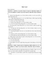
Fig. 1. High-performance thin-layer chromatographs (HPTLC) of methanol extracts and six natural deep eutectic solvent (NADES) extracts with 10% (w/w) water for
ginkgolides (a), ginkgo phenolics (b), and ginkgolic acids (c) of Ginkgo biloba leaves, and ginsenosides of Panax ginseng stems (d) and leaves (e). Ginkgolides were analysed after derivatisation with acetic anhydride at 366 nm using plates that were impregnated with an ethanolic solution of sodium acetate (see details in M&M). Ginkgo
phenolics were detected after derivatisation with the natural products reagent (NPR) and PEG 4000 at 366 nm. Ginsenosides were visualised under white light after treatment
with the anisaldehyde-sulfuric acid reagent. The detailed HPTLC conditions were provided in the experimental section. M: methanol extract, N1-N6: NADES extracts. N1:
malic acid-choline chloride (1:1), N2: malic acid- glucose (1:1), N3: choline chloride-glucose (5:2), N4: malic acid-proline (1:1, molar ratio), N5: glucose-fructose-sucrose
(1:1:1), N6: glycerol-proline-sucrose (9:4:1). Reference compounds: 1: bilobalide, 2: ginkgolide A, 3: ginkgolide B, 4: ginkgolide C, 5: quercetin, 6: chlorogenic acid, 7: rutin,
8: ginkgolic acids (C13:0, C15:1 and C17:1), 9: ginsenoside Rg3, 10: ginsenoside Rg2, 11: ginsenoside Rg1, 12: ginsenoside Re, 13: ginsenoside Rb3, 14: ginsenoside Rb2, 15:
ginsenoside Rb1.
the HPTLC methods for the four different groups of NPs. The presence of NADES caused severe tailing of spots in all of the systems.
It was clearly necessary to perform some sample clean-up procedures, hence SPE was selected as the method of choice for this.
Because of their ability to bind a wide range of secondary metabolites, including glycosides, Oasis HLB cartridges were tested for the
purification of ginsenosides from the NADES extracts composed of
glycerol, proline and sucrose (9:4:1) [25]. The NADES extract was
introduced onto the cartridge and the NADES components were
removed by an initial elution with water, after which the compounds of interest were eluted with ethanol. The quality of the
HPTLC separation improved as tailing completely disappeared.
The NADES extracts of G. biloba leaves and P. ginseng leaves
and stems were all treated in the same way and then analysed by
HPTLC. Fig. 1 shows the four groups of metabolites that each can
be well visualised in the different HPTLC chromatograms, clearly
showing its power to detect a wide range of chemically diverse
groups of metabolites [43]. The P. ginseng saponins without UVchromophores were able to be visualised at 254 nm and 366 nm
after treatment with the anisaldehyde-sulfuric acid derivatisation
reagent (Fig. 1d, e).
Visual examination of the HPTLC chromatograms showed that
the extraction efficiency of all NADES employed in this study,
except for the all-sugar NADES N5, was similar to that of methanol
for ginkgolides and phenolics from G. biloba and for ginsenosides
in P. ginseng leaves and stems (Fig. 1). Ginkgolic acids were not significantly extracted by any of the NADES. In general, plant aliphatic
phenols like ginkgolic acids have very low polarity, which makes
them difficult to be dissolved in polar solvents. Most NADES are
categorised as polar solvents and could not adequately extract the
non-polar ginkgolic acids.
In the cases of ginkgolides and ginsenosides, the NADES extracts
showed fewer bands than the methanol extracts, but all of the main
compounds (bilobalide, 3 ginkgolides, and 7 ginsenodides) in the
NADES extracts were still present in similar concentrations to that
of the methanol extracts with an exception for the sugar mixture
(N5) (Fig. 1a, d, e). In the case of G. biloba phenolics, NADES extracts
displayed more bands than the methanol extracts, for example,
within the 0.35–0.38 Rf range (Fig. 1b). The most striking feature,
however, is low extraction yield of the ginkgolic acids in all tested
NADES revealing clearly different extraction profiles for the NADES
and methanol (Fig. 1c). Ginkgolic acids are considered to be toxic
and the presence of these compounds is unwanted in G. biloba
extracts that are used for human consumption, so the extraction of
the leaves with NADES could result in high quality Ginkgo preparations with very low ginkgolic acid content.
X. Liu et al. / J. Chromatogr. A 1532 (2018) 198–207
203
Fig. 2. Score plot of principal component analysis (PCA) of natural deep eutectic solvent (NADES) extracts and methanol extracts of ginkgolides (a) and ginkgo phenolics
(b) in Ginkgo biloba leaves, and score plots of orthogonal partial least square discriminant analysis of ginkgolides (c) and ginkgo phenolics (d). M: methanol extract. 1–6:
NADES extracts. 1: malic acid-choline chloride (1:1, molar ratio), 2: NADES of malic acid- glucose (1:1), 3: choline chloride-glucose (5:2), 4: malic acid-proline (1:1), 5:
glucose-fructose-sucrose (1:1:1), 6: glycerol-proline-sucrose (9:4:1).
The results obtained in this study highlight once more the great
potential of NADES as a green alternative solvent for the extraction
of phenolics. This high extraction power of NADES for phenolics
may be related to H bonding interactions between the functional
groups of the components (e.g., hydroxyl and carboxyl groups) and
the hydroxyl groups in phenolics. We have reported the observation of H bonding interactions between quercetin and NADES in
previous studies [44].
Chromatographic profiles provide basic information about specific groups of compounds but, most importantly, can characterise
the chemical composition of a sample in a holistic way. In fact, that
is the paradigm of metabolomics; aiming at the unbiased analysis of all of the metabolites within an organism. In order to fully
take advantage of all of the information provided by this profiling
method, it is necessary to use biometric methods such as multivariate data analysis (MVDA) and multivariate image analysis to be able
to identify the similarities and differences between the measured
profiles and then combine these with other observations, including
the metabolic changes triggered by diseases or related to resistance
against herbivores.
All chromatographic profiling methods require the normalisation and alignment of signals prior to MVDA. For the normalisation,
three control samples (in this study a methanol extract was used)
were spotted alongside the other samples on each plate. The intensity at each Rf value of the samples were normalised to a methanol
extract in order to minimise the variation of replicates on different plates. This normalisation improved the quality of the MVDA
data quality (Fig. S1). The recently-developed open-source software, rTLC, was used for alignment which offers a standardised
procedure for image processing and the visualisation tools that are
required to compare HPTLC fingerprints via different pattern recognition and prediction techniques [32]. The processed data were
further analysed by PCA and OPLS.
The PCA data of ginkgolides and ginkgo phenolics in G. biloba
leaves is shown in Fig. 2. No further analysis of the ginkgolic acids
in NADES extracts of G. biloba leaves was performed due to their low
204
X. Liu et al. / J. Chromatogr. A 1532 (2018) 198–207
Fig. 3. Score plots of principal component analysis (PCA) of data corresponding to ginsenosides in Panax ginseng leaves (a) and stems (b) obtained from HPTLC chromatograms
using PC1 and PC2 comparing various natural deep eutectic solvents (NADES) extracts and methanol extracts. Orthogonal partial least square-discriminant analysis (OPLS-DA)
score plots of ginsenosides in P. ginseng leaves (c) and stems (d). The numbering of the extracts is the same as in Fig. 2.
yield as shown in Fig. 1c. In the PCA score plot of G. biloba samples,
NADES extracts were clearly distinct from the methanol extract but
there was no significant difference amongst the NADES solvents.
If any, in the case of phenolics (Fig. 2b), malic acid-choline chloride (N1) and glycerol-proline-sucrose (N6) appeared to be closer to
the methanol extracts than the other NADES extracts. A supervised
MVDA, OPLS-DA, was employed to obtain a more detailed comparison between both the methanol and NADES extracts, including all
of the six tested NADES. This showed a clear separation between
methanol and NADES extracts (Fig. 2d). For the identification of
the contributing metabolites, an S plot was used and it revealed
that methanol extracted higher amounts of most metabolites. All
of the tested ginkgolides, bilobalide (Rf 0.447), ginkgolides A (Rf
0.361), B (Rf 0.266) and C (Rf 0.114) were extracted less efficiently
with NADES (Fig. S2a). The HPTLC analysis of phenolics in G. biloba
leaves also acknowledged that methanol was more efficient than
NADES, for example, for chlorogenic acid (Rf 0.52), rutin (Rf 0.352)
and quercetin (Rf 0.99) (Fig. S2b).
In the case of P. ginseng leaves and stems, there were differences
amongst the extraction profiles obtained with methanol, but also
amongst the NADES extracts (Fig. 3a, b). The first cluster consisted
of the methanol extracts, the second of malic acid-choline chloride
(N1) and malic acid-glucose (N2), and the third grouped together
the three remaining NADES, choline chloride-glucose (N3), malic
acid-proline (N4) and glycerol-proline-sucrose (N6), implying a
similarity in their chemical profiles. Extracts made with glucosefructose-sucrose (N5) formed a fourth cluster (Fig. 3a and b). To
compare the NADES and methanol extracts, OPLS-DA was applied
to the P. ginseng samples (Fig. 3c and d) and the results showed
that all of the seven analysed ginsenosides were more efficiently
extracted from both P. ginseng leaves and stems with NADES (Fig.
S3).
Apart from their chemical composition, another factor that has
a great influence on the physicochemical properties of NADES is
their water content [45]. One of the positive effects of increasing
the water content is a decrease in their viscosity; one of the features
X. Liu et al. / J. Chromatogr. A 1532 (2018) 198–207
205
Fig. 4. Orthogonal partial least square-discriminant analysis (OPLS-DA) score plots of different natural deep eutectic solvents (NADES) extracts (a), OPLS score plots of various
water contents (0–40%, w/w) (b) and shared-and-unique-structures (SUS)-plots (c) of ginkgolides and ginkgo phenolics in Ginkgo biloba leaves, and ginsenosides in Panax
ginseng leaves and stems. SUS plots correlate the two OPLS-DA models with the X-axis of NADES composition and OPLS of water contents as Y-axis. The numbering of the
extracts in Fig. 4a and b is the same as in Fig. 2. The numbering of the identified compounds is the same as in Fig. 1.
that hinders their use as extraction solvents. To evaluate the effect
of the water ratio, different amounts of water was added (0–40%,
w/w) to the two NADES that had the highest extraction yields for
each plant in the first experiment. The extracts were analysed using
HPTLC-MVDA.
The content of ginkgolides and phenolics in G. biloba extracts
prepared with malic acid-choline chloride (1:1) (N1) and glycerol-
proline-sucrose (9:4:1) (N6) with varying water ratios were
compared. The data obtained from the HPLTC chromatograms (Xdata) were combined with the water content (Y-data) to evaluate
the effect of the water percentage (w/w) on the tested NADES. Two
different OPLS models with the water content or composition of
NADES as Y-data were set up (Fig. 4a, b). In the score plot of the
OPLS modeling, the two selected NADES extracts were clearly sep-
206
X. Liu et al. / J. Chromatogr. A 1532 (2018) 198–207
arated from each other (Fig. 4a). From the results, it was clear that
apart from the different chemical compositions of N1 and N6, the
water content also had a large effect on the extraction profiles.
These two OPLS models (NADES composition of N1 and N6, and
water content) were integrated by a shared-and-unique-structures
(SUS)-plot, in which diagonally-aligned metabolites are of equal
importance and shared by the two models, and the main factor
influencing extraction yield of each metabolite can be deduced
(Fig. 4c). In the SUS-plot, the effect of individual factors (X-axis
for NADES chemical compositions and Y-axis for water content)
were easily distinguished for each metabolite. As observed, the
two metabolites that are most affected by the NADES composition
changes were ginkgolide B and chlorogenic acid, though very differently, i.e., glycerol-proline-sucrose (N6) extracted the highest
amount of ginkgolide B but the least amount of chlorogenic acid.
The water content greatly influenced NADES extraction yields as
seen on the Y-axis in the SUS-plot (Fig. 4c). This was particularly
noticeable in the case of rutin.
Panax ginseng leaves and stems were extracted with the two
most efficient NADES, malic acid-choline chloride (N1) and malic
acid-glucose (N2), with varying volumes of added water and then
analyzsed by OPLS and SUS-plot, similarly to the G. biloba samples.
Interestingly, as seen in Fig. 4, the yield of the seven ginsenosides
were influenced in a different way, even though their structural
differences are minimal. The highest yield of ginsenosides Rg3 and
Rg2 in both P. ginseng leaves and stems were obtained with malic
acid-glucose (N2) whilst malic acid-choline chloride (N1) yielded
the most ginsenoside Rb1 from P. ginseng stems (Fig. 4c). All seven
ginsenosides of P. ginseng leaves and six ginsenosides of P. ginseng
stems (except for ginsenoside Rb1) were best extracted with NADES
with the highest added water content (Fig. 4c).
4. Conclusion
To develop a reliable analytical method for NADES extracts,
a HPTLC-based method was employed. This method was tested
on two well-known medicinal plants and proved to be able to
deliver reproducible chemical profiles from the NADES extracts.
The results verified that the yield of bioactive compounds obtained
with six different types of NADES is similar to that of methanol
in all cases with only one exception. The application of multivariate analysis revealed, however, some clear differences in
extraction selectivity amongst the different NADES. The addition
of water to the NADES had a large effect on the efficiency of their
extraction for the selected compounds, increasing their yield in
general. Maximum amounts of ginkgolides, phenolics and ginsenosides were obtained with an addition of approximately 20%
water to the NADES. It is worth noting that the most striking
difference between the methanol and NADES extracts was the
significant lack in ginkgolic acids in the NADES extracts. This
is very promising for further studies, since it suggests potential for obtaining practically ginkgolic acid-free preparations for
pharmaceutical use.
All of the results obtained in this study show NADES to be
promising extraction solvents. A deeper knowledge of the theoretical basis for the extraction mechanism of the NADES and their
interaction with solutes would greatly facilitate the development of
future applications. Aside from this, the HPTLC-analytical method
described here will be a useful tool for this process.
Acknowledgments
We thank Mr Dick Bruynzeel in BCON Instrument (Sint Annaland, the Netherlands) for technical support for HPTLC experiments
and Dr Erica G. Wilson for all valuable scientific discussion.
Appendix A. Supplementary data
Supplementary material related to this article can be found, in
the online version, at doi: />12.009
References
[1] P.T. Anastas, M.M. Kirchhoff, Origins, current status, and future challenges of
green chemistry, Acc. Chem. Res. 35 (2002) 686–694.
[2] S. Armenta, S. Garrigues, M. de la Guardia, Green analytical chemistry, TrAC
27 (2008) 497–511.
[3] L.S. Chua, A review on plant-based rutin extraction methods and its
pharmacological activities, J. Ethnopharmacol. 150 (2013) 805–817.
[4] R. Murga, R. Ruiz, S. Beltran, J.L. Cabezas, Extraction of natural complex
phenols and tannins from grape seeds by using supercritical mixtures of
carbon dioxide and alcohol, J. Agric. Food Chem. 48 (2000) 3408–3412.
[5] M.C. Henry, C.R. Yonker, Supercritical fluid chromatography, pressurized
liquid extraction, and supercritical fluid extraction, Anal. Chem. 78 (2006)
3909–3915.
[6] H.H. Wijngaard, M. Ballay, N. Brunton, The optimisation of extraction of
antioxidants from potato peel by pressurised liquids, Food Chem. 133 (2012)
1123–1130.
[7] M. Letellier, H. Budzinski, L. Charrier, S. Capes, A.M.J. Dorthe, Optimization by
factorial design of focused microwave assisted extraction of polycyclic
aromatic hydrocarbons from marine sediment, Fresenius J. Anal. Chem. 364
(1999) 228–237.
[8] C. Pronyk, G. Mazza, Design and scale-up of pressurized fluid extractors for
food and bioproducts, J. Food Chem. 95 (2009) 215–226.
[9] H. Zhao, S.Q. Xia, P.S. Ma, Use of ionic liquids as ’green’ solvents for
extractions, J. Chem. Technol. Biotechnol. 10 (2005) 1089–1096.
[10] P. Moriel, E.J. García-Suárez, M. Martínez, A.B. García, M.A. Montes-Morán, V.
˜
Synthesis, characterization, and catalytic
Calvino-Casilda, M.A. Banares,
activity of ionic liquids based on biosources, Tetrahedron Lett. 51 (2010)
4877–4881.
[11] W.L. Hough, M. Smiglak, H. Rodríguez, R.P. Swatloski, S.K. Spear, D.T. Daly, J.
Pernak, J.E. Grisel, R.D. Carliss, M.D. Soutullo, J.H. Davis Jr, R.D. Rogers, The
third evolution of ionic liquids: active pharmaceutical ingredients, New J.
Chem. 31 (2007) 1429–1437.
[12] A.P. Abbott, G. Capper, D.L. Davies, R.K. Rasheed, V. Tambyrajah, Novel solvent
properties of choline chloride/urea mixtures, Chem. Commun. (2003) 70–71.
[13] B. Tang, H. Zhang, K.H. Row, Application of deep eutectic solvents in the
extraction and separation of target compounds from various samples, J. Sep.
Sci. 38 (2015) 1053–1064.
[14] E.L. Smith, A.P. Abbott, K.S. Ryder, Deep eutectic solvents (DESs) and their
applications, Chem. Rev. 21 (2014) 11060–11082.
[15] T. Gorke, F. Srienc, R.J. Kazlauskas, Hydrolase-catalyzed biotransformations in
deep eutectic solvents, Chem. Commun. 138 (2008) 1235–1237.
[16] H. Zhao, G.A. Baker, S. Holmes, Protease activation in glycerol-based deep
eutectic solvents, J. Mol. Cat. B: Enzym. 72 (2011) 163–167.
[17] Y.H. Choi, J. van Spronsen, Y. Dai, M. Verberne, F. Hollmann, I.W.C.E. Arends,
G.-J. Witkamp, R. Verpoorte, Are natural deep eutectic solvents the missing
link in understanding cellular metabolism and physiology? Plant Physiol. 156
(2011) 1701–1705.
[18] Y. Dai, J. van Spronsen, G.-J. Witkamp, R. Verpoorte, Y.H. Choi, Natural deep
eutectic solvents as new potential media for green technology, Anal. Chim.
Acta 766 (2013) 61–68.
[19] Y. Dai, G.-J. Witkamp, R. Verpoorte, Y.H. Choi, Natural deep eutectic solvents
as new extraction media for phenolic metabolites in safflower, Anal. Chem. 85
(2013) 6272–6278.
[20] M. Nam, J. Zhao, M. Lee, J. Jeong, J. Lee, Enhanced extraction of bioactive
natural products using tailor-made deep eutectic solvents: application to
flavonoid extraction from Flos sophorae, Green Chem. 17 (2015) 1718–1727.
[21] Z. Wei, X. Qi, T. Li, Application of natural deep eutectic solvents for extraction
and determination of phenolics in Cajanus cajan leaves by ultra performance
liquid chromatography, Sep. Purif. Technol. 149 (2015) 237–244.
[22] Y. Dai, E. Rozema, R. Verpoorte, Y.H. Choi, Application of natural deep eutectic
solvents to the extraction of anthocyanins from Catharanthus roseus with high
extractability and stability replacing conventional organic solvents, J.
Chromatogr. A 1434 (2016) 50–56.
[23] K. Radosevic, N. Curko, V.G. Srcek, M.C. Bubalo, M. Tomaˇsevic, K.K. Ganic, I.R.
Redovnikovic, Natural deep eutectic solvents as beneficial extractants for
enhancement of plant extracts bioactivity, LWT -Food Sci. Technol. 73 (2016)
45–51.
[24] T. Bosiljkov, F. Dujmic, M.C. Bubalo, J. Hribar, R. Vidrih, M. Brncic, E. Zlatic, I.R.
Redounikavic, S. Jokic, Natural deep eutectic solvents and ultrasound-assisted
extraction: green approaches for extraction of wine lees anthocyanins, Food
Bioprod. Process. 102 (2017) 195–200.
[25] K.M. Jeong, M.S. Lee, M.W. Nam, J. Zhao, Y. Jin, D.K. Lee, S.W. Kwon, J.H. Jeong,
J. Lee, Tailoring and recycling of deep eutectic solvents as sustainable and
efficient extraction media, J. Chromatogr. A 1424 (2015) 10–17.
[26] L. Piemontese, F.M. Perna, A. Logrieco, V. Capriati, M. Solfrizzo, Deep eutectic
solvents as novel and effective extraction media for quantitative
X. Liu et al. / J. Chromatogr. A 1532 (2018) 198–207
[27]
[28]
[29]
[30]
[31]
[32]
[33]
[34]
[35]
determination of ochratoxin A in wheat and derived products, Molecules 22
(2017) 121–130.
M.A. Farajzadeh, M.R.A. Mogaddam, M. Aghanassab, Deep eutectic
solvent-based dispersive liquid-liquid microextraction, Anal. Methods 8
(2016) 2576–2583.
R.B. Cody, J.A. Laramee, H.D. Durst, Versatile new ion source for the analysis of
materials in open air under ambient conditions, Anal. Chem. 77 (2005)
2297–2302.
G.J. van Berkel, B.A. Tomkins, V. Kertesz, Thin-layer
chromatography/desorption electrospray ionization mass spectrometry:
investigation of goldenseal alkaloids, Anal. Chem. 79 (2007) 2778–2789.
G. Morlock, W. Schwack, Coupling of planar chromatography to mass
spectrometry, TrAC. 29 (2010) 1157–1171.
S.C. Cheng, M.Z. Huang, J. Shiea, Thin layer chromatography/mass
spectrometry, J. Chromatogr. A 19 (2011) 2700–2711.
´ G.E. Morlock, Proof-of-principle of rTLC, an
D. Fichou, P. Ristivojevic,
open-source software developed for image evaluation and multivariate
analysis of planar chromatograms, Anal. Chem. 88 (2016) 12494–12501.
T.A. van Beek, P. Montoro, Chemical analysis and quality control of Ginkgo
biloba leaves, extracts, and phytopharmaceuticals, J. Chromatogr. A 11 (2009)
2002–2032.
S. Shibata, O. Tanaka, M. NagaI, T. Ishit, Studies on the constituents of
Japanese and Chinese crude drugs. XII. Panaxadiol, a sapogenin of ginseng
roots, Chem. Pharm. Bull. (Tokyo) 11 (1963) 762–765.
J. Huang, X.H. Tang, T. Ikejima, X.J. Sun, X.B. Wang, R.G. Xi, L.J. Wu, A new
triterpenoid from Panax ginseng exhibits cytotoxicity through p53 and the
caspase signaling pathway in the HepG2 cell line, Arch. Pharm. Res. 31 (2008)
323–329.
207
[36] D.H. Kim, Chemical diversity of Panax ginseng, Panax quinquifolium, and Panax
notoginseng, J. Ginseng Res. 36 (2012) 1–15.
[37] J. Yang, J. Guo, J. Yuan, In vitro antioxidant properties of rutin, LWT-Food Sci.
Technol. 41 (2008) 1060–1066.
[38] CAMAG. HPTLC methods for the identification of medicinal plants. Available
at />[39] China Pharmacopoeia Committee, Pharmacopoeia of the People’s Republic of
China, Chemical Industry Press, Beijing, 2005.
[40] E. Reich, V. Widmer, Plant analysis 2008 – planar chromatography, Planta
Med. 75 (2009) 711–718.
[41] G. Morlock, Background mass signals in TLC/HPTLC-ESI-MS and practical
advices for use of the TLC-MS interface, J. Liq. Chromatogr. Relat. Technol. 37
(2014) 2892–2914.
[42] H.-R. Adhami, U. Scherer, H. Kaehlig, T. Hettich, G. Schlotterbeck, E. Reich, L.
Krenn, Combination of bioautography with HPTLC-MS/NMR: a fast
identification of acetylcholinesterase inhibitors from Galbanum dagger,
Phytochem. Anal. 24 (2013) 395–400.
[43] M. Waksmundzka-Hajnos, J. Sherma, T. Kowalska, Thin Layer
Chromatography in Phytochemistry, CRC Press, Boca Raton, 2008, pp. 3–14.
[44] Y. Dai, R. Verpoorte, Y.H. Choi, Natural deep eutectic solvents providing
enhanced stability of natural colorants from safflower (Carthamus tinctorius),
Food Chem. 159 (2014) 116–121.
[45] Y. Dai, G.-J. Witkamp, R. Verpoorte, Y.H. Choi, Tailoring properties of natural
deep eutectic solvents with water to facilitate their application, Food Chem.
187 (2015) 14–19.