Báo cáo hóa học: "Rac1-mediated signaling plays a central role in secretion-dependent platelet aggregation in human blood stimulated by atherosclerotic plaque" ppt
Bạn đang xem bản rút gọn của tài liệu. Xem và tải ngay bản đầy đủ của tài liệu tại đây (1.38 MB, 10 trang )
RESEARC H Open Access
Rac1-mediated signaling plays a central role in
secretion-dependent platelet aggregation in
human blood stimulated by atherosclerotic plaque
Suman Dwivedi
1
, Dharmendra Pandey
1,3
, Anna L Khandoga
1
, Richard Brandl
2
, Wolfgang Siess
1*
Abstract
Background: Platelet activation requires rapid remodeling of the actin cytoskeleton which is regulated by small
GTP-binding proteins. By using the Rac1-specific inhibitor NSC23766, we have recently found that Rac1 is a central
component of a signaling pathway that regulates dephosphorylation and activation of the actin-dynamising
protein cofilin, dense and a-granule secretion, and subsequent aggregation of thrombin-stimulated washed
platelets.
Objectives: To study whet her NSC23766 inhibits stimulus-induced platelet secretion and aggregation in blood.
Methods: Human platelet aggregation and ATP -secretion were measured in hirudin-anticoagulated blood and
platelet-rich plasma (PRP) by using multiple electrode aggregometry and the Lumi-aggregometer. Platelet
P-selectin expression was quantified by flow cytometry.
Results: NSC23766 (300 μM) inhibited TRAP-, collagen-, atherosclerotic plaque-, and ADP-induced platelet
aggregation in blood by 95.1%, 93.4%, 92.6%, and 70%, respectively. The IC
50
values for inhibition of TRAP-,
collagen-, and atherosclerotic plaque-, were 50 ± 18 μM, 64 ± 35 μM, and 50 ± 30 μM NSC23766 (mean ± SD,
n = 3-7), respectively. In blood containing RGDS to block integrin a
IIb
b
3
-mediated platelet aggregation, NSC23766
(300 μM) completely inhibited P-selectin expression and reduced ATP-secretion after TRAP and collagen stimulation
by 73% and 85%, respectively. In ADP-stimulated PRP, NSC23766 almost completely inhibited P-selectin expression,
in contrast to aspirin, which was ineffective. Moreover, NSC23766 (300 μM) decreased plaque-stimulated platelet
adhesion/aggregate formation under arterial flow conditions (1500s
-1
) by 72%.
Conclusions: Rac1-mediated signaling plays a central role in secretion-dependent platelet aggregation in blood
stimulated by a wide array of platelet agonists including atherosclerotic plaque. By specifically inhibiting platelet
secretion, the pharmacological targeting of Rac1 could be an interesting approach in the development of future
antiplatelet drugs.
Background
After rupture of atherosclerotic plaques thrombogenic
matrix components and lipids are locally exposed to cir-
culating platelets [1-5]. By adhering to these sites, plate-
lets rapidly become activated, leading t o secretion of
their granule contents such as ADP that recruits circu-
lating platelets into large aggregates culminating in the
formation of platelet thr ombi [5,6]. The latter are
potentially life-threatening by occluding coronary and
cerebral arteries.
The step-wise activation of platelets (adhesion, shape
change, secretion and aggregat ion) involves an organized
remodeling of the actin cytoskeleton. The major molecules
involved in actin dynamics are the small GTP-binding
proteins Rho, Rac, and Cdc42. These proteins differentially
regulate the reorganization of the actin cytoskeleton,
leading to the formation of different cellular structures.
In platelets, Rho activation mainly regulates the Ca
2+
-
independent cell spheration and contractility during shape
change through stimulation of the Rho-kinase ROCK,
* Correspondence:
1
Institute for Prevention of Cardiovascular Diseases, University of Munich,
Munich, Germany
Full list of author information is available at the end of the article
Dwivedi et al. Journal of Translational Medicine 2010, 8:128
/>© 2010 Dwivedi et al; licensee BioMed Central Ltd. This is an Open Access article distributed under the terms of the Creative Commons
Attribution License ( es/by/2.0), which permits unrestricted use, distribution, and reproduction in
any medium, provided the original work is properly cited.
whereas Rac1 has been reported to be essential for the
formation of lamellipodia during platele t spreading [7-9].
Rac1 activation in platelets is Ca
2+
-dependent [10,11], and
it has been shown to be involved in regulating secretion
and subsequent aggregation in human platelets stimulated
with thrombin [12,13]. However, in mice platelets, the
results regarding the role of Rac1 in thrombin-induced
aggregation and secretion are controversial [9,12,14]. By
using conditional Rac1 knock-out mice, only one study
showed impaired thrombin-induced aggregation [12]. In
the two other studies, thrombin-induced secretion and
aggregation were not affected; Rac1 was found to be
involved only in collagen/glycoprotein VI-mediated plate-
let activation [9,14].
An important tool in studying the functio n of Rac1 is
the compound NSC23766, a small-molecule inhibitor
that fits into a surface groove of Rac1 known to be criti-
cal for the binding of specific guanine nucleotide
exchange factors (GEFs) converting Rac-GDP into its
active Rac-GTP form. NSC23766 inhibits invitroRac1
binding and activation by the Rac-specific GEF Trio or
Tiam1 [15]. The specific Rac-inhibitor NSC23766 has
been used in more than 90 scienti fic studies in which
the results obtained have often been validated by Rac-
silencing and Rac knock-out experiments (see http://
www.ncbi.nlm.nih.gov/pubmed).
By using NSC23766, our group recently unraveled a
Ca
2+
-dependent pathway regulating secretion in throm-
bin-stimulated human platelets linking Rac1 activation
to actin dynamics: Calcineurin®Rac1 ®class-II PAKs
activation®cofilin dephosphorylation and activation
[13]. In the present study, we asked whether NSC23766
could inhibit human platelet secretion and aggregation
induced by other platelet stimuli, particularly athero-
sclerotic plaque, and also whether it could reduce plate-
let function under more physiological conditions such as
in bl ood. We report here that NSC23766 indeed blocks
secretion and secretion-dependent aggregation in PRP
and blood i nduced by ADP, TRAP, collagen and human
atherosclerotic plaque, and notab ly plaque-stimulated
platelet thrombi formation under arterial flow condi-
tions. Such a broad inhibitory profile of a Rac1 inhibitor
suggests that pharmacological targeting of Rac1 is an
interesting approach for developing future antiplatelet
drugs.
Methods
Materials
Acetylsalicylic acid was obtained from Fluka Chemie.
Adenosine 3’-phosphate 5’-phosphate (ADP) was from
Biopool (Wicklow, Ireland) . Arg-Gly-Asp-Ser (RGDS)
peptide was from Bachem Biochemica (Heidelberg,
Germany). Albumin (fatty acid free) was purchased from
Sigma. Collagen (Horm) was obtained from Nycomed
Pharma (Unterschleißeim, Germany). Luciferase luciferin
reagent was obtained from Chrono-Log corp (Haver-
town, PA). Microfluidic chambers were from Bioflux
(Fluxion, San Francisco, California, USA). NSC23766
was obtained from Tocris Bioscience (Bristol, UK). Red
blood cell (RBC) lysing buffer was from AbD Serotec
(Oxford, UK).Formaldehyde was obtained from Sigma
(Taufkirchen, Germany). Recombinant lepirudin was
obtained from Pharmion (Refludan®, Germany). TRAP-6
(SFLLRN-OH, thrombin activating peptide) was
from Bachem Biochemica (Heidelberg, Germany). T he
following monoclonal antibodies directly conjugated to
fluorochromes were purchased from BD Biosciences
(Heidelberg, Germany): phycoerythrin-(PE) conjugated
anti-CD41a (HIP8) and fluorescein isothiocyanate-
(FITC) conjugated anti CD62P (AK-4).
Isolation of human atheromatous plaques
Atherosclerotic tissue specimens were collected from
patients who underwent surgery for high grade carotid
arter y stenosis as described previously [16]. Patient con-
sent was obtained and approved by the Ethics Commit-
tee of the Faculty of Medicine of the University of
Munich. Plaque specimens were immediately frozen at
-80°C after surgical re moval. The atheromatous plaques,
macroscopically visible by their yellowish color, were
dis sected under sterile conditions from other regions of
atherosclerotic tissue. Calcified plaques were discarded.
The plaques were characterized by histological analysis
as atheroma with a thin fibrous capsule. Plaques were
homogenized and processed as described [5,17]. The
plaque concentration was adjusted to 100 mg/ml. Plaque
homogenates from individual patients were pooled and
used for the experiments.
Preparation of blood
After informed consent was g iven, blood was collected
from healthy volunteers using a 19-gauge needle and
plastic syringe containing hirudin (~200U/ml in blood).
In some of the experiments, acetylsalicylic acid (ASA)
was added to the anticoagulant [17]. The final concen-
tration of ASA in the blood was 1 mM.
Platelet aggregation and ATP-secretion in blood
Whole blood platelet aggregation was determined by
impedance aggreg ometry as described previously [18]. In
brief,a1:1mixtureof0.9%NaClandwholebloodwas
incubated for 5 min at 37°C whilst stirring in the presence
or absence of different c oncentr ations of NSC23766 and
was then stimulated with collagen (0.5 μg/ml), athero-
sclerotic plaque homogenate (0.42 mg/ml), TRAP (5 μM)
and ADP (5 μM). The increase in electrical impedance
was recorded for 5 min, and the mean value of the area
under the curve of two independent recordings (AU*min)
Dwivedi et al. Journal of Translational Medicine 2010, 8:128
/>Page 2 of 10
was taken. For some experiments, blood with aspirin
(1 mM) was taken and stimulated with ADP (5 μM) in the
presence and absence of NSC23766 (300 μM).
For measuring ATP-secretion, a 1:1 mixture of 0.9%
NaCl and whole blood was taken. The samples were pre-
incubated with NSC23766 (300 μM) or solvent (water)
for 5 min at 37°C whilst stirring ( 1000 rpm) in the
aggregometer cuvettes. Luciferase-luciferin reagent (50 μl
of 17.6 U/ml) was added for each reaction of 400 μl
blood-saline mixture, and the increase of luminescence
after e xposure of stirred blood to platelet stimuli
was recorded in the lumi-aggregometer (Chronolog,
Havertown, PA)[19]. To some of the samples, RGDS (2
mM) or solvent (water) was added.
Platelet aggregation and ATP-secretion in platelet
rich plasma
Platelet-rich plasma (PRP) was prepared from hirudin-
anticoagulated blood by centrifuging the blood at 160 × g
for 20 min at room temperature (RT). Luciferin-luciferase
was added, and aggregation of PRP and simultaneous
ATP-secretion were determinedat37°Cwhilststirring
(1000 rpm) in the lumi-aggregomete r. PRP whilst stirring
was pre-incubated with different concentrations of
NSC23766 or solvent (water) for 5 min at 37°C. In some
of the sam ples, RGDS (1 mM) or solvent (water) was
added 2 min before stimulation of PRP with ADP (5 μM),
collagen (1.25 μg/ml), or atherosclerotic plaque homoge-
nate (0.625 mg/ml). In some of the experiments, acetylsa-
licylic acid (1 M in ethanol) was added to the PRP (final
concentration 1 mM) and incubat ed for 30 min. PRP was
exposed to ADP (5 μ M) in the presence or absence of
NSC23766 (300 μM).
P-selectin expression in PRP and blood
All experiments were performed in the presence of
RGDS (1 mM). PRP (with and without aspirin pretreat-
ment), stirred in the LABOR-aggregometer (Hamburg,
Germany), was incubated with NSC23766 (300 μM) or
solv ent (water) for 5 min at 37°C before stimulation with
collagen (5 μg/ml) or ADP (5 μM) for 2 min. Samples
were fixed with equal volumes of D ulbecco’ s phosphate
buffered saline (PBS) containing 3.7% forma ldehyde for
30minatroomtemperature. After fixation, samples
were centrifuged in a microfuge for 5 min at 2300 × g.
Pellets were washed twice with PBS. The pellets were
incubated for 15 min in the dark at room temperature
with CD62P-FITC or IgG- FITC (6 μl). P-selectin positive
cells were quantified by flow cytometry (FACScan,
Becton Dickinson, NJ, USA) and CELLQuest software.
For each sample, a minimum of 10000 events was
counted. For analysis, the perc entage of positive cells was
counted, and isotype matched IgG-FITC labeled platelets
were subtracted from CD62P-FITC labeled platelets.
ForP-selectinexpressionin blood, all experiments
were performed in the presence of RGDS (2 mM).
Aliquots (600 μl) of blood (0.9% NaCl and blood 1:1
mixture) were incubated with NSC23766 (300 μM) or
solvent (water) for 5 min at 37°C whilst stirring in an
impedance aggregometer (Multiplate® analyzer, Dyna-
byte Medical; Munich) before stimulation with collagen
(5 μg/ml) or TRAP (5 μM).After2min,analiquotof
100 μl blood was added to 1.5 ml 1 × RBC lysis buffer,
and platelets were fixed for 1 hour at room tempera-
ture. After fixation, samples were centrifuged in a
microfuge for 8 min at 2300 × g. Pellets were washed
twice with PBS. The pellets were incubated for 15 min
in the dark at room temperature with CD41a-PE and
CD62P-FITC (6 μl each). Platelets were gated by
CD41a-PE fluorescence, and P-selectin positive cells
were quantified by flow cytometry (FACScan, Becton
Dickinson, NJ, USA) and CELLQuest software as
described above.
Analysis of platelet adhesion and thrombus formation
in flowing whole blood
For flow experiments, T-BIO-FLUX200 (Fluxion, San
Francisco, California, USA) with high shear plates
(48 wells, up to 200dyne/cm
2
) was used. The microflui-
dic chambers were coated with 20 μl of plaque homoge-
nate (5 mg/ml) dissolved in PBS containing 0.1% fatty
acid-free albumin from the outlet channel. Care was
taken to co at the viewing window of the channel and to
leave the inlet channel free. The p laque coating was
allowed to dry at room temperature overnight. Before
the experiment, the channels were perfused with PBS
(containing 0.3% albumin) for 10 min at a wall shear
rate of 500s
-1
. Then hirudin-anticoagulated blood con-
taining mepacrine (10 μM) in order to visualize platelets
was added to the inlet well, and chambers were perfused
for 10 min at a wall shear rate of 1500 s
-1
.
The plaque-coated microfluidic high shear plates were
mounted on the stage of an upright micro scope (Nikon
TE2000E-PFS, Tokyo, Japan). Control blood and blood
with NSC23766 (300 μM) was prewarmed to 37°C for
5 min prior to the start of flow, and experiments were
performed at 37°C. Platelet deposition was observed and
recorded in real-time (100 frames per sec) with a CCD
camera (CooLSNAP HQ2, Tuscon AZ; USA). We used
bright field and fluorescence microscopy for real-time
visualization of platelet adhesion and aggregation in
flowing blood. Control blood and blood containing
NSC23766 were observed simultaneously in parallel
channels. For each flow experiment, perfused surface
fields of the size of 237900 μm
2
(located in the middle
of the channels of the viewing window) were recorded,
and fluorescence images were later analyzed off-stage by
quantifying the area covered by platelets with the
Dwivedi et al. Journal of Translational Medicine 2010, 8:128
/>Page 3 of 10
software NIS-element 3.0 version. In each field, the
areas covered by platelets were quantified.
Statistical analysis
Results are reported as mean ± SD from 3-7 experi-
ments conducted with blood or PRP from different
donors. Statistical significance was assessed by e ither
paired Student’s t-test or signed rank test where appro-
priate. Differences were considered significant when
p was < 0.05.
Results
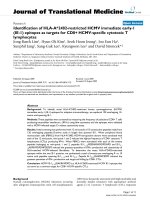
NSC23766 inhibits platelet aggregation upon stimulation
of blood and PRP by TRAP, collagen and atherosclerotic
plaque
Platelet aggregation in blood induced by TRAP (5 μM)
activating the PAR-1 receptor was reduced by 300 μM
NSC23766 from 644 ± 37 to 59 ± 40 AU*min (control
29 ± 13 AU*min; n = 3) which corresponds to 95.1%
inhibition (Figure 1). The IC
50
of NSC23766 for inhibi-
tion of TRAP-stimulated aggregation was 50 ± 18 μM.
Platelet aggregation stimulated by collagen (0.5 μg/ml)
was reduced by 300 μM NSC237 66 from 5 42 ± 181 to
76 ± 56 AU*min (control 43 ± 25 AU*min; n =7)
which amounts to 93.4% i nhibition of (Figure 1). The
IC
50
of NSC23766 for inhibition of collagen-stimulated
aggregation in blood was 64 ± 35 μM.
Plaques contain collagenous structures that directly
stimulate platelet adhesion and aggregation which is
mediated mainly by stimulation of GPVI [5]. Platelet
aggregation i nduced by plaque was reduced by 300 μM
NSC23766 from 289 ± 89 to 52 ± 26 AU*min (control
33 ± 13 AU*min; n = 3) which corresponds to 92.6%
inhibition (Figure 1). The IC
50
of NSC23766 for inhibi-
tion of plaque-stimulated aggregatio n in blood was
found to be 50 ± 30 μM.
We also found that NSC23766 dose-dependently
inhibited stimulus-induced aggregation of PRP (addi-
tional files 1 and 2, Figure s S1 and S2). Platelet aggrega-
tion stimulated by collagen and plaque was completely
inhibited by 300 μM NSC23766. The IC
50
of NSC23766
for inhibition of collagen and plaque-stimulated aggrega-
tion of PRP was found to be 47 ± 14 μM, and 57.5 ±
20 μM, respectively.
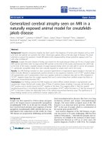
NSC23766 inhibits platelet ATP-secretion upon
stimulation of blood and PRP by TRAP, collagen, and
atherosclerotic plaque
Inhibition of stimulus-induced platelet aggregation in
blood by NSC23766 might be due to inhibition of secre-
tion as observed previously in our study of thrombin-
stimulated washed platelets [13]. Therefore, we studied
the effect of NSC23766 on dense granule secretion by
measuring the ATP-secretion in stirred blood. NSC23766
(300 μM) inhibited ATP-secretion induced by 5 μM
TRAP (Fi gure 2A) and 0.5 μg/ml collagen (Figure 2B) by
60 ± 31% (n =4)and78±7%(n = 6), respectively. In
order to study the effect of NSC23766 on secretion inde-
pendent of platelet aggreg ation, blood was pre-incubated
with RGDS (2 mM) to block the integrin a
IIb
b
3
.RGDS
reduced ATP-secretion by 26 ± 10% ( p <0.003;n =4)in
TRAP-stimulated blood and by 6 3 ± 14% (p <0.04;n =
6) in collagen-stimulated blood (Figure 2A, B). Further
pre-i ncubation with NSC23766 (300 μM) inhibited ATP-
secretion by 73 ± 15%(p< 0.03 n = 4) and by 85 ± 4% (p <
0.004 n = 6) after stimulation with TRAP and collagen,
respectively.
In PRP, RGDS reduced ATP-secretion by 92 ± 3%
when stimulated with collagen and by 86 ± 7% when sti-
mulated with plaque (additional files 1 and 2, Figure
S1B, Figure S2B). Additional pre-incubation wit h
NSC23766 (300 μM) inhibited ATP-secretion by 98 ±
1% in collagen-stimulated PRP (RGDS vs.RGDS
+NSC23766: p<0.03;n =4)andby99±1%inplaque-
stimulated PRP (p<0.04n = 4). The results in PRP sup-
port our findings in blood that NSC23766 inhibits plate-
let aggregation due to inhibition of secretion.
NSC23766 inhibits ADP-induced aggregation of platelets
in blood and PRP
The extent of inhibition of stimulus-induced ATP-
secretion in blood by NSC23766 (60-80%) was less
than that of inhibition of platelet aggregation (92-95%).
This discrepancy might be explained by an inhibitory
action of NSC23766 on the platelet stimulatory effect
of the remaining secreted ADP. Indeed, NSC23766
inhibited ADP-induced platelet aggregation in blood
and PRP; this inhibition was 70% and 75%, respective ly
(Figure 3A, B).
NSC23766 inhibits P-selectin expression on platelets upon
stimulation of blood and PRP
To study whether NSC23766 also inhibits a-granule secre-
tion, we examined the platelet surface expression of
P-selectin in the presence and absence of NSC23766 in
stirred blood containing RGDS. We found that NSC23766
completel y inhibited P-selectin expression after stimula-
tion with TRAP (5 μM) and collagen (5 μg/ml) (Table 1).
Also in PRP, NSC23766 effectively inhibited P-selectin
expression induced by ADP (5 μM) and collagen (5 μg/ml)
(Table 2).
NSC23766 inhibits P-selectin expression and platelet
aggregation stimulated by ADP independently of platelet
cyclooxgenase activity
Aspirin reduced P-selectin expression of PRP by 89.8%,
when stimulated with collagen but not when stimulated
with ADP (Figure 3B). NSC23766 (300 μM) almost
Dwivedi et al. Journal of Translational Medicine 2010, 8:128
/>Page 4 of 10
completely inhibite d ADP-induced P-selectin expression
in non-aspirin and aspirin-pretreated PRP (Table 2), and
reduced ADP-stimulated platelet aggregation of
untreated PRP and aspirin-pretreated PRP to a similar
degree, by 70% and 75%, respectively ( Figure 3B).
NSC23766 (300 μM) also inhibited ADP-induced plate-
let aggregation in blood by 70% and 75% in the abse nce
or presence of aspirin, respectively (Figure 3A).
The results indicate that NSC23766 eff ectively inhibits
a-granule secretion and platelet aggregation stimulated
by ADP, and that the mech anism is ind ependent of pla-
telet prostaglandin-endop eroxide and thromboxane
formation.
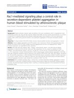
NSC23766 inhibits human plaque-induced platelet
thrombus formation under flow conditions
The effects of NSC23766 on human plaque-induced pla-
telet aggregation and thrombus formation under arterial
flow conditions are shown in Figure 4. After perfusion
of hirudin-anticoagulated blood over plaque-coated sur-
faces at 37°C with a wall shear rate of 1500 s
-1
,rapid
platelet adhesion and aggregate formation were observed
(additional file 3 Movie S1; Figure 4a). The platelet cov-
erage of the plaque-coated channels 10 min after start
of flow was 36314 ± 30013 μm
2
(mean ± SD; n =5).
NSC23766 (300 μM) reduced plaque-induced platelet
adhesion and aggregate formation. After NSC23766
incubation of blood, the platelet coverage was inhibited
by 72% to 10322 ± 9226 μm
2
(mean ± S D; n =5;p <
0.002).
Discussion
In the present study, we have provided further evidence
for a central role of Rac1 in the regulation of secretion
and aggregation of human platelets activated by a broad
range of platelet stimuli including atherosclerotic plaque.
Moreover, we have demonstrated the efficacy of
NSC23766 to inhibit platelet secretion and aggregation
induced by these stimuli in blood, and we have shown
that NSC2 3766 reduces plaque-induced platelet throm-
bus formation under arterial flow conditions.
Blood platelets are often studied after purifying plate-
lets from their milieu, which excludes the influence
exerted by other blood cells and factors present in
plasma (e.g., high concentrations of albumin a nd fibri-
nogen, lipids exposed on LDL and HDL particles) on
the physiological platelet response. Sometimes, pharma-
cological or physiological plat elet inhibitors even fail to
act on platelets in blood. For example, lysophosphatidic
acid-receptor antagonists effective in washed platelets
Figure 1 Effect of NSC23766 on stimulus-induced platelet aggregati on in blood. (A) Hirudin-anticoagulated blood was pretreated with
NSC23766 (300 μM) or solvent (H
2
O) for 5 min whilst stirring at 37°C before stimulation with TRAP (5 μM), collagen (0.5 μg/ml) or atherosclerotic
plaque homogenate (0.62 mg/ml) for 5 min; representative impedance tracings. (B) Dose-response curves of NSC23766; values are mean ± SD
(n = 4).
Dwivedi et al. Journal of Translational Medicine 2010, 8:128
/>Page 5 of 10
Figure 2 Effect of NSC23766 on stimulus-induced ATP-secretion in blood. Blood was pre-incubated with or without 300 μM NSC23766 (for
5 min), with or without 2 mM RGDS (for 2 min; added 3 min after NSC23766 or H
2
O) whilst stirring at 37°C before stimulation with (A) TRAP (5
μM) and (B) collagen (0.5 μg/ml). Top, tracings of ATP-secretion of blood. Bottom, bar diagrams; numbers are % of maximal ATP-secretion
induced by TRAP (5 μM) and collagen (0.5 μg/ml), respectively. Values are mean ± SD (n = 3-4). * p < 0.05.
Figure 3 Effect of NSC23766 on aggregation of platelets in blood and PRP stimulated with ADP. (A) Blood (with or without aspirin)
or (B) PRP (with or without aspirin) was pre-treated with 300 μM NSC23766 for 5 min whilst stirring at 37°C before stimulation with ADP (5 μM).
Aggregation values of PRP are % of maximal aggregation induced by collagen (5 μg/ml). Values are mean ± SD (n = 4). * p < 0.05.
Dwivedi et al. Journal of Translational Medicine 2010, 8:128
/>Page 6 of 10
do not inhibit lysophosphatidic acid stimulation of plate-
lets in PRP and blood (Rother E, Khandoga AL, Siess W,
unpublished data), and PGI
2
, in contrast to washed plate-
lets and PRP, was reported to be unable to inhibit platelet
aggregation induced by arachidonic acid in whole blood
[20]. Therefore, i t was important to study the effect o f
NSC23766 on platelet activation in blood and PRP.
NSC23766 (300 μM) was able to almost completely
block (~95% inhibition) p latelet aggregation induced
by TRAP (5 μM) in whole blood similar to thrombin-
(0.5 U/ml) induced aggregation of washed platelets [13].
Thrombin activates PAR-1 and PAR-4 receptors,
whereas TRAP only the PAR-1 receptor. A previous
study has shown rapid activation and redistribution of
Rac from the platelet interior to the cell periphery after
TRAP-induced activation of platelets indicating that
PAR-1 activation sti mulates Rac [21]. It is not known
whether PAR-4 activation also signals to Rac1 activation.
NSC23766 was also able to block human platelet
aggregation in blood induced by other platelet agonists,
such as fibrillar collagen, atherosclerotic plaque, and
ADP, sugge sting a central role of Rac1 signaling down-
stream of GPVI (collagen and atherosclerotic plaque) [5]
and ADP receptors. These results are in part supported
by studies of Rac1-deficient mice platelets, which
showed inhibition of GPVI-dependent platelet activation
[9,12,14]. However, in sharp contrast to two of these
studies which reported only inhibition of collagen-
stimulated, but not thrombin-induced platelet activation
in Rac1-deficient mice [9,14], our study shows that Rac1
plays a role in platelet activation induced by all stimuli
studied. Concerning the mechanism of ADP-receptor
signaling to Rac in human platelets, it was shown that
externally added ADP activate s Rac through the activa-
tion of the P2Y
1
receptor/G
q
pathway. However, when
ADP was secreted from TRAP-stimulated platelets acti-
vation of the P2Y
12
receptor/G
i
pathway played a central
role [22].
Dose-response curves showed that NSC23766 inhib-
ited human platelet aggregation i n blood and PRP sti-
mulated by all these agonists with a similar IC
50
ranging
between 50 to 70 μM. NSC23766 acts by disrupting the
interaction of Rac1 with TrioN or Tiam1 Rac-GEFs, and
it has been shown to inhibit in vitro both Rac1-TrioN
binding a nd GEF activity of TrioN in a dose dependent
manner, achieving 50% inhibition at 50 μM [15]. It is
puzzling that the IC
50
of NSC23766 for inhibition of sti-
mulus-induced platelet aggregation in blood was found
to be in the sam e range as the IC
50
of NSC23766 in the
in vitro re constitution system consisting only of the two
proteins Rac1 and TrioN. We expected that much
higher concentrations of NSC23766 would be needed to
inhibit Rac1 in platelets in blood considering the possi-
blebindingofthedrugtoplasmaproteinsandother
blood cells and its crossing of the cell membrane before
reaching its target Rac1 in the platelet interior. Platelet
proteome data do not indicate the expression of TrioN
or Tiam1 in human platelet (apps.
biozentrum.uni-wuerzburg.de). One possible reason that
μM concent rations of NSC23766 were effective in inhi-
biting Rac1 in platelets in blood is that other Rac1-GEFs
might be present in human platelets which have a lower
affinity to Rac1 than TrioN or Tiam1 and are thus dis-
placed by lower (nM) drug concentrations in vitro.
Experiments using RGDS to block the integrin a
IIb
b
3
showed that NSC23766 inhibited stimulus-induced secre-
tion of dense granule as well as alpha granule contents in
blood and PRP. These results indicate that NSC23766
Table 1 Effect of NSC23766 on P-selectin expression of
platelets in blood stimulated by TRAP and collagen
Agonist P-selectin expression (% positive
cells)
Control Stimulated
TRAP (5 μM) 1.6 ± 0.6 6.8 ± 3.4
TRAP+NSC23766 (300 μM) 1.4 ± 0.6
Collagen (5 μg/ml) 1.7 ± 0.9 8 ± 2.6
Collagen+NSC23766 (300 μM) 2.9 ± 2
Blood was incubated with NSC23766 (300 μM) or solvent (water) in the
presence of 2 mM RGDS for 5 min whilst stirring at 37°C before stimulation
with TRAP or collagen. P-selectin expression was measured by flow cytometry.
Values are mean ± SD, n =3.
Table 2 Effect of NSC23766 and aspirin on P-selectin expression of PRP stimulated by ADP and collagen
Agonist P-selectin expression
(% positive cells)
PRP Aspirin-PRP
Control Stimulated Control Stimulated
ADP (5 μM) 1.4 ± 0.7 6 ± 2.8 1 ± 0.5 5.4 ± 2.6
ADP+NSC23766 (300 μM) 1.2 ± 1 1.8 ± 1.3 0.9 ± 0.4 2.1 ± 1.5
Collagen (5 μg/ml) 3.3 ± 3.1 42.4 ± 16.9 2 ± 1.3 6 ± 3.6
Collagen+NSC23766 (300 μM) 1.8 ± 1.3 3.1 ± 2.7 2 ± 1.5 2 ± 1.8
PRP or aspirin-pretreated PRP was incubated with NSC23766 (300 μM) or solvent (water) in the presence of 1 mM RGDS for 5 min whilst stirring at 37°C in the
lumi-aggregometer before stimulation with ADP or collagen. P-selectin expression was measured by flow cytometry. Values are mean ± SD, n =4.
Dwivedi et al. Journal of Translational Medicine 2010, 8:128
/>Page 7 of 10
also primarily inhibits platelet secretion and subsequently
platelet aggregation in blood and PRP confirming pre-
vious studies in thrombin-stimulated washed platelet sus-
pensions [12,13]. NSC23766 (300 μM) completely
inhibite d platelet P-selectin expression stimulated by col-
lagen and TRAP in blood, but under the same experi-
mental conditions (stirring, presence of RGDS), it did not
inhibit completely ATP-secretion (inhibition of 73% after
TRAP stimulation and of 85% after collagen stimulation).
We reasoned that NSC23766 might be so effective in
inhibiting collagen- and TRAP-induced platelet aggrega-
tion and platelet P-selectin expression in blood because it
might inhibit the action of the residual secreted ADP on
platelets. Indeed, NSC23766 inhibited ADP-induced
aggregation by 70% and 75% in blood and PRP, respec-
tively and completely in P-selectin expression.
Another important observation of our study concerns
the role of integrin a
IIb
b
3
outside-in signaling in the
regulation of ATP-secretion in stirred activated blood.
RGDS reduced ATP-secretion of stirred blood stimu-
lated with collagen (0.5 μg/ml) and TRAP (5 μM) by
63% and 26%, respectively, indicating that integrin
a
IIb
b
3
signaling stimulated by platelet-to-platelet contact
plays a role that is more important in collagen- than in
TRAP-induced dense granule secretion of platelets in
blood. These results are in line with a previous study of
mice PRP showing the important role of the integrin
a
IIb
b
3
in mediating secretion after stimulation with low
level (2.5 μg/ml) collagen [23].
Aspirin, which reduced P-selectin expression of col-
lagen-stimul ated hirudin-anticoagulat ed PRP by 90%,
was ineffective in inhibiting P-selectin expression when
hirudin PRP was stimulated with ADP, confirming a
previous study in citrated PRP [24]. Thus, aspirin fails
to inhibit a-granule secretion after ADP stimulation of
platelets independent of the anticoagulant used. Th e
findings are in contrast to the results of dense granule
secretion in citrated PRP, where aspirin is well known
to inhibit dense granule secretion and the secondary
wave of platelet aggregation after ADP stimulation [25].
Interestingly, we found that NSC23766 was equally
effective in aspirin- and non-aspirin pretreated platelets
in reducing P-selectin expression as well as platelet
aggregation stimulated by ADP. Two conclusions can be
drawn from these results: (1) NSC23766 is much mor e
effective than aspirin in inhibiting the effect of ADP on
platelets in blood a nd (2) NSC23766 inhibits a-granule
secretion and platelet aggregation stimulated by ADP
independent of platelet prostaglandin-endoperoxide and
thromboxane formation.
Conclusion
Our data c learly demonstratethecentralroleofRac1
in secretion and subsequent platelet aggregation in
blood upon activation by a wide array of platelet sti-
muli including atherosclerotic plaque. Rac1 inhibition
by NSC23766 prevented platelet secretion from both
a-granules and dense granules. We suggest that by
inhibiting specifically platelet secretion, the pharmaco-
logical targeting of Rac1 could be an interesting
approach in the development of future antiplatelet
drugs.
Figure 4 Effect of NSC23766 on atherosclerotic plaque-induced platelet thrombus formation under arterial flow conditions.Hirudin-
anticoagulated blood pre-incubated with H
2
O or with NSC23766 (300 μM) for 5 min was perfused over plaque-coated surfaces for 10 min at 37°
C at a shear rate of 1500 s
-1
. (A) representative flow images of control (upper channel) and NSC23766 treated blood (lower channel) 10 min after
start of the flow; Platelets are visualized by mepacrine fluorescence; (B) bar diagram (values are mean ± SD; n = 5). * p < 0.002.
Dwivedi et al. Journal of Translational Medicine 2010, 8:128
/>Page 8 of 10
Additional material
Additional file 1: Figure S1. Effect of NSC23766 on ATP-secretion
and aggregation of PRP stimulated with collagen. PRP was pre-
incubated with or without 300 μM NSC23766 (for 5 min), with or
without 1 mM RGDS (for 2 min; added 3 min after NSC23766 or H
2
O)
whilst stirring at 37°C before stimulation with collagen (1.25 μg/ml). (A)
Top, tracings of light transmission and ATP-secretion of PRP stimulated
by collagen with or without NSC23766. Bottom, tracings of light
transmission and ATP-secretion of PRP stimulated by collagen with or
without NSC23766 in the presence of RGDS. (B) Dose-response curve of
NSC23766 on platelet aggregation and ATP-secretion induced by
collagen (1.25 μg/ml). Values are mean ± SD (n = 3).
Additional file 2: Figure S2. Effect of NSC23766 on ATP-secretion
and aggregation of PRP stimulated with plaque. PRP was pre-
incubated with or without 300 μM NSC23766 (for 5 min), with or
without 1 mM RGDS (for 2 min; added 3 min after NSC23766 or H
2
O)
whilst stirring at 37°C before stimulation with plaque (0.62 mg/ml). (A)
Top, tracings of light transmission and ATP-secretion of PRP stimulated
by plaque with or without NSC23766. Bottom, tracings of light
transmission and ATP-secretion of PRP stimulated by plaque with or
without NSC23766 in the presence of RGDS. (B) Dose-response curve of
NSC23766 on platelet aggregation and ATP-secretion induced by plaque
(0.62 mg/ml). Values are mean ± SD (n = 3).
Additional file 3: Movie S1. Effect of NSC23766 on human plaque-
induced platelet thrombus formation under arterial flow conditions.
Hirudin-anticoagulated blood was incubated with mepacrine to visualize
platelets by fluorescence. Blood was perfused (direction right to left) over
atherosclerotic plaque-coated microfluidic chambers and observed for 10
min. Upper channel, control; lower channel, blood pre-treated with 300
μM NSC23766. In the upper channel, rapid platelet adhesion and
aggregate formation (green fluorescence) occurred, mainly at the edges
of the channel, where also the majority of plaque material is present (as
seen by phase contrast microscopy before start of the flow experiments).
NSC23766 reduced platelet adhesion and aggregate formation. The
video is in. mov format and can be viewed using Quick time player on
different PCs with Windows XP or Vista.
Acknowledgements
We thank Kathrin von Oheimb for her technical assistance in this study. The
study was supported by grants from the Deutsche Forschungsgemeinschaft
(DFG Si 274/11), the August-Lenz-Stiftung, the University of Munich and the
Bayern University ("BayEFG"; to A.L.K.). The results are part of the doctoral
thesis of S.D. at the University of Munich.
Author details
1
Institute for Prevention of Cardiovascular Diseases, University of Munich,
Munich, Germany.
2
Department of Vascular Surgery, Clinic Schwabing,
Munich, Germany.
3
Max-Planck Institute of Biochemistry, Martinsried,
Germany.
Authors’ contributions
SD designed and performed the experiments, collected the results and
analyzed the data. DP contributed by designing some of the experiments
and interpreting the results. AKL participated in helping to perform the flow
experiments. RB provided human plaque material. WS planned the study,
assisted in designing the experiments, discussed and interpreted the results
throughout the study, and wrote together with SD and DP the paper. All
the authors have read and approved the final manuscript.
Competing interests
The authors declare that they have no competing interests.
Received: 17 September 2010 Accepted: 6 December 2010
Published: 6 December 2010
References
1. Fernandez-Ortiz A, Badimon JJ, Falk E, Fuster V, Meyer B, Mailhac A,
Weng D, Shah PK, Badimon L: Characterization of the relative
thrombogenicity of atherosclerotic plaque components: implications for
consequences of plaque rupture. J Am Coll Cardiol 1994, 23:1562-1569.
2. Vanzanten GH, Degraaf S, Slootweg PJ, Heijnen HFG, Connolly TM,
Degroot PG, Sixma JJ: Increased Platelet Deposition on Atherosclerotic
Coronary-Arteries. Journal of Clinical Investigation 1994, 93:615-632.
3. Siess W, Zangl KJ, Essler M, Bauer M, Brandl R, Corrinth C, Bittman R, Tigyi G,
Aepfelbacher M: Lysophosphatidic acid mediates the rapid activation of
platelets and endothelial cells by mildly oxidized low density lipoprotein
and accumulates in human atherosclerotic lesions. Proceedings of the
National Academy of Sciences of the United States of America 1999,
96:6931-6936.
4. Rother E, Brandl R, Baker DL, Goyal P, Gebhard H, Tigyi G, Siess W: Subtype-
selective antagonists of lysophosphatidic Acid receptors inhibit platelet
activation triggered by the lipid core of atherosclerotic plaques.
Circulation 2003, 108:741-747.
5. Penz S, Reininger AJ, Brandl R, Goyal P, Rabie T, Bernlochner I, Rother E,
Goetz C, Engelmann B, Smethurst PA, et al: Human atheromatous plaques
stimulate thrombus formation by activating platelet glycoprotein VI.
FASEB J 2005, 19:898-909.
6. Reininger AJ, Bernlochner I, Penz SM, Ravanat C, Smethurst P, Farndale RW,
Gachet C, Brandl R, Siess W: A 2Step Mechanism of Arterial Thrombus
Formation Induced by Human Atherosclerotic Plaques. Journal of the
American College of Cardiology 2010, 55:1147-1158.
7. Bauer M, Retzer M, Wilde JI, Maschberger P, Essler M, Aepfelbacher M,
Watson SP, Siess W: Dichotomous regulation of myosin phosphorylation
and shape change by Rho-kinase and calcium in intact human platelets.
Blood 1999, 94:1665-1672.
8. Klages B, Brandt U, Simon MI, Schultz G, Offermanns S: Activation of G12/
G13 results in shape change and Rho/Rho-kinase-mediated myosin light
chain phosphorylation in mouse platelets. J Cell Biol 1999, 144:745-754.
9. McCarty OJ, Larson MK, Auger JM, Kalia N, Atkinson BT, Pearce AC, Ruf S,
Henderson RB, Tybulewicz VL, Machesky LM, Watson SP: Rac1 is essential
for platelet lamellipodia formation and aggregate stability under flow. J
Biol Chem 2005, 280:39474-39484.
10. Soulet C, Gendreau S, Missy K, Benard V, Plantavid M, Payrastre B:
Characterisation of Rac activation in thrombin- and collagen-stimulated
human blood platelets. FEBS Lett 2001, 507:253-258.
11. Gratacap MP, Payrastre B, Nieswandt B, Offermanns S: Differential
regulation of Rho and Rac through heterotrimeric G-proteins and cyclic
nucleotides. J Biol Chem 2001, 276:47906-47913.
12. Akbar H, Kim J, Funk K, Cancelas JA, Shang X, Chen L, Johnson JF,
Williams DA, Zheng Y: Genetic and pharmacologic evidence that Rac1
GTPase is involved in regulation of platelet secretion and aggregation. J
Thromb Haemost 2007, 5:1747-1755.
13. Pandey D, Goyal P, Dwivedi S, Siess W: Unraveling a novel Rac1-mediated
signaling pathway that regulates cofilin dephosphorylation and
secretion in thrombin-stimulated platelets. Blood 2009,
114:415-424.
14. Pleines I, Elvers M, Strehl A, Pozgajova M, Varga-Szabo D, May F, Chrostek-
Grashoff A, Brakebusch C, Nieswandt B: Rac1 is essential for
phospholipase C-gamma2 activation in platelets. Pflugers Arch 2009,
457:1173-1185.
15. Gao Y, Dickerson JB, Guo F, Zheng J, Zheng Y: Rational design and
characterization of a Rac GTPase-specific small molecule inhibitor. Proc
Natl Acad Sci USA 2004, 101:7618-7623.
16. Brandl R, Richter T, Haug K, Wilhelm MG, Maurer PC, Nathrath W:
Topographic analysis of proliferative activity in carotid endarterectomy
specimens by immunocytochemical detection of the cell cycle-related
antigen Ki-67. Circulation 1997, 96:3360-3368.
17. Penz SM, Reininger AJ, Toth O, Deckmyn H, Brandl R, Siess W: Glycoprotein
Ibalpha inhibition and ADP receptor antagonists, but not aspirin, reduce
platelet thrombus formation in flowing blood exposed to atherosclerotic
plaques. Thromb Haemost 2007, 97:435-443.
18. Toth O, Calatzis A, Penz S, Losonczy H, Siess W: Multiple electrode
aggregometry: a new device to measure platelet aggregation in whole
blood. Thromb Haemost 2006, 96:781-788.
19. Ingerman CM, Smith JB, Silver MJ: Direct measurement of platelet
secretion in whole blood. Thromb Res 1979, 16:335-344.
Dwivedi et al. Journal of Translational Medicine 2010, 8:128
/>Page 9 of 10
20. Saniabadi AR, Lowe GD, Belch JJ, Barbenel JC, Forbes CD: Effect of
prostacyclin (epoprostenol) on the aggregation of human platelets in
whole blood in vitro. Haemostasis 1984, 14:487-494.
21. Azim AC, Barkalow K, Chou J, Hartwig JH: Activation of the small GTPases,
rac and cdc42, after ligation of the platelet PAR-1 receptor. Blood 2000,
95:959-964.
22. Soulet C, Hechler B, Gratacap MP, Plantavid M, Offermanns S, Gachet C,
Payrastre B: A differential role of the platelet ADP receptors P2Y1 and
P2Y12 in Rac activation. J Thromb Haemost 2005, 3:2296-2306.
23. Cho MJ, Liu J, Pestina TI, Steward SA, Thomas DW, Coffman TM, Wang D,
Jackson CW, Gartner TK: The roles of alpha IIb beta 3-mediated outside-in
signal transduction, thromboxane A2, and adenosine diphosphate in
collagen-induced platelet aggregation. Blood 2003, 101:26462651.
24. Rinder CS, Student LA, Bonan JL, Rinder HM, Smith BR: Aspirin does not
inhibit adenosine diphosphate-induced platelet alpha-granule release.
Blood 1993, 82:505-512.
25. Siess W: Molecular mechanisms of platelet activation. Physiol Rev 1989,
69:58-178.
doi:10.1186/1479-5876-8-128
Cite this article as: Dwivedi et al.: Rac1-mediated signaling plays a central
role in secretion-dependent platelet aggregation in human blood
stimulated by atherosclerotic plaque. Journal of Translational Medicine 2010
8:128.
Submit your next manuscript to BioMed Central
and take full advantage of:
• Convenient online submission
• Thorough peer review
• No space constraints or color figure charges
• Immediate publication on acceptance
• Inclusion in PubMed, CAS, Scopus and Google Scholar
• Research which is freely available for redistribution
Submit your manuscript at
www.biomedcentral.com/submit
Dwivedi et al. Journal of Translational Medicine 2010, 8:128
/>Page 10 of 10