Gãy xương cánh tay pot
Bạn đang xem bản rút gọn của tài liệu. Xem và tải ngay bản đầy đủ của tài liệu tại đây (514.68 KB, 5 trang )
Fractures of the Lateral
Condyle of the Humerus
M
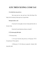
ilch described two types of lat-
eral condyle fractures (Figure
1). In Milch type I, the fracture ex-
tends through the ossification center
of the capitellum and enters the joint
lateral to the trochlear groove. In
Milch type II, the fracture extends
medially into the trochlear groove.
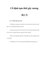
The most widely used system (not
identified by name) identifies three
fracture patterns (Figure 2). In a type
I fracture, the articular surface is in-
tact and the fracture is nondisplaced
and stable. In types II and III, the frac-
ture enters the joint. Type II fractures
are minimally displaced (2 to 3 mm);
type III fractures are displaced >4 mm
and may be rotated. (For additional
discussion of these systems, see
Milch,
1
Jakob et al,
2
Ogden,
3
Herring,
4
Wilkins et al,
5
and McIntyre.
6
)
Indications
Type I fractures and type II fractures
displaced <2 mm may be treated by
closed means.
7
Closed reduction and
percutaneous pinning should be at-
tempted in type II fractures displaced
2to3mm;
8
however, if anatomic re-
duction is not obtained, open reduc-
tion and internal fixation is required.
Type II fractures displaced >2 to 3 mm
and all type III fractures are unstable.
Displaced fractures have an increased
propensity to nonunion.
Properly managed fractures treat-
ed by closed manipulation and per-
cutaneous pinning or by open reduc-
tion and internal fixation have a
95% union rate, making these the
preferred methods of treatment.
Contraindications
There are very few contraindications
to performing percutaneous or open
reduction and internal fixation in
the properly selected patient. The
nondisplaced, stable fracture does
not require surgical treatment; cast
immobilization is sufficient. When
an underlying medical condition
prevents surgery or an anesthetic
risk, then either nonsurgical treat-
ment is required or the medical con-
dition must be managed before un-
dertaking a surgical procedure.
Surgical Technique
The patient is positioned supine on
the operating table and a general an-
esthetic is induced. A small child
should be positioned with the arm
and forear m lying on the operating
table but close enough to the edge
that the operative limb can be
brought over the edge of the table for
use with either standard fluoroscopy
or the mini C-arm fluoroscopy unit.
Some surgeons use the receiving
unit of the fluoroscopy unit as an op-
erating surface.
The arm is prepared and draped in
a sterile manner, then is exsan-
guinated and the tourniquet inflated.
When closed reduction and percuta-
neous pinning is considered, we per-
form this technique with fluoro-
scopic imaging (
video). If this
fails or if open treatment is the
method of choice, a curvilinear later-
al incision is made centered over the
lateral condyle. Minimal dissection
is preferred to avoid periosteal strip-
ping of the blood supply. The surgi-
cal approach involves directly enter-
ing the fracture hematoma and
visualizing the fragment. I recom-
mend the surgical interval between
the brachioradialis and the triceps.
Once the fragment is identified, the
fracture site is ir rigated thoroughly
J. Andy Sullivan, MD
The video that accom-
panies this article is
″Supracondylar Fractures
of the Humerus in Children,″ available
on the Orthopaedic Knowledge On-
line Website, at />oko/jaaos/surgical.cfm
Dr. Sullivan is Don H. O’Donoghue
Professor and Chief Medical Officer,
Department of Orthopedic Surgery &
Rehabilitation, University of Oklahoma
Health Sciences Center, Children’s
Hospital, Oklahoma City, OK.
Neither Dr. Sullivan nor the department
with which he is affiliated has received
anything of value from or owns stock in a
commercial company or institution
related directly or indirectly to the
subject of this article.
Reprint requests: Dr. Sullivan, Children’s
Hospital, Room 2MR2000, 940 NE
13th Street, Oklahoma City, OK 73104.
J Am Acad Orthop Surg 2006;14:
58-62
Copyright 2006 by the American
Academy of Orthopaedic Surgeons.
Surgical Techniques
58 Journal of the American Academy of Orthopaedic Surgeons
to remove all hematoma and to im-
prove visualization.
Dissection is kept to a minimum.
Usually no dissection is necessary on
the distal fragment. Distal and poste-
rior dissection should be avoided in
order to avert damage to the circula-
tion of the fragment, which can cause
osteonecrosis. The periosteum of the
proximal fragment, which overhangs
the fracture site, may have to be
stripped back slightly to remove it
from the fracture site.
It is important to adequately visu-
alize the joint articular surface. In
some cases, the fragment is rotated
180°; in these situations, I have found
it easier to visualize the fragment by
applying a varus movement to the el-
bow, which reproduces the mecha-
nism of injury and opens the fracture
site so that it may be easily seen.
Once the hematoma has been evac-
uated, the distal fragment is manip-
ulated into position onto the end of
the proximal fragment (distal hu-
merus). To accomplish this reduction,
the distal fragment is grasped with a
bone-holding forceps or a towel clip
and rotated back into proper align-
ment. This maneuver is facilitated by
flexing the elbow in order t o take ten-
sion off the distal fragment.
My preferred technique is to place
a large towel clip or a small-bone
point-to-point forceps with one tong
in the lateral condyle, then hook the
other tong into the periosteum of the
proximal fragment (Figure 3). The el-
bow is flexed and the clamp gently
closed. It is important at this point
to place retraction in the wound to
directly visualize the joint articular
surface. The key component of this
procedure is to ensure anatomic re-
duction of the joint articular surface.
At times, the joint surface cannot be
entirely visualized. By palpation an-
teriorly and posteriorly, one is usual-
ly able to get a good idea of the suit-
ability of the reduction.
After anatomic reduction is
achieved, the arm is brought out
over the fluoroscopy machine, and
anteroposterior and lateral radio-
graphic views are obtained. In my
experience with displaced fractures,
when anatomic reduction has not
been obtained, the most common
position is lateral displacement.
Usually reduction can be improved
by loosening the clamp, placing pres-
sure on the fragment to push it more
medially, then closing the clamp.
Once the anatomic reduction is
obtained, internal fixation should be
secured with Kirschner wires (K-
wires) (Figure 4). Other fixation tech-
niques include compression screws
and absorbable pins. I have found
that K-wires are simple, efficient, in-
expensive, and effective. In most of
these patients, a 0.62-in K-wire is
sufficient. In a very small child, I
recommend 0.45-in K-wires.
Some surgeons advocate placing
all fixation pins or screws in the
metaphyseal fragment, thus avoiding
the ossific nucleus and physis of the
lateral condyle. With a very large
fragment, this is usually possible.
However, in many of these condylar
fractures, the reduction and articular
surface are difficult to visualize. A
sufficiently large fragment is needed
to confirm that the pin has adequate
purchase. However, when the frac-
ture fragment is small, I do not hes-
itate to place the pins through the
condyle and across the physis into
the medial aspect of the distal hu-
Figure 1
A, Milch type I fracture. The fracture line is through the ossific nucleus of the
capitellum. B, Milch type II fracture. The fracture line is lateral to the ossific nucleus.
Figure 2
Fracture types I through III. A, In a type I fracture, the fracture line does not violate
the articular surface and therefore is stable. B, A type II fracture is through the
articular surface but minimally displaced. C, A type III fracture is displaced and often
rotated.
J. Andy Sullivan, MD
Volume 14, Number 1, January 2006 59
merus. The anatomy of the distal hu-
merus is such that most of the
growth occurs in the lateral condyle
and the trochlea. Occasionally, pa-
tients end up with a fishtail appear-
ance to the distal humerus, which
does not interfere with function. In
my experience, I have never seen a
true arrest of the entire lateral
condyle.
A variety of pin configurations
may be used. Some advocate parallel
pins, as in the video (
video); how-
ever, I prefer convergent pins placed
through the lateral condyle and up
into the shaft of the humerus. A sec-
ond pin is placed transversely across
the fracture line through the meta-
physeal fragment. This provides good
stability and divergence of the pins.
Parallel pins or diverging pins are
more stable than converging pins.
What is to be avoided is having the
pins converge at the fracture site be-
cause this is a less stable construct.
An important intraoperative tech-
nique is to pick up the skin of the pos-
terior aspect of the incision gently
with forceps and start the K-wires
away from the incision in order to
avoid having the pins come out
through the incision. In most in-
stances, the pins can be cut subcuta-
neously to bring them retrograde
through the skin (by pressing the skin
over them).
Pin placement and adequacy of re-
duction should be confirmed and doc-
umented radiographically. This pro-
vides a baseline for postoperative care.
The fracture site should be inspected
visually and by palpation to ensure
continuity of the joint articular sur-
face. The wound is irrigated, and layer
closure is accomplished with absorb-
able sutures, including an absorbable
subcuticular suture. When the frac-
ture treatment has been delayed and
there is excessive swelling or concern
about skin tension, then mattress or
tension-releasing sutures should be
used in the skin. Either absorbable or
nonabsorbable may be used, depend-
ing on surgeon preference.
The K-wires previously inserted
are cut off outside the skin and bent
over (
video). Nonadherent gauze
is placed around the pins, and a felt
pad is cut and placed over the pin.
The arm is wrapped with cotton cast
padding, and a posterior splint is ap-
plied. For elevation to prevent depen-
dent edema, the arm is placed in a
sling after the procedure.
Follow-up Care
The patient is seen at 1 week after pri-
mary treatment; the cast is removed,
the pin sites are examined, and radio-
graphs are obtained. If the pins are in
satisfactory position and the reduc-
tion maintained, a fiberglass long arm
cast is applied. In children younger
than age 6 or 7 years, an additional 2
weeks of immobilization is recom-
mended, although 3 weeks o r more of
immobilization is preferred after a
cast application, giving a total of 4
Figure 3
A, A type III fracture with displacement and rot ation. B, The fracture has been
reduced and is being held with a clamp.
Figure 4
Anteroposterior (A) and lateral (B) views of fixation secured with K-wires.
Fractures of the Lateral Condyle of the Humerus
60 Journal of the American Academy of Orthopaedic Surgeons
weeks of immobilization. (I have not
had any problems with the children
regaining elbow motion after lateral
condylar fractures and find that I sleep
a little easier with the extra week of
immobilization.)
At 4 weeks, anteroposterior and
lateral radiographs are obtained. If
there is new bone formation indicat-
ing the early stage of healing, the
K-wires are removed. If there is no ev-
idence of bone formation (which is
extremely uncommon, in my experi-
ence), pins still can be removed and
additional cast immobilization per-
formed. Reexamination at 6 weeks is
necessary. Failure to demonstrate
union of the fracture at that point
would require an additional 6 weeks
of cast or splint immobilization. Be-
yond 12 weeks, I would consider the
fracture to be nonunited and would
proceed with bone grafting.
Although I have not done a formal
review, I do not recall having had a
nonunion in a lateral condylar frac-
ture that was treated acutely, nor do
I recall one that was not sufficiently
united at 4 weeks in which I could
not remove the pins and begin mobi-
lization.
Delayed Presentation or
Nonunion
Nonunion of the lateral condyle can
result in cubitus valgus deformity and
in a tardy ulnar nerve palsy.
9
If the
fracture is not united in a patient who
presents between 6 and 12 weeks, the
standard surgical approach described
is recommended. W ith nonunion, the
surgical procedure is more difficult.
In the established nonunion, the frac-
ture site is separated from the prox-
imal fragment with sharp dissection
with a scalpel or a small osteotome.
Once the two fragments are sepa-
rated, curets are used to remove the
reactive fibrous tissue, taking care to
dissect the distal fragment as mini-
mally as possible. The surfaces of the
proximal and distal fragments must
be cleaned of this fibrous tissue. Once
this is achieved, I t ry to obtain as near
an anatomic reduction as possible.
There are no good anatomic land-
marks at this point, and achieving an-
atomic reduction is more difficult
than in an acute case.
Once the anatomic position is de-
termined, the fracture fragments are
reduced with a towel clip and pins
are inserted. I still prefer iliac crest
bone as the bone donor site. There
are bone graft substitutes available
that may be equally effective.
With an established nonunion, I
prefer internal fixation and bone
graft, followed by 6 weeks of cast im-
mobilization. Most fractures in my
experience have united with this
treatment at 6 to 12 weeks. Patients
may present with established non-
unions that are in an older age group.
Roye et al
10
have demonstrated that,
even in adolescents, treatment can
be successful by means of open re-
duction, cleaning the bony surfaces,
inserting a cancellous screw, and
making use of bone grafting. With
surgical intervention, the stability of
the elbow is improved, and the risk
of cubitus valgus is reduced.
11
References
1. Milch H: Fractures and fracture dislo-
cations of the humeral condyles.
J Trauma 1964;15:592-607.
2. Jakob R, Fowles JV, Rang M, Kassab
MT: Observations concerning frac-
tures of the lateral humeral condyle in
children. J Bone Joint Surg Br 1975;
57:430-436.
3. Ogden JA: Skeletal Injury in the
Child, ed 2. Philadelphia, PA: WB
Pearls
• Position the patient close enough to the edge of the operating table
that the arm can be visualized by fluoroscopy, but with sufficient
room that the arm is completely supported during the surgical pro-
cedure.
• In a larger child, I recommend use of a hand table. During surgery,
extend the elbow and apply varus movement to visualize the frac-
ture. Flex the arm at the moment of reduction to relax the distal frag-
ment.
• Warn the family preoperatively of potential complications, includ-
ing nonunion, cubitus varus, osteonecrosis, and protuberance of the
lateral condyle. Protuberance of the lateral condyle is the most com-
mon. The other complications are extremely rare but are serious.
Clinical series report that up to half of cases of lateral condyle frac-
ture have a lateral protuberance. Although there is no functional dis-
ability from the protuberance and no surgical treatment is required,
it is disconcerting to parents to see the deformity, so it is best to have
warned them preoperatively. Although the exact cause is not
known, it is thought that dissection of the periosteum may increase
the likelihood of this result. For this reason, dissection should be
limited to that necessary to expose the fracture site.
Pitfalls
• Failure to visualize the intra-articular component of the fracture and
obtain anatomic reduction
• Inadequate internal fixation or fracture reduction with rotational
displacement
• Failure to recognize displacement or rotation in fractures treated
closed or with percutaneous pinning
• Aggressive early return to sports. It is best to keep patients out of
sports for 2 months following fracture treatment.
J. Andy Sullivan, MD
Volume 14, Number 1, January 2006 61
Saunders, 1990.
4. Herring JA: Tachdjian’s Pediatric Or-
thopedics, ed 3. Philadelphia, PA: WB
Saunders, 2002.
5. Wilkens KE, Beaty JH, Chambers
HG,Toniolo RM: Fractures and dislo-
cations of the elbow region, in Rock-
wood CA Jr, Wilkens KE, Beaty JH
(eds): Fractures in Children. Philadel-
phia, PA: Lippincott-Raven, 1996, vol
3, pp 653-904.
6. McIntyre W: Lateral condylar frac-
tures of thehumerus, in Letts RM(ed):
Management of Pediatric Fractures.
New York, NY: Churchill Living-
stone, 1994, pp 241-258.
7. Foster DE, Sullivan JA, Gross RH: Lat-
eral humeral condylar fractures in
children. J Pediatr Orthop 1985;5:16-
22.
8. Mintzer CM, Waters PM, Brown DJ,
Kasser JR: Percutaneous pinning in
the treatment of displaced lateral
condyle fractures. J Pediatr Orthop
1994;14:462-465.
9. Masada K, Kawai H, Kawabata H,
Masatomi T, Tsuyuguchi Y,Yamamo-
to K: Osteosynthesis for old, estab-
lished non-union of the lateral
condyle of the humerus. J Bone Joint
Surg Am 1990;72:32-40.
10. Roye DP, Bini SA, Infosino A: Late
surgical treatment of lateral condylar
fractures in children. J Pediatr
Orthop 1991;11:195-199.
11. Morrissy RT, Wilkins KE: Deformity
following distal humeral fracture in
childhood. J Bone Joint Surg Am 1984;
66:557-562.
Fractures of the Lateral Condyle of the Humerus
62 Journal of the American Academy of Orthopaedic Surgeons