Human Musculoskeletal Biomechanics Part 9 pot
Bạn đang xem bản rút gọn của tài liệu. Xem và tải ngay bản đầy đủ của tài liệu tại đây (584.55 KB, 20 trang )
Cervical Spine Anthropometric and Finite Element Biomechanical Analysis
151
5. Acknowledgments
The authors would like to thank Miami Valley Hospital (Dayton, OH) for support on this
project. Specifically Dr. David Udin in the Clinical Research Office, Scott Calvin manager of
the Miami Valley Imaging Group, and Matt Binkley fourth year medical student, for
assistance in collecting CT images.
6. References
Ambard, D. and Cherblanc, F. (2009) 'Mechanical Behavior of Annulus Fibrosus: A
Microstructural Model of Fibers Reorientation', Annals of Biomedical Engineering.
Adam, C. Pearcy, M. McCombe, P. 2003. Stress analysis of interbody fusion – Finite element
modeling of inetervertebral implant and vertebral body. Clinical Biomechanics 18,
265-272
Bogduck, N. and Yoganandan, N. (2001) 'Biomechanics of the cervical spine Part 3: minor
injuries', Clinical Biomechanics, pp. 267 - 75.
Boos, N., Aebi, M., 2008 Spinal Disorders: Fundamentals of Diagnosis and Treatment.
Springer, Zurich, pp. 41-62
Human Musculoskeletal Biomechanics
152
Bozic, K.J., Keyak, J.H., Skinner, H.B., Beuff, H.U. and Bradford, D.S. (1994) 'Three-
dimensional finite element modeling of a cervical vertebra: An investigation of
burst fracture mechanism', Journal of Spinal Disorders, pp. 102 - 10.
Bozkus, H.A.K.M.H.M.U.E.B.A.S. (2001) 'Finite element model of the Jefferson fracture:
comparison with a cadaver model', Eur Spine J, pp. 257-263.
Bozkus, H., Karakas, A., Hanci, M., Uzan, M., Bozdag, E. and Sarioglu, A. (2001) 'Finite
element model of the Jefferson fracture: comparison with a cadaver model',
European Spine Journal, pp. 257-263.
Brolin, K. and Halldin, P. (2004) 'Development of a finite element model of the upper
cervical spine and a parameter study of ligament characteristics', Spine.
Brolin, K. and Halldin, P. (2004) 'Development of a Finite Element Model of the Upper
Cervical Spine and a Parameter Study of Ligament Characteristics', Spine, pp. 376-
385.
Brolin, K. and Halldin, P. (2005) 'The effect of muscle activation on neck response', Traffic
Injury Prevention.
Camacho, D.L.A., Nightingale, R.W., Robinette, J.J., Vanguri, S.K., Coates, D.J. and Myers,
B.S. (1997) 'Experimetnal flexibility measurements for the development of a
computational head-neck model validated for near-vertex head impact', Proceedings
from the 41st Stapp car Crash Conference, pp. 473 - 486.
Choi, J. Sung, K., 2006. Subsidence after anterior lumbar interbody fusion using paired
stand-alone rectangular cages. European Spine Journal 15, 16-22
Dai, L., 1998. The relationship between vertebral body deformity and disc degeneration in
lumbar spine of the senile. European Spine Journal 7, 40-44
Denoziere, G. Ku, D., 2006. Biomechanical comparison between fusion of two vertebrae and
implantation of an artificial disc. Journal of Biomechanics 39, 766-775
Doherty, B., Heggeness, M. and Esses, S. (1993) 'A biomechanical study of odontoid
fractures and fracture fixation', Spine, pp. 178 - 84.
Douglas C. Montgomery, a.E.A.P. (1982) Introduction to Linear Regression Analysis, New York:
John Wiley & Sons, Inc.
Ebara, S., Iatridis, J.C., Setton, L.A., Foster, R.J., Mow, V.C. and Weidenbaum, M. (1996)
'Tensile properties of nondegenerate human lumbar annulus fibrosus', Spine, pp.
452 - 61.
Eberlein, R., Holzapfel, G.A. and Froelich, M. (2004) 'Multi-segment FEA of the human
lumbar spine including the heterogeneity of the annulus fibrosus', Computational
Mechanics.
Edwards, W.T. Ferrarra, L.A. Yuan, H.A., 2001. The effect of the vertebral cortex in the
thoracolumbar spine. Spine 26, 218-225
Esat, V.L.D.A.M. (2005) 'Combined Multi-Body Dynamic and FE Models of Human Head
and Neck', IUTAM Proceedings on Impact Biomechanics, 91-100.
Francis, C.C. (1955) 'Dimensions of the cervical vertebrae', Anat Rec, vol. 122, pp. 603-609.
Francis, C.C. (1955) 'Dimensions of the cervical vertebrae', Anat Rec, vol. 122, pp. 603-609.
Galbusera, F., Bellini, C.M., Raimondi, M.T., Fornari, M. and Assietti, R. (2008) ' Cervical
spine biomechanics following implantation of a disc prosthesis', Medical Engineering
and Physics, pp
. 1127-1133.
Cervical Spine Anthropometric and Finite Element Biomechanical Analysis
153
Gamst, G., Meyers, L.S. and Guarino, A.J. (2008) Analysis of Variance Designs: A conceptual and
Computational Approach with SPSS and SAS, New York: Cambridge University Press.
Gilad, I. and Nissan, M. (1986) 'A study of vertebra and disc geometic relations of the human
cervical and lumbar spine', Spine, pp. 154 - 7.
Glenn Gamst, L.S.M.A.J.G. (2008) Analysis of Variance Designs: A conceptual and Computational
Approach with SPSS and SAS, New York: Cambridge University Press.
Goel, V.K., Clark, C.R., Harris, K.G. and Schulte, K.R. (1988) 'Kinematics of the cervical
spine: effects of multiple total laminectomy and facet wiring', Jouranl of Orhopaedic
Research, pp. 611 - 19.
Goel, V.K. and Clausen, J.D. (1998) 'Prediction of Load Sharing Among Spinal Components
of a C5-C6 Motion Segment Using the Finite Element Approach', Spine, pp. 684-691.
Graham, R., Oberlander, E., Stewart, J. and Griffiths, D. (2000) 'Validation and use of a finite
element model of C-2 for determination of stress and fracture patterns of anterior
odontoid loads', Journal of Neurosurgery.
Grant, J. Oxland, T. Dvorak, M., 2001. Mapping the structural properties of the lumbosacral
vertebral endplates. Spine 8, 889-896
Greaves, C.Y., Gadala, M.S. and Oxland, T.R. (2008) 'A three-dimensional finite element
model of the cervical spine with spinal cord: an investigation of three injury
mechanisms', Annals of Biomedical Engineering, pp. 396-405.
Ha, S.K. (2006) 'Finite element modeling of multi-level cervical spinal segments (C3-C6) and
biomechanical analysis of an elastomer-type prosthetic disc', Medical Engineering &
Physics, pp. 534-541.
Haghpanahi, M.M.R. (2006) 'Development of a Parametric Finite Element Model of Lower
Cervical Spine in Sagital Plane', Proceedings of the 28th IEEE EMBS Annual
International Conference, New York City, USA, 1739-1741.
Holzapfel, G.A. (2000) Nonlinear Solid Mechanics, New York: Wiley.
Jost, B. et. al., 1998. Compressive strength of interbody cages in the lumbar spine: the eddect
of cage shape, posterior instrumentation and bone density. European Spine Journal
7, 132-144
Kallemeyn, N.A., Tadepalli, S.C. and Shivanna, K.H. (2009) ' An interactive multiblock
approach to meshing the spine', Computer Methods and Programs in Biomedicine, pp.
227-235.
KenethS. Saladin PhD, a.L.M.M.S.N. (2004) Anatomy & Physiology: The Unity of Form and
Function, Third Ed. edition, New York: McGraw-Hill.
Kulkarni, A. D’Orth Hee, H. Wong, H., 2007. Solis cage (PEEK) for anterior cervical fusion:
preliminary radiological results with emphasis on fusion and subsidence. The
Spine Journal 7, 205-209
Kumaresan, S., Yoganandan, N. and Pintar, F. (1999) 'Finite Element Analysis of the Cervical
Spine: A Material Property Sensitivity Study', Clinical Biomechanics, pp. 41-53.
Kumaresan, S., Yoganandan, N., Pintar, F. and Maiman, D. (1999) 'Finite element modeling
of the cervical spine: role of intervertebral disc under axial and eccentric loads',
Medical Engineering & Physics, pp. 689-700.
Human Musculoskeletal Biomechanics
154
Kumaresan, S., Yoganandan, N., Pintar, F. and Maiman, D. (1999) 'Finite element modeling
of the lower cervical spine: role of intervertebral disc under axial and eccentric
loads', Medical Engineering & Physics, pp. 689-700.
Kumaresan, S., Yoganandan, N., Pintar, F. and Maiman, D. (1999) 'Finite element modeling
of the lower cervical spine: role of intervertebral disc under axial and eccentric
loads', Medical Engineering & Physics, pp. 689-700.
Kumaresan, S., Yoganandan, N., Pintar, F., Maiman, D. and Kuppa, S. (2000) 'Biomechanical
study of pediatric human cervical spine: a finite element approach', Journal of
Biomechanical Engineering, pp. 60-71.
Kumaresan, S., Yoganandan, N., Voo, L. and Pintar, F. (1998) 'Finite Element Analysis of the
Human Lower Cervical Spine', Journal of Biomechanics, pp. 87-92.
Langrana, N. Kale, S. Edwards, T. Lee, C. Kopacz, K., 2006. Measurement and analyses of
the effects of adjacent
Li, Y. and Lewis, G. (2010) 'Influence of surgical treatment for disc degeneration diseases at
C5-C6 on changes in some biomechanical parameters of the cervical spine', Medical
Engineering & Physics, pp. 593 - 503.
Linde, F., 1994. Elastic and viscoelastic properties of trabecular bone by a compression
testing approach. Danish Medical Bulletin 41, 119–138.
Liu YK, C.C.K.K. (1986) 'Quantitative geometry of young human male cervical vertebrae',
Mechanism of Head and Spine Trauma, pp. 417-431.
Liu, Y.K., Clark, C.R. and Krieger, K.W. (1986) 'Quantitative geometry of young human male
cervical vertebrae', Mechanism of Head and Spine Trauma, pp. 417-431.
Manohar M. Panjabi, P.J.D.M.V.G.P.T.O.M.a.K.T.M. (1991) 'Cervical Human Vertebrae:
Quantitative Three-Dimensional Anatomy of the MIddle and Lower Regions',
Spine, vol. 16, no. 8, pp. 861-869.
Maurel, N., Lavaste, F. and Skalli, W. (1997) 'A three-dimensional parameterized finite
element model of the lower cervical spine. Study of the influence of the posterior
articular facets', Journal of Biomechanics, pp. 921-931.
Meakin, J. and Huskins, D.W.L. (2001) 'Replacing the nucleus pulposus of the intervertebral
disk: prediction of suitable properties of a replacement material using finite
element analysis', Journal of Materials Science.
Montgomery, D.C. and Peck, E.A. (1982) Introduction to Linear Regression Analysis, New York:
John Wiley & Sons, Inc.
Moroney, S.P., Schultz, A.B., Miller, J.A.A. and Andersson, G.B.J. (1988) 'Load-displacement
properties of lower cervical spine motion segments', Journal of Biomechanics, vol. 21,
pp. 769-779.
Muller-Gerbl, M. Weiber, S. Linsenmeier, U., 2008. The distribution of mineral density in the
cervical vertebral endplates. European Spine Journal 17, 432-438
Nigg, B.M., Herzog, W.H., 1994. Biomechanics of the musculo- skeletal system (Eds.), John
Wiley & Sons, Chichester.
Ng, H W. and Teo, E C. (2001) 'Nonlinear Finite-Element Analysis of the Lower Cervical
Spine (C4-C6) Under Axial Loading', Journal of Spinal Disorders, pp. 201-10.
Cervical Spine Anthropometric and Finite Element Biomechanical Analysis
155
Ng, H W., Teo, E C., Lee, K K. and Qiu, T X. (2003) 'Finite element analysis of cervical
spinal instability under physiologic loading', Journal of Spinal Disorders &
Techniques, pp. 55 - 63.
Nightingale, R.W., Chancey, V.C., Otaviano, D., Luck, J.F., Tran, L., Prange, M. and Myers,
B.S. (n.d) 'Flexion and extension structural properties and strenghts for male
cervical spine segments', Journal of Biomechanics, pp. 534 - 42.
Nissan M, G.I. (1984) 'The cervical and lumbar vertebrae: An anthropometric model', Eng
Med, vol. 13, no. 3, pp. 111-114.
Nissan, M. and Gilad, I. (1984) 'The cervical and lumbar vertebrae: An anthropometric
model', Eng Med, vol. 13, no. 3, pp. 111-114.
Noailly, J., Lacoix, D. and Planell, J.A. (2005) 'Finite element study of a novel intervertebral
disc substitute', Spine.
Noailly, J., Wilke, H J., Planell, J.A. and Lacroix, D. (2007) 'How does the geometry affect
the internal biomechanics of a lumbar spine bi-segment finite element model?
Consequences on validation process', Journal of Biomechanics, pp. 2414-2425.
Ordway, N. Lu, Y. Zhang, X. Cheng, C. Fang. H, Fayyazi, A., 2007. Correlation of the
cervical endplate strength with CT measured subchondral bone density. European
Spine Journal 16, 2104-2109
Oxland, T.R., 2003. Effects of endplate removal on the structural properties of the lower
lumbar vertebral bodies. Spine 8, 771-777
Palomar, A., Calvo, B. and Doblare, M. (2008) 'An accurate finite element model of the
cervical spine under quasi-static loading', Journal of Biomechanics, pp. 523-531.
Panagiotopoulou, O. (2009) 'Finite element analysis (FEA): applying an engineering method
to functional morphology in anthropology and human biology', Annals of Human
Biology, pp. 609 - 23.
Panjabi, M., White. A., 1990. Clinical Biomechanics of the Spine. Lippencott, Philidelphia
Panjabi, M. Chen, M.C. Wang, J.L., 2001. The cortical architecture of the human cervical
vertebral bodies. Spine 22, 2478-2484
Panjabi, M.M., Crisco, J.J., Vasavada, A., Oda, T., Cholewicki, J., Nibu, K. and Shin, E. (2001)
'Mechanical properties of the human cervical spine as shown by three-dimensional
load-displacement curves', Spine, pp. 2692 - 2700.
Panjabi, M., Duranceau, J., Goel, V., Oxland, T. and Takata, K. (1991) 'Cervical human
vertebrae. Quanatative three-dimensional anatomy of the middle and lower
regions', Spine, pp. 861 - 9.
Panjabi, M.M., Duranceau, J., Goel, V., Oxland, T. and Takata, K. (1991) 'Cervical Human
Vertebrae: Quantitative Three-Dimensional Anatomy of the MIddle and Lower
Regions', Spine, vol. 16, no. 8, pp. 861-869.
Panzer, M.B. and Cronin, D.S. (2009) 'C4-C5 segment finite element model development,
validation, and load-sharing investigation', Journal of Biomechanics, pp. 480-490.
Pintar, F., Yoganandan, N., Pesigan, M., Reinartz, J., Sances, A. and Cusick, J. (1995)
'Cervical vertebral strain measurements under axial and eccentric loading', Journal
of Biomechanical Engineering, pp. 474 - 8.
Human Musculoskeletal Biomechanics
156
Pintar, F.A., Yoganandan, N., Pesigan, M., Reinartz, J., Sances, A. and Cusick, J.F. (1995)
'Cervical vertebral strain measurements under axial and eccentricl loading', Journal
of Biomechanical Engineering, pp. 474 - 8.
Pitzen, T., Geisler, F., Matthis, D., Muller-Storz, H., Barbier, D., Steudel, W I. and Feldges,
A. (2002) 'A finite element model for predicting the biomechanical behaviour of the
human lumbar spine.', Control Engineering Practice, pp. 83-90.
Pitzen, T., Geisler, F.H., Matthis, D., Muller-Storz, H., Pedersen, K. and Steudel, W I. (2001)
'The influence of cancellous bone density on load sharing in human lumbar spine: a
comparison between an intact and a surgically altered motion segment', European
Spine Journal, pp. 23-29.
Pitzen, T., Matthis, D. and Steudel, W I. (2002) 'Posterior Element Injury and Cervical Spine
Flexibility Following Anterior Cervical Fusion and Plating', European Journal of
Trauma, pp. 24-30.
Pitzen, T. et. al., 2004. Variation of endplate thickness. European Spine Journal 13, 235-240
Polikeit
1
, A. et. al., 2003. Factors influencing stresses in the lumbar spine after the insertion
of intervertebral cages: Finite element analysis. European Spine Journal 12, 413-420
Polikeit
2
, A. et. al., 2003. The importance of the endplate for interbody cages in the lumbar
spine. European Spine Journal 12, 556-561
Richter, M., Wilke, H.J., Kluger, P., Claes, L. and Puhl, W. (2000) 'Load-displacement
properties of the normal and injured lower cervical spine in vitro', European Spine
Journal, pp. 104 - 8.
Rohlmann, A. Zander, T. Bergmann, G., 2006. Effects of fusion-bone stiffness on the
mechanical behavior of the lumbar spine after vertebral body replacement. Clinical
Biomechanics 21, 221-227
S.H. Tan, E.C.T.a.H.C.C. (2004) 'Quantitative three-dimensional anatomy of cervical,
thoracic and lumbar vertebrae of Chinese Singaporeans', European Spine Journal,
vol. 13, pp. 137-146.
Sairyo, K., Goel, V.K., Masuda, A., Vishnubhotla, S., Faizan, A., Biyani, A., Ebraheim, N. and
Yonekura, D.e.a. (2006) ' Three-dimensional finite element analysis of the pediatric
lumbar spine. Part 1: pathomechanism of apophyseal bony ring fracture', European
Spine Journal, pp. 923-929.
Sairyo, K., Goel, V.K., Masuda, A., Vishnubhotla, S., Faizan, A., Biyani, A., Ebraheim, N. and
Yonekura, D.e.a. (2006) 'Three-dimensional finite element analysis of the pediatric
lumbar spine. Part II: biomechanical change as the initiating factor for pediatric
ishmic spondylolisthesis after growth plate', European Spine Journal, pp. 930-935.
Sairyo, K., Goel, V.K., Masuda, A., Vishnubhotla, S., Faizan, A., Biyani, A., Ebraheim, N. and
Yonekura, D.e.a. (206) ' Three-dimensional finite element analysis of the pediatric
lumbar spine. Part II: biomechanical change as the initiating factor for pediatric
ishmic spondylolisthesis after growth plate', European Spine Journal, pp. 930-935.
Saladin, K.S. and Miller, L. (2004) Anatomy & Physiology: The Unity of Form and Function,
Third Ed. edition, New York: McGraw-Hill.
Schmidt, H.H.F.D.J.K.Z.C.L.W.H. (2007) 'Application of a calibration method provides more
realistic results for a finite element model of a lumbar spinal segment', Clinical
Biomechanics, pp. 377-384.
Cervical Spine Anthropometric and Finite Element Biomechanical Analysis
157
Shirazi-Adl, A. (2006) ' Analysis of large compression loads on lumbar spine in flexion and
in torsion using a novel wrapping element.', Journal of Biomechanics, pp. 267-275.
Stemper, B.D., Yoganandan, N., Pintar, F.A. and Rao, R.D. (2006) 'Anterior longitudinal
ligament injuries in whiplash may lead to cervical instability', Medical Engineering &
Physics.
Tan, S.H., Teo, E.C. and Chua, H.C. (2004) 'Quantitative three-dimensional anatomy of
cervical, thoracic and lumbar vertebrae of Chinese Singaporeans', European Spine
Journal, vol. 13, pp. 137-146.
Teo, J.C.M., Chui, C.K., Wang, Z.L., Ong, S.H., Yan, C.H., Wang, S.C., Wong, H.K. and Teoh,
S.H. (2007) 'Heterogenous meshing and biomechanical modeling of human spine.',
Medical Engineering and Physics, pp. 277-290.
Teo, E.C. and Ng, H.W. (2001) 'Evaluation of the role of ligaments, facets and disc nucleus in
lower cervical spine under compression and sagittal moments using finite element
method', Medical Engineering & Physics, pp. 155 - 64.
Teo, E.C. and Ng, H.W. (2001) 'First cervical vertebra (atlas) fracture mechanism studies
using finite element method', Journal of Biomechanics, pp. 13-21.
Traynelis, P.A., Donaher, R.M., Roach, R.M. and Goel, v.K. (1993) 'Biomechanical
comparison of anterior caspar plate and three-level posterior fixation techniques in
human cadaveric model', Journal of Neurosurgery, pp. 96 - 103.
Van Jonbrgen, H.P. Spruit, M. Anderson, P. Pavlov, P., 2005. Anterior cervical interbody
fusion with a titanium box cage: early radiological assessment of fusion and
subsidence. The Spine Journal 5, 645-649
Voo, L.M., Kumaresan, S., Yoganandan, N., Pintar, F. and Cusick, J.F. (1997) 'Finite element
analysis of cervical facetectomy', Spine.
Wheeldon, J.A., Pintar, F.A., Knowles, S. and Yoganandan, N. (2006) 'Experimental
flexion/extension data corridors for validation of finite element models of the
young, normal cervical spine', Journal of Biomechanics, pp. 375 - 80.
Wheeldon, J.A., Stempter, B.D., Yoganandan, N. and Pintar, F.A. (2008) 'Validation of finite
element model of the young normal lower cervical spine', Annals of Biomedical
Engineering, pp. 1458-1469.
Yoganandan, N., Kumaresan, S. and Pintar, F.A. (2000) 'Geometric and mechanical
properties of human cervical spine ligaments', Journal of Biomechanical Engineering,
pp. 623 - 29.
Yoganandan, N., Kumaresan, S. and Pintar, F.A. (2001) 'Biomechanics of the cervical spine
part 2. Cervical spine soft tissue responses and biomechanical modeling', Clinicals
Biomechanics, pp. 1-27.
Yoganandan, N., Kumaresan, S., Voo, L. and Pintar, F.A. (1996) 'Finite elemnt applications in
human cervical spine modeling', Spine.
Yoganandan, N., Kumaresan, S., Voo, L. and Pintar, F.A. (1997) 'Finite Element Model of the
Human Lower Cervical Spine: Parametric Analysis of the C4-C6 Unit', Journal of
Biomechanical Engineering, pp. 81-92.
Yoganandan, N., Kumaresan, S., Voo, L., Pintar, F. and Larson, S. (1996) 'Finite Element
Analysis of the C4-C6 Cervical Spine Unit', Medical Engineering & Physics, pp. 569-
574.
Human Musculoskeletal Biomechanics
158
Zander, T. Rohlmann, A. Klockner, C. Bergmann, G., 2002. Effect of bone graft
characteristics on the mechanical behavior of the lumbar spine. Journal of
Biomechanics 35, 491-497
Zhang, Q.H., Teo, E.C., Ng, H.W. and Lee, V.S. (2006) 'Finite element analysis of moment-
rotation relationships for human cervical spine', Journal of Biomechanics.
Zheng, P.D N., Young-Hing, M.D K. and Watson, P.D L.G. (2000) 'Morphological and
biomechanical studies of pedicle screw fixation for the lower cervical spine', Journal
of Systems Integration, pp. 55-66.
7
Biomechanics of the Temporomandibular Joint
Shirish M. Ingawalé
1
and Tarun Goswami
1,2
1
Biomedical, Industrial and Human Factors Engineering, Wright State University,
Dayton, OH
2
Orthopaedic Surgery and Sports Medicine, Wright State University, Dayton, OH
U.S.A.
1. Introduction
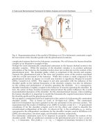
Temporomandibular joint (TMJ) connects the mandible or the lower jaw to the skull and
regulates the movement of the jaw (see Figure 1). The TMJ is one of the most complex,
delicate and highly used joints in a human body (Alomar et al., 2007). The most important
functions of the TMJ are mastication and speech. Temporomandibular disorder (TMD) is a
generic term used for any problem concerning the jaw joint. Injury to the jaw, the TMJ, or
muscles of the head and neck can cause TMD. Other possible causes include grinding or
clenching the teeth; dislocation of the disc; presence of osteoarthritis or rheumatoid arthritis
in the TMJ; stress, which can cause a person to tighten facial and jaw muscles or clench the
teeth; aging (Bakke et al., 2001; Detamore et al., 2007; Ingawalé and Goswami, 2009; Tanaka
et al., 2000). The most common TMJ disorders are pain dysfunction syndrome, internal
derangement, arthritis, and traumas (Breul et al., 1999; Chen et al., 1998). TMDs are seen
most commonly in people between the ages of 20 and 40 years, and occur more often in
women than in men (Detamore and Athanasiou, 2003; Detamore et al., 2007; Tanaka et al.,
2008a). Some surveys have reported that 20-25% of the population exhibit one or more
symptoms of TMD (Detamore et al., 2007; Ingawalé and Goswami, 2009).
With a large part of population suffering from TMDs, it is a problem that should be looked
at more fully. Relations between muscle tensions, jaw motions, bite and joint force, and
craniofacial morphology are not fully understood. A large fraction of TMD causes are
currently unexplained. There is a great need of better understanding of the etiology of
TMDs to develop methods to prevent, diagnose, and cure joint disorders (Beek et al., 2003;
Ingawalé and Goswami, 2009). This chapter provides a state-of-the-art review of TMJ
anatomy, disorders, and biomechanics; and briefly discusses our approach toward three-
dimensional (3D) anatomical and finite element (FE) modeling to understand the interaction
between structure and function of the TMJ.
2. TMJ anatomy and function
TMJ is a bi-condylar joint in which the condyles, the movable round upper ends of the
mandible, function at the same time (see Figure 1). Between the condyle and the articular
fossa is a disc made of fibrocartilage that acts as a cushion to absorb stress and allows the
condyle to move easily when the mouth opens and closes (AAOMS, 2007; Ide et al., 1991).
Human Musculoskeletal Biomechanics
160
The bony structures consist of the articular fossa; the articular eminence, which is an
anterior protuberance continuous with the fossa; and the condylar process of the mandible
that rests within the fossa. The articular surfaces of the condyle and the fossa are covered
with cartilage (Ide et al., 1991). The disc divides the joint cavity into two compartments -
superior and inferior (Ide et al., 1991; Tanaka et al., 2008b). The two compartments of the
joint are filled with synovial fluid which provides lubrication and nutrition to the joint
structures (Tanaka et al., 2008b). The disc distributes the joint stresses over broader area
thereby reducing the chances of concentration of the contact stresses at one point in the joint.
The presence of the disc in the joint capsule prevents the bone-on-bone contact and the
possible higher wear of the condylar head and the articular fossa (Beek et al., 2001; Tanaka
et al., 2008b). The bones are held together with ligaments. These ligaments completely
surround the TMJ forming the joint capsule.
Source: American Association of Oral and Maxillofacial Surgeons (AAOMS, 2007).
Fig. 1. Anatomical structure of the temporomandibular joint (TMJ)
Strong muscles control the movement of the jaw and the TMJ. The temporalis muscle which
attaches to the temporal bone elevates the mandible. The masseter muscle closes the mouth
and is the main muscle used in mastication (see Figure 2) (Hylander, 1979). Movement is
guided by the shape of the bones, muscles, ligaments, and occlusion of the teeth. The TMJ
undergoes hinge and gliding motion (Alomar et al., 2007). The TMJ movements are very
complex as the joint has three degrees of freedom, with each of the degrees of freedom
associated with a separate axis of rotation. Rotation and anterior translation are the two
primary movements. Posterior translation and mediolateral translation are the other two
possible movements of TMJ (Dutton, 2004).
The Temporomandibular
j
oint
Cond
y
le
Li
g
ament
Disc
Articular fossa
Muscle
TMJ Biomechanics
161
Source: Scrivani et al., 2008
Fig. 2. Normal anatomy of the jaw. The lateral view of the skull (Panel A) shows the normal
position of the mandible in relation to the maxilla, the TMJ capsule, and the masticatory
muscles – temporalis, masseter, mylohyoid, anterior and posterior digastrics, hyglossus, and
stylohyoid. Also shown (Panels B and C) are the deep muscles associated with jaw function
and the TMJ intra-articular disc.
3. TMJ disorders and treatment
Temporomandibular disorder (TMD) is a generic term used for any problem concerning the
jaw joint. Injury to the jaw, temporomandibular joint, or muscles of the head and neck can
cause TMD. Other possible causes include grinding or clenching the teeth, which puts a lot
of pressure on the TMJ; dislocation of the disc; presence of osteoarthritis or rheumatoid
arthritis in the TMJ; stress, which can cause a person to tighten facial and jaw muscles or
clench the teeth; aging, etc (Bakke et al., 2001; Detamore et al., 2007; Ingawalé and Goswami,
2009; Tanaka et al., 2000). The most common TMJ disorders are pain dysfunction syndrome,
internal derangement, arthritis, and traumas (Detamore and Athanasiou, 2003; Detamore et
al., 2007). TMD is seen most commonly in people between the ages of 20 and 40 years, and
occurs more often in women than in men (Detamore and Athanasiou, 2003; Detamore et al.,
2007). Some surveys have reported that 20-25% of the population exhibit symptoms of TMD
while it is estimated that 30 million Americans suffer from it, with approximately one
Human Musculoskeletal Biomechanics
162
million new patients diagnosed yearly (Detamore and Athanasiou, 2003; Detamore et al.,
2007; Tanaka et al., 2008b; Wolford, 1997).
Disc displacement is the most common TMJ arthropathy and is defined as an abnormal
relationship between the articular disc and condyle (Tanaka et al., 2000).
As the disc is
forced out of the correct position, there is often bone on bone contact which creates
additional wear and tear on the joint, and often causes the TMD to worsen (Tanaka et al.,
2000). Almost 70% of TMD patients have disc displacement (Detamore and Athanasiou,
2003). Different types of functional malocclusion have been shown to be partly responsible
for signs and symptoms of TMD. The functional unilateral posterior cross-bite, habitual
body posture during sleep, juvenile chronic arthritis - a chronic arthritis in childhood with
an onset before the age of 16 years and a duration of more than three months – are also
reported as TMD risk (Bakke et al., 2001; Hibi and Ueda, 2005; Pellizoni et al., 2006).
Treatments for the various TMJ disorders range from physical therapy and nonsurgical
treatments to various surgical procedures. Usually the treatment begins with conservative,
nonsurgical therapies first, with surgery left as the last option. The majority of TMD
patients can be successfully treated by non-surgical therapies and surgical interventions
may be required for only a small part of TMD population (Ingawalé and Goswami, 2009).
The initial treatment does not always work and therefore more intense treatments such as
joint replacement may be a future option (Ingawalé and Goswami, 2009). The non-surgical
treatment options include medication; self-care; physical therapy, to keep the synovial joint
lubricated and to maintain full range of the jaw motion; wearing splints, the plastic
mouthpieces that fit over the upper and lower teeth to prevent the upper and lower teeth
from coming together, lessening the effects of clenching or grinding the teeth (Ingawalé and
Goswami, 2009). Splints are used to help control bruxism – a TMD risk factor in some cases
(Glaros et al., 2007; Kalamir et al., 2007; Tanaka et al., 2000a). However, the long-term
effectiveness of this therapy has been widely debated and remains controversial (Glaros et
al., 2007; Kalamir et al., 2007). Surgery can play an important role in the management of
TMDs. Conditions that are always treated surgically involve problems of overdevelopment
or underdevelopment of the mandible resulting from alterations of condylar growth,
mandibular ankylosis, and benign and malignant tumors of the TMJ (Laskin et al., 2006).
The surgical treatments include arthrocentesis, arthroscopy, discectomy, and joint
replacement. While more conservative treatments are preferred when possible, in severe
cases or after multiple operations, the current end stage treatment is joint replacement
(Tanaka et al., 2008b). However, before a joint replacement option is ever considered for a
patient, all non-surgical, conservative treatment options must be exhausted; and all
conservative surgical methodologies should be employed (Quinn, 199; Quinn, 2000).
4. Biomechanical behavior of the TMJ
Mandibular motions result in static and dynamic loading in the TMJ. During natural loading
of the joint, combinations of compressive, tensile, and shear loading occur on the
articulating surfaces (Tanaka et al., 2008b).
The analysis of mandibular biomechanics helps
us understand the interaction of form and function, mechanism of TMDs; and aids in the
improvement of the design and the behavior of prosthetic devices, thus increasing their
treatment efficiency (Hansdottir and Bakke, 2004; Ingawalé and Goswami, 2009; Korioth
and Versluis, 1997)
TMJ Biomechanics
163
4.1 In-vivo assessment
Very few studies which report in-vivo biomechanical assessment of the TMJ can be found in
the literature. In contrast to some earlier studies which reported the TMJ to be a force-free
joint, Hylander
(1979)
demonstrated that considerable forces were exerted on the TMJ
during occlusion as well as mastication. In face of these contrary reports, Breul et al. (1999)
showed that the TMJ was subjected to pressure forces during occlusion as well as during
mastication and it was slightly eccentrically loaded in all positions of occlusion.
Korioth and Hannam (1994)
indicated that the differential static loading of the human
mandibular condyle during tooth clenching was task dependent and both the medial and
lateral condylar thirds were heavily loaded.
Huddleston Slater et al. (1999) suggested that
when the condylar movement traces coincide during chewing, there is compression in the
TMJ during the closing stroke. However, when the traces do not coincide, the TMJ is not or
only slightly compressed during chewing. Naeije and Hofman (2003) used these
observations to study the loading of the TMJ during chewing and chopping tasks. Their
analysis showed that the distances traveled by the condylar kinematic centers were shorter
on the ipsilateral side than on the contralateral. The kinematic centers of all contralateral
joints showed a coincident movement pattern during chewing and chopping. The indication
that the ipsilateral joint is less heavily loaded during chewing than the contralateral joint
may explain why patients with joint pain occasionally report less pain while chewing on the
painful side.
Hansdottir and Bakke (2004) evaluated the effect of TMJ arthralgia on mandibular mobility,
chewing, and bite force in TMD patients (categorized as disc derangements, osteoarthritis,
and inflammatory disorders) compared to healthy control subjects. The pressure pain
threshold (PPT), maximum jaw opening, and bite force were significantly lower in the
patients as compared to that in controls. The patients were also found to have longer
duration of chewing cycles. The bite force and jaw opening in patients were significantly
correlated with PPT. The most severe TMJ tenderness (i.e., lowest PPT) and the most
impeded jaw function with respect to jaw opening and bite force were found to be more
severe in the patients with inflammatory disorders than the patients with disc derangement
or osteroarthritis (Hansdottir and Bakke, 2004).
4.2 In-vitro assessment – mechanical testing and finite element modeling
As the TMJ components are difficult to reach and as the applications of experimental
devices inside the TMJ cause damage to its tissue, the direct methods are not used often.
Indirect techniques utilized to evaluate mandibular biomechanics have had limited success
due to their ability to evaluate only the surface stress of the model but not its mechanical
properties (Ingawalé and Goswami, 2009). Mechanical testing and finite element modeling
(FEM) have been progressively used by TMJ researchers.
Excessive shear strain can cause degradation of the TMJ articular cartilage and collagen
damage eventually resulting in joint destruction (Tanaka et al., 2008).
Tanaka et al. (2008)
attempted to characterize the dynamic shear properties of the articular cartilage by studying
shear response of cartilage of 10 porcine mandibular condyles using an automatic dynamic
viscoelastometer. The results showed that the shear behavior of the condylar cartilage is
dependent on the frequency and amplitude of applied shear strain suggesting a significant
role of shear strain on the interstitial fluid flow within the cartilage. Beek et al. (2001)
performed sinusoidal indentation experiments and reported that the dynamic mechanical
Human Musculoskeletal Biomechanics
164
behavior of disc was nonlinear and time-dependent. Beek et al. (2003) simulated these
experiments using axisymmetric finite element model and showed that a poroelastic
material model can describe the dynamic behavior of the TMJ disc. Tanaka et al. (2006)
carried out a series of measurements of frictional coefficients on 10 porcine TMJs using a
pendulum-type friction tester. The results showed that the presence of the disc reduces the
friction in the TMJ by reducing the incongruity between the articular surfaces and by
increasing synovial fluid lubrication. This study highlighted the importance of preserving
the disc through alternatives to discectomy to treat internal derangement and osteoarthritis
of the TMJ.
The finite element modeling (FEM) has been used widely in biomechanical studies due to its
ability to simulate the geometry, forces, stresses and mechanical behavior of the TMJ
components and implants during simulated function (Beek et al., 2001; Chen et al., 1998;
Koolstra and van Eijden, 2005, 2006; Perez del Palomar and Doblare, 2006b, 2008; Reina et
al., 2007; Tanaka et al., 2000). Chen et al. (1998) performed stress analysis of human TMJ
using a two-dimensional FE model developed from magnetic resonance imaging (MRI). Due
to convex nature of the condyle, the compressive stresses were dominant in the condylar
region whereas the tensile stresses were dominant in the fossa-eminence complex owing to
its concave nature. Beek et al. (2001) developed a 3D linear FE model and analyzed the
biomechanical reactions in the mandible and in the TMJ during clenching under various
restraint conditions. Nagahara et al. (1999) developed a 3D linear FE model and analyzed
the biomechanical reactions in the mandible and in the TMJ during clenching under various
restraint conditions. All these FE simulations considered symmetrical movements of
mandible, and the models developed only considered one side of the joint. Hart et al. (1992)
generated 3D FE models of a partially edentulated human mandible to calculate the
mechanical response to simulated isometric biting and mastication loads. Vollmer et al.
(2000) conducted experimental and finite element study of human mandible to investigate
its complex biomechanical behavior. Tanaka et al. (2001, 2004) developed a 3D model to
investigate the stress distribution in the TMJ during jaw opening, analyzing the differences
in the stress distribution of the disc between subjects with and without internal
derangement. Tanaka et al. (2008c) suggested, from the results of finite element model of the
TMJ based on magnetic resonance images, that increase of the frictional coefficient between
articular surfaces may be a major cause for the onset of disc displacement. Sellers and
Crompton, (2004) used sensitivity analysis to validate the predictions of 3D FE simulations.
In 2005, Koolstra and van Eijden developed a combination of rigid-body model with a FE
model of both discs and the articulating cartilaginous surfaces to simulate the opening
movement of the jaw. Using the same model, Koolstra and van Eijden (2006) performed FEA
to study the load-bearing and maintenance capacity of the TMJ. The results indicated that
the construction of the TMJ permitted its cartilaginous structures to regulate their
mechanical properties effectively by imbibitions, exudation and redistribution of fluid.
Perez-Palomar and Doblare (2006a) used more realistic FE models of both TMJs and soft
components to study clenching of mandible. Perez del Palomar and Doblare (2006b)
developed a 3D FE model that included both discs ligaments and the three body contact
between all elements of the joints, and analyzed biomechanical behavior of the soft
components during a nonsymmetrical lateral excursion of the mandible to investigate
possible consequences of bruxism. This study suggested that a continuous lateral movement
of the jaw may lead to perforations in the lateral part of both discs, conforming to the
TMJ Biomechanics
165
indications by Tanaka et al. (2001; 2004). Later, in 2007, Perez del Palomar and Doblare
suggested that unilateral internal derangement is a predisposing factor for alterations in the
unaffected TMJ side. However, it would be necessary to perform an exhaustive analysis of
bruxism with the inclusion of contact forces between upper and lower teeth during
grinding.
Whiplash injury is considered as a significant TMD risk factor and has been proposed to
produce internal derangements of the TMJ (Kasch et al., 2002; Perez del Palomar and
Doblare, 2008). However, this topic is still subject to debate (Detamore et al., 2007). In 2008,
Perez del Palomar and Doblare, published the results of finite element simulations of the
dynamic response of TMJ in rear-end and frontal impacts to predict the internal forces and
deformations of the joint tissues. The results, similar to suggested by Kasch et al. (2002),
indicated that neither a rear-end impact at low-velocity nor a frontal impact would produce
damage to the soft tissues of the joint suggesting that whiplash actions are not directly
related with TMDs. However; since this study has its own limitations such as analysis of
only one model, for low-velocity impacts, without any restrictions like contact with some
component of the vehicle; there is a need for more reliable finite element simulations to
obtain more accurate numerical results.
A theoretical model developed by Gallo et al. (2000) for estimating the mechanical work
produced by mediolateral stress-field translation in the TMJ disc during jaw
opening/closing suggested that long-term exposure of the TMJ disc to high work may result
in fatigue failure of the disc. In 2001, Gross et al. proposed a predictive model of occlusal
loading of the facial skeleton while May et al. (2001) developed a mathematical model of the
TMJ to study the compressive loading during clenching. Effect of mandibular activity on
mechanical work in the TMJ, which produces fatigue that may influence the
pathomechanics of degenerative disease of the TMJ, was studied by Gallo et al. (2006).
Nickel et al. (2002) validated numerical model predictions of TMJ eminence morphology
and muscle forces, and demonstrated that the mechanics of the craniomandibular system
are affected by the combined orthodontic and orthognathic surgical treatments. Using this
validated numerical model to calculate ipsilateral and contralateral TMJ loads for a range of
biting positions and angles, Iwasaki et al. (2009) demonstrated that TMJ loads during static
biting are larger in subjects with TMJ disc displacement compared to subjects with normal
disc position.
4.3 Post-surgery assessment
TMJ reconstruction using the partial or total TMJ prosthetics, in most cases, improves range
of motion and mouth opening in the TMJ patients. However, loss of translational
movements of the mandible on the operated side has been often observed, especially in
anterior direction, owing to various factors like loss of pterygoid muscle function, scarring
of the joint region and the muscles of mastication (Yoon et al., 2007). Komistek et al. (1998)
assessed in-vivo kinematics and kinetics of the normal, partially replaced, and totally
replaced TMJs. Less translation was reported in the implanted fossa and total TMJs than in
the normal joints. The study suggests that total TMJ implants only rotate and do not
translate; and the muscles do not apply similar forces at the joint when the subject has a total
TMJ implant, compared to a subject who has a normal, healthy TMJ.
In the post TMJ replacement follow-up studies, Mercuri et al. (2008) obtained the
measures of mandibular interincisal opening and lateral excursions. The assessment
Human Musculoskeletal Biomechanics
166
showed a 24% and a 30% improvement in mouth opening after 2 years and 10 years,
respectively. On the other hand, at 2 years post-implantation there was a 14% decrease in
left lateral excursion and a 25% decrease in right lateral excursion from the pre-
implantation data.
As the loss of lateral jaw movement is a great disadvantage to total
TMJ prosthesis replacement, a future prosthesis must allow some lateral translation as
well as the anterior movement of mandible on the operated side when the mouth is
opened (van Loon et al., 1995).
Yoon et al., (2007) followed a kinematic method that
tracked the condylar as well as incisors path of the TMJ motion. An electromagnetic
tracking device and accompanying software were used to record the kinematics of the
mandible relative to temporal bone during opening-closing, protrusive, and lateral
movements (Yoon et al., 2007). Mean linear distance (LD) of incisors during maximal
mouth opening for the surgical patient group was 18% less than the normal subjects.
Mean LD for mandibular right and left condyles was symmetrical in the normal group;
however, in the surgical patient group, measurements for operated condyle and
unoperated condyle were asymmetric and reduced as compared with normal subjects by
57% and 36%, respectively (Yoon et al., 2007).
In protrusive movements, operated and
unoperated condyles of surgical patients traveled less and significantly differently as
compared with condyles of normal subjects, which moved almost identically. For the
surgical patient group, the mean incisor LD away from the operated side and toward the
operated side as compared with the normal group incisors were reduced by 67% and 32%,
respectively (Yoon et al., 2007).
5. Anatomical modeling and finite element analysis
The TMJ and associated components of masticatory system represent a complicated
combination of several muscles and a mandible supported by two interlinked joints.
Relations between muscle tensions, jaw motions, bite and joint forces, and craniofacial
morphology are not fully understood, and critical information is often difficult to obtain
by conducting experiments on living humans (Langenbach et al., 2002; Pileicikiene et al.,
2007). Hence the mechanical forces, their distribution and impact in the TMJ and its
associated structures cannot be measured directly in a non-destructive way. Therefore, to
study mechanical behavior of the TMJ and attached artificial devices – to better
understand the form and function, and to improve the design and performance of the
prosthetic devices –, it is necessary to create an anatomically viable representation of the
mandible, the TMJ and its associated structures. The TMJ surgeons, clinicians, and patient
community have collectively expressed great interest in understanding the forces
associated with translation, chewing, clenching, etc. Anatomical 3D models can be used to
determine the relationships between the masticatory forces and the performance of the
natural and/or reconstructed TMJ. The patient-specific force models would be highly
valuable for comparison of pre- and post-operative conditions, and also to obtain data
from people with healthy TMJ as a baseline group (Detamore et al., 2007). Our research
focuses on developing computerized 3D models from medical images of the mandible and
TMJs of men and women of different age groups. FEA of these models can provide useful
information about contact stresses that possibly contribute to dysfunction of the mandible
and the TMJ. Patient-specific FEMs are expected to add another dimension to TMD
diagnosis, which is currently based on clinical, radiographic and morphological
evaluations (Singh and Detamore, 2009).
TMJ Biomechanics
167
5.1 Modeling approaches
Determining the actual shape of the TMJ components through medical images greatly
increases the accuracy of the model. We tried two approaches for 3D reconstruction of
mandible and the TMJ from computed tomography (CT) images. In the first method, a
software tool, MATLAB, was used for image processing. The MATLAB code was developed
in such a way that it converts the original gray scale CT images into binary images thus
separating the region of interest from rest of the data in the images (see Figure 3). The
MATLAB code, then, finds the co-ordinates of the boundary pixels of the region of interest
in each slice of the scan. These co-ordinates were imported into another software package,
ANSYS, to plot contours corresponding to each CT slice and, subsequently, to develop a 3D
model by connecting the consecutive contours to form closed areas and, subsequently, the
closed volume mesh (see Figure 4). This modeling approach is very time consuming and
involves a lot of manual tasks for image processing and further modeling. Accuracy of
image processing is affected when the CT images have scatter due to dental implants. This
requires making approximations about the actual shape of the object of interest.
Fig. 3. Processing the CT images in MATLAB. Each gray-scale image (slice) in the scan is
converted into a binary image after segmentation. After performing series of morphological
operations to form the skeleton of the feature of interest (i.e., mandible in this example), the
code returns the co-ordinates of the boundary pixels of the skeleton. These co-ordinates are
then exported to ANSYS to create a 3D representation of the object of interest.
Due to the time consuming procedures and inaccuracies in the resultant models in the first
approach, later we used a 3D modeling software Mimics
®
(Materialise, Ann Arbor, MI).
Using Mimics one can translate CT or MRI data into complete 3D models for a variety of
applications. Mimics
®
interactively reads CT/MRI data in the DICOM format. Once an area
of interest is separated, it can be visualized in 3D. The segmentation task is made easier due
to the ability to see the images in three different views: axial, sagittal, and coronal. We
developed several subject-specific models of the mandible and TMJ. Improper segmentation
Human Musculoskeletal Biomechanics
168
of the medical images during reconstruction as well as less than optimal quality of medical
images used for modeling hampers quality of the 3D models. Shorter the inter-slice distance
in the medical images, better is the quality of resultant model. The inter-slice distance for the
CT scans used to develop model 1 (see Figure 5) is 2 mm while that for the CT scans used for
model 2 (see Figure 8) is 0.67mm.
Fig. 4. The co-ordinates for each CT slice are imported in ANSYS (with the z-co-ordinate =
the slice thickness) and plotted manually to form a contour that represents shape of the
object in the CT slice. After plotting such contours for all slices, the consecutive slices are
connected to form the solid model. Such a model can be meshed and used for FEA.
5.2 Model 1 - FEA
A subject-specific 3D model of mandible was developed in Mimics using CT data (see
Figure 5). A surface mesh was formed from this solid model using 7074 triangular elements.
More the number of elements, more exact is the FEA solution. However, the large number of
elements means the model requires higher computing power and more time to run the FEA
simulations. Therefore, we try to reduce the number of elements to an appropriate extent in
such a way that the quality of the elements and the accuracy of the estimated FEA solution
are not affected by the reduction in number of elements. In this process, it is made sure that
the mesh has more elements in the areas of complex geometry.
Estimating the stresses occurring over the mandible and the TMJ during different bite
patterns and bite forces can be useful in understanding the function of joint and the possible
mechanism of TMDs. The 3D surface mesh of mandible was imported into ANSYS to
investigate comparative stress development and distribution in the mandible as a result of
bite forces during four different loading conditions: normal/balanced occlusion versus three
parafunctional loading conditions – which are believed to contribute to the TMDs –
unbalanced loading, teeth grinding (bruxism), and teeth clenching. Since von Mises failure
criterion has been widely used for mechanical testing of the ductile materials and bone, we
TMJ Biomechanics
169
considered von Mises stress to assess stress profile of the mandible. Linear and isotropic
material properties were assigned to the solid model. The Young’s modulus of 15 GPa and
Poisson’s ratio of 0.3 were selected (Korioth and Versluis, 1997). The model was fixed at
both the condylar heads. Ideally, for the condylar heads, some anterior-posterior and
mediolateral displacement, and rotation should be allowed. The magnitudes for bite forces
were selected based on the literature (Pizolato et al., 2007) and authors’ judgment from
discussions with clinicians.
In all loading conditions, maximum von Mises stress was observed at the condylar head, a
component of the TMJ. FEA results showed the least maximum von Mises stress during
balanced loading of the mandible. The maximum von Mises stress of increasing order were
observed for unbalanced loading, teeth grinding, and clenching respectively (see Table 1
and Figure 6). Higher stresses were observed over the condylar region compared to the rest
of the mandible. Overall, the results indicate two features: considerably more stress
development at the condylar head; and relatively higher stress at the condylar head during
unbalanced loading, bruxism, and clenching compared to the loading during balanced bite
forces.
Fig. 5. 3D finite element surface mesh, with 7074 triangular elements, of a subject-specific
mandibular model.
Loading
Condition
Applied load (N)
Max. von Mises
Stress (Pa)
Location of Max. von
Mises stress
Left side Right side
Balanced 400 400 0.884E+05 Right condylar head
Unbalanced 250 400 2.30E+05 Right condylar head
Teeth grinding
400(vertical),
300(transverse)
500(vertical),
300(transverse)
2.79E+05 Left condylar head
Clenching 600 600 8.96E+05 Right condylar head
Table 1. The maximum von Mises stress on the mandible for different loading conditions.
Human Musculoskeletal Biomechanics
170
Two more FEA simulations were performed using the same 3D model with the same
loading and boundary conditions; but Young’s modulus of 10 GPa and 7 GPa. This was
done to see if the bone quality has any effect on the stress development in mandible and,
especially, the condylar head – a TMJ component. Both of these simulations resulted in the
least and highest maximum stress on the condylar head during balanced loading and teeth
clenching, respectively, in accordance with the first simulation (see Figure 7). However, in
contradiction with the previous simulation, the new simulations showed lower maximum
von Mises stress during teeth grinding than that during unbalanced loading.
Fig. 6. Maximum von Mises stress developed over the mandibular 3D model during finite
element simulation of teeth loading under four different bite conditions.
5.3 Model 2 - FEA
The second subject-specific anatomical 3D model of the mandible was developed in Mimics
®
from CT scan of a subject, aged 54 years, who reported moderate and intermittent pain in
both TMJs. The CT images had ultra-high resolution with inter-slice thickness of 0.67mm.
After importing the CT images in Mimics
®
; independent masks were created each for the
cortical bone, cancellous bone, teeth, and articular fibrocartilage. After calculating 3D
equivalent of the mandible, a volume mesh was generated using 37439 nodes and 23156 ten-
node quadratic tetrahedral elements of type C3D10 (see Figures 8 and 9). Appropriate
material properties were assigned to each component of the mandible using corresponding
masks (see Table 2). The mandibular 3D volume mesh was, then, exported to a software
package ABAQUS
®
(version 6.8) to perform comparative stress investigation in condylar
cartilage under different loading conditions as in case of model-1.