Optoelectronics Materials and Techniques Part 6 ppt
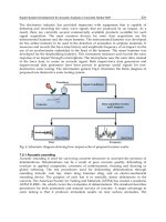
Bạn đang xem bản rút gọn của tài liệu. Xem và tải ngay bản đầy đủ của tài liệu tại đây (1.56 MB, 30 trang )
4 Will-be-set-by-IN-TECH
Γ is a phenomenological damping rate. Equations (1) and (2) denote the photonic and
excitonic parts of a polariton wave, where the coupling coefficient η is proportional to k for
a quadrupole exciton. The solution to the above equations yields the quadrupole polariton
dispersion [see Fig. 12(b)]. The propagating nature of the quadrupole polariton was first
observed in the variation of the beat period using coherent quantum beat spectroscopy under
resonant one-photon excitation [Frohlich et al. (1991); Langer et al. (1995)]. By contrast, a dark
orthoexciton does not directly couple to the radiation field. When both excitonic matter
species are generated under resonant excitation, the initial coherence of the laser light is
essentially carried by them. These resonantly created dark orthoexcitons and quadrupole
polaritons are potentially important in semiconductor-based coherent quantum information
science [Yoshioka & Kuwata-Gonokami (2006)].
Excitons in Cu
2
O can be created by conventional one-photon over-the-gap excitation. Under
this excitation condition, electron-hole (e-h) pairs are initially generated which subsequently
combine to form excitons via a screened Coulomb interaction. This “nonresonant” excitation
results in excitons that initially have an excess kinetic energy and the exciton gas temperature
can be much higher than the lattice temperature. Both orthoexcitons and paraexcitons can
recombine via indirect phonon-assisted processes [Elliot (1961); Petroff et al. (1975)], but only
the bright orthoexciton states can radiatively recombine by direct quadrupole transition,
displaying a sharp Lorentzian peak.
1
Due to the flat dispersion relation of optical phonons,
the phonon-assisted PL line can sample excitons having all possible kinetic energies, yielding
a kinetic energy distribution of excitons [Beg & Shapiro (1976)]. At temperatures lower
than about 20 K, the lifetime of orthoexcitons is basically limited by down-conversion
into lower-lying paraexcitons, which is on the order of several nanoseconds [Jang et al.
(2004); Wolfe & Jang (2005)]. Paraexcitons can have a lifetime up to several milliseconds in
high-purity natural-growth samples but is extrinsically limited by the impurity concentration,
i.e., the sample quality [Jang et al. (2006)]. Most of the previous experiments directed at
excitonic BEC in Cu
2
O were carried out using one-photon excitation [Fortin et al. (1993);
Hulin et al. (1980); Snoke et al. (1987; 1990); Snoke & Negoita (2000); Wolfe et al. (1995)].
In contrast, quadrupole polaritons can be generated using resonant excitation involving
either one or two photons [Frohlich et al. (1991); Goto et al. (1997); Ideguchi et al. (2008);
Jang & Ketterson (2007); Jang et al. (2008a); Langer et al. (1995); Sun et al. (2001); Tayagaki et al.
(2006)]. Rather than trying to cool the highly nonequilibrium state which follows nonresonant
excitation, thermalization of the system under resonant excitation involves a subsequent
heating induced by acoustic phonon absorption. Once resonantly generated, the lifetime (total
coherence time) of quadrupole polaritons is basically limited by various elastic and inelastic
dephasing processes [Takagahara (1985)]. Inelastic energy relaxation processes include
irreversible damping arising from radiative recombination, thermalization to orthoexcitons,
down-conversion to paraexcitons, and capture by ambient impurities, whereas elastic
processes are caused by pure transverse dephasing mechanisms, affecting the phase only. All
excitons and quadrupole polaritons undergo a density-dependent Auger-type decay process at
high densities [Jang & Ketterson (2008); Tayagaki et al. (2006)]. According to the recent model
[Jang & Wolfe (2005; 2006a;c)], it seems to arise due to formation of optically inactive biexcitons
though their existence has not been confirmed spectroscopically yet.
1
Details on various relaxation processes of excitons in Cu
2
O are discussed in Jang (2005).
140
Optoelectronics - Materials and Techniques
Cuprous Oxide (Cu
2
O): A Unique System Hosting Various Excitonic Matter and Exhibiting Large Third-Order Nonlinear Optical Responses 5
Fig. 2. High-quality synthetic crystals of Cu
2
O grown by thermal oxidation with various
structures: (a) Platelet with macroscopic grain boundaries, (b) hollow cylinder (inset: cross
section), and (c) spheroid.
3. Experimental methods
In order to obtain shiny, ruby-red colored, large-area single crystals of Cu
2
O, we utilize
conventional thermal oxidation of metallic Cu with platelet, wire, and shot structures
followed by a high-temperature annealing protocol. The oxidation parameters and annealing
procedure are obtained from Toth et al. (1960) and carefully adjusted to refine the Cu
2
O
crystal quality. During the growth process, we carefully maintain O
2
pressure and
temperature to lie within the middle of the Cu
2
OphaseintheCu−Cu
2
O−CuO phase
diagram [Schmidt-Whitley et al. (1974)]. It is noted that elevated annealing temperatures near
the melting temperature of Cu
2
O and slower rates of oxidation, annealing, and cooling of
the samples play key roles in diminishing the concentration of macroscopic defects such as
voids and CuO precipitates.
2
Figure 2 shows as prepared, (a) platelet, (b) hollow tube, and
(c) spherical structures of Cu
2
O, respectively. It is interesting that the oxidation of Cu wire at
high temperatures leads to the formation of hollow tubules of Cu
2
O. Together with a spheroid
form, such unconventional structures could be utilized to confine propagating quadrupole
polaritons within a whispering gallery mode [Vollmer & Arnold (2008)]. Our natural-growth
samples used in the experiments were donated by the Smithsonian Institute.
Our one- and two-photon experiments are performed on both natural-growth and synthetic
Cu
2
O crystals. For resonant two-photon excitation, the samples are properly oriented
relative to the laser polarization (E-field direction) to maximize optical transition. The
cryogenic temperatures are produced with a Janis variable-gas-flow optical cryostat and an
accompanying temperature controller. We use the frequency-tripled output of a mode-locked
Nd:YAG laser (EKSPLA PL 2143 series) with a pulse width of about 30 ps and a repetition
rate of 10 Hz in order to synchronously pump an optical parametric amplifier (OPA). The
OPA generates vertically polarized pulses in the range of 400 - 2000 nm. At the two-photon
resonance energy
2p
= 1016.5 meV (1219.4 nm), the spectral bandwidth of the laser light
from the OPA is rather broad, about 8 meV full width at half maximum. However, the phase
space compression phenomena [Kuwata-Gonokami et al. (2002)] ensure an effective creation
of quadrupole polaritons or dark orthoexcitons since the lower energy portions
(
2p
− δ
2p
)
are exactly compensated by higher parts (
2p
+ δ
2p
), thereby satisfying both energy and
2
See Mani et al. (2009a) for detailed growth procedures and X-ray and optical characterizations.
141
Cuprous Oxide (Cu
2
O): A Unique System Hosting
Various Excitonic Matter and Exhibiting Large Third-Order Nonlinear Optical Responses
6 Will-be-set-by-IN-TECH
Fig. 3. Time-integrated PL spectrum at 2 K under resonant two-photon excitation along a
(100) direction that initially generates dark orthoexcitons. The bound exciton PL is
×10
magnified.
momentum conservations. In order to verify the one- and two-photon selection rules, a pair of
polarization analyzers is placed in front of and behind the samples. The incident laser pulse is
focused onto a spot 500 μm in diameter using a 15 cm focal-length lens. The PL from excitonic
matter is collected and focused onto a fiber optic bundle mounted on a goniometer, thereby
allowing us to measure the angular dependence (φ)ofthePL.Theoutputofthefiberoptic
bundle is coupled to the entrance slit of a Spex Spec-One 500 M spectrometer and detected
using a nitrogen-cooled CCD camera. The collection efficiency of our optical system as a
function of the collection angle φ is explained in Jang & Ketterson (2007).
The Z-scan technique is traditionally employed to probe the third-order nonlinearity χ
(3)
by translating a test sample through the beam waist of a focused Gaussian-laser profile
and measuring the corresponding variation of the transmitted beam intensity in the far
field [Sheik-Bahae et al. (1990; 1991)]. For our Z-scan experiments [Mani et al. (2009b; 2010)],
the laser pulses from the OPA is first spatially filtered using a 100 μm pinhole, insuring
transmission of only the TEM
00
Gaussian mode. This Gaussian beam is focused on Cu
2
O
using a converging lens with a 7.5 cm focal length, which is mounted on a computer-controlled
stage that is translated relative to the window of the optical cryostat. This allows us to
continuously change the input irradiance I as a function of the lens position Z; I can be varied
more than a factor of 400 simply by translating Z inour1-inchscanrange. Thechangein
the far-field image of the transmitted beam with Z is minimized by using a combination of
collection lenses prior to entering a photomultiplier tube (PMT). The output of the PMT is fed
into a boxcar integrator and read out using a data acquisition system.
4. Resonant two-photon excitation and selection rules
According to k-dependent exchange interactions [Dasbach et al. (2004)], two-photon
excitation along highly symmetric crystal orientations does not generate quadrupole
polaritons but dark orthoexcitons. For example, Table 1 shows the selection rules for a
(100) direction, ensuring that two-photon excitation along this direction initially creates dark
orthoexcitons, the O
yz
state, whose one-photon transition is not allowed. This can be a crucial
issue for achieving a polariton-based whispering gallery mode, where the direction of the
142
Optoelectronics - Materials and Techniques
Cuprous Oxide (Cu
2
O): A Unique System Hosting Various Excitonic Matter and Exhibiting Large Third-Order Nonlinear Optical Responses 7
Fig. 4. (a) Dots (circles) correspond to the observed polarization dependence of the X
o
line
obtained using analyzers in front of (behind) the sample. Superimposed solid curve (line) is
the two-photon (one-photon) selection rules. Inset: schematic of the excitation geometry.
(b) Time-integrated PL spectra at 2 K as a function of the collection angle φ
= 0,5, 10, and 15
o
.
polariton propagation is arbitrarily reflected and guided by curved interfaces. However,
quadrupole polaritons can be indirectly generated although dark states are initially created.
Figure 3 shows a typical time-integrated PL spectrum under resonant two-photon excitation
at 2 K along a (100) direction. Considering that optically inactive “singlet” O
yz
dark
orthoexcitons are initially generated in this excitation geometry, it seems rather surprising
to observe several PL lines. Once created, however, these excitons undergo various relaxation
processes and can recombine accompanied with the emission of a single photon. For example,
they can: (i) inelastically scatter from optical phonons, causing the phonon replica (X
o
−Γ
−
12
),
(ii) be captured by ambient impurities, where the symmetry of an exciton is broken and
the parent selection rules do not apply, resulting in the broad bound exciton PL, and (iii)
convert into the bright orthoexciton states that directly recombine, yielding a sharp X
o
line.
They also can either nonradiatively decay due to phonon cascade or down-convert into the
lower-lying paraexcitons. Compared with other inelastic energy relaxation processes that
cause irreversible damping of dark orthoexcitons, we find that the conversion into the bright
state is the most dominant mechanism based on the observed strong X
o
line.
In order to verify that the direct X
o
line arises from two bright “doublet” O
xy
and O
zx
states,
which are subsequently converted from the dark “singlet” state, we examine the one- and
two-photon selection rules using two analyzers. The dots in Fig. 4(a) correspond to the
observed two-photon selection rules for dark orthoexcitons inferred from the bright-state PL
(X
o
line) obtained with the analyzer in front of the sample. Considering that the sample
orientation is 45
o
as shown in the inset of Fig. 4(a), the overall two-photon polarization
dependence is shown as the solid curve and is given by P
2p
∝ sin
2
[2(θ − 45
o
)] cos
4
θ,
where the extra cos
4
θ term accounts for two-photon excitation of the incident laser intensity
that decreases with cos
2
θ, as the analyzer rotates from θ = 0
o
. The circles correspond
to the observed one-photon selection rules for bright orthoexcitons, converted from dark
orthoexcitons, obtained with the analyzer behind the sample. Note that the measured X
o
intensity barely depends on the analyzer angle. Considering the total polarization of the two
bright states, O
xy
∝ cos
2
θ and O
zx
∝ sin
2
θ, this implies that the two-fold degenerate bright
states are equally populated: P
1p
∝ cos
2
θ + sin
2
θ = constant [solid line in Fig. 4(a)]. Clearly,
143
Cuprous Oxide (Cu
2
O): A Unique System Hosting
Various Excitonic Matter and Exhibiting Large Third-Order Nonlinear Optical Responses
8 Will-be-set-by-IN-TECH
the observed polarization dependencies support that the strong direct PL line arises from
dark-to-bright conversion.
This dark-to-bright conversion was first observed by Yoshioka & Kuwata-Gonokami (2006)
using two-photon absorption along the (110) direction, and the measured conversion rate
wasabout5ns
−1
. The contribution to this conversion rate due to phonon scattering can be
estimated by the deformation potential theory [Trebin et al. (1981); Waters et al. (1980)]:
3
γ(T)=
Ξ
2
xy
m
2
δ
3πρv
T
¯h
4
1
+
2k
B
T
v
T
√
2mδ
,(3)
where Ξ
xy
= 0.18 eV is the shear deformation potential, m = 2.7m
e
is the exciton mass,
ρ
= 6.1 g/cm
3
is the mass density of Cu
2
O, and v
T
= 1.3 km/s is the TA-phonon velocity.
With the measured splitting δ
= 2 μeV along this direction [Dasbach et al. (2004)], Eq. (3) yields
a conversion rate γ
0.7 ×10
−4
ns
−1
at 2 K. This implies that dark-to-bright conversion via
phonon scattering is negligible. Therefore, it most likely arises from state mixing caused by
the so-called cross relaxation, where two dark states elastically scatter to equally populate two
bright states by satisfying angular momentum conservation. Although the dark orthoexcitons
may lose their initial coherence, this implies that their phase information can be partially
carried by subsequently generated bright states, because elastic scattering only induces
a phase shift in the total ensemble coherence [Takagahara (1985)]. This cross relaxation
mechanism is currently under investigation using two-photon quantum beat spectroscopy
as a function of the incident laser intensity.
Figure 4(b) shows the PL spectra under the same conditions for several collection angles
φ in the range of 0
− 15
o
,whereφ is the angle between the laser beam direction and the
PL collection direction. Note that the direct PL intensity sharply depends on φ and is well
correlated with the laser-propagation direction, whereas the indirect phonon line does not;
i.e. it is essentially isotropic. This clearly indicates that the initial momentum of a dark
orthoexciton inherited from the laser is nearly conserved after the conversion. This leads the
momentum of a subsequently generated bright orthoexciton being near the light cone to form
a quadrupole polariton, which propagates along the initial laser direction. Based on highly
directional PL properties, this strongly indicates that propagating quadrupole polaritons are
indirectly generated. This implies that two-photon excitation in Cu
2
O eventually generates
quadrupole polaritons regardless of the crystal orientation.
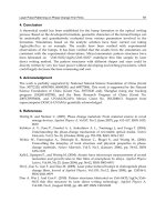
5. Half-matter/half-light characteristics of quadrupole polaritons
Near the quadrupole resonance in Cu
2
O, light propagating through the medium is
accompanied by quadrupolar polarization through the excitonic component. Ideally, the
angular distribution of the quadrupole polariton PL should be same as the angular divergence
for the incident laser because its propagation direction is inherently determined by the
incident laser direction. However, these quadrupole polaritons can lose their initial coherence
because the excitonic component of the mode, a tightly bound e-h pair, is subject to
wide-angle scattering by atomic-scale imperfections within the crystal. Therefore, we employ
angle-resolved spectroscopy to examine scattering by ambient impurities, which results
in decoherence, and monitor the angular divergence of quadrupole polaritons generated
3
See, for example, Jang & Wolfe (2006b) for the derivation of the rate due to off-diagonal shear scattering.
144
Optoelectronics - Materials and Techniques
Cuprous Oxide (Cu
2
O): A Unique System Hosting Various Excitonic Matter and Exhibiting Large Third-Order Nonlinear Optical Responses 9
Fig. 5. (a) Time-integrated PL spectra at 2 K as a function of φ = 0,5, 10, and 15
o
obtained
from the (111) oriented sample. (b) Angular distributions of the X
o
intensities from the
(100)-cut (dots) and (111)-cut (circles) samples, respectively. The solid red and blue curves
correspond to our simplified model for ka
= 14 and 6. The dashed curve denotes the angular
distribution of the transmitted laser measured just below the quadrupole resonance.
by resonant two-photon transition. In fact, Fig. 4(b) displays such angle-resolved spectra
obtained from a (100)-cut sample for several collection angles. This angle dependence can
differ from sample to sample.
Figure 5(a) plots time-integrated spectra obtained from a (111)-cut sample
4
under the same
conditions as Fig. 4(b). For this direction, the observed X
o
line is caused by quadrupole
polaritons both directly and indirectly generated by two-photon absorption. The series of
peaks in a range from 1980 to 2015 meV arise from excitons bound to ambient impurities
that are essentially isotropic (no φ dependence). Considering much enhanced bound exciton
PL intensity, this sample apparently contains more impurities and the X
o
intensity from
quadrupole polaritons remaining after transmission through the sample is strongly attenuated
due to ambient impurity scattering. This is clearly indicated by much more gradual drop in
the X
o
intensity as φ changes from 0
o
, compared with that in Fig. 4(b). This implies that
the photonic character (straight propagation with a definite k) of a quadrupole polariton is
obstructed by impurities, significantly affecting its excitonic component and thus deflecting
its initial path which, in turn, affects the photonic component by the exciton-photon coupling
terms in Eqs. (1) and (2).
From the fact that this wide-angle impurity scattering originates from the particle nature of a
quadrupole polariton, our problem reduces to a “propagating” (not diffusive
5
) exciton that
is most likely scattered by ambient charged impurities. The 1s exciton is uncharged and has
no higher multipole moments. However, a charged impurity can induce a dipole moment in
the excitonic part of a quadrupole polariton. The potential between an induced dipole and an
ion has the form V
(r)=−αe
2
/2r
4
for large r,whereα is the polarizability [Landau & Lifshitz
(1977)]. But the scattering amplitude calculated with this potential is divergent due to the
behavior of V
(r) at small r. To avoid this problem we assume the interaction approaches a
4
This sample contains high impurity levels and was used for studying bound excitons [Jang et al. (2006)].
5
Highly diffusive nature of excitons in Cu
2
O are described in Trauernicht & Wolfe (1986).
145
Cuprous Oxide (Cu
2
O): A Unique System Hosting
Various Excitonic Matter and Exhibiting Large Third-Order Nonlinear Optical Responses
10 Will-be-set-by-IN-TECH
constant at small r. Including a phenomenological “cutoff radius” a, the model potential is
V
(r)=−
αe
2
2r
4
(r > a) and V
o
≡−
αe
2
2a
4
(r < a).(4)
Since the observed angular divergence depends on the impurity concentration, the trajectory
of a quadrupole polariton is mainly determined by successive small-angle scattering, leading
to a Gaussian-like distribution. In order to obtain the angular distribution due to multiple
scattering, one needs to numerically add each stochastic process considering many parameters
[Amsel et al. (2003)]. In the absence of information on the nature and distribution of the
scattering centers we model the behavior as arising from single scattering events which are
parameterized by a cutoff radius a. By neglecting the long-range contribution, which is
very small compared with the one for r
< a, the quantum mechanical scattering amplitude
produced by Eq. (4) is given in the first-order Born approximation by
f
(Ω)=−
2m
¯h
2
V
o
q
a
0
r sin (qr )dr = −
2m
¯h
2
V
o
q
3
{sin (qa) − qacos (qa)},(5)
where we take m
to be the effective mass of a quadrupole polariton and q = |k −k
| =
2k sin(θ/2) is the associated momentum transfer with the incident wavevector k.Sincethe
interaction potential is spherically symmetric, the scattering amplitude f
(Ω)= f (θ) does
not contain any azimuthal-angle dependence. The corresponding differential cross section is
analytic and given by the absolute square of the scattering amplitude. The observed angular
distribution is then proportional to this differential cross section.
In Fig. 5(b), we plot the angular distributions of the quadrupole polariton PL intensities
from Figs. 4(b) (dots) and 5(a) (circles), where these intensity distributions are normalized
at φ
= 0
o
for comparison. The superimposed fits are generated using our model potential
with ka
= 14 (red) and 6 (blue), respectively. The dashed curve is the angular divergence of
the incident laser. Note that the only adjustable parameter is the effective screening radius
a since the wavevector of a quadrupole polariton is given by k
2.63 × 10
5
cm
−1
with a
minor spreading Δk, which is a measure of the polariton bottleneck. Although our model
might oversimplify the light character of a quadrupole polariton that actually undergoes
multiple scattering, therefore affecting macroscopic ensemble coherence in a complicated
way, we believe that it captures the essence of the dominant polariton-impurity scattering
mechanism, where the charged-impurity concentration is parameterized by a cutoff radius
a. Obviously, a stronger X
o
signal with a narrower angular distribution would occur for
samples containing lower impurity level. This also implies that the total coherence time can
be extrinsically limited by scattering from impurities. Minimizing such extrinsic effects is
crucial for preserving coherence. This angle-resolved technique can also be used as a sensitive
path-averaged (and by some deconvolution perhaps a local) impurity detector allowing some
degree of optimization for the coherence time of propagating quadrupole polaritons.
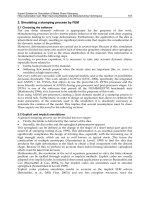
Another striking effect
6
arising from the dual character of quadrupole polaritons is anomalous
Fresnel coefficients at the quadrupole resonance, resulting in resonantly enhanced reflection of
quadrupole polaritons at crystal boundaries [Jang et al. (2008b)]. As originally suggested by
6
Unlike polaritonic effects discussed in this section, which result from the half-matter character,
suppressed collisional loss of quadrupole polaritons arises basically due to their half-light character
and this is discussed in Sec. 7.
146
Optoelectronics - Materials and Techniques
Cuprous Oxide (Cu
2
O): A Unique System Hosting Various Excitonic Matter and Exhibiting Large Third-Order Nonlinear Optical Responses 11
Fig. 6. (a) Schematic diagram of the PL collection geometry for two different boundary
conditions. The incident IR beam (solid arrows) excites Cu
2
O to create a traveling
quadrupole polariton wave (red dashed arrows) inside the medium via two-photon
absorption. As this wave leaves Cu
2
O, it converts into photons (red solid arrows), yielding
PL signals that we detect. The time-integrated PL measured from the incoming surface R
(blue trace) and the opposite surface T (red trace) under (b) condition 1 and (c) condition 2,
respectively.
Hopfield & Thomas (1963), polariton propagation in a dielectric medium is rather different
from classical light propagation. The complexity basically arises from the fact that there are
two propagating modes in the crystal associated with upper- and lower-branch polaritons.
Therefore, the usual Maxwell boundary conditions are not enough to determine the field
amplitudes for these two modes, requiring so-called additional boundary conditions.The
special case of quadrupole polaritons was theoretically studied by Pekar et al. (1981) assuming
a Frenkel-type excitation that vanishes at the vacuum-crystal boundary. However, the
correction to the “effective” index of refraction at the quadrupole resonance is predicted to be
negligible due to relatively small quadrupole coupling. In order to check this resonance effect,
we experimentally investigate the “total” reflectance (R) and transmittance (T) of traveling
quadrupole polaritons arising from multiple internal reflections at the sample surfaces. In
our excitation geometry, we define R and T as the X
o
intensities collected from the incoming
and the opposing (outgoing) surfaces, respectively [see Fig. 6(a)]. Surprisingly, our principal
finding indicates that the experimental value of T/R at the quadrupole resonance differs
significantly from the prediction of Pekar et al. (1981).
Figure 6(a) shows a schematic diagram for measuring R and T for the two boundary
conditions using (100)- and (111)-oriented natural-growth samples, respectively. Since we use
resonant two-photon excitation in which the excitation energy is the half of the quadrupole
polariton energy, the measured PL is decoupled from the incident laser. In order to measure
R we use a dichroic mirror, which is an efficient IR filter transmitting the excitation light
but reflecting visible light. The measured reflectivity in our observation range (1980
−
2040 meV) is about 0.485. Two-photon generated quadrupole polaritons propagate through
the crystal along the incident laser direction. Therefore, the opposite sur f ace is the first
boundary encountered. For condition 2, the sample is attached to a glass slide to impose
a different boundary condition. In this case, there is one more interface formed by the
147
Cuprous Oxide (Cu
2
O): A Unique System Hosting
Various Excitonic Matter and Exhibiting Large Third-Order Nonlinear Optical Responses
12 Will-be-set-by-IN-TECH
glass and the superfluid He bath. When the quadrupole polariton wave leaves Cu
2
O, it is
converted into transmitted light and a portion of that is reflected from this extra boundary
by satisfying usual Fresnel relations. These reflected photons will resonantly excite Cu
2
Ovia
one-photon excitation at the glass and Cu
2
O interface, thereby producing a counterpropagating
quadrupole polariton wave in Cu
2
O.
In Fig. 6(b) we plot the observed PL spectrum (red trace) for condition 1 as collected from
the opposite surface, corresponding to T. The blue trace shows the light transmitted at the
incoming surface (corrected for the reflectivity of the IR filter), corresponding to R.The
measured T/R is about 2.75
±0.05. Considering multiple internal reflections, this ratio can be
analytically calculated and is given by
T
R
=
(
te
−γ
)[1 +(re
−γ
)
2
+ ]
(re
−γ
)(te
−γ
)[1 +(re
−γ
)
2
+ ]
=
1
re
−γ
,(6)
where e
−γ
is a phenomenological damping factor which includes all irreversible losses during
a“one-waytrip”,andr and t are the reflection and transmission coefficients at the Cu
2
Oand
superfluid He interface, which are approximately given by
r
=
n
−1
n + 1
2
and t =
4n
(n + 1)
2
.(7)
Note that R in Eq. (6) contains t because of transmission at the incoming surface. Also,
Eq.(6)showsthatthemeasuredT/R is only affected by a single damping factor because the
accumulative damping due to multiple internal reflections exactly cancels out in this ratio. In
fact, e
−γ
is negligible for our relatively thin samples (d < 1 mm) considering a much longer
decoherence length l
= v
g
τ 2−20 mm, where v
g
is the quadrupole polariton group velocity
(on the order of 10
6
−10
7
m/s) and τ 2 ns is the measured decoherence time [Frohlich et al.
(1991)]. Assuming e
−γ
= 1andusingn = 2.65 for Cu
2
O, the simple Fresnel prediction yields
T/R
= 4.89, which does not agree with our measurement. Note that this damping factor, if
significant, induces a larger discrepancy between the theoretical and measured T/R.
Figure 6(c) plots the measured R and T for condition 2 in which the sample attached to
the glass contains a higher impurity concentration as indicated by the bound exciton PL.
The isotropic bound exciton PL from two different collections overlap each other, verifying
the scaling factor introduced by the IR filter. Because of an extra boundary formed by
the glass and superfluid He, there are numerous combinations of multiple reflections and
transmissions. In our analysis, we consider up to the 4th order, involving 8 combined
reflections and transmissions at the boundaries. Using the measured index of refraction for
the glass, n
g
= 1.48, the calculation yields T/R = 9.77. However, the measured T/R for
the condition 2 is about 5.46
± 0.15, again significantly different from the classical Fresnel
prediction.
The present theory [Pekar et al. (1981)] based on the additional boundary conditions predicts
a slight modification in the effective index of refraction n
eff
for a propagating quadrupole
polariton wave depending on the wavevector direction. For example, n
eff
for normal
incidence is given by
n
eff
=
ε +
2m
¯h
2
4πq
2
Ω
≡
ε +
1
ζ
,(8)
148
Optoelectronics - Materials and Techniques
Cuprous Oxide (Cu
2
O): A Unique System Hosting Various Excitonic Matter and Exhibiting Large Third-Order Nonlinear Optical Responses 13
where ε = 7 is the background dielectric constant, m
is the effective mass for a quadrupole
polariton that depends on the wavevector direction, q is the exciton quadrupole moment,
and Ω is the unit-cell volume. The microscopic calculation yields 1/ζ
−0.46 and −0.17
for (100) and (111) directions, respectively. Therefore, the predicted index of refraction
at the quadrupole resonance is about n
eff
=
√
7 − 0.46 2.56 for the (100) direction.
This negligible correction apparently does not explain our measurements and n
eff
must be
significantly larger than n
= 2.65. Based on the series of experiments, we have confirmed
that our experimental results can be explained by introducing the effective index of refraction
n
eff
= 4.0 ±0.1 for the boundary conditions we employed. This increased index of refraction
in turn implies a significantly enhanced reflection of quadrupole polaritons at the crystal
boundary.
The failure of the present theory might result from assuming localized Frenkel excitons,
whereas Cu
2
O is well known for hosting weakly bound Mott-Wannier excitons. Alternatively,
the amplitude of the orthoexciton may not vanish at the boundary as discussed below.
This significantly enhanced reflection arises most likely from the behavior of the matter
component (exciton). Although thermal excitons may break down at the crystal boundary,
the quasi-ballistic excitonic component of moving quadrupole polaritons will most likely
be reflected at the surface with minimal surface recombination, presumably hindering
quadrupole polaritons from exiting Cu
2
O and thus causing enhanced reflection. In the
absence of a proper theory, we propose that the phase shift associated with this reflection be
regarded as a free parameter. It may be that the behavior can be Fresnel-like, however with a
modified index of refraction. We believe that this anomalous reflection is universal, arising
from the half-matter/half-light property of polaritons, regardless of host materials. Our
results have implications for the optoelectronic design of polariton waveguides and resonators
in which a larger (effective) index of refraction implies a larger angle of total internal reflection
which in turn affects the cutoff wavelength and with it the confinement of polaritons inside
the medium.
6. Efficient quadrupole polariton generation with unconventional approaches
As a bound state of an electron and a hole, an exciton in Cu
2
O is electrically neutral and
only weakly magnetic.
7
Therefore, conventional electromagnetic external perturbations do
not cause a significant modification in its electronic properties. But, mechanical strain affects
the electronic states of Cu
2
O in two ways: (i) it induces a bandgap shift and, more important,
(ii) it lowers the crystal symmetry, resulting in splitting of orthoexciton levels depending
on the stress direction. Although numerous studies on excitons under external stress were
performed [Jang & Wolfe (2006b); Lin & Wolfe (1993); Liu & Snoke (2005); Mysyrowicz et al.
(1983); Naka & Nagasawa (2002); Snoke & Negoita (2000); Trauernicht & Wolfe (1986)], the
effect of external stress on quadrupole polaritons is essentially a virgin territory, potentially
full of unexplored interesting physics.
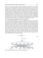
Under spatially inhomogeneous Hertzian stress [Snoke & Negoita (2000)], a strain well forms
a potential minimum for excitons inside the crystal. This technique has been extensively
used in attempts to create trapped high-density excitons. Figures 7(a) and (b) illustrate the
potential well formed in a Lucite crystal under Hertzian contact stress and a schematic of
7
One needs more than 10 T to observe noticeable exciton-level splitting in Cu
2
O induced by external
magnetic field [Fishman et al. (2009)].
149
Cuprous Oxide (Cu
2
O): A Unique System Hosting
Various Excitonic Matter and Exhibiting Large Third-Order Nonlinear Optical Responses
14 Will-be-set-by-IN-TECH
Fig. 7. (a) Hertzian contact at Lucite showing potential minimum and equipotential contours
monitored by cross polarizer. (b) Schematic of potential-trap experiments and (c) image of
excitons in Cu
2
O effectively confined by a potential well just below the stressor after fast
drift from the excitation spot (left).
stress experiments at low temperatures, respectively. Exciton drift into such a potential well
in Cu
2
O at 2 K is clearly shown in Fig. 7(c). One can then ask how this harmonic potential well
affects the propagating quadrupole polaritons. They could be attracted by the well due to the
excitonic component, as shown in Fig. 7(c), or not because the photonic component is little
affected. It will be interesting to study the influence of the potential well on the quadrupole
polariton propagation.
As a preliminary, we first conduct a rather simple experiment using uniaxial stress along a
(001) direction and collect the PL from a (110) surface of a natural-growth Cu
2
Osample.
8
Surprisingly, our results indicate that the quadrupole polariton PL (X
o
line) is significantly
enhanced with external stress. Figures 8(a)
−(c) plot the observed X
o
intensities (red traces) as
afunctionofstressintherangeofσ
= 0 − 0.3 kbar. The heavy solid traces are fits using
a single or double Gaussian function, considering the spectral resolution of our detection
system. As we increase stress, the triply-degenerate quadrupole state splits into the singlet
and doublet states where the latter lies lower [Jang & Wolfe (2006b)]. The measured splitting
is consistent with our theoretical prediction. In Figs. 8(d)
−(f), we plot the corresponding
polarization dependence of the X
o
intensities obtained using an analyzer behind the sample,
indicating a significant modification of the one-photon selection rules. Most of all, it is very
interesting that the quadrupole polariton PL rapidly increases with σ and its brightness at σ =
0.3 kbar is more than 10 times that obtained under no stress. We have also performed the same
experiments using one-photon over-the-gap excitation to check the exciton PL as a function of
σ and confirmed that no such a strong enhancement is observed. It implies that this is solely
related to either "coherent" polaritonic effects or enhanced two-photon excitation, arising from
modification of the electronic structure (mixing between dark and bright states) induced by
external stress. In order to clarify the underlying mechanism, one needs to time-resolve the
population and relaxation dynamics of quadrupole polaritons as a function of σ. Clearly, this
stress technique is promising for generating high-density quadrupole polaritons for BEC.
Previous experiments based on two-photon absorption were conducted using a single-beam
laser tuned to the two-photon quadrupole resonance (1219.4 nm). However, quadrupole
polariton generation can be also accomplished using two independent beam sources as long
as (i) the sum of beam frequencies matches with the quadrupole resonance and (ii) the
8
This sample was previously used for studying paraexcitons under stress [Trauernicht & Wolfe (1986)].
150
Optoelectronics - Materials and Techniques
Cuprous Oxide (Cu
2
O): A Unique System Hosting Various Excitonic Matter and Exhibiting Large Third-Order Nonlinear Optical Responses 15
Fig. 8. Time-integrated quadrupole polariton PL (red traces) at (a) σ = 0 kbar, (b) 0.2 kbar, and
(c) 0.3 kbar, respectively, superimposed with Gaussian fits (heavy solid curves). (d) Measured
polarization dependence (dots) under no stress, well explained by the one-photon selection
rules (solid curve). The corresponding polarization dependence under external stress are
plotted by dots (doublet) and circles (singlet) in (e) and (f). Superimposed are empirical fits.
conservation of momentum is fulfilled inside the crystal (phase matching). This two-beam
technique has been initially triggered by the idea of mixing much stronger pulses from
the pump YAG laser (1064 nm) and those from the OPA (tuned to 1428 nm) in order to
generate high-density quadrupole polaritons. Moreover, we can independently control the
polarizations of the two incident beams and their propagation directions, and therefore, the
resulting wavevector of quadrupole polaritons inside the sample.
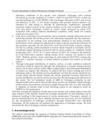
Unlike one-beam two-photon technique, however, there are number of issues to optimize
two-beam two-photon excitation such as pulse synchronization, OPA wavelength tuning, and
precise optical alignments, etc. For example, the dots in Fig. 9(a) correspond to the quadrupole
polariton signal when the delay arm of the OPA is varied near the pulse synchronization
position. The superimposed curve is a fit to the data, yielding a temporal overlap of
30 ps, which is consistent with the pulse widths of two beams. In Fig. 9(b), we plot the
quadrupole polariton signal (dots) observed when we vary the wavelength of the OPA near
1428 nm. The solid curve is a fit that basically reflects the spectral linewidth of the OPA at
this wavelength. These clearly show that two-beam two-photon efficiency strongly depends
on both time and wavelength detuning of the OPA. The dots (and superimposed curve) in
Fig. 9(c) show the relative polarization dependence of the two-beam two-photon efficiency
when the polarization angle of the OPA is varied in the range from
−90
o
to 90
o
, indicating
that orthogonal polarization is not favorable, as expected. Employing this two-beam
two-photon technique, we can also study “impact ionization” of quadrupole polaritons by
varying two incident beam powers independently, which arises from additional absorption
151
Cuprous Oxide (Cu
2
O): A Unique System Hosting
Various Excitonic Matter and Exhibiting Large Third-Order Nonlinear Optical Responses
16 Will-be-set-by-IN-TECH
Fig. 9. Measured X
o
intensity from quadrupole polaritons as a function of (a) OPA time
detuning, (b) OPA wavelength detuning, and (c) OPA polarization relative to the fixed
vertical polarization of the 1064 nm output from the YAG laser, respectively.
of an incident photon by a quadrupole polariton that ionizes the excitonic component. In
fact, this mechanism can mimic Auger-type collisional loss of quadrupole polaritons, and
therefore, measuring and controlling this process could be an important issue for achieving a
high-density polariton system.
Another interesting direction is to use “quadrupole-induced” second harmonic generation
(SHG) to efficiently generate high-density quadrupole polaritons using a non-collinear
orthogonal polarization geometry [Figliozzi et al. (2005)]. Interestingly, the corresponding SHG
polarization is largest when the incident electric fields are mutually orthogonal and is
proportional to sinψ,whereψ is the angle between two wavevectors inside the crystal. This
condition is quite different from that for two-beam two-photon absorption as explained above.
The technique was developed to investigate the surface structure of Si nanocrystals embedded
in SiO
2
matrix using SHG signals, which is enhanced by several orders of magnitude
[Figliozzi et al. (2005)]. Since Cu
2
O has a centrosymmetric crystal structure, SHG is not viable
in the dipole approximation. However, one can turn on SHG in this semiconductor by
exploiting this technique. Clearly, it is an interesting question whether quadrupole polariton
generation can be further improved via enhanced quadrupole SHG.
7. Third-order nonlinearity and nonlinear processes at quadrupole resonance
Although Cu
2
O has a rich history as a prototype material for studying fundamental exciton
physics, its nonlinear optical properties have received little attention presumably because
of its vanishing second-order susceptibility χ
(2)
stemming from its centrosymmetric crystal
structure. Consequently, the lowest-order optical nonlinearity in Cu
2
Oarisesfromthe
third-order susceptibility χ
(3)
. Precise characterization of the nonlinear optical parameters
such as χ
(3)
and the two-photon absorption coefficient β is crucial in evaluating its potential
for nonlinear optical applications and estimating the densities of excitonic matter under
two-photon excitation. Recently, we have reported the first measurement of the nonlinear
refractive index n
2
∝ Re[χ
(3)
/n](n = 2.65) and β based on the Z-scan technique [Mani et al.
(2009b)].
The single-beam Z-scan technique relies on the phenomenon of self-focusing of an intense
Gaussian laser beam in the presence of a nonlinear medium [Sheik-Bahae et al. (1990; 1991)].
152
Optoelectronics - Materials and Techniques
Cuprous Oxide (Cu
2
O): A Unique System Hosting Various Excitonic Matter and Exhibiting Large Third-Order Nonlinear Optical Responses 17
Fig. 10. (a) Normalized closed-aperture Z-scan data (dots) obtained from a (110)-oriented
natural-growth Cu
2
O sample with 20% aperture transmittance, superimposed by a
theoretical fit (solid trace). (b) Wavelength-dependent THG from Cu
2
O (red) and GaAs
(blue).
One can characterize both n
2
and β using the closed- and open-aperture Z-scan configurations,
respectively. The dots in Fig. 10(a) correspond to the normalized closed-aperture Z-scan
trace showing a valley-peak configuration, indicating positive nonlinearity of Cu
2
O, when
the on-axis irradiance at the focus is set to I
(Z = 0)=0.86 GW/cm
2
at λ = 1064 nm.
At this relatively low irradiance level, with negligible e-h pair generation by two-photon
transition, the closed-aperture Z-scan accounts for purely refractive nonlinearity due to the
bound electronic Kerr effect; Δn
= n
2
I,whereΔn is the on-axis index change at focus.
Transmittance change at the detector (ΔT) is related to Δn by
ΔT
0.406(1 −S)
0.25
2π
λ
d
eff
Δn,(9)
where S
= 20% is the aperture transmittance and d
eff
=(1 − e
−αd
)/α, with the linear
absorption coefficient α =47cm
−1
at λ = 1064 nm for the sample thickness d = 100 μm. The
solid curve is a least-square fit to the data, yielding n
2
= 1.32 × 10
−10
esu. We have found
that similar values of n
2
are obtained from our synthetic samples [Mani et al. (2009b)]. This
measured n
2
value of Cu
2
O seems comparable with those of other conventional nonlinear
semiconductors with large n
2
values. However, it is important to note that n
2
∝ 1/E
4
g
,where
E
g
is the bandgap, and that the best χ
(3)
materials have bandgap energies far below that
for Cu
2
O[seeforexampleTableIIIofSheik-Bahaeet al. (1991)]. This implies that the matrix
elements entering χ
(3)
are very large in Cu
2
O but the overall response is scaled down by its
relatively large bandgap energy. Considering this factor, we believe that Cu
2
Oisapotential
χ
(3)
material with a bandgap energy lying in the visible region.
This is further confirmed by Fig. 10(b), showing the measured THG signals from Cu
2
Oand
GaAs, both oriented along a (111) direction and 0.5 mm thick, when the input OPA wavelength
is varied from 1300 nm to 1800 nm. Considering that n
2
of GaAs is about two times that of
Cu
2
O, it is initially surprising that THG from Cu
2
O is more intense. This basically arises from
two reasons: (i) since χ
(2)
of GaAs is very large, the incident laser most strongly contributes
to the lower-order SHG process and (ii) GaAs is a dipole-allowed semiconductor in which
153
Cuprous Oxide (Cu
2
O): A Unique System Hosting
Various Excitonic Matter and Exhibiting Large Third-Order Nonlinear Optical Responses
18 Will-be-set-by-IN-TECH
Fig. 11. (a) Normalized ω Z-scan (red dots) for 10 μJ/pulse, superimposed by a theoretical fit
(solid trace) with β
= 0.217 cm/GW. (b) 2ω Z-scan traces (colored dots) for various excitation
levels in the range of 10.4
−226 μJ/pulse, fit by the theoretical model (solid traces). (c) 3ω
Z-scan traces (colored dots) for 79.5, 122, and 226 μJ/pulse. The solid curves correspond to a
model assuming no fundamental depletion.
the THG light being above the bandgap is strongly absorbed compared with the case for a
dipole-forbidden semiconductor of Cu
2
O. In support of the latter, Fig. 10(b) shows that the
THG intensity from GaAs sharply dependson the OPA input wavelength, whereas for Cu
2
Oit
does not. Most importantly, it is noteworthy that measurable THG signals can be transmitted
through a 0.5 mm-thick Cu
2
O sample used in the measurements since the absorption lengths
for the converted THG wavelengths shown in Fig. 10(b) are known to be less than a few
microns [O’Hara et al. (1999b)]. This implies that Cu
2
O could be utilized for an active THG
medium working at ω
IR
in the mid-IR range such that 3ω
IR
is still below the bandgap of Cu
2
O
without any complication from χ
(2)
contributions.
Recently, it has been suggested that three-photon excitation [Ideguchi et al. (2008)] and THG
[Mani et al. (2009b)] can affect population dynamics of quadrupole polaritons in Cu
2
Ounder
strong two-photon excitation. Considering possible complications caused by these high-order
processes, the feasibility of quadrupole polariton BEC remains an open question. In pursuit
of this question we systematically investigate various nonlinear optical processes such as
two-photon absorption, Auger-type recombination, and THG under resonant two-photon
excitation at 2 K. By extending the standard open-aperture Z-scan method (that only monitors
the intensity at ω), we also keep track of the frequency-doubled (2ω) and tripled (3ω)
Z-scan outputs to probe the quadrupole polariton PL and THG responses using appropriate
band-pass filters [Mani et al. (2010)].
In order to estimate the absolute number of quadrupole polaritons generated under
two-photon excitation, it is essential to precisely determine β, basically arising from Im[χ
(3)
]
at the quadrupole resonance. The red dots in Fig. 11(a) correspond to the normalized ω
Z-scan trace, showing two-photon absorption for 10.4 μJ/pulse. Note that only 0.4% of
the incident beam is absorbed at the focus (Z
= 0). The solid trace is a theoretical fit
[Sheik-Bahae et al. (1990)] with β
= 0.217 cm/GW and the beam waist of ω
0
= 15.1 μmat
Z
= 0. This ω
0
is consistent with the standard Gaussian width σ
s
= 0.19 cm of the incident
beam through σ
s
/ f = λ/πω
0
,wheref = 7.5 cm and λ = 1219.4 nm. Unlike conventional
band-to-band two-photon absorption, the effect at the narrow quadrupole resonance depends
on the spectral width δω of the incident laser. In our case of δω
8meV,wehaveconfirmed
154
Optoelectronics - Materials and Techniques
Cuprous Oxide (Cu
2
O): A Unique System Hosting Various Excitonic Matter and Exhibiting Large Third-Order Nonlinear Optical Responses 19
that β = 0.217 cm/GW within a 50% uncertainty based on the series of ω Z-scan experiments.
We also find that the measured β value persists up to 226 μJ/pulse without any evidence
for higher-order multiphoton absorption. This implies that one-photon transition induced by
subsequent absorption of the THG light apparently mimics three-photon excitation.
With β determined, depletion of the fundamental intensity I along the beam path z due to
two-photon absorption can be calculated and is given by
dI
dz
= −βI
2
→ I(z; Z, t)=
I
0
(Z, t)
I
0
(Z, t)βz + 1
, (10)
where I
0
(Z, t) is the photon flux at the incident sample surface, which is a function of the
focusing lens position Z and given by
I
0
(Z, t)=
2P(t)
πω
2
(Z)
→
2P
πω
2
(Z)
=
2P
πω
2
0
(1 + Z
2
/Z
2
0
)
, (11)
where P
(t) is the input pulse power with a 30 ps temporal profile and Z
0
= πω
2
0
/λ
0.06 cm is the confocal parameter. Since Z-scan yields the time-averaged data, we use the
time-integrated pulse power P to evaluate I
0
(Z). In Eq. (11), a factor of 2 is correctly introduced
for the averaged power of the TEM
00
mode Gaussian beam.
Figure 11(b) plots the 2ω Z-scan traces (colored dots) under several excitation levels from
10.4 μJ/pulse to 226 μJ/pulse, showing quadrupole polaritons generated by resonant
two-photon excitation. Note that we plot the time-averaged absolute number of quadrupole
polaritons using the measured β as explained below. As predicted for two-photon absorption
in a finite-thickness sample, for a given excitation level, the measured quadrupole polariton
number increases quadratically with the corresponding I
0
(Z) as we sweep Z.
9
However, a
striking dip-like feature develops in the vicinity of Z
= 0 as we increase the pulse energy and
quadrupole polariton generation severely saturates at the focus. In order to check whether
any signal was lost due to the finite aperture of the PMT collector, we have probed the
quadrupole polariton spatial profile in the far field as a function of Z using a gated intensified
CCD camera and verified that this mechanism is negligible [Mani et al. (2010)]. Together
with ω Z-scan indicating negligible higher-order contributions, this implies that quadrupole
polaritons undergo an Auger-type two-body decay process at high densities.
In order to explain 2ω Z-scan, we now model the population and relaxation dynamics of
quadrupole polaritons. The quadrupole polariton generation rate G should match the laser
absorption profile; G
(r; Z)=−(dI/dz)/2 = βI
2
/2, where a factor of 1/2 accounts for energy
conservation during two-photon absorption. The temporal behavior of the local quadrupole
polariton density n
(r; Z, t) is described by
dn
dt
= G(r; Z) −
n
τ
− An
2
, (12)
where τ is the quadrupole polariton lifetime and A is an Auger coefficient [Jang & Wolfe
(2005; 2006a;c); Jang & Ketterson (2008)]. The analytical solution to Eq. (12) exists and the
9
If the sample is infinitely thick, the incident IR photons N should be all absorbed and the number of
quadrupole polaritons created is simply N/2, independent of I
0
(Z).
155
Cuprous Oxide (Cu
2
O): A Unique System Hosting
Various Excitonic Matter and Exhibiting Large Third-Order Nonlinear Optical Responses
20 Will-be-set-by-IN-TECH
time-averaged density n(r; Z) is given by
n
(r; Z)=
n(r; Z, t)dt
dt
=
ln[1 + An
0
(r; Z)τ]
Aτ
, (13)
where the initial density is well approximated by n
0
(r; Z)={[1 + 4G(r; Z)Aτ
2
p
]
1/2
−1}/2Aτ
p
with the pulse width τ
p
= 30 ps. While n
0
is essentially Gτ
p
at low excitation, it approaches
to
(G/A)
1/2
and is limited by fast Auger-type decay during the 30 ps buildup time when
GAτ
2
p
1. To obtain the time-averaged quadrupole polariton number N(Z) for a given Z,
we numerically integrate n
(r; Z) over the sample dimension (d = 100 μm):
N
(Z)=
n(r; Z)d
3
r = πω
2
(Z)
d
0
n(z; Z)dz. (14)
Note that N
(Z) contains only two independent fit parameters of τ and A,sinceG is accurately
determined with β
= 0.217 cm/GW. The solid traces in Fig. 11(b) show N(Z) using a single
fit-parameter set of τ
= 2nsandA = 0.55 ×10
−16
cm
3
/ns, showing excellent fits to the series
of 2ω Z-scan data. A value τ
= 2 ns is consistent with that obtained from coherent quantum
beat spectroscopy [Frohlich et al. (1991)] and A is about 2 times smaller than that reported
based on Lyman absorption spectroscopy [Jang & Ketterson (2008); Tayagaki et al. (2006)].
Most of all, it is remarkable that the measured A value for quadrupole polaritons is more
than 10 times smaller than that for thermalized excitons [Jang & Wolfe (2005; 2006a;c)]. This
significantly reduced A seems puzzling but we can qualitatively explain it based on the
unique property a quadrupole polariton possesses but an exciton does not. This two-body
decay process for excitons is exclusively caused by “matter-matter collision” via Coulomb
interaction between two e-h pairs consisting of two excitons. Since this process arises from
random collision of excitons, it depends on the exciton gas temperature [Jang & Wolfe (2005;
2006a;c)]. However, as a quantum superposition of a photon (light) and a bright orthoexciton
(matter), a quadrupole polariton also carries the light character that is not relevant to this
collisional process. Unlike diffusive excitons, quadrupole polaritons all move in the same
direction with a definite nonzero wavevector primarily determined by the incident laser beam.
Therefore, they should have a greatly reduced probability for random collision. In this view,
it is not surprising that quadrupole polaritons have a much suppressed Auger coefficient.
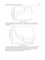
Figure 11(c) displays 3ω Z-scan traces (colored dots) for 79.5, 122, and 226 μJ/pulse, resulting
from THG of the input laser. We confirmed that 3ω Z-scan responses were very small for
lower excitation. Again, considering a submicron absorption length at 406.5 nm in Cu
2
O,
it is remarkable that measurable THG signals are transmitted through the sample. Since
fundamental depletion due to two-photon absorption is negligible, the THG field intensity
E
3ω
as a function of Z is given by [Boyd (2008)]
E
3ω
(Z)=
i3ω
2nc
χ
(3)
E
3
(Z)J
3ω
(Δkd), (15)
where n is the index of refraction for Cu
2
O, c is the speed of light in vacuum, E(Z)=
[
I
0
(Z)/2nc]
1/2
,andJ
3ω
(Δkd) is the phase-matching factor. The solid traces in Fig. 11(c) are the
predicted THG photon counting
[∝ πω
2
(Z)|E
3ω
(Z)|
2
] properly scaled to match the overall
data, simply assuming phase matching (J
3ω
= d)andusingI
0
(Z) in Eq. (11). While this
156
Optoelectronics - Materials and Techniques
Cuprous Oxide (Cu
2
O): A Unique System Hosting Various Excitonic Matter and Exhibiting Large Third-Order Nonlinear Optical Responses 21
Fig. 12. (a) Time-averaged quadrupole polariton densities (colored dots) as a function of Z,
superimposed by our Auger model (solid traces). Calculated quadrupole polariton
dispersion for (b) 3D bulk and (c) 10 μm-thick cavity (m
= 84) modes in Cu
2
O for a (110)
direction.
simple model basically corresponds to a I
3
fit, the observed 3ω Z-scan data reveal a different
power dependence of I
1.8
. We believe that this rather unusual power dependence stems
from complicated processes involving (i) strong absorption of THG beam that crucially affects
phase coherence between the fundamental and THG lights inside Cu
2
O and/or (ii) possible
contribution due to the generation of higher harmonics. Although a full understanding of
detailed THG mechanism in Cu
2
O awaits more experimental and theoretical studies, it is clear
that high-density quadrupole polariton generation is more affected by an Auger-type process
rather than THG, as indicated by our Auger model explaining 2ω Z-scan data without a χ
(3)
parameter [see Fig. 11(b)].
Unlike typical dipole coupling encountered in other semiconductors, quadrupole coupling
near the crossover is much smaller in bulk Cu
2
O, causing relatively large curvature of
the dispersion near the quadrupole polariton bottleneck; the associated group velocity is
relatively fast and the effective mass is very small, several orders of magnitude smaller than
the exciton mass [see Fig. 12(b)]. This should result in a significant reduction in the BEC
transition density. Furthermore, the Auger-type cross section is significantly reduced for
quadrupole polaritons owing to their half-matter/half-light characters. This implies that
the experimentally achievable quadrupole polariton densities can be well above the critical
density for BEC.
In order to estimate experimental quadrupole polariton densities attainable, we plot the
time-averaged density n
(Z)=N(Z)/πω
2
(Z)d (colored dots) in Fig. 12(a) using N(Z) in
Fig. 11(b). The solid traces correspond to our Auger model with A
= 0.55 × 10
−16
cm
3
/ns.
In our excitation range, the spatial extent of quadrupole polaritons along the propagation
direction is limited by the sample thickness of d
= 100 μm, which is smaller than the
two-photon absorption length
(βI)
−1
. The corresponding areal densities N(Z)/πω
2
(Z) are
also plotted in Fig. 12(a). Despite considerable decrease in N
(Z) around Z = 0 in Fig. 11(b),
it is important to note that the maximum density still locates at the focus in which it saturates
around 3
×10
16
cm
−3
under high excitation levels. This is more than 10 times higher than the
maximum thermal exciton density (
10
15
cm
−3
) [O’Hara & Wolfe (2000)], basically due to
the suppression of A by the same amount.
157
Cuprous Oxide (Cu
2
O): A Unique System Hosting
Various Excitonic Matter and Exhibiting Large Third-Order Nonlinear Optical Responses
22 Will-be-set-by-IN-TECH
Fig. 13. (a) Time-integrated PL recorded using a reflection geometry, showing effective
confinement of quadrupole polaritons from a silvered region . Inset depicts the sample and
collection geometry. (b) Schematic diagram for a Cu
2
O Fabry-Perot cavity.
Although the areal densities for quadrupole polaritons we observe in Cu
2
Oaremuchhigher
than the critical BEC density (
10
9
cm
−2
) in the 2D cavity-polariton structures [Balili et al.
(2007); Deng et al. (2003); Kasprzak et al. (2006)], BEC is not expected to arise in practice,
since the absence of a local minimum precludes condensation as shown in Fig. 12(b); of
course such minima have been engineered into the polariton dispersion curves of the 2D
microcavities. This limitation might be circumvented by depositing partially transmitting
mirrors on opposing sides of a flat platelet of Cu
2
O, thereby forming a Fabry-Perot cavity
[see Fig.13(b)]. In Fig. 12(c), we plot our simulation result for the m
= 84 cavity-photon
mode assuming a 10 μm-thick Cu
2
O platelet that forms a Fabry-Perot cavity (blue dashed
curve) sitting just above the exciton mode (red dashed curve).
10
Then, the lower-polariton
branch (red solid curve) would develop a local minimum via quadrupole coupling associated
with a very small effective mass (
10
−5
m
e
) in which long-lived quadrupole polaritons can
condense.
Recently, we have experimentally confirmed that depositing a silver film on Cu
2
O
significantly enhances the quadrupole polariton confinement without altering underlying
excitonic structures, as demonstrated by Fig. 13(a). The quadrupole polariton PL from a
silvered region (blue trace) is about 4 times stronger than that from an unsilvered region
(red trace). This behavior persists when we move the excitation position around the
silvered portion. Figure 13(b) illustrates a detailed schematic of the proposed cavity system
for quadrupole polariton BEC. The excitation surface of a Cu
2
O platelet is attached to a
commercial dichroic mirror that transmits the incident excitation beam in the IR (1219.4 nm),
but strongly reflects the quadrupole polariton PL in the visible range (609.7 nm), thereby
forming a high-Q cavity with efficient quadrupole polariton generation. Optimally coupling
the OPA output to the cavity poses a more challenging problem, as it requires a way to
continuously vary this coupling. We have two strategies for matching of mode frequency
10
Smaller node numbers and large mode spacings would require a thinner sample. A promising
technology to make thin Cu
2
O films showing sharp exciton lines was reported by Markworth et al.
(2001).
158
Optoelectronics - Materials and Techniques
Cuprous Oxide (Cu
2
O): A Unique System Hosting Various Excitonic Matter and Exhibiting Large Third-Order Nonlinear Optical Responses 23
either by (i) adjusting the propagation direction of quadrupole polaritons relative to the plane
normal or (ii) placing a silver mirror on the far side of the sample with a variable thickness.
Unlike our previous experiments based on time-integrated methods, we plan to directly
time-resolve the population and relaxation dynamics of two-photon generated quadrupole
polaritons as a function of the input IR power and the angle of incidence. This will clearly
reveal not only the impact of two-body decay on high-density quadrupole polaritons but
also the coherent temporal evolution of long-lived quadrupole polariton condensate once
achieved.
8. Concluding remarks
The goal of this chapter is to study both fundamental and technological aspects of
natural-growth/synthetic cuprous oxide (Cu
2
O) crystals that host long-lived excitonic
matter and exhibit large third-order nonlinear optical responses. All these remarkable
physical properties primarily originate from the centrosymmetric crystal structure and
the dipole-forbidden bandgap of Cu
2
O. Bright orthoexcitons intersecting the light cone
quadrupole couple to the electromagnetic field and split into two polariton branches. A
quadrupole polariton in Cu
2
O is special in that it propagates over a macroscopic distance
with a very long coherence time, compared with polaritons in conventional dipole-allowed
semiconductors.
Based on the series of experiments under resonant two-photon excitation, we
have demonstrated various interesting phenomena essentially arising from unique
half-matter/half-light characteristics of quadrupole polaritons. However, our current
understanding of the properties of a quadrupole polariton is rather empirical and accessible
only experimentally, calling for a more rigorous microscopic description of this quasiparticle.
More importantly, our findings imply that high-density quadrupole polaritons can be
effectively confined inside Cu
2
O and we have proposed a promising direction for long-lived
polariton Bose-Einstein condensation (BEC). Clearly, BEC in a forbidden-gap semiconductor
would be a valuable addition to the current BEC class from the perspective of fundamental
physics. Also, polariton BEC on the order of several nanoseconds at nominal cryogenic
temperatures will be a revolutionary step toward quantum computers and information
sciences.
Various nonlinear optical parameters have been quantitatively characterized using Z-scan
and third harmonic generation (THG) techniques. Distinctive and interesting properties
of this semiconductor allow us to consider possible optoelectronic applications involving
polariton-based waveguides and whispering gallery resonators, polariton lasers, and active
third-order nonlinear optical devices. We have also discussed a set of particularly
interesting and timely issues. This includes detailed research directions such as (i)
high-density quadrupole polariton dynamics near BEC regime, (ii) impact of external stress
on quadrupole polaritons, (iii) nonlinear optics at the quadrupole resonance using both
two-beam two-photon absorption and quadrupole-induced second harmonic generation, and
(iv) clarification of underlying THG mechanism.
159
Cuprous Oxide (Cu
2
O): A Unique System Hosting
Various Excitonic Matter and Exhibiting Large Third-Order Nonlinear Optical Responses
24 Will-be-set-by-IN-TECH
9. Acknowledgments
The author acknowledges the essential collaboration of J. B. Ketterson at Northwestern
University, S. Mani at Intel Corporation, and J. P. Wolfe at the University of Illinois. This
work is supported by the National Science Foundation under (i) the Northwestern Materials
Research Center; Grant DMR-0520513, (ii) the U.S./Ireland cooperation; Grant 0306731, and
(iii) the Integrative Graduate Education and Research Training program; Grant 0801685.
10. References
Amsel, G.; Battistig, G. & L’Hoir, A. (2003). Small angle multiple scattering of fast ions, physics,
stochastic theory and numerical calculations, Nucl. Instrum. Methods Phys. Res. B 201,
325.
Balili, R.; Hartwell, V.; Snoke, D.; Pfeiffer, L. & West, K. (2007). Bose-Einstein condensation of
microcavity polaritons in a trap, Science 316, 1007.
Beg, M. M. & Shapiro, S. M. (1976). Study of phonon dispersion relations in cuprous oxide by
inelastic neutron scattering, Phys.Rev.B 13, 1728.
Boyd, R. W. (2008). Nonlinear Optics, 3rd ed. Academic Press, San Diego. pp. 119.
Christopoulos,S.;vonHogersthal,G.B.H.;Grundy,A.J.D.;Lagoudakis,P.G.;Kavokin,A.
V.; Baumgerg, J. J.; Christmann, G.; Butte, R.; Feltin, E.; Carlin, J. -F. & Grandjean,
N. (2007). Room-temperature polariton lasing in semiconductor microcavities, Phys.
Rev. Lett. 98, 126405.
Dahl, J. P. & Switendick, A. C. (1966). Energy bands in cuprous oxide, J. Phys. Chem. Solids 27,
931.
Dasbach, G.; Frohlich, D.; Klieber, R.; Suter, D.; Bayer, M. & Stolz, H. (2004).
Wave-vector-dependent exchange interaction and its relevance for the effective
exciton mass in Cu2O, Phys. Rev. B 70, 045206.
Deng, H.; Weihs, G.; Snoke, D.; Bloch, J. & Yamamoto, Y. (2003). Polariton lasing vs. photon
lasing in a semiconductor microcavity, Proc. Natl. Acad. Sci. U.S.A. 100, 15318.
Deng, H.; Haug, H. & Yamamoto, Y. (2010). Exciton-polariton Bose-Einstein condensation,
Rev. Mod. Phys. 82, 1489.
Elliot, R. J. (1961). Symmetry of excitons in cuprous oxide, Phys. Rev. 124, 340.
Figliozzi, P.; Sun, L.; Jiang, Y.; Matlis, N.; Mattern, B.; Downer, M. C.; Withrow, S. P.; White, C.
W. & Mendoza, B. S. (2005). Single-beam and enhanced two-beam second-harmonic
generation from silicon nanocrystals by use of spatially inhomogeneous femtosecond
pulses, Phys. Rev. Lett. 94, 047401.
Fishman, D.; Faugeras, C.; Potemski, M.; Revcolevschi, A. & van Loosdrecht, P. H. M. (2009).
Magneto-optical readout of dark exciton distribution in cuprous oxide, Phys. Rev. B
80, 045208.
Fortin, E.; Farfard, S. & Mysyrowicz, A. (1993). Exciton transport in Cu
2
O: Evidence for
excitonic superfluidity?, Phys. Rev. Lett. 70, 3951 and references therein.
Frohlich, D.; Kulik, A.; Uebbing, B.; Mysyrowicz, A.; Langer, V.; Stolz, H. & von der Osten, W.
(1991). Coherent propagation and quantum beats of quadrupole polaritons in Cu
2
O,
Phys.Rev.Lett. 67, 2343.
Goto, T.; Shen, M. Y.; Koyama, S. & Yokouchi, T. (1997). Bose-Einstein statistics of
orthoexcitons generated by two-photon resonant absorption in cuprous oxide, Phys.
Rev. B 55, 7609.
160
Optoelectronics - Materials and Techniques
Cuprous Oxide (Cu
2
O): A Unique System Hosting Various Excitonic Matter and Exhibiting Large Third-Order Nonlinear Optical Responses 25
Grun, J. B.; Seiskind, M. & Nikitine, S. (1961). Etude spectrophotometrique des spectres
continus de Cu
2
O a diverses temperatures, J. Phys. Chem. Solids 19, 189.
Hayashi M. & Katsuki, K. (1952). Hydrogen-like absorption spectrum of cuprous oxide, J.
Phys. Soc. Jpn. 7, 599.
Hopfield, J. J. (1958). Theory of the contribution of excitons to the complex dielectric constant
of crystals, Phys. Rev. 112, 1555.
Hopfield, J. J. & Thomas, D. G. (1963). Theoretical and experimental effect of spatial dispersion
on the optical properties of crystals, Phys. Rev. 132, 563.
Hulin, D.; Mysyrowicz, A. & Benoit a la Guillaume, C. (1980). Evidence for Bose-Einstein
statistics in an exciton gas, Phys. Rev. Lett. 45, 1970.
Ideguchi, T.; Yoshioka, K.; Mysyrowicz, A. & Kuwata-Gonokami, M. (2008). Coherent
quantum control of excitons at ultracold and high density in Cu
2
Owithphase
manipulated pulses, Phys. Rev. Lett. 100, 233001.
Inoue, M. & Toyozawa, Y. (1965). Two-photon absorption and energy band structure, J. Phys.
Soc. Jpn. 20, 363.
Jang, J. I.; O’Hara, K. E. & Wolfe, J. P. (2004). Spin-exchange kinetics of excitons in Cu
2
O:
Transverse acoustic phonon mechanism, Phys. Rev. B 70, 195205.
Jang, J. I. & Wolfe, J. P. (2005). Biexcitons in the semiconductor Cu
2
O: An explanation of the
rapid decay of excitons, Phys. Rev. B 72, 241201(R).
Jang, J. I. (2005). Lifetimes of excitons in cuprous oxide, Ph.D. thesis, University of Illinois.
Jang, J. I. & Wolfe, J. P. (2006). Exciton decay in Cu
2
O at high density and low temperature:
Auger recombination, spin-flip scattering, and molecule formation, Solid State
Commun. 137, 91.
Jang, J. I. & Wolfe, J. P. (2006). Relaxation of stress-split orthoexcitons in Cu
2
O, Phys.Rev.B 73,
075207.
Jang, J. I. & Wolfe, J. P. (2006). Auger recombination and biexcitons in Cu
2
O: A case for dark
excitonic matter, Phys. Rev. B 74, 045211.
Jang, J. I.; Sun, Y.; Watkins, B. & Ketterson, J. B. (2006). Bound excitons in Cu
2
O: Efficient
internal free exciton detector, Phys. Rev. B 74, 235204.
Jang, J. I. & Ketterson, J. B. (2007). Impact of impurities on orthoexciton-polariton propagation
in Cu
2
O, Phys. Rev. B 76, 155210.
Jang, J. I. & Ketterson, J. B. (2008). Suppression of molecule formation for
orthoexciton-polaritons in Cu
2
O, Solid State Commun. 146, 128.
Jang, J. I.; Sun, Y. & Ketterson, J. B. (2008). Indirect generation of quadrupole polaritons from
dark excitons in Cu
2
O, Phys. Rev. B 77, 075201.
Jang, J. I.; Sun, Y.; Mani, S. & Ketterson, J. B. (2008). Resonantly enhanced reflection of
quadrupole polaritons in Cu
2
O, Appl. Phys. Lett. 93, 121111.
Kasprzak, J.; Richard, M.; Kundermann, S.; Baas, A.; Jeambrun, P.; Keeling, J. M. J.; Marchetti,
F. M.; Szymanska, M. H.; Andre, R.; Staehli, J. L.; Savona, V.; Littlewood, P. B.;
Deveaud, B. & Dang, L. S. (2006). Bose-Einstein condensation of exciton polaritons,
Nature 443, 409.
Kavoulakis, G. M.; Chang, Y. -C. & Baym, G. (1997). Fine structure of excitons in Cu
2
O, Phys.
Rev. B 55, 7593.
Kuwabara, G.; Tanaka, M. & Fukutani, H. (1977). Optical absorption due to paraexciton of
Cu
2
O, Solid State Commun. 21, 599.
161
Cuprous Oxide (Cu
2
O): A Unique System Hosting
Various Excitonic Matter and Exhibiting Large Third-Order Nonlinear Optical Responses
26 Will-be-set-by-IN-TECH
Kuwata-Gonokami, M.; Shimano, R. & Mysyrowicz, A. (2002). Phase-coherent manipulation
of cold biexcitonic waves, J. Phys. Soc. Jpn. 71, 1257.
Lagoudakis, K. G.; Wouters, M.; Richard, M.; Baas, A.; Carusotto, I.; Andre, R.; Dang, L. S. &
Deveaud-Pledran, B. (2008). Quantized vortices in an exciton-polariton condensate,
Nat. Phys. 4, 706.
Lai, C. W.; Kim, N. Y.; Utsunomiya, S.; Roumpos, G.; Deng, H.; Fraser, M. D.; Byrnes, T.;
Recher, P.; Kumada, N.; Fujisawa, T. & Yamamoto, Y. (2007). Coherent zero-state and
π-state in an exciton-polariton condensate array, Nature 450, 529.
Landau, L. D. & Lifshitz, E. M. (1977). Quantum Mechanics, 3rd ed. Pergamon, New York.
pp. 339.
Langer, V.; Stolz, H. & von der Osten, W. (1995). Picosecond quantum-beat spectroscopy of
quadrupole polaritons in Cu
2
O, Phys.Rev.B 51, 2103.
Lin, J. L. & Wolfe, J. P. (1993). Bose-Einstein condensation of paraexcitons in stressed Cu
2
O,
Phys.Rev.Lett. 71, 1222.
Liu, Y. & Snoke, D. (2005). Resonant two-photon excitation of 1s paraexcitons in cuprous
oxide, Solid State Commun. 134, 159.
Mani, S.; Jang, J. I.; Ketteron, J. B. & Park, H. Y. (2009). High-quality Cu
2
O crystals with various
morphologies grown by thermal oxidation, J. Cryst. Growth 311, 3549.
Mani, S.; Jang, J. I. & Ketterson, J. B. (2009). Large third-order susceptibility and
third-harmonic generation in centrosymmetric Cu
2
O crystal, Opt. Lett. 34, 2817.
Mani, S.; Jang, J. I. & Ketterson, J. B. (2010). Nonlinear optical processes at quadrupole
polariton resonance in Cu
2
O as probed by a Z-scan technique, Phys.Rev.B 82, 113203.
Markworth, P. R.; Chang, R. P. H.; Sun, Y.; Wong, G. K. L. & Ketterson, J. B. (2001). Epitaxial
stabilization of orthorhombic cuprous oxide films on MgO (110), J. Mater. Res. 16,
914.
Mysyrowicz, A.; Trauernicht, D. P.; Wolfe, J. P. & Trebin, H. -R. (1983). Stress dependence of
the paraexciton in Cu
2
O, Phys. Rev. B 27, 2562.
Naka, N. & Nagasawa, N. (2002). Nonlinear paraexciton kinetics in a potential trap in Cu
2
O
under two-photon resonance excitation, Phys. Rev. B 65, 245203.
O’Hara, K. E.; Suilleabhain, L. O. & Wolfe, J. P. (1999). Strong nonradiative recombination of
excitons in Cu
2
O and its impact on Bose-Einstein statistics, Phys. Rev. B 60, 10565.
O’Hara, K. E.; Gullingsrud, J. R. & Wolfe, J. P. (1999). Auger decay of excitons in Cu
2
O, Phys.
Rev. B 60, 10872.
O’Hara, K. E. & Wolfe, J. P. (2000). Relaxation kinetics of excitons in Cu
2
O, Phys. Rev. B 62,
12909.
Olsen, L. C.; Addis, F. W. & Miller, W. (1982). Experimental and theoretical studies of Cu
2
O
solar cells, Solar Cells 7, 247.
Pekar, S. I.; Piskovoi, V. N. & Tsekvava, B. E. (1981). Transmission and reflection of light at the
vacuum-crystal interface in the region of a quadrupole exciton transition, Sov. Phys.
Solid State 23, 1113.
Pestov, D.; Wang, X.; Ariunbold, G. O.; Murawski, R. K.; Sautenkov, V. A.; Dogariu, A/;
Sokolov, A. V. & Scully, M. O. (2008). Single-shot detection of bacterial endospores
via coherent Raman spectroscopy, Proc. Natl. Acad. Sci. U.S.A. 105, 422.
Petroff, Y. P.; Yu, P. Y. & Shen, Y. R. (1975). Study of photoluminescence in Cu
2
O, Phys. Rev. B
12, 2488.
162
Optoelectronics - Materials and Techniques
Cuprous Oxide (Cu
2
O): A Unique System Hosting Various Excitonic Matter and Exhibiting Large Third-Order Nonlinear Optical Responses 27
Pushkarsky, M. B.; Webber, M. E.; Macdonald, T. & Patel, C. K. N. (2006). Sub-parts-per-billion
level detection of NO
2
using room-temperature quantum cascade lasers, Proc. Natl.
Acad. Sci. U.S.A. 103, 10846.
Schmidt-Whitley, R. D.; Martinez-Clemente, M. & Revcolevschi, A. (1974). Growth and
microstructural control of single crystal cuprous oxide Cu
2
O, J. Cryst. Growth 23,
113.
Serebryakov, V. A.; Boiko, E. V.; Petrishchev, N. N. & Yan, A. V. (2010). Medical applications of
mid-IR lasers. Problems and prospects, J. Opt. Technol. 77, 6.
Sheik-Bahae, M.; Said, A. A.; Wei, T.; Hagan, D. J. & Van Stryland, E. W. (1990). Sensitive
measurement of optical nonlinearities using a single beam, IEEE J. Quantum Electron.
26, 760.
Sheik-Bahae, M.; Hutchings, D. C. ; Hagan, D. J. & Van. Stryland, E. W. (1991). Dispersion of
bound electronic nonlinear refraction in solids, IEEE J. Quantum Electron. 27, 1296.
Snoke, D. W.; Wolfe, J. P. & Mysyrowicz, A. (1987). Quantum saturation of a bose gas; excitons
in Cu
2
O, Phys. Rev. Lett. 59, 827.
Snoke, D. W.; Wolfe, J. P. & Mysyrowicz, A. (1990). Evidence for Bose-Einstein condensation
of excitons in Cu
2
O, Phys.Rev.B 41, 11171.
Snoke, D. W. & Negoita, V. (2000). Pushing the Auger limit: Kinetics of excitons in traps in
Cu
2
O, Phys. Rev. B 61, 2904.
Sun, Y.; Wong, G. K. L. & Ketterson, J. B. (2001). Production of 1s quadrupole-orthoexciton
polaritons in Cu
2
O by two-photon pumping, Phys.Rev.B 63, 125323.
Takagahara, T. (1985). Theory of dephasing relaxation of excitonic polaritons, Phys.Rev.B 31,
8171.
Tayagaki, T.; Mysyrowicz, A. & Kuwata-Gonokami, M. (2006). Collision between supercooled
excitons in Cu
2
O studied by time-resolved Lyman spectroscopy, Phys. Rev. B 74,
245127.
Toth, R. S.; Kilson, R. & Trivich, D. (1960). Preparation of large area single-crystal cuprous
oxide, J. Appl. Phys. 31, 1117.
Trauernicht, D. P. & Wolfe, J. P. (1986). Drift and diffusion of paraexcitons in Cu
2
O:
Deformation-potential scattering in the low-temperature regime, Phys.Rev.B 33,
8506.
Trebin, H. -R.; Cummins, H. Z. & Birman, J. L. (1981). Excitons in cuprous oxide under uniaxial
stress, Phys. Rev. B 23, 597.
Utsunomiya, S.; Tian, L.; Roumpos, G.; Lai, C. W.; Kumada, N.; Fujisawa, T.;
Kuwata-Gonokami, M.; Loffler, A.; Hofling, S.; Forchel, A. & Yamamoto, Y. (2008).
Observation of Bogoliubov excitations in exciton-polariton condensates, Nat. Phys.
4, 700.
Vollmer, F. & Arnold, S. (2008). Whispering-gallery-mode biosensing: label-free detection
down to single molecules, Nature 5, 591.
Waters, R. G.; Pollak, F. H.; Bruce, R. H. & Cummins, H. Z. (1980). Effects of uniaxial stress on
excitons in Cu
2
O, Phys. Rev. B 21, 1665.
Wolfe, J. P.; Lin, J. L. & Snoke, D. W. (1995). Bose-Einstein condensation of a nearly ideal gas:
Excitons in Cu
2
O, In: Bose-Einstein Condensation,Griffin,A.;Snoke,D.W.&Stringari,
S. (Ed.), Cambridge University Press, Cambridge, England. pp. 281-329.
Wolfe, J. P. & Jang, J. I. (2005). New perspectives on kinetics of excitons in Cu
2
O, Solid State
Commun. 134, 143.
163
Cuprous Oxide (Cu
2
O): A Unique System Hosting
Various Excitonic Matter and Exhibiting Large Third-Order Nonlinear Optical Responses