báo cáo hóa học:" The bioenergetic signature of isogenic colon cancer cells predicts the cell death response to treatment with 3-bromopyruvate, iodoacetate or 5-fluorouracil" ppt
Bạn đang xem bản rút gọn của tài liệu. Xem và tải ngay bản đầy đủ của tài liệu tại đây (591.85 KB, 9 trang )
RESEARC H Open Access
The bioenergetic signature of isogenic colon
cancer cells predicts the cell death response to
treatment with 3-bromopyruvate, iodoacetate or
5-fluorouracil
María Sánchez-Aragó, José M Cuezva
*
Abstract
Background: Metabolic reprogramming resulting in enhanced glycolysis is a phenotypic trait of cancer cells, which is
imposed by the tumor microenvironment and is linked to the down-regulation of the catalytic subunit of the
mitochondrial H
+
-ATPase (b-F1-ATPase). The bioenergetic signature is a protein ratio (b-F1-ATPase/GAPDH), which
provides an estimate of glucose metabolism in tumors and serves as a prognostic indicator for cancer patients. Targeting
energetic metabolism could be a viable alternative to conventional anticancer chemotherapies. Herein, we document
that the bioenergetic signature of isogenic colon cancer cells provides a gauge to predict the cell-death response to the
metabolic inhibitors, 3-bromopyruvate (3BrP) and iodoacetate (IA), and the anti-metabolite, 5-fluorouracil (5-FU).
Methods: The bioenergetic signature of the cells was determined by western blotting. Aerobic glycolysis was
determined from lactate production rates. The cell death was analyzed by fluorescence microscopy and flow
cytometry. Cellular ATP concentrations were determined using bioluminiscence. Pearson’s correlation coefficient
was applied to assess the relationship between the bioenergetic signature and the cell death response. In vivo
tumor regression activities of the compounds were assessed using a xenograft mouse model injected with the
highly glycolytic HCT116 colocarcinoma cells.
Results: We demonstrate that the bioenergetic signature of isogenic HCT116 cancer cells inversely correlates with
the potential to execute necrosis in response to 3BrP or IA treatment. Conversely, the bioenergetic signature directly
correlates with the potential to execute apoptosis in response to 5-FU treatment in the same cells. However,
despite the large differences observed in the in vitro cell-death responses associated with 3BrP, IA and 5-FU, the in
vivo tumor regression activi ties of these agents were comparable.
Conclusions: Overall, we suggest that the determination of the bioenergetic signature of colon carcinomas could
provide a tool for predicting the therapeutic response to various chemotherapeutic strategies aimed at combating
tumor progression.
Background
Colorectal cancer (CRC) is a common neoplasia which
poses a heavy burden on public health systems world-
wide [1]. Despite the establishment of CRC screening
protocols, tailored therapeutic approaches are required
to minimize the significant s ocial impact of this disease
[1]. At present, KRAS mutation status is the only vali-
dated predictive marker for targeted CRC therapy [2].
Thus, the development and clinical implementation of
new predictive molecular markers are needed to aid in
the selection of patients likely to respond to therapy and
rationalized CRC treatments [2].
Cancer cells and tumors have a predominant glycolytic
metabolism, even under aerobic conditions [3,4].
Although the altered energetic metabolism of cancer
* Correspondence:
1
Departamento de Biología Molecular, Centro de Biología Molecular Severo
Ochoa, Consejo Superior de Investigaciones Científicas-Universidad
Autónoma de Madrid (CSIC-UAM), Centro de Investigación Biomédica en
Red de Enfermedades Raras CIBERER-ISCIII, Instituto de Investigación Hospital
12 de Octubre, Universidad Autónoma de Madrid, 28049 Madrid, Spain
Sánchez-Aragó and Cuezva Journal of Translational Medicine 2011, 9:19
/>© 2011 Sánchez-Aragó and Cuezva; li censee BioMed Central Ltd. This is an Open Access article distributed under the terms of the
Creative Commons Attribution License ( which permits unres tricted use, distribution, and
reproduction in any medium, provided the original work is prope rly cited.
cells has be en proposed as a potential target for cancer
treatment [3,5-7], it could also represent a therapeutic
obstacle, because of its contribution to chemo- and
radio-resistance [8]. In some tumors, this glycolytic phe-
notype is accompanied by a loss of b ioenergetic activity
in mitochondria [9,10], which can be estimated by
determining it s bioenergetic signature [10,11]. The bioe-
nergetic signature is a protein ratio (b-F1-ATPase/
GAPDH ratio) that assesses the expression of the cataly-
tic subunit of mitochondrial H
+
-ATP synthase (b-F1-
ATPase), a bottle-neck component required for the
synthesis of biological energy, relative to the expression
of glycolytic glyceraldehyde-3-phosphate dehydrogenase
(GAPDH) [10]. Consistently, the bioenergetic signature
has been observed to be significantly down-regulated in
diff erent human tumors compared to paired normal tis-
sues [10,12-19]. Recent findings indicate that the bioe-
nergetic signature also represents a functional index of
metab olic activity because it correlates, both in vivo and
in vitro, with the rate of glucose utilization by cancer
cells and t umors [9,11]. Moreover, according to large
cohort studies of colon [10,19], lung [9,14] and breast
[16,20] cancer patients, low tumor bioenergetic signa-
tures are associated with poor patient prognosis,
strongly suggesting that impaired mitochondrial bioe-
nergetics is at the heart of cancer progression.
Remarkably, down-regulation of b-F1-ATPase has
been widely associated with the r esistance of cancer
cells to standard anticancer therapies [21-23]. In the
specific case of colon cancer cells, chemotherapeutic
response to 5-fluorou racil (5-FU) [11,21], as well as sev-
eral metabolic inhibitors [23,24], was assessed in cells
with different genetic backgrounds: a condition that is
likely to affect the cellular response to chemotherapeutic
agents. The recent development of isogenic HCT116
colon cancer cell lines, representing different bioener-
getic signatures [11], has provided an opportunity to
unambiguously assess the influence of energetic metabo-
lism on colon cancer therapy. In this study, we investi-
gated cell death responses in metabolically different
isogenic HCT116 cells and the regressio n of tumor
xenographs, in response to the glycolytic inhibitors 3-
bromopyruvate (3BrP) and iodoacetate (IA), and the
classic chemotherapeutic agent, 5-FU. The small alkylat-
ing 3BrP and IA target the enzymes of glycolysis hexoki-
nase [25] and GAPDH [26], respectively, although recent
findings suggest that 3BrP also targets GAPDH [27].
Methods
Cell cultures and treatments
Human colorectal carcinoma HCT116 cells were grown
in McCoy ’ s 5A media supplemented with 10% fetal
bovine serum. Twenty four h after seeding, cells were
left untreated (M-type), treated with 6 μMoligomycin
(G-type), or treated with 10 mM 2-DG (SM-type) for
48h. On the day of the experiment, culture medium was
replaced without the addition of any drug and cells were
used at ~ 60% confluence for experiments. Where indi-
cated, cells were incubated with 10 μM 5-FU for 48h, or
8 μM 3BrP or 100 μM IA for 7h.
Protein electrophoresis and Western blot analysis
Cells were resuspended in lysis buffer (25 mM Hepes,
2.5 mM EDTA, 1% Triton X-100, 1 mM PMSF and
5 μg/mL leupeptin). Cell lysates were clarified by centri-
fugation at 11000 × g for 15 min. Resulting supernatants
were fractionated on SDS-PAGE and transferred onto
PVDF membranes for immunoblot analysis (Inmobilon-
P, Millipore). Protein concentrations were determined
using Bradford reagent (Bio-Rad protein assay). The pri-
mary monoclonal antibodies used were: anti-Hsp60
(Stressgene SPA-807, 1:2000) and anti -GAPDH (Abcam,
1:20000). The polyclonal rabbit anti-b-F1-ATPase
(1:15000) [10] was also used. Peroxidase-conjugated
anti-mouse or anti-rabbit IgGs (Nordic Immunonology,
1: 3000) were used as secondary antibodies. The blots
were developed using the ECL reagent.
Aerobic glycolysis
For determination of the rates of aerobic glycolysis,
0.1 mL aliquots of culture media were collected and
used for enzymatic determination of lactate [11].
Cell death assays
Exposure of phosphatidylserine on the cell surface was
analyzed after various cellular treatments using the
annexin V-FITC assay (Sigma-Aldrich). Briefly, cells
were washed twice in PBS and incubated in the dark for
10minatroomtemperaturewith FITC-conjugated
annexin-V (50 μg/mL) and propidium iodide (100 μg/
mL) solutions. For each analysis, 10,000 events were
recorded in a FACScan (Becton-Dickinson). Cell death
was also determined using fluorescence microscopy. In
brief, cells treated with the different compounds
described were harvested, washed with PBS and incu-
bated in the dark for 5 min at r oom temperature wit h
Hoechst 33342 (1 mg/mL) and propidium iodide (1 mg/
mL) solutions. After washing, samples were observed
under a Leica DM-IRB fluorescence microscope (UV).
The percentage of dead (red stained) cells was calculated
from 10-20 different randomly selected fields for each
condition assayed.
Caspase activity assays
Caspase 3/7 activity was determined using the lumino-
genic Ac-DEVD-pNA substrate included in the caspase-
Glo 3/7 assay kit, according to the manufacturer’ s
instructions (Promega). The reaction product was
Sánchez-Aragó and Cuezva Journal of Translational Medicine 2011, 9:19
/>Page 2 of 9
detected at 405 nm using a FLUOstar OPTIMA (BMG
Labtech) plate luminometer.
Determination of ATP
Approximately 6 × 10
4
cells were seeded and treated as
indicat ed. Ce llular ATP concentrations were determined
using an ATP Bioluminiscence Assay Kit (Roche).
In vivo tumorigenesis and treatments
Approximately, 1 × 10
7
G-type HCT116 cells were
injected into the flank of 6-week-old male nude mice
(National Cancer Institute, Frederick, Maryland). Tumor
size was determined using a standard caliper and tumor
volume was calculated using the formula: (width
2
×
length) × 0.52, where width represents the shortest
dimension of the tumor [11]. Twenty days after tumor
induction, when tumors reached ~ 1,000 mm
3
of
volume, animals were randomly allocated into four dif-
ferent groups for daily intraperitoneal injections (100
μL) with inhibitors of glycolysis (8 μM 3BrP or 100 μM
IA), a conventi onal treatment for colon cancer (0.5 mM
5-FU) or 0.9% NaCl as a control group. All treatments
were performed for six consecutive days. Following
treatment, animals were weighted and killed and the
tumors extracted. All animal experiments were con-
ducted according to the ethical rules established by the
Universidad Autónoma de Madrid Review Board.
Statistical analysis
Statistical analysis was performed by Student’ sttest.
Statistical tests were two-sided at the 5% level of si gnifi-
cance. Pearson’s c orrelation coefficient, p-value (p)and
ANOVA with post hoc test (Dunnett’s test) were calcu-
lated using the SPSS 17.0 software package.
Results
Because of the regulated expression of b-F1-ATPase,
development of HCT116 colon cancer cell lines, display-
ing low (G-cells), medium (M-cells) or high (SM-cells)
bioenergetic signatures (see Figure 1, and additional file 1)
was accomplished by modification of cell culture con-
ditions [11]. As recently detailed [11], the bioenergetic
signature of each cell line was found to inversely corre-
late with the rate of aerobic gly colysis, where G-cells >
M-cells > SM-cells (Figure 1A-C). Evaluation of cell
death responses were assessed using fluorescence micro-
scopy after double labeling with Hoechst 33342 and
propidium iodide (PI) (Figure 1A-C). Our results show
that death responses (% PI positive cells) to both meta-
bolic inhibitors (3Br P and IA) decreased as the bioener-
getic signature of the cells increased. Thus, the lower
the bioenergetic signature of a cell the greater the death
response to the glycolytic inhibitor treatment (G > M >
SM) (Figure 1D). In fact, significant inverse correlations
were uncovered between the bioenergetic signature of a
cell and the extent of cell death following 3BrP (R =
-0.633; n = 36, P < 0.01) and IA (R = -0.616; n = 36, P
< 0.01) treatment, supporting the relevance of these gly-
colytic inhibitors in cancer treatment [7,23]. Specifically,
in M-cells, 3BrP treatment was m ore effective than IA
treatment at triggering cell death (Figure 1). In contrast,
cell death in response to 5-FU treatment was found to
directly correlate with bioenergetic signature (R = 0.519;
n = 27, P < 0.01) (Figure 1D): as the activity of aerobic
glycolysis is diminished cell death in response to 5-FU
treatment is augmented (SM-cells > M-cells > G-cells),
suggesting the participation of mitochondrial oxidative
phosphorylation in the mechanism of 5-FU mediated
cell death.
Flow cytometric analysis of plasma membrane exposure
of phosphatidylserine (detected using an annexin V-
FITC assay) was used as an index of apoptotic (annexin-
positive) versus necrotic (PI positive) cell death [28]
(Figure 2). This approach enables simultaneous estima-
tion of the cell death pathway preferentially induced by
each type of treatment. Upon treatment with the m eta-
bolic inhibitors 3BrP and IA, G-, M- and SM-cells all
display a very large increase in the percentage of PI-
positive cells (coupled with the absence of relevant
changes in the percentage of annexin-positive cells) and
thus appear to die by necrosis (Figure 2) [29,30]. In
agreement with these results, no activation of caspase 3
(an apoptotic indicator) was observed following any of
these above treatments in any of the cell lines tested
(data not shown). Therefore, inhibition of the activity of
glycolytic enzymes appears to trigger necrotic cell death.
Furthermore, this effect was observed to be more pro-
nounced in cells that rely more heavily on glyc olysi s as
a pathway for energy provision. In contrast, 5-FU treat-
ment of G-, M- and especially SM-cells resulted in a sig-
nificant percentage of annexin-positive stained cells
compared to controls (Figure 2), suggesting induction of
apoptosis in response to 5-FU treatment. Importantly,
this induction of apoptosis following 5-FU treatment
appears to be more pronounced in cells that rely less
heavily on glycolysis. In agreement with this finding,
caspase 3 activity was found to be significantly increased
in 5-FU treated SM-cells (1.0 ± 0.2 vs. 2.2 ± 0.1 a.u./
15000 cells for control and 5-FU treated cells, respec-
tively,P<0.05).Therefore,efficientactivationofapop-
tosis following 5-FU treatment may be associated with
cellular reliance on mitochondrial energetic metabolism
for cellular energy provision, in agreement with previous
reports [11,21,31].
To further confirm the cell death pathway activated in
response to each of the treatments studied cellular ATP
concentrations were determined (Figure 3). We observed
that treatment of cells with 3BrP or IA was a ssociated
Sánchez-Aragó and Cuezva Journal of Translational Medicine 2011, 9:19
/>Page 3 of 9
A
0
20
40
60
80
100
C
3BrP
IA
5-FU
% IP positive cells
G- cells
Control
5-FU
3BrP
IA
B
M- cells
Control
5-FU
3BrP
IA
SM- cells
Control
5-FU
3BrP
IA
C
0
20
40
60
80
100
C
3BrP
IA
5-FU
% IP positive cells
0
20
40
60
80
100
% IP positive cells
C
3BrP
IA
5-FU
0
1
2
3
ȕF1/ GAPDH ratio
0
2
4
6
8
10
12
14
Aerobic glycolysis
0
2
4
6
8
10
12
14
0
1
2
3
ȕF1/ GAPDH ratio
Aerobic glycolysis
0
2
4
6
8
10
12
14
0
1
2
3
Aerobic glycolysis
ȕF1/ GAPDH ratio
(nmol lactate/μg protein/h)
(nmol lactate/μg protein/h)(nmol lactate/μg protein/h)
D
% 3BrP-induced cell death
% IA-induced cell death
ȕ- F1/GAPDH ratio
M
SM
G
% 5-FU-induced cell death
ȕ- F1/GAPDH ratio
G
M
SM
ȕ- F1/GAPDH ratio
M
SM
G
*
*
*
*
*
*
*
*
*
0
20
40
60
80
100
120
0123
0
20
40
60
80
100
120
0123
0
10
20
30
40
50
012
3
p<0.01 p<0.01 p<0.01
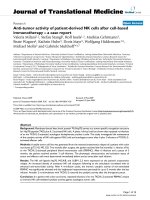
Figure 1 The Bioene rgetic Signature correlates with the cell-death response to chemotherapy . HCT116 cells were treated as indicated
[11] to produce cells with low (G-cells) (A), medium (M-cells) (B) and high (SM-cells) (C) bioenergetic signatures ( b-F1/GAPDH ratio). The rates of
aerobic glycolysis in G-, M- and SM-cells are also indicated. Cells were exposed to the following agents: 8 μM 3BrP, 100 μM IA, 10 μM 5-FU or
were left untreated (Control). Cells were double-stained with Hoechst 33342 and propidium iodide and visualized using fluorescence microscopy
at 20x magnification. The percentage of dead cells (red cells, PI positive) was determined by examination of different randomly selected fields.
Histograms shown (A-C) represent the means ± SEM of 10-25, 10-24 and 10-23 independent determinations in G-, M-, and SM-cells respectively.
*, P < 0.05 for multiple comparisons by ANOVA and post hoc Dunnett’s test. Plots in (D) illustrate the inverse (3BrP and IA) and direct (5-FU)
correlation that exists between the bioenergetic signature of the cells and the death-response to the chemotherapeutic agents.
Sánchez-Aragó and Cuezva Journal of Translational Medicine 2011, 9:19
/>Page 4 of 9
G cells
Control
3BrP
IA 5-FU
10
0
10
1
10
2
10
3
10
4
FL1-H
10
0
10
1
10
2
10
3
10
4
FL1-H
10
0
10
1
10
2
10
3
10
4
FL1-H
10
0
10
1
10
2
10
3
10
4
FL1-H
10
0
10
1
10
2
10
3
10
4
FL1-H
10
0
10
1
10
2
10
3
10
4
FL1-H
10
0
10
1
10
2
10
3
10
4
FL1-H
10
0
10
1
10
2
10
3
10
4
FL1-H
M cells
10
0
10
1
10
2
10
3
10
4
FL1-H
10
0
10
1
10
2
10
3
10
4
FL1-H
10
0
10
1
10
2
10
3
10
4
FL1-H
10
0
10
1
10
2
10
3
10
4
FL1-H
10
0
10
1
10
2
10
3
10
4
FL1-H
10
0
10
1
10
2
10
3
10
4
FL1-H
10
0
10
1
10
2
10
3
10
4
FL1-H
10
0
10
1
10
2
10
3
10
4
FL1-H
SM cells
10
0
10
1
10
2
10
3
10
4
FL1-H
10
0
10
1
10
2
10
3
10
4
FL1-H
10
0
10
1
10
2
10
3
10
4
FL1-H
10
0
10
1
10
2
10
3
10
4
FL1-H
10
0
10
1
10
2
10
3
10
4
FL1-H
10
0
10
1
10
2
10
3
10
4
FL1-H
10
0
10
1
10
2
10
3
10
4
FL1-H
10
0
10
1
10
2
10
3
10
4
FL1-H
A
B
0
10
20
30
BrP IA
5
-F
U
Apoptotic cell death
(fold change vs. control)
0
5
10
15
BrP IA FU
Necrotic cell death
(fold change vs. control)
C
Figure 2 The Bioenergetic Signature correlates with the cell-death pathway in response to chemotherapy . HCT116 cells were treated as
indicated [11] to produce cells with low (G-cells), medium (M-cells) and high (SM-cells) bioenergetic signatures. Cells were then treated with the
following agents: 8 μM 3BrP, 100 μM IA, 10 μM 5-FU or left untreated (Control). A, Representative FACS analysis of cells after annexinV-FITC (50
μg/mL) and propidium iodide (100 μg/mL) staining are shown. The lower left quadrant corresponds to viable cells; the lower right quadrant
early-apoptotic (annexin-positive) cells and the upper right and left quadrants corresponds to dead (PI positive) cells. Histograms shown are the
means ± SEM of the percentage of apoptotic (annexin V positive) (B) and necrotic (PI positive) cells (C) from 4-6 independent determinations in
G- (open bars), M- (closed bars) and SM-cells (hatched bars). *, p < 0.05 for multiple comparisons by ANOVA and post hoc Dunnett’s test.
0
0.3
0.6
0.9
1.2
Control 3BrP IA 5-FU
G
- cells
*
*
*
*
ATP levels
(f
old change vs.
C
ontrol
)
0
0.3
0.6
0.9
1.2
Control 3BrP IA 5-FU
*
*
*
*
ATP levels (fold change vs. Control)
M- cells
0
0.3
0.6
0.9
1.2
ATP levels (fold change vs. Control)
Co
n
t
r
o
l
3
BrP IA
5
-F
U
*
*
*
*
*
*
S
M- cells
ns
ns
Figure 3 Cellular ATP concentrations in response to chemotherapy . HCT116 cells were treated as indicated [11] to produce cells with low
(G-cells), medium (M-cells) and high (SM-cells) bioenergetic signature. Cells were then treated with the following agents: 8 μM 3BrP, 100 μM IA,
10 μM 5-FU or left untreated (Control) and ATP concentrations were determined. Histograms shown are means ± SEM. *, p < 0.05 compared to
controls by Student’s t test. ns, no significant.
Sánchez-Aragó and Cuezva Journal of Translational Medicine 2011, 9:19
/>Page 5 of 9
with a very large deple tion of cellular ATP concentra-
tions in all cell lineages (Figure 3), consistent with activa-
tion of necrosis by metabolic catastrophe in response to
treatment with these metabolic inhibitors. In contrast,
treatment of cells with 5-FU only marginally affected cel-
lular A TP c oncentration s in G- and M-cells (Figure 3)
and slightly, but significantly, promoted a 50% reduction
in cellular ATP concentrations in SM-cells (Fi gure 3),
indicating the absence of a compromised metabolic state
following 5-FU treatment.
Previous findings have suggested that only highly gly-
colytic G-cells are a ble to develop tumors in nude mice
[11]. Therefore, in order to test the in vivo tumor
regression activity of the metabolic inhibitors analyzed
in vitro, animals were implanted with G-cells. Animals
that developed ~ 1 cm
3
tumors were treated with daily
doses of 8 μM 3BrP, 100 μM IA or 0.5 mM 5-FU over
six consecutive days (Figure 4A). A control NaCl-treated
group was also included for comparison (Figure 4A).
Interestingly, from both a macroscopic (Figure 4B) and
behavioral point of view, all treatments tested (except
controls) seemed to affe ct the mice in a similar manner.
Specifically, control animals developed a rapid 2.5-fold
increase in tumor volume during the treatment period
(Figure 4A). In contrast, animals treated with either
5-FU or IA revealed a significant ~ 30% decrease in
tumor volume after 6 days of treatment (Figure 4A),
while maximum tumor regression (> 50%) was observed
in mice treated with 3BrP (Figure 4A), consistent with
the higher cell-death trend associated with 3BrP treat-
ment in vitro (Figure 1). However, the large differences
in cell death triggered by 3BrP and IA compared to
5-FU in G-type cells (Figure 1A) were largely absent fol-
lowing in vivo treatments despite the fact that the
tumors had a G-phenotype [11]. These results suggest
that additional mechanisms may play a role in promot-
ing tumor regression in vivo and that in vitro data
should be extrapolated with caution.
Discussion
In an effort to translate the bioenergetic signature to
clinical practice, we have recently developed monoclonal
antibodies against various markers of energetic metabo-
lism [32]. We found that cancer abolishes cell-type spe-
cific d ifferences in the bioenergetic signature [32],
supporting its use as a generic target to combat different
Body weight (g)
*
*
0
7
14
21
28
35
NaCl
5-FU
3BrP
IA
*
AB
Da
y
s of treatment
Relative Tumor Volume
NaCl (0.9 %)
3BrP (8 ȝM)
5-FU (0.5 mM)
IA (100 ȝM)
*
#
0
0.5
1.0
1.5
2.0
2.5
3.0
0123456
3BrP
5-FU
IA
*
*
Figure 4 Metabol ic inhibitors effectively promote tumor regression . HCT116 cells (10
7
cells per animal) with low bioenergetic signature (G-
cells) were injected into nude mice for tumor development. Twenty days after, when tumor volume reached ~1 cm
3
, the animals received daily
100 μL intraperitoneal injections, containing 8 μM 3BrP (n = 10, open square), 0.5 mM 5-FU (n = 5, grey square) or 100 μM IA (n = 7, closed
square) for six consecutive days. A 0.9% NaCl-treated control group (n = 6, hatched square) was also included for comparison. (A) Tumor volume
is presented normalized to its volume before initiation of the treatments. * and #, p < 0.05 when comparing 3BrP with 5-FU- and IA-treated
mice, respectively. **, p < 0.05 for multiple comparisons by ANOVA. Inserts provide representative examples of the differences in tumor size
compared to controls. (B) Mice body weight (g) after treatments. Results shown are means ± SEM. *, p < 0.05 when compared to NaCl-treated
controls by Student’s t test.
Sánchez-Aragó and Cuezva Journal of Translational Medicine 2011, 9:19
/>Page 6 of 9
neoplasias [32]. Indeed, b-F1-ATPase expression has
been shown to be a therapeu tic response marker in dif-
ferent cancer cell lines, both for single and combined
chemotherapy [19,21-24,33]. In the present study, we
document the cor relation between the bioenergetic sig-
nature of a cell, which represents an index of the rela-
tive relevance of cellular energy provision pathways
[9,11], and the potential to execute cell death in
response to the metabolic inhibitors, 3BrP and IA, and
the anti-metabolite 5-FU. The correlations observed in
this study cannot be ascribed to differences in the
genetic background of the cells because: (i) all of the
cells were derived from the same parental HCT116 cells
and (ii) the energetic metabolism of HCT116 cells is a
reversible phenotypic trait amenable to regulation
[11,34]. Furthermore, although some cancer cells can
oxidize glutamine for energy production purposes [3-5],
glutamine contributes very little to the energetic meta-
bolism of the highly glycolytic HCT116 cells used in
this study. In fact, oxygen consumption rates, aerobic
glycolysis rates and the bioenergetic signature of
HCT116 cells are not affected by the presence of gluta-
mine in the culture medium (see additional file 2).
Mechanistically, we propose that the cell death and
tumor regression observed following administration of
glycolytic inhibitors (3BrP and IA) may be due to induc-
tion of necrosis, whereas the cell death activity observed
upon 5-FU treatment may occur through apoptosis (Fig-
ures 2 and 3). This later finding is consistent with the
relevant roles played by oxidative phosphorylation [35]
and mitochondrial H
+
-ATP synthase activity [33,36] in
the efficient execution of cell death. Indeed, the bioener-
getic activity of mitochondria in colon cancer cells
[11,21] and tumors [19], has been associated with the
ability to execute a ROS-mediated cell death response
upon 5-FU treatment [11].
On the other hand, small alkylating agents have been
shown to be able to kill cancer cells resistant to apopto-
sisbyaprocessknownas“ programmed necrosis”
through depletion of NAD
+
via PARP1 activation [30].
However, the induction of necrosis in response to the
glycolytic inhibitors 3BrP and IA is exerted indepen-
dently of PARP1 processing (data not shown), and most
likely results from a metab olic catastrophe due to cellu-
lar ATP depletion (Figure 3) [23,24]. Overall, our studies
suggest that the enzymes of glycolysis could represent
therapeutic targets for t he treatment of colon cancer
thatmaybeaseffectiveasconventionaltreatments
(5-FU) at promoting tumor regression, in agreement
with findings by others [25,37].
The use of glycolytic inhibitors as chemotherapeutic
agents has pros and contras. One problem is the deleter-
ious effects that the se agents could trigger in cell types
strictly dependent on aerobic glycolysis for energy (e.g.
neurons, lymphocytes, erythrocytes, retina, renal
medulla, etc). However , glycolytic enzymes do have
highly specific active site residues that, in principle,
could provide more specific drug targets than those of
proteins involved in signal transduction pathways. Thus,
the use of such inhibitors may be beneficial in combina-
tion therapy as enhancers of the action of current che-
motherapeutic drugs [7,23]. Targeting energetic
metabolism might represent an alternative cancer treat-
ment route in the near future, because tumor cells that
are resistant to chemotherapy could effectively die by
necrosis in response to different metabolic inhibit ors.
Whatever the case, the bioenergetic signature offers a
reliable gauge to predict the cell death response (apop-
totic or necrotic cell death) to cancer therapy.
Conclusions
In summary, we have demonstrated that the bioenergetic
signature of colon cancer cells inversely c orrelates with
the potential to execute necrosis in response to treat-
ments with glycolytic inhibitors. In contrast, the bioener-
getic signature directly correlates with the apoptotic
response to 5-FU treatm ent. Overall, our results support
the use of the bioenergetic signature as a gauge for pre-
dicting cell death in response to different therapeutic
strategies in colon cancer.
Additional material
Additional file 1: The bioenergetic signature of HCT116-derived cell
lines. Representative western blot analysis. Representative western
blots of the expression of b-F1-ATPase, Hsp60 and GAPDH in two
different preparations (lanes 1-2) of (A) 2DG-treated (SM) and (B) OL-
treated (G) cells when compared to non-treated (M) HCT116 cells.
Additional file 2: Effect of glutamine (Gln) in the energetic
metabolism of HCT116 cells. (A) Representative western blots of the
expression of b- F1-ATPase, Hsp60 and GAPDH in two different
preparations of HCT116 cells grown in the presence (+) or absence (-) of
glutamine (Gln). The histogram illustrates the bioenergetic signature (b-F1/
GAPDH ratio) in the presence (open bar) or absence (closed bar) of
glutamine. (B) HCT116 cells were processed for the determination of the
rates of aerobic glycolysis in the presence (open bar) or absence (closed
bar) of glutamine. The rates of aerobic glycolysis were also determined
after the addition of 6 μM oligomycin (hatched bars). (C) Determination
of the rates of oxygen consumption. The results shown are the mean ±
SEM of 6-15 independent determinations. No statistical significant
differences were observed by Student’s t-test in any of the parameters
determined.
List of abbreviations
β-F1-ATPase: β catalytic subunit of the mitochondrial H
+
-ATP synthase;
bioenergetic signature: β-F1-ATPase/GAPDH ratio; GAPDH: Glyceraldehyde 3-
phosphate dehydrogenase; IA: iodoacetate; 3BrP: 3-bromopyruvate; 5-FU: 5-
fluorouracil; PI: propidium iodide; CRC: colorectal cancer.
Acknowledgements
We thank Mrs. M. Chamorro and C. Nuñez de Arenas for expert technical
assistance and J. Palacín (CBMSO) for his support in nude mice studies. This
work was supported by grants from the Ministerio de Educación y Ciencia
Sánchez-Aragó and Cuezva Journal of Translational Medicine 2011, 9:19
/>Page 7 of 9
(BFU2010-18903), the Centro de Investigación Biomédica en Red de
Enfermedades Raras (CIBERER), ISCIII, Madrid and Comunidad de Madrid (S-
GEN-0269), Spain. The CBMSO receives an institutional grant from the
Fundación Ramón Areces.
Authors’ contributions
MSA carried out experiments. MSA and JMC designed experiments and
wrote the manuscript. All authors read and approved the final manuscript.
Competing interests
JMC as inventor and the Universidad Autónoma de Madrid hold the
following patents on “the bioenergetic signature of cancer”, which has been
licensed to Fina Biotech, S.L. (Spain): US 10/514.771, Japanese 4235610,
Canadian 2,487,176 and EU 03 727 509.6. MSA declares no competing
interests.
Received: 6 September 2010 Accepted: 8 February 2011
Published: 8 February 2011
References
1. Gellad ZF, Provenzale D: Colorectal cancer: national and international
perspective on the burden of disease and public health impact.
Gastroenterology 2010, 138:2177-2190.
2. Cunningham D, Atkin W, Lenz HJ, Lynch HT, Minsky B, Nordlinger B,
Starling N: Colorectal cancer. Lancet 2010, 375:1030-1047.
3. Cuezva JM, Ortega AD, Willers I, Sanchez-Cenizo L, Aldea M, Sanchez-
Arago M: The tumor suppressor function of mitochondria: Translation
into the clinics. Biochim Biophys Acta 2009, 1792:1145-1158.
4. DeBerardinis RJ, Lum JJ, Hatzivassiliou G, Thompson CB: The biology of
cancer: metabolic reprogramming fuels cell growth and proliferation.
Cell Metab 2008, 7:11-20.
5. Kroemer G, Pouyssegur J: Tumor cell metabolism: cancer’s Achilles’ heel.
Cancer Cell 2008, 13:472-482.
6. Tennant DA, Duran RV, Gottlieb E: Targeting metabolic transformation for
cancer therapy. Nat Rev Cancer 2010, 10:267-277.
7. Pelicano H, Martin DS, Xu RH, Huang P: Glycolysis inhibition for anticancer
treatment. Oncogene 2006, 25:4633-4646.
8. Diehn M, Cho RW, Lobo NA, Kalisky T, Dorie MJ, Kulp AN, Qian D, Lam JS,
Ailles LE, Wong M, et al: Association of reactive oxygen species levels and
radioresistance in cancer stem cells. Nature 2009, 458:780-783.
9. Lopez-Rios F, Sanchez-Arago M, Garcia-Garcia E, Ortega AD, Berrendero JR,
Pozo-Rodriguez F, Lopez-Encuentra A, Ballestin C, Cuezva JM: Loss of the
mitochondrial bioenergetic capacity underlies the glucose avidity of
carcinomas. Cancer Res 2007, 67:9013-9017.
10. Cuezva JM, Krajewska M, de Heredia ML, Krajewski S, Santamaria G, Kim H,
Zapata JM, Marusawa H, Chamorro M, Reed JC: The bioenergetic
signature of cancer: a marker of tumor progression. Cancer Res 2002,
62:6674-6681.
11. Sanchez-Arago M, Chamorro M, Cuezva JM: Selection of cancer cells with
repressed mitochondria triggers colon cancer progression. Carcinogenesis
2010, 31:567-576.
12. Bi X, Lin Q, Foo TW, Joshi S, You T, Shen HM, Ong CN, Cheah PY, Eu KW,
Hew CL: Proteomic analysis of colorectal cancer reveals alterations in
metabolic pathways: mechanism of tumorigenesis. Mol Cell Proteomics
2006, 5:1119-1130.
13. Chen J, Kahne T, Rocken C, Gotze T, Yu J, Sung JJ, Chen M, Hu P,
Malfertheiner P, Ebert MP: Proteome analysis of gastric cancer metastasis
by two-dimensional gel electrophoresis and matrix assisted laser
desorption/ionization-mass spectrometry for identification of metastasis-
related proteins. J Proteome Res 2004, 3:1009-1016.
14. Cuezva JM, Chen G, Alonso AM, Isidoro A, Misek DE, Hanash SM, Beer DG:
The
bioenergetic signature of lung adenocarcinomas is a molecular
marker of cancer diagnosis and prognosis. Carcinogenesis 2004,
25:1157-1163.
15. He QY, Chen J, Kung HF, Yuen AP, Chiu JF: Identification of tumor-
associated proteins in oral tongue squamous cell carcinoma by
proteomics. Proteomics 2004, 4:271-278.
16. Isidoro A, Casado E, Redondo A, Acebo P, Espinosa E, Alonso AM, Cejas P,
Hardisson D, Fresno Vara JA, Belda-Iniesta C, et al: Breast carcinomas fulfill
the Warburg hypothesis and provide metabolic markers of cancer
prognosis. Carcinogenesis 2005, 26:2095-2104.
17. Isidoro A, Martinez M, Fernandez PL, Ortega AD, Santamaria G,
Chamorro M, Reed JC, Cuezva JM: Alteration of the bioenergetic
phenotype of mitochondria is a hallmark of breast, gastric, lung and
oesophageal cancer. Biochem J 2004, 378:17-20.
18. Unwin RD, Craven RA, Harnden P, Hanrahan S, Totty N, Knowles M,
Eardley I, Selby PJ, Banks RE: Proteomic changes in renal cancer and co-
ordinate demonstration of both the glycolytic and mitochondrial
aspects of the Warburg effect. Proteomics 2003, 3:1620-1632.
19. Lin PC, Lin JK, Yang SH, Wang HS, Li AF, Chang SC: Expression of beta-F1-
ATPase and mitochondrial transcription factor A and the change in
mitochondrial DNA content in colorectal cancer: clinical data analysis
and evidence from an in vitro study. Int J Colorectal Dis 2008,
23:1223-1232.
20. Ortega AD, Sala S, Espinosa E, Gonzalez-Baron M, Cuezva JM: HuR and the
bioenergetic signature of breast cancer: a low tumor expression of the
RNA-binding protein predicts a higher risk of disease recurrence.
Carcinogenesis 2008, 29:2053-2061.
21. Shin YK, Yoo BC, Chang HJ, Jeon E, Hong SH, Jung MS, Lim SJ, Park JG:
Down-regulation of mitochondrial F1F0-ATP synthase in human colon
cancer cells with induced 5-fluorouracil resistance. Cancer Res 2005,
65:3162-3170.
22. Li RJ, Zhang GS, Chen YH, Zhu JF, Lu QJ, Gong FJ, Kuang WY: Down-
regulation of mitochondrial ATPase by hypermethylation mechanism in
chronic myeloid leukemia is associated with multidrug resistance. Ann
Oncol 2010, 7:1506-1514.
23. Hernlund E, Hjerpe E, Avall-Lundqvist E, Shoshan M: Ovarian carcinoma
cells with low levels of beta-F1-ATPase are sensitive to combined
platinum and 2-deoxy-D-glucose treatment. Mol Cancer Ther 2009,
8:1916-1923.
24. Hernlund E, Ihrlund LS, Khan O, Ates YO, Linder S, Panaretakis T,
Shoshan MC: Potentiation of chemotherapeutic drugs by energy
metabolism inhibitors 2-deoxyglucose and etomoxir. Int J Cancer 2008,
123:476-483.
25. Geschwind JF, Ko YH, Torbenson MS, Magee C, Pedersen PL: Novel therapy
for liver cancer: direct intraarterial injection of a potent inhibitor of ATP
production. Cancer Res 2002, 62:3909-3913.
26. Bickis IJ, Quastel JH: Effects of Metabolic Inhibitors on Energy Metabolism
of Ehrlich Ascites Carcinoma Cells. Nature 1965, 205:44-46.
27. Ganapathy-Kanniappan S, Geschwind JF, Kunjithapatham R, Buijs M,
Vossen JA, Tchernyshyov I, Cole RN, Syed LH, Rao PP, Ota S, Vali M:
Glyceraldehyde-3-phosphate dehydrogenase (GAPDH) is pyruvylated
during 3-bromopyruvate mediated cancer cell death. Anticancer Res 2009,
29:4909-4918.
28.
Pigault C, Follenius-Wund A, Schmutz M, Freyssinet JM, Brisson A:
Formation of two-dimensional arrays of annexin V on
phosphatidylserine-containing liposomes. J Mol Biol 1994, 236:199-208.
29. Jaattela M: Multiple cell death pathways as regulators of tumour
initiation and progression. Oncogene 2004, 23:2746-2756.
30. Zong WX, Ditsworth D, Bauer DE, Wang ZQ, Thompson CB: Alkylating DNA
damage stimulates a regulated form of necrotic cell death. Genes Dev
2004, 18:1272-1282.
31. Munoz-Pinedo C, Ruiz-Ruiz C, Ruiz de Almodovar C, Palacios C, Lopez-
Rivas A: Inhibition of glucose metabolism sensitizes tumor cells to death
receptor-triggered apoptosis through enhancement of death-inducing
signaling complex formation and apical procaspase-8 processing. J Biol
Chem 2003, 278:12759-12768.
32. Acebo P, Giner D, Calvo P, Blanco-Rivero A, Ortega AD, Fernandez PL,
Roncador G, Fernandez-Malave E, Chamorro M, Cuezva JM: Cancer
abolishes the tissue type-specific differences in the phenotype of
energetic metabolism. Transl Oncol 2009, 2:138-145.
33. Santamaria G, Martinez-Diez M, Fabregat I, Cuezva JM: Efficient execution
of cell death in non-glycolytic cells requires the generation of ROS
controlled by the activity of mitochondrial H+-ATP synthase.
Carcinogenesis 2006, 27:925-935.
34. Rossignol R, Gilkerson R, Aggeler R, Yamagata K, Remington SJ, Capaldi RA:
Energy substrate modulates mitochondrial structure and oxidative
capacity in cancer cells. Cancer Res 2004, 64:985-993.
35. Tomiyama A, Serizawa S, Tachibana K, Sakurada K, Samejima H, Kuchino Y,
Kitanaka C: Critical role for mitochondrial oxidative phosphorylation in
the activation of tumor suppressors Bax and Bak. J Natl Cancer Inst 2006,
98:1462-1473.
Sánchez-Aragó and Cuezva Journal of Translational Medicine 2011, 9:19
/>Page 8 of 9
36. Matsuyama S, Xu Q, Velours J, Reed JC: The Mitochondrial F0F1-ATPase
proton pump is required for function of the proapoptotic protein Bax in
yeast and mammalian cells. Mol Cell 1998, 1:327-336.
37. Fantin VR, St-Pierre J, Leder P: Attenuation of LDH-A expression uncovers
a link between glycolysis, mitochondrial physiology, and tumor
maintenance. Cancer Cell 2006, 9:425-434.
doi:10.1186/1479-5876-9-19
Cite this article as: Sánchez-Aragó and Cuezva: The bioenergetic
signature of isogenic colon cancer cells predicts the cell death response
to treatment with 3-bromopyruvate, iodoacetate or 5-fluorouracil.
Journal of Translational Medicine 2011 9:19.
Submit your next manuscript to BioMed Central
and take full advantage of:
• Convenient online submission
• Thorough peer review
• No space constraints or color figure charges
• Immediate publication on acceptance
• Inclusion in PubMed, CAS, Scopus and Google Scholar
• Research which is freely available for redistribution
Submit your manuscript at
www.biomedcentral.com/submit
Sánchez-Aragó and Cuezva Journal of Translational Medicine 2011, 9:19
/>Page 9 of 9