Advances in Lasers and Electro Optics Part 18 potx
Bạn đang xem bản rút gọn của tài liệu. Xem và tải ngay bản đầy đủ của tài liệu tại đây (846.85 KB, 8 trang )
Application of Ultrafast Laser Optoperforation for Plant Pollen Walls and Endothelial Cell Membranes
831
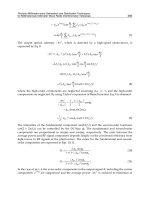
Fig. 17. Representation of retinal segments irradiated with a single-shot ultrafast lasers. They
were tentatively grouped into three types of lesions:
A. No change, B. The ablations at the
ILM and
C. The optoperforation of blood vessel walls. The arrow head indicates the point
of irradiations on the blood vessels.
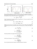
Fig. 18. Linear plot of the percent probability for inner limiting membrane (ILM) damage
(solid rectangles) and vessel perforation (solid circles) as a function of the laser fluence. The
ablation threshold fluence for ILM and blood vessels was found to be 2.19 ± 1.08 J/cm
2
and
5.85 ± 1.49 J/cm
2
, respectively. With increasing fluence, the percent probability of blood
vessel perforation monotonically increases. The lines represent an extrapolation to
determine the ablation thresholds for perforation of retinal primary blood vessels and for
ILM damage of a porcine eye.
4.3 Discussion
Recent development in advanced laser technology transiently facilitates to perform
transaction, ablation, and coagulation of tissues via delivery of laser irradiation into a small
focal volume are providing an attractive possibilities for new laser surgical technologies.
The laser beam is a potential candidate that has already undergone a multi-center clinical
Advances in Lasers and Electro Optics
832
trial to evaluate the feasibility for its use in vitreoretinal surgery (Schastak et al., 2007).
Limited precision and significant damage by lasers with relative long pulse durations does
not allow partial or selective tissue ablation with high precision. If such damage is to be
overcome, infrared laser sources, such as CO
2
, Er:YAG and Holmium:YAG lasers, have
undergone several trials via optical fiber delivery in intraocular surgery. However, apparent
collateral damage to surrounding tissue due to significant thermal and shockwave effects
have been reported (Paula- Yu et al., 2006).
Laser ablation of tissues could be described using either an optical breakdown model or a
thermal confinement models. The optical breakdown model considers plasma formation
and subsequent shock wave formation, cavitation, and tissue disruption. The thermal
confinement model recognizes the competing thermal effects of the vaporization of water
driving an explosive ablation and thermal diffusion leading to collateral damage. This
model accounts for the observation that collateral damage is limited if the pulse duration is
less than the thermal relaxation time of the ablated tissue volume (Vogel et al., 2003; Apitz et
al., 2005).
Fig. 19. Interplay of photoionization, inverse Bremsstrahlung absorption, and impact
ionization in the process of plasma formation. Recurring sequences of inverse
Bremsstrahlung absorption events and impact ionization lead to an avalanche growth in the
number of free electrons. (Vogel et al., 2005)
The process of plasma formation through laser induced breakdown in transparent biological
media is schematically depicted in Fig. 19. It essentially consists of the formation of quasi-
free electrons by interplay of photoionization and avalanche ionization. It’s a well known
fact that the optical breakdown threshold in water is very similar to that in ocular and other
biological media (Docchio et al., 1986). Irradiation by an intense ultrafast laser beam further
leads to multiphoton excitation of a target material. The absorbed energy might be
transported to the electrons without thermal diffusion to adjacent material because the pulse
width is shorter than the vibrational relaxation time constant of several picoseconds. As a
result, thermal damage on the surrounding tissues could be minimized, and the biological
tissue remains unaffected by the subsequent photoinduced mechanical shock process. This
Application of Ultrafast Laser Optoperforation for Plant Pollen Walls and Endothelial Cell Membranes
833
effectively renders the fs-laser surgical process non-thermal. The formation of a high density
of free electrons could result in a local plasma formation in the targeted materials. This hot
plasma formation results in a permanently damaged region, even inside a cell with a sub-
micron size (Vogel et al., 2005). Furthermore, a previous on tissues like the corneal stroma
revealed that the ablation threshold fluence decreased with increasing pulse width of the
applied laser (Preuss et al., 1995). These uniquely show that ultrafast lasers can be utilized
for precise treatment of tissues while minimizing any apparent thermal damage or shock
pressure to biological tissues (Kohli et al., 2005). The results illustrated in the current work
made the above hypothesis true for the retinal tissues, where retinal blood vessels were
selectively perforated with wide range of laser fluence (1.42 ~ 99.4 J/cm
2
) with an ultra fast
laser in near infra red region.
From the past literature values for the ablation thresholds for various tissues, including the
corneal stroma, axons, the eye’s anterior chamber, and hard tissue (under a single-shot
configuration, as in current work), the ablation threshold of the corneal stroma for an
ultrafast laser is in the range of 1 J/cm
2
to 2 J/cm
2
. Meanwhile, the ablation threshold for
axons of C. elegans is reported to be about 4.4 J/cm
2
. It is of great interest to note that the
value for the femtosecond laser ablation threshold of the ILM of the porcine retina, 2.19 ±
1.08 J/cm
2
as determined in the current work, is in the same range of reported values for the
soft tissues. It is also interesting to compare the ablation threshold of the retina upon
irradiation by a femtosecond laser to the values for irradiation with an ultraviolet (UV) laser
with a nanosecond pulse width, including ArF excimer lasers and higher-harmonic Nd:YAG
lasers. The ablation threshold is reported to be in the range of between 0.6 J/cm
2
and 1
J/cm
2
when irradiating single-pulsed UV light into the retina tissue, which is slightly lower
than that for femtosecond laser ablation threshold. Considering the remarkable difference in
the linear optical absorption coefficients of the retina tissue in the UV and the NIR ranges, it
is reasonable to suppose that an ultrafast laser operating in the NIR region would be able to
ablate the ILM layer in the retina with much lower deposited energy per unit volume
compared to UV nanosecond lasers. The perforation threshold of the underlying primary
retinal blood vessels (5.85 ± 1.49 J/cm
2
) is significantly higher than the literature values.
The thickness of the ILM, which is essentially a basement membrane consists of retina
müller cells, is only 6 µm to 10 µm. The thickness of the ILM is thinnest at the fovea region
of the retina. However, the thickness is larger at the posterior pole of retina (Hoerauf et al.,
2006). Furthermore, the ILM is also present over the retinal blood vessels. If only the ILM is
to be ablated selectively without any alterations in the underlying layers, the energy
delivered by the laser irradiation must be confined in thin layers without any apparent
diffusion of the deposited energy into other parts of the retina. To evoke this topic, we have
examined the dependence of the ablation depth for transparent materials, like retinal tissue,
on the laser fluence (Fig. 20). If there is high free electron density due to optical absorption
processes, we suppose that the underlying mechanism for the ablation by fs-laser irradiation
is not directly governed by the optical and the electronic properties of the materials. Even if
the absorption mechanism of the NIR fs-laser is dependent on the optical band gap of each
material, two different slopes under fs laser irradiation have already been reported for
metals, semiconductors, and dielectrics (Nolte et al., 1997; Furusawa et al., 1999). For a lower
laser fluence, F, the ablation depth can be described by the expression L = δln(F/F
th
(
δ
)
),
where δ is the optical penetration depth and F
th
(
δ
)
is the threshold laser fluence of ablation
[Preuss et al., 1995, Jia et al., 2006]. A fit to the experimental data results in F
δ
th
= 2.2 ± 0.9
Advances in Lasers and Electro Optics
834
J/cm
2
and δ = 8.2 ± 2.2 μm. It should be notified that the optical penetration depth is
governed by a nonlinear optical transition, if multi-photon absorption plays an important
role in photo-excitation of the materials. Therefore, the optical penetration depth estimated
from the current work is difficult to reconcile with the literature value of the optical
absorbance of retina tissue at a wavelength of 810 nm. Due to the strong dependence of the
multiphoton absorption on the energy density, the value of δ should be relatively small. At
any rate, it is of great interest to compare the observed optical penetration depth with the
thickness of the ILM in the porcine retina. This comparison led us to propose that the energy
delivered by femtosecond laser irradiation under the controlled laser fluence can be
confined in the ILM layer, followed by a selective ablation of the layers only if the optical
penetration depth of 8.2 ± 2.2 μm is comparable to the thickness of the ILM of the retina.
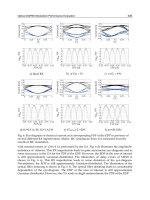
Fig. 20. The lesion depth of a porcine retina caused by fs-laser irradiation as a function of the
laser fluence. The blue solid and the red dotted lines represent linear fit. About 300
sectioned slices from more than 10 eyeballs were examined for each laser fluence.
With increasing laser fluence, however, the mechanism underlying the retina ablation can
no longer be expressed by the optical penetration depth. As shown in Figure 16, the retina
surface treated with a high laser fluence of 99.4 J/cm
2
is very much roughened compared to
the surface treated with a low fluence of 7.1 J/cm
2
. Based on the changes in the slopes of the
semi-logarithmic plot of the ablation depth as function of the fluence, we have supposed
that at laser fluence higher than 25.3 J/cm
2
, the electronic heat diffusion process plays an
important role, even in an ultrafast laser ablation. The ablation depth in this region can be
described with the expression of L = l ln(F/F
th
(l)
), where l is the electronic heating depth, and
F
th
(l)
is the corresponding threshold fluence. The electronic heating depth and F
th
(l)
are
estimated to be 69.7 ± 8.7 μm and 25.3 ± 13.9 J/cm
2
, respectively, which means that the
thickness of the retina tissue affected by fs-laser irradiation might be abruptly increased for
Application of Ultrafast Laser Optoperforation for Plant Pollen Walls and Endothelial Cell Membranes
835
the laser fluence higher than 25.3 J/cm
2
. As a result, we have to control the laser fluence
very precisely to achieve a selective peeling of the ILM layer without any visible thermal
damage being induced by the laser irradiation.
The probability of retina blood vessel damage shows a linear relationship with the laser
fluence. With the progressive increase in the laser fluence, selective ablations of concerted
retina layers even including primary blood vessels is possible without any apparent damage
to the underlying layers of the porcine retina. The threshold fluence to perforate the walls of
the primary blood vessels embedded in the porcine retina is estimated to be 5.85
± 1.49
J/cm
2
. If the ablation depth depends on the laser fluence as δln(F/F
th
(
δ
)
), the thickness of the
tissues ablated by a single-shot fs-laser pulse can be estimated to be 8.0 ± 3.0 μm, by using
the parameters of δ and F
th
(
δ
)
from this work. Meanwhile, the thickness of the tissues
covering the primary blood vessels is tentatively determined to be about 25 μm by
examining the sectioned slices shown in Fig. 15. If the current interpretation for the ablation
depth of the tissues by fs-laser irradiation is correct, the fluence to perforate the primary
blood vessels should be about 46 J/cm
2
. However, the ablation depth per pulse in the high-
laser-fluence region should be described in terms of electronic heating depth with the
relation of L = δln(F/F
th
(l)
). With the parameters of l and F
th
(l)
, we are able to estimate the
fluence to fully perforate the primary blood vessels of the retina to be 36.2 J/cm
2
. This value
for blood vessel perforation is very close to the laser fluence at 1/e
2
percent perforation
probability, as shown in Fig. 18.
5. Conclusion
In summary, all the observations from the present work reveals that fs-laser irradiation on
pollen walls to make an evident physical hole with an outside diameter of about 1 μm well
conserves the physiological state of the cell including its viability and pollen tube
germination capability. Furthermore, from the successful delivery of foreign DNA into
pollen through the hole reveals that the current method has an evident potential in the field
of plant genetic engineering.
Topographical imaging as well as optical imaging of the plasma membranes led us to
observe a self-healing process for live cells within several minutes of time after the fs-laser
ablation on the live cells. A simple viscoelastic model for both the hole opening and closing
process was found to be applicable to interpret its dynamics. The very slow dynamics could
be explained in terms of high surface viscosity due to the presence of cytoskeleton network
bound to the plasma membrane. The irregular feature in plasma topography observed in the
final stage of the healing process might be due to a slice of the assembled lipid, which
resulted from the reconstruction of not only the plasma membrane itself but also F-actin
network as a cytoskeleton structure of live cells. Although two-dimensional plug flow
model adapted in the current work fairly well interpret the experimental observations in
macroscopically, the presence of transmembrane proteins, transbilayer interactions, and
adhesion sites, etc., in addition to the bound cytoskeleton structure, produces a variety of
restrictions on the flow dynamics of the plasma membrane through an alterations in many
microscopic physico-chemical properties including thickness and hydrodynamic properties
of the fluidic films.
We have developed a new method for elucidating more exact mechanism on the interesting
topic of self-healing process based on ultrafast laser perforation of the plasma membrane of
the animal cell. A mechanical stimulus to live-cell plasma membrane by the induced surface
Advances in Lasers and Electro Optics
836
tension as well as surface line energy can be also applied by the current methods with high
spatial resolution and unattainable speed of perforation. So interesting is the spatiotemporal
characterization of the plasma membrane movement associated with the healing process
that is closely related with the cell migration and transmission of the mechanical stimuli into
biochemical signals, which might be mainly governed by cytoskeleton structure (Wang et
al., 2005; Yamazaki et al., 2005; Supatto et al., 2005).
We have also successfully applied the current fs- laser technology to selectively perforate the
retinal blood vessels without any apparent damage in the concerted retina layers. It
provides a major breakthrough for the retinal vein occlusion therapy and removal of
abnormal blood vessels (Choroidal Neovascularization (CNV)) grown during numerous
retinal diseases.
6. Acknowledgements
This work was financially supported by the Ministry of Knowledge Economy of Korea and
KRISS program.
7. References
Apitz, I. & Vogel, A. (2005) Material ejection in nanosecond Er: YAG laser ablation of water,
liver, and skin, Appl. Phys. A 81, 329–338
Aronen, T. S.; Nikkanen, T. O. & Haggman, H. M. (2003). The production of transgenic Scots
pine (Pinus sylvestris L.) via the application of transformed pollen in controlled
crossing, Transgenic Res. 12, 375-378
Benkert, R.; Obermeyer, G. & Bentrup, F. W. (1997). The turgor pressure of growing lily
pollen tubes, Protoplasm 198, 1-8
Debregeas, G.; Martin, P. & Brochard-Wyart, F. (1995). Viscous bursting of suspended films,
Phys. Rev. Lett. 75, 3886-3889
Docchio, F.; Sachhi, C. A. & Marshall, J. (1986) Experimental investigation of optical
breakdown thresholds in ocular media under single pulse irradiation with different
pulse durations, Lasers Ophthalmol. 1, 83-91
Engelman, D. M. (2005). Membrane are more mosaic than fluid, Nature 438, 578-570
Fernado, D. D.; Richards, J. L. & Kikkert, J. R. (2006). In vitro germination and transient GFP
expression of American chestnut (Castanea dentate) pollen, Plant Cell Rep. 25, 450-
456
Furusawa, K.; Takahashi, K.; Kumagai, H.; Midorikawa, K. & Obara, M. (1999) “Ablation
characteristics of Au, Ag, and Cu metals using a femtosecond: Ti Sapphire laser,
Appl. Phy. A. 69(7), S359-S366
Gonzalez-Serratos, H.; Rozycka, M.; Cordoba-Rodriguez, R. & Ortega, A. (1996). Membrane
healing and restoration of contractility after mechanical injury in isolated skeletal
muscle fibers of the frog, Proc. Natl. Acad. Sci. USA 93, 5996-6001
Greulich, K. O. & Weber, G. (1992). The light microscope on its way from an analytical to a
preparative tool, J. Microsc. 167, 127-151
Heilbrunn, L. V. (1956). The surface precipitation reaction, In: The Dynamics of Living
Protoplasm 62-84, Academic, New York
Higashiyama, T.; Yabe, S.; Sasaki, N.; Nishimura, Y.; Miyagishima, S.; Kuroiwa, H. & Kuroiwa,
T. (2001). Pollen Tube Attraction by the Synergid Cell, Science 293, 1480-1483
Application of Ultrafast Laser Optoperforation for Plant Pollen Walls and Endothelial Cell Membranes
837
Hoerauf, H.; Brix, A.; Winkler, J.; Droege, G.; Winter, C.; Birngruber, R.; Laqua, H.; and
Vogel, A. (2006) A Photoablation of inner limiting membrane and inner retinal
layers using the Erbium: YAG-laser: An in vitro study, Lasers Surg. Med. 38(1), 52-61
Hoffmann, F. (1996). Laser microbeams for the manipulation of plant cells and subcellular
structures, Plant Science 113, 1-11
Jeoung, S. C.; Kim, H. S.; Park, M. I.; Lee, J.; Kim, C. S. & Park, C. O. (2005). Preparation of
room-temperature photoluminescent nanoparticles by ultrafast laser processing of
single-crystalline Ge, Jap. J. Appl. Phys. 44, 5278-5281
Jia, T. Q.; Chen, H. X.; Huang, M.; Zhao, F. L.; Li, X. X.; Xu, S. Z.; Sun, H. Y.; Feng, D. H.; Li,
C. B.; Wang, X. F.; Li, R. X.; Xu, Z. Z.; He, X. K. and Kuroda, H. (2006) Ultraviolet-
infrared femtosecond laser-induced damage in fused silica and CaF
2
crystals Phys.
Rev. B 73 (5) 054105-1 - 054105-9
Kobayashi, N.; Rivas-Carrillo, J. D.; Soto-Gutierrez, A.; Fukazawa, T., Chen, Y.; Navarro-
Alvarez, N. & Tanaka, N. (2005). Gene delivery to embryonic stem cells, Birth
Defects Research (Part C) 75, 10-18
Kohli, V.; Elezzabi, A. Y.; & Acker, J. P. (2005) Cell nanosurgery using ultrashort
(femtosecond) laser pulses: applications to membrane surgery and cell isolation,
Lasers Surg. Med. 37, 227–230
König, K.; Riemann, I.; Fischer, P. & Halbhuber, K. J. (1999). Intracellular nanosurgery with
near infrared femtosecond laser pulses, Cell. Mol. Biol. 45, 195-201
Krautwig, B & Lörz, H. (1995). Cereal protoplasts, Plant Science 111, 1-10
Lee, Y. J.; Kim, D. H.; Kim, Y. & Hwang, I. (2001). Identification of a signal that distinguishes
between the chloroplast outer envelope membrane and the endomembrane system
in vivo,” Plant Cell 13, 2175–2190
Lovy-Wheeler, A.; Cardenas, L.; Kunkel, J. G. & Hepler, P. K. (2007). Differential organelle
movement on the actin cytoskeleton in lily pollen tubes. Cell Motil Cytoskeleton. 64,
217-232
Nelson, J. S. & Berm, M. W. (1989). Laser application in biomedicine. Part II: Clinical
applications, J. Laser Appl. 1, 9-20
Nolte, S.; Momma, C.; Jacobs, H.; Tünnermann, A.; Chichkov, B. N.; & Wellegehausen, B.
(1997) Ablation of metals by ultrashort laser pulses , J. Opt. Soc. Am. B 14, 2716-2722
Oliver, J. M.; King, J. R.; Mckinlay, K. J.; Brown, P. D.; Grant, D. M.; Scotchford, C. A. &
Wood, J. V. (2005). Thin-film theories for two-phase reactive flow models of active
cell motion, Mathematical Medicine and Biology 22, 53-98
Parpura, V.; Haydon, P. G. & Henderson, E. (1993). Three-dimensional imaging of living
neurons and glia with the atomic force microscope, J. Cell Sci. 104, 427-432
Paula-Yu, K.; Miller, J.; Cringle, S. J.; & Yu, D-Y. (2006) Experimental retinal ablation using a
fourth-harmonic 266 nm laser coupled with an optical fiber probe, Invest.
Ophthalmol. Vis. Sci. 47(4), 1587-1593.
Preuss, S.; Demchuk, A.; & Stuke, M. (1995) Sub-picosecond UV laser ablation of metals,
Appl. Phys. A 61, 33–37
Sandre, O.; Moreaux, L. & Brochard-Wyart, F. (1999). Dynamics of transient pores in
stretched vesicles, Proc. Natl. Acad. Sci. USA 96, 10591-10596
Schastak, S.; Yafai, Y.; Yasukawa, T.; Wang, Y. S.; Hillrichs, G. & Wiedemann, P. (2007)
Flexible UV light guiding system for intraocular laser microsurgery, Lasers Surg.
Med. 39, 353-357
Senz, R. & Miiller, G. (1989). Laser in Medicine, Ber. Bunsenges. Phys. Chem. 93, 269 –277
Advances in Lasers and Electro Optics
838
Shen, N.; Datta, D.; Schaffer, C. B.; LeDuc, P.; Ingber, D. E. & Mazur, E. (2005). Ablation of
cytoskeletal filaments and mitochondria in live cells using a femtosecond laser
nanoscissor, Mechanics and Chemistry of Biosystems 2, 17-26
Sidhu, M. S.; Kim, E. K; Woo, S. Y; Song. M. C.; Jeoung, S. C. & Park, Y. I. (2009)
Femtosecond – laser - assisted optoperforation of the primary retinal blood vessel
and retina tissue of porcine eyes, J. Kor. Phys. Soc. 55(2) (in Press)
Singer, S. J. & Nicolson, G. L. (1972). The fluid mosaic model of the structure of cell
membranes, Science 175, 720-731
Strubinska, J. & Sniezko, R. (2000). Localization of vegetative nucleus and generative cell
nuclei in branching pollen tubes of Oenothera hookeri L. grown in vitro, Acta
Biologica Cracoviensia Series Botanica 42, 107-112
Supatto, W.; Debarre, D.; Moulia, B.; Brouzes, E.; Martin, J. L.; Farge, E. & Beaurepaire, E.
(2005). In vivo modulation of morphogenetic movements in Drosophila embryos
with femtosecond laser pulses, Proc. Natl. Acad. Sci. USA 102, 1047-1052
Tang, W.; Weidner, D. A.; Hu, B. Y.; Newton, R. J. & Hu, X. H. (2006). Efficient delivery of
small interfering RNA to plant cells by a nanosecond pulsed laser-induced stress
wave for posttranscriptional gene silencing, Plant Science 171, 375-381
Taylor, L. P. & Hepler, P. K. (1997). Pollen germination and tube growth, Anuu. Rev. Plant
Physiol. Plant Mol. Biol. 48, 461-491
Tirlapur, U. K. & König, K. (2002). Targeted transfection by femtosecond laser, Nature 418,
290-291
Touraev, A.; Stoger, E.; Voronin, V. & Heberle-Bors, E. (1997). Plant male germ line
transformation, Plant Journal 12, 949-956
Van der Leede-Plegt, L. M.; van den Ven, B. C. E.; Schilder, M.; Franker, J. & van Tunen, A. J.
(1995). Development of a pollen-mediated transformation method for Nicotiana
glutinosa, Transgenic Res. 4, 77-86
Velegol, S. B.; Pardi, S.; Li, X.; Velegol, D. & Logan, B. E. (2003). AFM imaging artifacts due
to bacterial cell height and AFM tip geometry, Langmuir 19, 851-857
Vervaeke, I.; Londers, E.; Piot, G.; Deroose, R. & Deproft, M. P. (2005). The division of the
generative nucleus and the formation of callose plugs in pollen tubes of Aechmea
fasciata (Bromeliaceae) cultured in vitro. Sexual plant reproduction 18, 9-19
Vogel, A. & Venugopalan, V. (2003) Mechanism of pulsed laser ablation of biological tissues,
Chem. Rev. 103, 577-644
Vogel, A.; Noack, J.; Huttman, G. & Paltauf, G. (2005) Mechanism of femtosecond laser
nanosurgery of cells and tissues, Appl. Phys. B 81, 1015–1047
Wang, Y.; Botvinick, E. L.; Zhao, Y.; Berns, M. W.; Usami, S.; Tsien, R. Y. & Chien, S. (2005).
Visualizing the mechanical activation of Src, Nature 434, 1040-1045
Yahng, J. S.; Jeoung, S. C.; Choi, D. S.; Cho, D.; Kim, J. H.; Choi, H. M. & Paik, J. S. (2005).
Fabrication of microfluidic devices by using a femtosecond laser micromachining
techniques and μ-PIV studies on its fluid dynamics, J. Korean Phys. Soc. 47, 977-981
Yamazaki, D.; Kurisu, S. & Takenawa, T. (2005). Regulation of cancer cell motility through
actin reorganization, Cancer. Sci. 96, 379-386
Zeira, E.; Manevitch, A.; Khatchatouriants, A.; Pappo, O.; Hyam, E.; Darash-Yahana, M.;
Tavor, E.; Honigman, A.; Lewis, A. & Galun, E. (2003). Femtosecond infrared laser -
An efficient and safe in vivo gene delivery system for prolonged expression,
Molecular Therapy 8, 342-350