báo cáo hóa học: " Single-step processing of copper-doped titania nanomaterials in a flame aerosol reactor" ppt
Bạn đang xem bản rút gọn của tài liệu. Xem và tải ngay bản đầy đủ của tài liệu tại đây (1.67 MB, 14 trang )
NANO EXPRESS Open Access
Single-step processing of copper-doped titania
nanomaterials in a flame aerosol reactor
Manoranjan Sahu and Pratim Biswas
*
Abstract
Synthesis and characterization of long wavelength visible-light absorption Cu-doped TiO
2
nanomaterials with well-
controlled properties such as size, composition, morphology, and crystal phase have been demonstrated in a
single-step flame aerosol reactor. This has been feasible by a detailed understanding of the formation and growth
of nanoparticles in the high-temperature flame region. The important process parameters controlled were: molar
feed ratios of precursors, temperature, and residence time in the high-temperature flame region. The ability to vary
the crystal phase of the doped nanomaterials while keeping the primary particle size constant has been
demonstrated. Results indicate that increasing the copper dopant concentration promotes an anatase to rutile
phase transformation, decreased crystalline nature and primary particle size, and better suspension stability.
Annealing the Cu-doped TiO
2
nanoparticles increased the crystalline nature and changed the morphology from
spherical to hexagonal structure. Measurements indicate a band gap narrowing by 0.8 eV (2.51 eV) was achieved at
15-wt.% copper dopant concentration compared to pristine TiO
2
(3.31 eV) synthesized under the same flame
conditions. The change in the crystal phase, size, and band gap is attributed to replacement of titanium atoms by
copper atoms in the TiO
2
crystal.
Introduction
Nanosized TiO
2
has been widely used because of its sta-
bility in aqueous environments and low production cost.
However, its light absorption range is limited to the
ultraviolet (UV) spectrum of light due to its wide band
gap (approximately 3.2 eV). To shift the absorption
range to the visible spectrum, various approaches have
been pursued in the past involving size optimization [1],
compositional variation to make sub-oxides [2], surface
modification [3], and doping [4-6] to modify the TiO
2
structure. Among these methods, tailoring the band
structures by incorporating a dopant into the host nano-
material is a promising approach [6-8]. Several studies
have reported enhancement of absorbtion in the visible
range and photocatalytic activity on doping TiO
2
by
transition metal ions like Cu, Co, V, Fe, Nb, and non-
metal like N, S, F [4,5,9-11]. However, a major challenge
is to process low-cost, and stable doped nanomaterials
with well-controlled properties that can effectively
absorb visible light.
Recently, copper has been increasingly investigated as a
dopant for titania [12]. Copper oxide is a narrow band
gap (cupric oxi de, 1.4 eV; cuprous oxide, 2.2 eV) material
which has a high-absorption coefficient, but suffers from
UV-induced photocorrosion [12]. However, copper oxide
coupled with TiO
2
has been demonstrated to be stable
with improved photocatalytic degradation properties
[9,13,14], effective CO
2
photoreduction [15,16], improved
gas sensing, and enhanced H
2
production [17,18]. It has
been shown that Cu-doped TiO
2
induces more toxicity
compared to TiO
2
[19]. Though a large number of stu-
dies on Cu-doped TiO
2
nanomaterials have been
reported, there is little information available on the effect
of dopant concentration on TiO
2
properties. Dopants
can replace Ti in the substitutional sites or be incorpo-
rated in the interstitial sites. In some cases, they may seg-
regate on the surface [20]. The creation of new energy
states due to the incorporation of the dopant in the host
TiO
2
alters the particle properties, electronic structure,
and light absorption properties. These affect their func-
tionality, and hence can be used in different applications
[3,8,20,21]. In summary, there is a need to synthesize Cu-
doped nanomaterials with controlled properties (inde-
pendently) which will help understand in detail the rol e
* Correspondence:
Aerosol and Air Quality Research Laboratory, Department of Energy,
Environmental and Chemical Engineering, Washington University in St. Louis,
St. Louis, MO 63130, USA
Sahu and Biswas Nanoscale Research Letters 2011, 6:441
/>© 2011 Sahu and Biswas; licensee Springer. This is an Open Access article distributed under the terms of the Creative Commons
Attribution License ( s/by/2.0), which permits unrestricted use, distri bution, and reproduction in
any medium, provided the original work is properly cited.
of the dopant in altering TiO
2
properties. It is essential to
have samples wherein one characteristic is varied, keep-
ing the others the same. For example, samples of varying
crystal phases while maintaining the size the same will
allow to establish the dependence of biological activity
with the crystal phase.
Studies have reported the preparation of various
doped TiO
2
nanomaterials by multi-step liquid-phase
synthesis [5], gas-phase spray pyrolysis, and flame synth-
esis methods [22-24]. Flame aerosol synthesis is a sin-
gle-step process and allows independent control of the
material properties such as particle size, crystallinity,
homogeneity, and degree of aggregation [25,26]. At ele-
vated temperatures encountered in the flame synthesis
process, most dopants can diffuse rapidly [27] and be
uniformly distributed due to excellent precursor vapor
mixing at the molecular level [22,20]. Furthermore,
flame aerosol processing is a scalable technique that is
commercially used to manufacture large quantities of
nanomaterials [28].
The synthesis of Cu-doped TiO
2
in a single-step flame
aerosol process is reported in this paper. A detailed
characterization of the as-produced samples to under-
stand the influen ce of process parameters on material
properties is done. The role of key process parameters
such as molar feed ratio of precursors and dopant con-
centration on TiO
2
nanomaterial properties such as size,
composition, crystallinity, stability in suspension, and
morphology are thoroughly investigated. A method to
control the crystal phase of the Cu-doped T iO
2
nano-
material has been discussed. The effect of annealing
temperature on crystal phase and microstructure of the
Cu-doped TiO
2
material is reported. A formation
mechanism of Cu-doped TiO
2
nanomaterial in the
flame aerosol reactor is elucidated.
Experimental
Nanomaterial synthesis
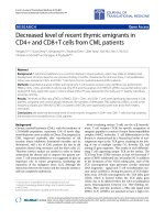
Figure 1 shows the schematic diagram of the flame aero-
sol reactor system used for the synthesis of the Cu-
doped TiO
2
nanomaterials. The main components of
the flame aerosol reactor system are: a diffusion burner,
a precursor feeding system, and a quenching and collec-
tion system. The design details of the diffusion burner
use d for this study is given in Jiang et al. [26]. Nitrogen
was passed through titanium tetra-ispopropoxide (TTIP,
99.7%, Aldrich, Steinheim, Germany) in a bubbler, and
the saturated vapor was introduced into the central port
of the burner. The bubbler containing the liquid TTIP
precursor was placed in an oil bath and was maint ained
at a temperature of 98°C. The precursor delivery tube
was maintained at a temperature of 210°C by a heating
tape. This avoided the condensation of the precursor
TTIP vapor in the delivery tube. Copper nitrate
trihydrate (99.5%, VWR International, Radnor, PA,
USA) was used as the dopant precursor. The dopant
precursor solution was prepared by dissolving a known
amount of copper nitrate in distilled water. A stainless
steel collison nebulizer was used to generate fine spray
droplets (less than 2 μm), which were then carried by
nitrogen gas into the high-temperature zone of the
flame. The doping percentage was varied by introducing
different molar ratios of both the precursors. The overall
doping concentration was varied from 0 to 15 wt.%.
Methane and oxygen were introduced into the second
and third ports of the burner respectively to create a dif-
fusion flame zone. The volumetric flow rates of N
2
through the TTIP bubbler and the O
2
were precisely
controlled by mass flow controllers at 2 and 7.5 lpm,
respectively. The methane flow rate was maintained at
1.8 lpm, and varied for few of the tests. A 20-lpm flow
of compressed air was supplied in a radial direction to
the quenching ring for cooling. The entrained air diluted
the aerosol stream and suppressed particle growth. The
synthesized materials were collected using a glass micro-
fiber filter paper (Whatman) for further characterization.
Material characterization
The size, morphology, and microstructure of the nanopar-
ticles were determined by a transmission electron micro-
scope (TEM; Model: JEOL 2100F FE-(S) TEM, JEOL Ltd.,
Tokyo, Japan) with an accelerating voltage of 200 kV and
by a field emission scanning electron microscope (SEM)
(Model: JEOL 7001LVF FE-SEM, JEOL Ltd.). The elemen-
tal analysis of the do ped nanomaterial was d one using
energy dispersive spectroscopy (EDS) analysis integrated
with a SEM. Pha se structures of the material were deter-
mined using an X-ray diffractometer (XRD) with Cu Ka
radiation (l = 1.5418 A) (Rigaku D-MAX/A9). Zeta poten-
tial, an indicator of the stability of nanoparticles in suspen-
sions, was measured by using a ZetaSizer Nano ZS
(Malvern Instruments Ltd., Worcestershire, UK) dynamic
light scattering instrument. Nanoparticles were dispersed
in de-ionized water at a concentration of 30 μg/ml and
sonicated for 25 min using a bath sonicator (40 W, 50
kHz, 5 Fisher Scientific, Fairlawn, New Jersey, USA) before
zeta potential measurements. UV-visible absorption spec-
troscopy (Perkin Elmer Lambda 2S, Perkin Elmer, Wal-
tham, MA, USA) was used to analyze the absorbance
spectrum of the nanomaterials over wavelengths ran ging
from 200 to 800 nm at room temperature. From the
absorption spectrum, the band gap was estimated. The
absorptio n ed ge was estimated to be the point where the
absorption was 30% of the maximum, corresponding to
where 50% of the photons were absorbed. This appr oach
was used because of the difficulty i n finding the linear
region of the absorption spectrum according to conven-
tional methods of band gap estimation [21].
Sahu and Biswas Nanoscale Research Letters 2011, 6:441
/>Page 2 of 14
Experimental test plan
The list of experiments performed is outlined in Table 1.
The flow rates were controlled to maintain the same resi-
dence time in the high-temperature flame (test 1). TiO
2
was synthesized under the same experimental conditions
using only TTIP as th e precursor (test 1A). Addition of
dopant influences nanomaterial properties such as size,
crystal structure, stability in suspension, and optical
properties. The copper dopant concentration was varied
from0to15wt.%toprocessCu-dopedTiO
2
Figure 1 Schematic diagram of the FLAR experimental setup used to synthesize Cu-doped TiO
2
nanoparticles.
Table 1 Summary of the experimental test plan
Test
no.
Dopant
concentration (wt
%)
CH
4
(lpm) Objective
1 A 0 1.8 Study the influence of dopant concentration on TiO
2
material properties such as size, crystal
phase, suspension stability, and light absorption.
B 0.5
C1
D3
E5
F15
2 A 3 0.8 Study the effect of methane flow rate on size and crystal phase of the material.
B 1.2
C 1.5
D 1.8
3 A 1 Annealing temperature,
400°C, 600°C
Examine the effect of annealing on phase and microstructure characteristics of Cu-doped
TiO
2
nanoparticles
B 15 Duration of annealing
under air, 4 h
All the particles were synthesized by diffusion flame aerosol reactor. Annealing was done in a furnace under air atmosphere.
Sahu and Biswas Nanoscale Research Letters 2011, 6:441
/>Page 3 of 14
nanomaterials (test 1(B-F)) t o investigate the impact on
properties. The copper dopant concentration was e sti-
mated based on the precursors feed rate to the flame.
The temperature-time history in the flame impacts the
particle formatio n and growth rates. This was varied by
altering the methane flow rate from 0.8 to 1.8 lpm at a
constant dopant level of 3 wt.% (test 2). Annealing of the
1 and 15-wt.% Cu-doped TiO
2
was conducted for 4 h at
400 and 600°C in an atmosphere of air to examine prop-
erty alterations (test 3).
Results and discussion
Doping TiO
2
with other atoms changes properties such
as particle size, crystal structure, stability in suspension,
and light absorption. The mechanism of Cu-doped TiO
2
nanoparticle formation in the flame aerosol reactor is
discussed first. The effect of copper dopant on TiO
2
particle properties are discusse d followed by crystal
structure control of the doped TiO
2
nanomaterials.
Finally, microstructure changes of Cu-doped TiO
2
are
discussed under different annealing conditions.
Particle formation mechanism
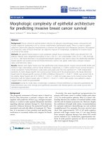
The proposed Cu-doped TiO
2
particle formation
mechanism is illustrated in Figure 2. This is similar to
the pathways proposed by Basak [24] for multi-compo-
nent nanomaterial system s. To understand the formati on
mechanism of the Cu-doped TiO
2
nanoparticles in the
flame aerosol react or, pristine TiO
2
was synthesized first
using TTIP only as the precursor. TTIP decomposes to
form TiO
2
monomers, which then undergo subsequent
growth by collision followed by sintering to form nano-
particles (test 1A). For synthesizing Cu-doped TiO
2
parti-
cles, both the TTIP and copper nitrate precursor are fed
to the high-temperature flame. The nanoparticle proper-
ties such as size and composition depend on the relative
decomposition kinetics and molar feed ratios of the pre-
cursors (see Figure 2). The decomposition rate of TTIP is
given by, k
a
= 3.9 6 × 10
5
exp((-7.05 × 10
4
)/RTs
-1
[29].
Since the kinetic data for copper nitrate precursor is not
available, the decomposition rate reported for copper
acetyl acetonate was assumed (k
b
= 3.02 × 10
7
exp((-1.15
×10
5
)/RT)s
-1
) [30]. The two precursors form TiO
2
(formed from TTIP molecular decomposition) and CuO
(formed by decomposition of copper nitrate followed by
evaporation) monomers at similar time instants as their
decomposition rates are similar (k
1, Cu
/k
1, Ti
to approxi-
mately 5, at 2,200°C). Dependi ng on the molar feed ratio
of the precursors, a variety of morphologies can be
formed, ranging from particles consisting of only copper
oxide, particles of only TiO
2
, and the particles of mixed
TiO
2
and CuO. At low copper concentration s (1-5 wt.%),
CuO monomers are readily incorporated into the higher
concentration TiO
2
clusters by a scavenging process.
This is similar to the phenomenon demonstrated by
Wang et al. [22]. Subsequent collisional growth and sin-
tering result in a homog enous mix of Cu-doped TiO
2
particles. However, at higher Cu feed concentration
(approximately 15wt%), apart from the collision and sin-
tering of the CuO monomers and TiO
2
cluster s, some of
the CuO oxide monomers also condense onto the formed
Cu-doped TiO
2
particles. The HR-TEM image of the
synthesized 15-wt.% Cu-TiO
2
nanoparticles indicates
regions of amorphous CuO on the particle surface. The
explanation of CuO monomer condensation on the parti-
cle surface is thus corroborated (test 1F). The nanoma-
terials synthesized at various dopant concentration were
verified by single particle EDS analysis to be comprised
of both copper and titania. No particles were found con-
sisting of only Ti or only copper species.
Effect of copper dopant concentration on TiO
2
properties
Particle size analysis
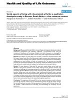
Figure 3 shows the TEM, HR-TEM images, and primary
particle size distribution of 1 wt.% Cu-TiO
2
(test 1B) and
15 wt.% Cu-TiO
2
(test 1F) samples. The particl e siz e dis-
tribution was obtained by measuring the diameter of 200
particles from representative TEM images. As shown in
the size distribution of these samples (see Figure 3), the
particles were spherical and size decreased with increas-
ing doping c oncentration. The geometric mean primary
particle size obtained at 1 wt.% doping was approximately
47 nm compared to approximately 33 nm obtained at 15
wt.% doping. The peak broadening observed in XRD pat-
tern (see Figure 4) also qualitatively explained the change
in particle size and lattice expansion with doping. The
crystallite size was estimated from the XRD pattern
obtained usi ng Scherrer formula. T he crystallite size
obtainedat1wt.%dopingwas33nmcomparedto25
and 23 nm at 5 and 15-wt.% doping concentration. It is
important to note that crystallite size estimation from
XRD is different from the particle size observed from the
microscopic analysis. XRD measures the size of t he small
domains within the grains and one particle may consist
of several crystallites based on the preparation methods
[31]. The decreased particle size with increasing doping
concentration is due to the inhibition of the grain
growth. As evident from the HR-TEM images of the 15
wt.% Cu-TiO
2
(see Figure 3), an enhanced amorphous
layer is observed on the surface. The excess CuO mono-
mers condense on to the existing Cu-doped TiO
2
parti-
cles. Thus, p article crystallinity decreases and also
prevents grai n growth. Wang et al. [22] observed a n
amorphous crystal structure and decreased grain size
with an increasing Fe
2+
/Ti
4+
ratios consistent with our
Cu-doped TiO
2
materials. Reduction in size was also
observed when Li et al. [3] syn thesized Zn-doped SnO
2
nanomaterials. Norris e t al. [27] proposed a process
Sahu and Biswas Nanoscale Research Letters 2011, 6:441
/>Page 4 of 14
called self-purification by which dopants diffuse from
inside to the surface sites of TiO
2
nanocrystals. This
change in particle si ze with doping concentrat ion is fun-
damentally a very importa nt phenomenon for electronic
structure modification. These results indicate that the
particle size of the Cu-doped TiO
2
can be controlled by
manipulating the dopant concentration in addition to the
methods demonstrated by other researchers by control-
ling the precursor feed concentration and residence time
of the particle in the high-temperature flame [26,32].
Crystal phase
The functionality of TiO
2
nanomaterials for various
applications depends on its crystal phase. The anatase
phase of TiO
2
is preferred for photoc ataytic applications,
whereas rutile phase is preferred for applications in pig-
ments [1]. It is, therefore, necessary to understand the
modifications in the crystal structure by incorporation of
the dopants in TiO
2
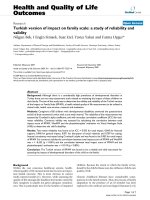
. The XRD diffraction pattern of the
Cu-doped TiO
2
nanomaterials synthesized at various
concentrations is shown in Figure 4. The pristine and
Cu-doped TiO
2
nanoparticles wer e prepared at the same
flame conditions for comparison. The pristine TiO
2
was
primarily anatase under the chosen processing condi-
tions. However, with increasing dopant concentration,
the transformation from anatase to rutile phase occurred,
as shown in Figure 4a from the (110) rutile peak, consis-
tent with other studies [18,33]. The anatase and rutile
fraction were calculated accordin g to the formula pro-
posed by Spurr and Myers [34]. The pristine TiO
2
had
1.2% rutile content, but with increasing doping concen-
tration to 15 wt.%, the rutile phase increased to 21.8%.
Even at high dopant concentration (15 wt.%), no pure
dopant-related crystal phase was observed within the
XRD detection limit. The same anatase to rutile phase
transformation was observed for synthesis of Cu-doped
TiO
2
by other methods [9,35].
The similarity in ionic radius of Cu
2+
(0.73 Å) to that of
Ti
4+
(0.64 Å) enable copper to substitutionally replaces
Ti in the titanium lattice in the flame environment,
where particles are formed from the atomistic state. In
the high-temperature flame synthesis of Cu-doped TiO2
nanomaterial, the copper dopant creates a higher number
Figure 2 Cu -doped TiO
2
nanopartic les formation mechanisms in a FLAR. Top represents TiO
2
formation mechanism, middle is for low
copper dopant concentration and bottom is for high dopant concentration.
Sahu and Biswas Nanoscale Research Letters 2011, 6:441
/>Page 5 of 14
of defects inside the anatase phase, resulting in a faster
formation and growth of a higher number of rutile nuclei
[36]. At elevated temperatures, the substitution of Ti
4+
by Cu
2+
increases the oxygen vacancy concentration and
decreases the free el ectron concentration. The excess of
oxygen vacancies created in the TiO
2
crystal lattice is the
responsible for anatase to rutile phase transition [36,37].
Nair et al. [36] found that a dopant with an oxidation
state above 4+ will reduce the oxygen vacancy concentra-
tion in the titania lattice as an interstitial impurity.
Dopants with an oxidation state of 3+ o r lower when
placed in the titan ia lattice points create a charge-com-
pensating anion vacancy [36] and cause a transformation
to the rutile phase as also found in this study. At higher
dopant concentration (15 wt.%) amorphous phase was
also observed on the surface as well as in the bulk. The
TEM and HR-TEM images 1 and 15-wt.% Cu-dope d
TiO
2
nanoparticles (see Figure 3) shows that particles at
lower doping concentrations are fully crystallized, and
the crystal lattice sp acing corresponds to the anatase
phase of TiO
2
(0.331 ± 0.03 nm), whereas the particle
synthesized at 15-wt. % copper concentration shows both
crystalline and amorphous phases of the material. The
HR-TEM images confirm that Cu
2+
doping retards the
grain growth of T iO
2
nanoparticles. Similar results of
decreasing crystalline nature of material were observed
when Fe
2+
-andZn
2+
-doped TiO
2
were synthesized
[3,22]. In a similar doping study, Wang et al. [22] found
that at higher Fe
2+
/Ti
4+
ratios of 0.12, more rutile and
amorphous crystal structure was observed, consistent
with our Cu-doped TiO
2
materials.
Figure 4b and 4c repres ent the XRD spectra for (101)
and (201) anata se peaks scanned at a very small steps of
0.004 degree for pristine and doped TiO
2
nanomaterials.
It is important to note that with increasing dopant con-
centration, broadening of the major anatase peaks (101)
and (201) was observed, which indicates a dec rease in
crystallite size. The shift in peak position to the right [8]
with increasing dopant concentration indicates that Cu
2
+
ions replaced some Ti
4+
ions along with the lattice
expansion. The results clearly indicate that addition of
dopant alters the crystal phase of the host nanomaterial
and the degr ee of phase transition depends on dopant
types and their concentrations.
Zeta potential and suspension stability
The dispersion characteristics of nanoparticles in aqu-
eous suspensions influence the fate and transport, cata-
lytic reactivity in the envi ronmental system as well as
critical in un derstanding for toxicological applications
[38,39]. The stability of the synthesized Cu-doped TiO
2
(
B
)
(A)
Figure 3 TEM images and particle size di stributions of as synthesized Cu-doped TiO
2
nanoparticles.(a) 1 wt.% Cu-TiO
2
and (b)15wt.%
Cu-TiO
2
. Inset is the HR-TEM image of the crystal fringes (test 1). Size distribution of particles is determined from measurement of 200 particles
from representative TEM images (test 1B, F).
Sahu and Biswas Nanoscale Research Letters 2011, 6:441
/>Page 6 of 14
nanoparticles was analyzed through the measurement of
zeta potential in aque ous system using de-ionized water
suspension (Figure 5) and compared with pure TiO
2
(test 1A) and commercial CuO. When metal oxide
nanoparticles are dispersed in water, the hydration of
the nanoparticle surface followed by protonation and
deprotonation of the surface groups from the oxide sur-
face results in a surface charge. The effective surface
charge on the particle depends on the isoelectric point
(IEP) in the suspension [39,40]. The zeta potential
observed for pure TiO
2
particle was +3.4 mV in the sus-
pension, as the measured pH of the suspension was
5.06, which is less than the IEP of the TiO
2
(pH approxi-
mately 6.0) and consistent with other studies [40]. How-
ever, for Cu-doped TiO2 nanoparticles, the zeta
potential value decreased to -3.4 mV and -25.6 mV at 1-
wt.% (test 1B) and 15-wt.% (test 1F) copper dopant con-
centration. The zeta potential measured for the
2Theta [degree]
20 30 40 50 60
Intensity [a.u]
5 wt% Cu-TiO
2
(1E)
15 wt% Cu-TiO
2
(1F)
3 wt% Cu-TiO
2
(1D)
Pristine TiO
2
(1A)
1 wt% Cu-TiO
2
(1C)
A(101)
A(004)
A(201)
R(101)
R(211)
2Theta [degree]
24.5 25.0 25.5 26.0 26.5
I
ntens
i
ty
[
a.u
]
Pristine TiO
2
(1A)
1 wt% Cu-TiO
2
(1C)
5 wt% Cu-TiO
2
(1D)
15 wt% Cu-TiO
2
(1E)
2Theta [degree]
47.5 48.0 48.5 49.0 49.
5
Intensity [a.u]
Pristine TiO
2
(1A)
1 wt% Cu-TiO
2
(1C)
5 wt% Cu-TiO
2
(1D)
15 wt% Cu-TiO
2
(1E)
(
c
)
(b)
(a)
Figure 4 TheXRDdiffractionpatternoftheCu-dopedTiO
2
nanomaterials.(a) XRD spectra of as-prepared Cu-TiO
2
nanoparticles with
different dopant concentrations (A anatase, R rutile). (b) Comparison of the XRD anatase peaks of Cu-TiO
2
nanoparticles: anatase (101) peaks and
(c) anatase (201) peaks (test 1).
Sahu and Biswas Nanoscale Research Letters 2011, 6:441
/>Page 7 of 14
commercial CuO was -27.3 mV which is close to the
zeta potential value observed for 15-wt.% Cu-TiO
2
sam-
ples (test 1F). The high surface charge on the 15 wt.%
Cu-TiO
2
indicates better stability of these particles over
pristine TiO
2
nanoparticles in aqueous suspension. The
higher zeta potential value and suspension stability of
the doped nanopartic les compared to TiO
2
is attributed
to charge imbalance created due to substitution of Ti
4+
atoms by Cu
2+
in the TiO
2
structure resulting in a more
negatively charged surface. Furthermore, zeta potential
values for 15-wt.% Cu-TiO
2
samples being similar to
pure CuO supports the presence of a copper oxide layer
on the outer surface of the particles.
Light absorption properties
The absorption spectra of the resulting Cu-doped TiO
2
nanomaterials was determi ned by a diffusive reflectanc e
spectroscopy measurement. The absorption spectrum of
Cu-doped T iO
2
nanomaterials prepared at various
dopant concentrations are shown in Figure 6. With
increasing dopant concentration, an increased absor-
bance in the visible spectrum is observed. The estimated
Eg for pristine TiO
2
was3.31eVwhichisconsistent
with the reported value for anatase TiO
2
[21]. With
increasing dopant c oncentration, the band gap energy
decreased and was estimated to be 2.51 eV at the high-
est dopant concentration of 15 wt.%. This change of
approximately 0.8 eV was due to the incorporation of
Cu
2+
ions into TiO
2
crystal structure, and CuO forming
a layer on the particle surface. From an experimental
and theoretical study of band s tructure estimation of
metal oxides, The results are consistent with findings of
Thimsen et al. [21] that the band gap energy decreases
with increasing Fe concentration in anatase-based TiO
2
materials.
Change in the optical absorption is due to the defect
centers created by the substitution of Ti
4+
by Cu
2+
atoms
in the TiO
2
crystal lattice. Earlier studies indicated that
doping with aliovalent ions changes the local lattice sym-
metr y and defect characteristics , which could change the
absorption properties and the material properties. In Cu-
dopedTiO
2
, when copper ions are either located inside the
bulk TiO
2
or on the surface sites, a rearrangement of the
neighbor atoms take place to compensate the charge defi-
ciency, resulting in lattice deformation. The lattice defor-
mation affects the electronic structure causing the band
gap shift [3]. Furthermore, small amounts of Cu
2+
dopant
in the lattice sites of TiO
2
introduce oxygen vacancies due
to the charge compensatio n effect [36,41]. Incr easing the
copper doping concentration increases the oxygen vacan-
cies and probably form a newly doubly occupied oxygen
vacancy as discussed in Li et al. [3]. Therefore absorption
of the doped nanomaterial and band gap shift may be con-
trolled by sur face effects, doping-ind uced vacancies, and
lattice strain. It can be said that the copper modified TiO
2
structure extends its absorption to the visible spectrum of
sunlight (400-700 nm) effectively. Hence, these copper-
Figure 5 Zeta potential measurements of Cu-doped TiO
2
nanoparticles in aqueous suspension.
Sahu and Biswas Nanoscale Research Letters 2011, 6:441
/>Page 8 of 14
doped materials can be utilized for various visible-light
photocatalytic applications, which have been demonstrated
in several other studies [9,18].
Crystal phase control of Cu-doped TiO
2
nanoparticle
The functionality of the nanomaterials d epends on their
properties such as particle size, crystal phase, morphology,
and agglomeration [38,40]. A recent study by Braydich-
Stolle et al. [42 ] showed that cytotoxicity in the cells is
both size and crystal structure dependent. They demon-
strated that mechanism of cell death varied with different
crystal structure; the anatase phase of TiO
2
being more
toxic than the rutile phase. To understand the role of crys-
tal phase of the doped nanomaterials on its functionality, it
is important to independently control the crystal phase
without varying the other material properties such as size.
Previous st udies have demonstrated that cryst al phase of
Photon Energy [eV]
2.0 2.5 3.0 3.5 4.0
Normalized Absorbance [-]
0.0
0.2
0.4
0.6
0.8
1.0
1
.
2
CuO
15 wt % Cu-TiO
2
(1F)
)
1 wt % Cu-TiO
2
(1C)
TiO
2
(1A)
5 wt % Cu-TiO
2
(1E)
Cu
[
wt %
]
0 2 4 6 8 10 12 14 16
B
an
d
gap
[
e
V]
2.0
2.2
2.4
2.6
2.8
3.0
3.2
3.4
Anatase Fraction
[
%
]
75
80
85
90
95
100
(b)
VISIBLE
UV
(a)
Figure 6 Absorption spectrum of Cu-doped TiO
2
nanomaterials prepared at various dopa nt concentra tions.(a) Normalized UV-visible
absorption spectra measured by diffuse reflectance spectroscopy. (b) Estimated band gap as a function of dopant concentrations (test 1).
Sahu and Biswas Nanoscale Research Letters 2011, 6:441
/>Page 9 of 14
the TiO
2
nanoparticle can be controlled by varying the
temperature in the flame (changing the methane flow
rates) and q uenching rate downstream of the flame
[25,26]. A similar methodology was adopted to control the
crystal phase of the Cu-doped TiO
2
materials. The dopant
concentration was kept constant at 3 wt.% and methane
flow was varied from 0.8 to 1.8 lpm (test 2, Figure 7a). The
anatase phase varied from 39% to 95%, when the methane
flo w was increased from 0.8 to 1.2 lpm, whereas the pri-
mary particle sizes for all the cases were similar. The
repre sentative TEM micrographs and corresponding size
distribution of the particles synthesized at 0.8 and 1.8 lpm
are shown in Figure 7b, c. The geometric mea n size of
31.5and32.3nmwerenearlythesameforthetwoflow
2Theta[degree]
20 30 40 50 60
Intensity [a.u]
0.8 lpm CH
4
: 39% anatase (2A)
1.2 lpm CH
4
: 50% anatase (2B)
1.5 lpm CH
4
: 69% anatase (2C)
1.8 lpm CH
4
: 95% anatase (2D)
A (101)
R(101)
R(211)
A (004)
A (201)
(a)
(b
)
(
c
)
Figure 7 Dopant concentration, representative TEM micrographs and corresponding size distribution of the particles.(a) XRD spectra at
different methane flow rates (A anatase, R rutile) and particle size distributions at (b) 0.8 lpm, (c) 1.2 lpm methane flow rates for 3-wt.% Cu-TiO
2
nanoparticles (test 2).
Sahu and Biswas Nanoscale Research Letters 2011, 6:441
/>Page 10 of 14
rate conditions. The size remained similar due to the bal-
ance between temperature profileandresidencetimein
the flame at different methane flow rates. For a fixed flame
operating parameters, increasing the methane flow rate
increases the flame temperature but at the same time
reduces the r esidence time in the flame. For lower
methane flow rate the temperature dec reases and resi-
dencetimeincreases.ThusthecrystalphaseoftheCu-
doped TiO
2
nanoparticles was independently varied while
keeping the primary particle size the same. These well-
controlled Cu-doped TiO
2
samples will be of significant
importance in biological studies to elucidate the role of
crystal phases without interferences from the other parti-
cle properties su ch as size.
Effect of annealing on Cu-doped TiO
2
nanoparticle
properties
The morphological and structura l transformation of the
doped nanoparti cles plays important role in photocataly-
tic activity by modifying the surface chemistry, crystal
and electronic structure [43] . Since both amorphous and
crystalline phases were observed in HR-TEM images at
higher dopant conc entration, the as-prepared Cu-doped
TiO
2
samples were annealed at different temperatures to
investigate the effect on crystal structure and morphol-
ogy. The 1 and 15-wt.% Cu-doped TiO
2
samples were
annealed at temperatures of 400°C and 600°C for 6 h. No
phase transformation was observed at 400°C. At 600°C,
the transformation from anatase to rutile phase was
observed as shown in Figure 8, which is consistent with
other studies [18,44]. The anatase weight fraction
decreased from 75% to 21% for the 15-wt.% Cu-doped
TiO
2
sample. However, the morphology of the particles
changed from spheric al to hexagonal structure for nano-
particles prepared at both the dopant concentrations.
The crystallite size increased with annealing. For 15-wt.%
Cu-doped TiO
2
sample, the phase related to CuO was
observed based on the peaks recorded at Bragg angle of
35.5 and 39 from the XRD pattern (Figure 8). The amor-
phous CuO present in the outer layers were annealed to
form the crystalline phase in the presence of air.
The HR-TEM images of samples annealed at 600°C are
shown in Figure 9. The figure indicates that the annealed
1-wt.% Cu-doped TiO
2
particle was completely crystal-
lized with no discontinuity in the crystal fringes as
observed from HR-TEM images, similar to the as-pre-
pared 1-wt.% Cu-doped TiO
2
particles. However, for the
15-wt.% dopant sample, some amorphous r egions were
still detected as shown in F igure 9 (highlighted with the
white squares). More detailed investigations are needed
to understand the effect of dopant concentration and
reaction environments on morphology change during
post-synthesis treatment of the initially synthesized sphe-
rical particles. The UV-vis measurements of absorption
spectra of 1- and 15-wt.% Cu-doped TiO
2
annealed
samples are shown in Figure 10 and compared with the
commercially available CuO nanoparticles. Annealing of
the 15 wt.% Cu-TiO
2
increased the absorpti on compared
to the as prepared samples in the visible spectrum mainly
because of enhance d crystalline CuO formation. It is
clear from the results that post-synthesis annealing can
alter the doped TiO
2
nanomaterial properties such as
size, crystal structures as well as absorption properties,
thus influencing eventual functionality and performance.
Conclusions
Cu-doped TiO
2
nanoparticles were synthesized in a dif-
fusion flame aerosol reactor and the properties were
2Theta [degree]
30 35 40 45 50 55 60
I
ntens
i
ty
[
a.u
]
As prepared
400
o
C
600
o
C
R
A
R
A
R
R
R-Rutile
A-Anatase
C-CuO
(a)
2Theta [de
g
ree]
30 35 40 45 50 55 6
0
Intensity [a.u]
As prepared
CuO
R
C
C
R
A
A
R
600
o
C
(b)
Figure 8 XRD pattern of the annealed Cu-TiO
2
nanoparticles.
(a) 1-wt.% Cu-TiO
2
(b) 15-wt.% Cu-TiO
2
. A anatase, R rutile. Samples
were annealed for 4 h in a furnace at constant temperature (test 3).
Sahu and Biswas Nanoscale Research Letters 2011, 6:441
/>Page 11 of 14
Figure 9 TEM images of annealed C u-doped TiO
2
samples.(a)1wt.%Cu-TiO
2
and (b)15wt.%Cu-TiO
2
. Annealing temperature, 600°C;
duration of annealing, 4 h (test 3).
Photon Ener
gy
[eV]
2.0 2.5 3.0 3.5 4.
0
Normalized Absorbance [-]
0.0
0.2
0.4
0.6
0.8
1.0
1
.
2
15 wt % Cu-TiO
2
(1F)
)
1 wt % Cu-TiO
2
(1C)
15 wt % Cu-TiO
2
(3B)
CuO
1 wt % Cu-TiO
2
(3A)
Figure 10 Normalized UV-visible absorption spectra measured by diffuse reflectance spectroscopy o f the annealed Cu-doped TiO
2
nanomaterials. Samples were annealed for 4 h at 600°C (test 3).
Sahu and Biswas Nanoscale Research Letters 2011, 6:441
/>Page 12 of 14
readily varied by controlling the processing conditions.
The increase in dopant concentration caused the trans-
formation from anatase to rutile phase of TiO
2
due to
replacement of Ti
4+
by Cu
2+
in the crystal structure of
TiO
2
. A decrease in primary particle size was also
observed. The doped nanomaterials exhibited better
aqueous suspension stability compared to pristine TiO
2
due to charge imbalance created. T he annealing o f the
doped samples resulted in the phase segregation and
crystallization of CuO for the higher dopant concentra-
tion samples. Spectroscopymeasurementsconfirma
shift in the absorption to visible frequencies, due to
crystal structure modification.
Acknowledgements
This work was partially supported by a grant from the NIEH S, Grant No.
100030N. The authors thank the McDonnell International Scholars Academy
and the McDonnell Academy Global Energy and Environment Partnership
() for providing partial support for this work.
Authors’ contributions
PB and MS both participated in the design of the study. MS carried out all
the experiments. MS and PB participated in results analysis. MS drafted the
manuscript and PB provided comments/suggestions to revise it.
Competing interests
The authors declare that they have no competing interests.
Received: 10 October 2010 Accepted: 6 July 2011 Published: 6 July 2011
References
1. Almquist CB, Biswas P: Role of synthesis method and particle size of
nanostructured TiO2 on its photoactivity. J Catal 2002, 212:145-156.
2. Dhumal SY, Daulton TL, Jiang J, Khomami B, Biswas P: Synthesis of visible
light-active nanostructured TiOx (x < 2) photocatalysts in a flame
aerosol reactor. Appl Catal B 2009, 86:145-151.
3. Li LP, Liu JJ, Su YG, Li GS, Chen XB, Qiu XQ, Yan TJ: Surface doping for
photocatalytic purposes: relations between particle size, surface
modifications, and photoactivity of SnO2:Zn2+ nanocrystals. Nanotechnol
2009, 20:155706.
4. Asahi R, Morikawa T, Ohwaki T, Aoki K, Taga Y: Visible-light photocatalysis
in nitrogen-doped titanium oxides. Science 2001, 293:269-271.
5. Choi WY, Termin A, Hoffmann MR: The role of metal-Ion dopants in
quantum-sized TiO2 - correlation between photoreactivity and charge-
carrier recombination dynamics. J Phys Chem 1994, 98:13669-13679.
6. Li W, Wang Y, Lin H, Shah SI, Huang CP, Doren DJ, Rykov SA, Chen JG,
Barteau MA: Band gap tailoring of Nd3-doped TiO2 nanoparticles. Appl
Phys Lett 2003, 83:4143-4145.
7. Bhattacharyya K, Varma S, Tripathi AK, Bharadwaj SR, Tyagi AK: Effect of
vanadia doping and its oxidation state on the photocatalytic activity of
TiO2 for gas-phase oxidation of ethene. J Phys Chem C 2008,
112:19102-19112.
8. Li W, Frenkel AI, Woicik JC, Ni C, Shah SI: Dopant location identification in
Nd3+-doped TiO2 nanoparticles. Phys Rev B 2005, 72:155315-155316.
9. Arana J, Dona-Rodriguez JM, Gonzalez-Diaz O, Rendon ET, Melian JAH,
Colon G, Navio JA, Pena JP: Gas-phase ethanol photocatalytic
degradation study with TiO2 doped with Fe, Pd and Cu. J Mol Catal A:
Chem 2004, 215:153-160.
10. Rane KS, Mhalsiker R, Yin S, Sato T, Cho K, Dunbar E, Biswas P: Visible light-
sensitive yellow TiO2-xNx and Fe-N co-doped Ti1-yFeyO2-xNx anatase
photocatalysts. J Solid State Chem 2006, 179:3033-3044.
11. Asahi R, Morikawa T: Nitrogen complex species and its chemical nature in
TiO2 for visible-light sensitized photocatalysis. Chem Phys 2007, 339:57-63.
12. Mor GK, Varghese OK, Wilke RHT, Sharma S, Shankar K, Latempa TJ, Choi KS,
Grimes CA: p-Type Cu-Ti-O nanotube arrays and their use in self-biased
heterojunction photoelectrochemical diodes for hydrogen generation.
Nano Lett 2008, 8:3555-3555.
13. Park HS, Kim DH, Kim SJ, Lee KS: The photocatalytic activity of 2.5 wt%
Cu-doped TiO2 nano powders synthesized by mechanical alloying. J
Alloys Compd 2006, 415:51-55.
14. Xu YH, Liang DH, Liu ML, Liu DZ: Preparation and characterization of
Cu2O-TiO2: Efficient photocatalytic degradation of methylene blue.
Mater Res Bull 2008,
43:3474-3482.
15. Tseng IH, Wu JCS, Chou HY: Effects of sol-gel procedures on the
photocatalysis of Cu/TiO2 in CO2 photoreduction. J Catal 2004,
221:432-440.
16. Li Y, Wang WN, Zhan Z, Woo MH, Wu CY, Biswas P: Photocatalytic
reduction of CO2 with H2O on mesoporous silica supported Cu/TiO2
catalysts. Appl Catal B 2011, 100:386-392.
17. Sakata Y, Yamamoto T, Okazaki T, Imamura H, Tsuchiya S: Generation of
visible light response on the photocatalyst of a copper ion containing
TiO2. Chem Lett 1998, 1253-1254.
18. Teleki A, Bjelobrk N, Pratsinis SE: Flame-made Nb- and Cu-doped TiO2
sensors for CO and ethanol. Sens Actuators, B 2008, 130:449-457.
19. Wu B, Huang R, Sahu M, Feng X, Biswas P, Tang YJ: Bacterial responses to
Cu-doped TiO2 nanoparticles. Sci Total Environ 2010, 408:1755-1858.
20. Nowotny MK, Sheppard LR, Bak T, Nowotny J: Defect chemistry of
titanium dioxide. application of defect engineering in processing of
TiO2-based photocatalysts. J Phys Chem C 2008, 112:5275-5300.
21. Thimsen E, Biswas S, Lo CS, Biswas P: Predicting the band Structure of
mixed transition metal oxides: theory and experiment. J Phys Chem C
2009, 113:2014-2021.
22. Wang ZM, Yang GX, Biswas P, Bresser W, Boolchand P: Processing of iron-
doped titania powders in flame aerosol reactors. Powder Technol 2001,
114:197-204.
23. McMillin BK, Biswas P, Zachariah MR: In situ characterization of vapor
phase growth of iron oxide-silica nanocomposites.1. 2-D planar laser-
induced fluorescence and Mie imaging. J Mater Res 1996, 11:1552-1561.
24. Basak S: Synthesis and characterization of magentic iron oxide for
nanomaterial and nanosystem fabricatiom. Ph.D Dissertation, Washington
University in St. Louis, Saint Louis, MO, USA; 2008.
25. Tiwari V, Jiang J, Sethi V, Biswas P: One-step synthesis of noble metal-
titanium dioxide nanocomposites in a flame aerosol reactor. Appl Catal,
A 2008, 345:241-246.
26. Jiang J, Chen DR, Biswas P: Synthesis of nanoparticles in a flame aerosol
reactor with independent and strict control of their size, crystal phase
and morphology. Nanotechnol 2007, 18:285603.
27. Norris DJ, Efros AL, Erwin SC: Doped nanocrystals. Science 2008,
319:1776-1779.
28. Swihart MT: Vapor-phase synthesis of nanoparticles. Curr Opin Colloid
Interface Sci 2003, 8:127-133.
29. Tsantilis S, Pratsinis SE: Soft- and hard-agglomerate aerosols made at high
temperatures. Langmuir 2004,
20:5933-5939.
30. Tsyganova EI, Mazurenko GA, Drobotenko VN, Dyagileva LM,
Aleksandrov YA: Kinetic principles of the thermolysis of yttrium, barium
and copper acetylacetonates. Zh Obshch Khim 1992, 62:499-504.
31. Narayan H, Alemu H, Macheli L, Thakurdesai M, Rao TKG: Synthesis and
characterization of Y3+-doped TiO2 nanocomposites for photocatalytic
applications. Nanotechnol 2009, 20:255601.
32. Thimsen E, Biswas P: Nanostructured photoactive films synthesized by a
flame aerosol reactor. AlChE J 2007, 53:1727-1735.
33. Francisco MSP, Mastelaro VR: Inhibition of the anatase-rutile phase
transformation with addition of CeO2 to CuO-TiO2 system: Raman
spectroscopy, X-ray diffraction, and textural studies. Chem Mater 2002,
14:2514-2518.
34. Spurr RA, Myers H: Quantitative analysis of anatse-rutile mixtures with an
X-Ray diffractometer. Anal Chem 1957, , 29: 760-762.
35. Colon G, Maicu M, Hidalgo MC, Navio JA: Cu-doped TiO2 systems with
improved photocatalytic activity. Appl Catal, B 2006, 67:41-51.
36. Nair J, Nair P, Mizukami F, Oosawa Y, Okubo T: Microstructure and phase
transformation behavior of doped nanostructured titania. Mater Res Bull
1999, 34:1275-1290.
37. Yuan SB, Meriaudeau P, Perrichon V: Catalytic combustion of diesel soot
particles on copper-catalysts supported on TiO2 - effect of potassium
promoter on the activity. Appl Catal, B 1994, 3:319-333.
Sahu and Biswas Nanoscale Research Letters 2011, 6:441
/>Page 13 of 14
38. Jiang J, Oberdorster G, Elder A, Gelein R, Mercer P, Biswas P: Does
nanoparticle activity depend upon size and crystal phase?
Nanotoxicology 2008, 2:33-42.
39. Suttiponparnit K, Jiang J, Sahu M, Suvachittanont S, Charinpanitkul T,
Biswas P: Role of surface area, primary particle size, and crystal phase on
titanium dioxide nanoparticle dispersion properties. Nanoscale Res Lett
2011, 6:1-8.
40. Jiang JK, Oberdorster G, Biswas P: Characterization of size, surface charge,
and agglomeration state of nanoparticle dispersions for toxicological
studies. J Nanopart Res 2009, 11:77-89.
41. Liu G, Sun CH, Yan XX, Cheng L, Chen ZG, Wang XW, Wang LZ, Smith SC,
Lu GQ, Cheng HM: Iodine doped anatase TiO2 photocatalyst with ultra-
long visible light response: correlation between geometric/electronic
structures and mechanisms. J Mater Chem 2009, 19:2822-2829.
42. Braydich-Stolle LK, Schaeublin NM, Murdock RC, Jiang J, Biswas P,
Schlager JJ, Hussain SM: Crystal structure mediates mode of cell death in
TiO2 nanotoxicity. J Nanopart Res 2009, 11:1361-1374.
43. Zhao YX, Qiu XF, Burda C: The effects of sintering on the photocatalytic
activity of N-doped TiO2 nanoparticles. Chemistry of Materials 2008,
20:2629-2636.
44. Xin BF, Wang P, Ding DD, Liu J, Ren ZY, Fu HG: Effect of surface species
on Cu-TiO2 photocatalytic activity. Appl Surf Sci 2008, 254:2569-2574.
doi:10.1186/1556-276X-6-441
Cite this article as: Sahu and Biswas: Single-step processing of copper-
doped titania nanomaterials in a flame aerosol reactor. Nanoscale
Research Letters 2011 6:441.
Submit your manuscript to a
journal and benefi t from:
7 Convenient online submission
7 Rigorous peer review
7 Immediate publication on acceptance
7 Open access: articles freely available online
7 High visibility within the fi eld
7 Retaining the copyright to your article
Submit your next manuscript at 7 springeropen.com
Sahu and Biswas Nanoscale Research Letters 2011, 6:441
/>Page 14 of 14