Advances in Applied Biotechnology Part 9 docx
Bạn đang xem bản rút gọn của tài liệu. Xem và tải ngay bản đầy đủ của tài liệu tại đây (659.99 KB, 20 trang )
Improvement of Heterologous Protein Secretion by Bacillus subtilis
149
We showed that the secretion production and activity of hIFN-α2b with propeptide
increased by more than 3-fold, compared to that without propeptide. The amount of
secreted hIFN-α2b with propeptide was 15mg /L. This result indicated that the propeptide
of AmyE enhanced the secretion of hIFNα-2b (Fig. 3, Kakeshita et al., 2011a).
Fig. 4. Western blot analysis of hIFN-β production by B. subtilis Dpr8 with pHKK3111
(AmyE SP-hIFN-β) or pHKK3211 (AmyE SP-Pro hIFN-β). Samples were collected at 20 h
after xylose induction, separated by 15% SDS-PAGE, and stained with Western blotting
using anti hIFN-β polyclonal antibodies. Dpr8 with pHKK3111 (lanes 1 and 2); Dpr8 with
pHKK3211 (lanes 3 and 4); 0.6% xylose induced (lanes 1 and 3), none induced (lanes 2 and
4), and commercially purified hIFN-β 50 ng (lane 5). Arrowheads indicate the positions of
the Pro-hIFN-β and hIFN-β. (adapted from Kakeshita et al., 2011b)
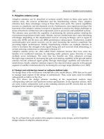
In L. lactis, directed mutagenesis experiments demonstrated that the positive effect of
LEISSTCDA on protein secretion was due to the insertion of negatively charged residues in
the N-terminus of the mature moiety (Le Loir et al., 2001). In hIFN-α2b with AmyE
propeptide, the first 10 amino acid residues of this mature protein have a net charge of -1.
On the other hand, hIFN-α2b without propeptide has a net charge of 0. In addition, we
demonstrated that propeptide mutants of neutral or positive charge resulted in a reduction
in the amount of secreted hIFN-α2b, compared with propeptides of negative charge. This
result suggested that negative charges in the mature protein can enhance the secretion of
hIFN-α2b (Kakeshita et al., 2011a).
We then indicated that the AmyE propeptide enhanced the secretion of the hIFN-β protein
from B. subtilis, as well. The secretion production and activity of hIFN-β with propeptide
increased by more than 4-fold (Fig. 4, Kakeshita et al., 2011b). The amount of secreted hIFN-
Advances in Applied Biotechnology
150
β with propeptide was 3.7mg /L. These results indicated that the propeptide of AmyE
enhanced the secretion and extracellular production of a heterologous protein in B. subtilis.
2.3 Deletion of the C-terminus of SecA
In B. subtilis, most secreted proteins are translocated across the cytoplasmic membrane via
the Sec system (Tjalsma et al., 2000; Tjalsma et al., 2004; Yamane et al., 2004). Nearly all of
the components of the Sec system identified in E. coli have also been identified in B. subtilis
and are biochemically well-characterized (van Wely et al., 2001; Harwood et al., 2008).
Among these components, the peripheral membrane protein, SecA is considered to play a
pivotal role in secretion. The SecYEG complex acts as a receptor for SecA, and functions as a
preprotein conducting channel (Hartl et al., 1990; Fekkes et al., 1997). In E. coli, SecB is a
molecular chaperone that functions in the post-translational protein translocation pathway,
and binds to the C-terminal SecB binding site of E. coli SecA. In B. subtilis, this region of SecA
is highly conserved. However, genome sequencing revealed that SecB is absent in B. subtilis
(Kunst et al., 1997). B. subtilis Ffh interacts directly with SecA, and promotes the formation of
soluble SecA-preprotein complexes (Bunai et al., 1999). These results suggest that the signal
recognition particle (SRP) of B. subtilis not only acts as a targeting factor in co-translational
translocation, but also stimulates the process of post-translocation across the membrane
(Harwood & Cranenburgh, 2008; Ling et al., 2007; Tjalsma et al., 2000; Yamane et al., 2004).
In additon, it has been shown that SecB binding site of B. subtilis SecA is not essential for
viability and protein secretion (van Wely et al., 2000). The SecB binding site is connected by
a C-terminal Linker (CTL) with the α-helical scaffold domain (HSD) in SecA. A cross-species
comparison of the amino acid sequence of SecA revealed that the CTL is not well-conserved
between B. subtilis and other species, including E. coli. We examined the effects of modifying
the C-terminal region of SecA on growth and the extracellular production of heterologous
proteins in B. subtilis, and demonstrated that the C-terminal domain (CTD) of SecA is not
essential for viability or protein secretion. Furthermore, we showed that the productivity of
hINF-α2b increased by 2.2-fold, compared to wild type SecA (Kakeshita et al., 2010). The
crystal structure of B. subtils SecA indicated that CTL binds to the surface of NBF-I. The
CTL-binding grove is a highly conserved and hydrophobic surface, and this grove is
predicted to be one of the mature preprotein binding sites in SecA (Hunt et al., 2002).
Therefore, deletion of the CTL of SecA is likely to affect SecA - preprotein interaction, and
likely caused an increase in the secretion of heterologous proteins.
2.4 Co-expression of PrsA
PrsA is essential for viability and protein secretion. In protein secretion, PrsA is suggested to
mediate protein folding at the late stage of secretion (Konitinen et al., 1991; Kontinen &
Sarvas, 1993; Vitikainen et al., 2001). We examined the effect of co-expression of an extra-
cytoplasmic molecular chaperone, PrsA. It is known that co-expression of an extra-
cytoplasmic molecular chaperone, PrsA enhances the secretion of several model proteins: α -
amylase, Single-chain antibody (SCA), and recombinant Protective antigen (rPA) (Kontinen
& Sarvas, 1993; Vitikainen et al., 2001; Wu et al., 1998; Williams et al., 2003).
We demonstrated that co-expression of PrsA can act in concert with the AmyE propeptide to
enhance the secretion production of hIFN-β. The amount of secreted hIFN-β with
propeptide was 5.5mg /L. (Fig. 5, Kakeshita et al., 2011b).
Improvement of Heterologous Protein Secretion by Bacillus subtilis
151
Fig. 5. Comparison of the amounts of secreted hIFN-β from B. subtilis D8C and D8PA, PrsA
co-expressing strains. (a) Schematic representation of the gene structure around the amyE
locus in the B. subtilis mutant strains D8PA and D8C. P
spoVG
and prsA represent the B. subtilis
spoVG promoter and B. subtilis PrsA, respectively. P
cat
and Cmr represent the
chloramphenicol-resistant gene promoter and coding region, respectively. (b) Western blot
analysis of PrsA protein from B. subtilis D8C, D8PA, and Dpr8. (c) Western blot analysis of
hIFN-β production by B. subtilis D8C, D8PA, and Dpr8. D8C with pHKK3211 (lane 1); D8PA
with pHKK3211 (lane 2); Dpr8 with pHKK3211 (lane 3). Arrowheads indicate the positions
of Pro-IFN-β. (Adapted from Kakeshita et al., 2011b).
3. Tat pathway
The majority of bacterial secreted proteins are translocated across the cytoplasmic
membrane via the Sec pathway, which acts on unfolded proteins using the energy provided
by ATP hydrolysis (Tajalsma et al., 2000; Antelman et al., 2000). Recently, a novel and
different secretion protein translocation pathway, the twin-arginine translocation (Tat)
pathway was discovered (Santini et al., 1998; Berks et al., 2000; van Dijl et al., 2002). The
bacterial twin-arginine translocation (Tat) machinery is able to transport folded proteins
across the cytoplasmic membrane (Robinson et al., 2001). The Tat pathway might have
advantages over the Sec pathway for the production of heterologous proteins, because many
proteins fold tightly before they reach the Sec machinery, and thus cannot engage with it for
translocation across the cytoplasmic membrane.
B. subtilis contains two substrate specific Tat systems, TatAyCy and TatAdCd. The TatAyCy
translocase is required for translocation of YwbN. On the other hand, a TatAdCd translocase
translocates the phosphodiesterase PhoD (Jongbloed JD et al., 2002; Pop et al., 2002).
Advances in Applied Biotechnology
152
3.1 Twin-arginine signal peptide
Proteins are targeted to the Tat pathway by tripartite N-terminal signal peptides, the amino-
terminal portion (n region) of which contain a conserved twin-arginine (RR) motif (R-R-X-#-
#, where # is a hydrophobic residue).
In a previous study by Jongbloed et al., a database search for the presence of this motif in
amino-terminal protein sequences identified a total number of 27 putative RR-signal
peptides.
Fig. 6. Schematic representation of the signal sequences used for secretion of human
Interferon-α in B. subtilis. Schematic structure of the proteins encoded by each indicated
plasmid. The twin-arginine motif is boxed, and the residues at positions -3 to -1 relative to
the predicted SPase I cleavage site are underlined. The six base pairs of the KpnI site add the
amino acids Gly–Thr to the end of each signal peptide coding sequence; therefore, in the
table, each sequence ends with GT. Numbers under the signal peptides refer to the
respective locations of the encoded amino acid residues.
We therefore selected six candidate Tat signal peptides, shown in Fig. 6, from the list
generated by Jongbloed et al. for testing in the hIFN-α secreted assay. To determine the
secretion ability for hINF-α2b, the six signal peptide genes considered to belong to the Tat
pathway of B. subtilis were PCR-amplified. The PCR-amplified signal peptide genes were
inserted upstream of the hIFN-α mature peptide gene in pHKK3101, yielding six secretion
expression vectors. pHKK3101 expressing hIFN-α with the AmyE signal peptide, as the
Sec-type signal peptide, was used as the control plasmid. The resultant recombinants
were transformed into B. subtilis Dpr8, respectively, and the secretion expression of hIFN-
α mediated by these signal peptides was detected by immunoblotting analysis. The hIFN-
α was expressed in these strains and hIFN-α production was induced with the addition of
0.6% of xylose to the exponentially growing cultures (OD660 = 0.3), and both culture
supernatants and intracellular lysates were analyzed as described in Kakeshita et al.
(2010). As shown in Fig. 7a, in the extracellular fraction, only one band corresponding to
mature protein (16 kDa) was detected for the samples of B. subtilis Dpr8 cells harboring
Improvement of Heterologous Protein Secretion by Bacillus subtilis
153
pHKK3101 (AmyE signal), pHKK4004 (WprA), pHKK4005 (LipA), and pHKK4006
(WapA) by Western blot and immunoblot. This result suggested that the obtained three
signal peptides (WprA, LipA, WapA) directed efficient secretion expression.
Fig. 7. Comparison of the amounts of secreted hIFN-α using the Twin arginine signal
peptides from B. subtilis Dpr8. (a) Western blot analysis of hIFN-α production in B. subtilis
Dpr8 harboring seven recombinants. Cells were grown at 30 °C in 2xL medium. Samples
were collected at 20 h after xylose induction, separated by 15% SDS-PAGE, and subjected to
Western blotting using anti hIFN-β polyclonal antibodies. Protein samples present in the
supernatant (lanes 1, 2, 3, 4, 5, and 6) and cell fractions (lanes 7, 8, 9, 10, 11, and 12) of
stationary-phase cultures were prepared by centrifugation, analyzed by SDS-PAGE, and
immunodetected with anti-hIFN-α antibodies. Dpr8/pHKK3101 (lanes 1 and 8);
Dpr8/pHKK4001 (lanes 2 and 9); Dpr8/pHKK4002 (lanes 3 and 10); Dpr8/pHKK4003 (lanes
4 and 11); Dpr8/pHKK4004 (lanes 5 and 12); Dpr8/pHKK4005 (lanes 6 and 13);
Dpr8/pHKK4006 (lanes 7 and 14); precursor, pre hIFN-α; mature, hIFN-α. S, supernatant; C,
cell fractions. (b) Quantification of secreted hIFN-α mature form in the culture medium and
cell fraction. The hIFN-α production corresponding to the supernatant of B. subtilis Dpr8
carrying pHKK3101 (AmyE signal peptide) was set as 100%. Data represent the mean of
three experiments, and error bars represent standard error.
Advances in Applied Biotechnology
154
Especially, WapA demonstrated the highest efficiency of hIFN-α secretion expression,
which was 1.5-fold as high as the Sec dependent signal peptide, AmyE (Fig. 7b).
However, No hIFN-α was detected in the supernatants of Dpr8/pHKK4001 (YvhJ),
Dpr8/pHKK4002 (YwbN), or Dpr8/pHKK4003 (PhoD). In the intracellular lysates of
Dpr8/pHKK3101, Dpr8/pHKK4004, Dpr8/pHKK4005, and Dpr8/pHKK4006, two bands
were detected. As deduced from the molecular mass of each band, these bands ware
assigned to the unprocessed precursor (17 kDa) and the mature protein (16 kDa),
respectively. On the other hand, only one band corresponding to the unprocessed protein
was detected for the samples of Dpr8/pHKK4001 (YvhJ), Dpr8/pHKK4002 (YwbN), and
Dpr8/pHKK4003 (PhoD).
These results suggested that the three obtained signal peptides, YvhJ, YwbN, and PhoD
cannot be secreted hIFN-α2b into the supernatant.
3.2 Co-expression of the tat system
We examined the effect of co-expression of the Tat-machinary, TatAd/Cd or TatAy/Cy. To
examine the effects of the co-expression of B. subtilis tat genes on hIFN-α secretion, we
constructed TatAd/TatCd and TatAy/TatCy under the control of the spoVG promoter in
plasmids. It is known that the spoVG promoter is a powerful promoter (Zuber & Losick
1983). The resulting constructs were subsequently integrated into the chromosome of B.
subtilis strain Dpr8 via a double crossover event at the amyE locus, leaving the native tat
genes intact (Fig. 8a).
The resultant strains, D8tatD and D8tatY were transformed with pHKK3101, pHKK4001,
pHKK4002, pHKK4003, pHKK4004, pHKK4005, and pHKK4006 for expression of hIFN-α.
As shown in Fig. 8b and c, when the LipA signal peptide was fused to hIFN-α, a densitometric
analysis of the western blotting demonstrated that the amounts of hIFN-α secreted by D8tatD
and D8tatY were increased by roughly 2-fold, compared with that in strain Dpr8 (Fig. 8c).
When the WprA signal peptide was fused to hIFN-α, in D8tatD, the amount of secreted hIFN-
α was increased by 71% compared with that in the parental strain, Dpr8, whereas the
enhanced production of hIFN-α increased by 29%. On the other hand, When the WapA signal
peptide was fused to hIFN-α, the amounts of hIFN-α secreted by D8tatD and D8tatY were
increased by only 10-20%, compared with that in strain Dpr8 (Fig. 8c). Then, when the AmyE
signal peptide was fused to hIFN-α, the amounts of hIFN-α secreted by D8tatD and D8tatY
were increased by 37% and 25%, respectively compared with that in strain Dpr8 (Fig. 8c).
Therefore, WapA signal peptide and AmyE signal peptide are not able to enhance of secretion
by co–expression of Tat system. In addition, when the YvhJ, YwbN, and PhoD signal peptides,
respectively were fused to hIFN-α, the bands of hIFN-α secreted by D8tatD and D8tatY could
not be detected in the resulting supernatants (data not shown).
We demonstrated that co-expression of TatAd/Cd or TatAy/Cy with LipA signal peptide
can act in concert to enhance the secretion production of hIFN-α. In addition, WprA signal
peptide was enhanced the secretion production of hIFNα by co-expression of TatAd/Cd,
not TatAy/Cy. On the other hands, AmyE signal peptide and WapA peptide are Tat
pathway independent.
Improvement of Heterologous Protein Secretion by Bacillus subtilis
155
Fig. 8. Comparison of the amounts of secreted hIFN-α from B. subtilis Dpr8 and Tat
overexpressing strains. (a) Schematic representation of the gene structure around the amyE
locus in the B. subtilis D8tatD and D8tatY mutant strain genomes. Construction of strains
D8tatD and D8tatY was by double crossover integration of plasmids pHKK2001 (tatAd-Cd)
and pHKK2002 (tatAy-Cy) into the amyE locus of B. subtilis Dpr8. The resulting strain
contains the native phoD-tatAd-tatCd locus, as well as one copy of tatAd-Cd and tatAy-Cy
under the control of the P
spoVG
promoter. The stem-loop structures and the bent arrows
indicate the putative Rho-independent terminators and promoters, respectively. (b) Western
blot analysis of hIFN-α production by B. subtilis Dpr8, D8tatD, and D8tatY (carrying
pHKK3101, pHKK4004, pHKK4005, or pHKK4006) was performed in the same manner as
for hIFN-α. (c) Quantification of secreted hIFN-α in mature form in the culture medium.
The hIFN-α production corresponding to the B. subtilis Dpr8 strain was set as 100%. Data
represent the mean of three experiments, and error bars represent standard error.
Advances in Applied Biotechnology
156
4. Conclusions
In recent years, considerable efforts have been targeted at developing B. subtilis as a host for
the production of heterologous proteins. However, the secretion of heterologous proteins
from eukaryotes by these species produces small yields and is frequently inefficient.
Initially, we considered the major problem to be the presence of high levels of extracellular
protease in B. subtilis. Nevertheless, even after obtaining many depleted protease strains, the
problem of inefficient secretion was not resolved. Currently, it is considered that the largest
problem is the detection of the pre-mature form of human protein in cell lysate, when
human proteins with signal peptide are over expressed in B. subtilis (Fig. 7a). Normally, the
pre-mature forms of target secretion proteins are not detected in cell lysates. If the pre-
mature form of target a secretion protein is detected, it indicates a problem in the secretion
pathway, for example, non-functional or depleted SecA, SecY, Ffh, or FtsY (Sadaie et al.
1991; Takamatsu et al., 1992; Honda et al., 1993; Oguro et al., 1995; Tjalsma et al., 2000;
Tjalsma et al., 2004; Yamane et al., 2004). Therefore, we must solve this primary problem,
which is the accumulation of the precursor of human proteins in B. subtilis cells.
We indicated that the propeptide of AmyE enhanced the secretion of the extracellular
production of a heterologous protein in B. subtilis. In L. lactis, the nine-residue synthetic
propeptide, LEISSTCDA, which is fused immediately after the signal peptide cleavage site,
is known to enhance heterologous protein secretion (Le Loir et al., 1998). In addition,
LEISSTCDA enhances secretion efficiency (Le Loir et al., 2001). Therefore, it is considered
that a short type propeptide may be one answer to improve the accumulation of precursor.
On the other hand, we indicated that the deletion of the C-terminal domain of SecA
enhanced the secretion of heterologous proteins. secA is an essential gene, and SecA is
considered to play a pivotal role in secretion (Sadaie et al. 1991; Takamatsu et al., 1992;
Tjalsma et al., 2000; Tjalsma et al., 2004; Yamane et al., 2004). In addition, we exhibited that
the co-expression of PrsA or the Tat system can be able to enhance the secretion production.
In the future, it may be necessary to modify the components of the secretion machinery for
higher secretion efficiency.
5. Acknowledgments
We are grateful to Naotake Ogasawara, Junichi Sekiguchi, Fujio Kawamura, Kunio Yamane
and members of MGP group in Kao Corporation for valuable discussions.
This work is the subproject, ‘Development of a Technology for Creation of a Host Cell’
included within the industrial technology project, ‘Development of a Generic Technology
for Production Process Starting Productive Function’ of the Ministry of Economy, Trade and
Industry, entrusted by the New Energy and Industrial Technology Development
Organization (NEDO), Japan.
6. References
Antelmann H, Tjalsma H, Voigt B, Ohlmeier S, Bron S, van Dijl JM, Hecker M (2001) A
proteomic view on genome-based signal peptide predictions. Genome Research
Vol.11, pp.1484-1502, ISSN 1088-9051 (Print), 1549-5469 (Electronic).
Improvement of Heterologous Protein Secretion by Bacillus subtilis
157
Baneyx F, Mujacic M (2004) Recombinant protein folding and misfolding in Escherichia coli.
Nature Biotechnology, Vol.11, pp.1399-1408, ISSN 1087-0156, EISSN 1546-1696.
Berks BC, Sargent F, Palmer T (2000) The Tat protein export pathway. Molecular
Microbiology, Vol.35, pp.260-274, ISSN 0950-382X(Print), 1365-2958 (Electronic).
Braun P, Tommassen J, Filloux A (1996) Role of the propeptide in folding and secretion of
elastase of Pseudomonas aeruginosa. Molecular Microbiology Vol.19, pp.297-306,
ISSN 0950-382X, EISSN: 1365-2958.
Braun P, Gerritse G, van Dijl JM, Quax WJ (1999) Improving protein secretion by
engineering components of the bacterial translocation machinery. Current Opinion
in Biotechnology, Vol.10, pp.376–381, ISSN 0958-1669.
Brockmeier U, Caspers M, Freudl R, Jockwer A, Noll T, Eggert T (2006) Systematic screening
of all signal peptides from Bacillus subtilis: a powerful strategy in optimizing
heterologous protein secretion in Gram-positive bacteria. Journal of Molecular
Biology, Vol.362, pp.393-402, ISSN 0022-2836.
Bunai K, Yamada K, Hayashi K, Nakamura K, Yamane K (1999) Enhancing effect of Bacillus
subtilis Ffh, a homologue of the SRP54 subunit of the mammalian signal recognition
particle, on the binding of SecA to precursors of secretory proteins in vitro. Journal
of Biochemistry, Vol.125, pp151-159, ISSN 0021-924X (Print), 1756-2651 (Electronic).
Davis A, Moore IB, Parker DS, Taniuchi H (1977) Nuclease B: a possible precursor of
nuclease A, an extracellular nuclease of Staphylococcus aureus. The Journal of
Biological Chemistry, Vol.252, pp.6544-6553, ISSN 0021-9258 (Print), 1083-351X
(Electronic).
Fekkes P, van der Does C, Driessen AJ (1997) The molecular chaperone SecB is released from
the carboxy-terminus of SecA during initiation of precursor protein translocation.
The EMBO Journal, Vol.16, pp.6105-6113, ISSN 0261-4189.
Hartl FU, Lecker S, Schiebel E, Hendrick JP, Wickner W (1990) The binding cascade of SecB
to SecA to SecY/E mediates preprotein targeting to the E. coli plasma membrane.
Cell Vol. 63, pp. 269-279, ISSN 0092-8674.
Harwood, CR, Cranenburgh R (2008) Bacillus protein secretion: an unfolding story. Trends
in Microbiology, Vol.16, pp.73-79, ISSN 0966-842X.
Heng C, Chen Z, Du L, Lu F (2005) Expression and secretion of an acid-stable -amylase
gene in Bacillus subtilis by SacB promoter and signal peptide, Biotechnol ogy
Letters, Vol.27, pp.1731-1737, ISSN 0141-5492 (Print), 1573-6776 (Electronic).
Honda K, Nakamura K, Nishiguchi M, Yamane K (1993) Cloning and characterization of a
Bacillus subtilis gene encoding a homolog of the 54-kilodalton subunit of
mammalian signal recognition particle and Escherichia coli Ffh. Journal of
Bacteriology, Vol.175, pp.4885-4894, ISSN 0021-9193 (Print), 1098-5530 (Electronic).
Hunt JF, Weinkauf S, Henry L, Fak JJ, McNicholas P, Oliver DB, Deisenhofer J (2002)
Nucleotide control of interdomain interactions in the conformational reaction cycle
of SecA. Science, Vol.297, pp.2018-2026, ISSN 0036-8075.
Ikemura H, Inouye M (1988) In vitro processing of prosubtilisin produced in Escherichia coli.
The Journal of Biological Chemistry, Vol.263, pp.12959-12963, ISSN 0021-9258
(Print), 1083-351X (Electronic).
Jongbloed JD, Antelmann H, Hecker M, Nijland R, Bron S, Airaksinen U, Pries F, Quax WJ,
van Dijl JM, Braun PG (2002) Selective contribution of the twin-arginine
translocation pathway to protein secretion in
Bacillus subtilis. The Journal of
Advances in Applied Biotechnology
158
Biological Chemistry, Vol.277,pp.44068-44078, ISSN 0021-9258 (Print), 1083-351X
(Electronic).
Kakeshita H, Kageyama Y, Ara K, Ozaki K, Nakamura K (2010) Enhanced extracellular
production of heterologous proteins in Bacillus subtilis by deleting the C-terminal
region of the SecA secretory machinery. Molecular Biotechnology Vol.46, pp.250-
257, ISSN 1073-6085 (Print), 1559-0305 (Electronic).
Kakeshita H, Kageyama Y, Ara K, Ozaki K, Nakamura K (2011a) Propeptide of Bacillus
subtilis Amylase Enhances Extracellular Production of Human Interferon-α in
Bacillus subtilis. Applied Microbiology and Biotechnology, Vol.89, pp.1509-1517,
ISSN 0175-7598 (Print), 1432-0614 (Electronic). .
Kakeshita H, Kageyama Y, Endo K, Tohata M, Ara K, Ozaki K, Nakamura K (2011b)
Secretion of biologically-active human interferon-β by Bacillus subtilis.
Biotechnology Letters, Vol.33, pp.1847-1852, ISSN 0141-5492 (Print), 1573-6776
(Electronic)
Kapust RB, Waugh DS (1999) Escherichia coli maltose-binding protein is uncommonly
effective at promoting the solubility of polypeptides to which it is fused. Protein
Science, Vol.8, pp.1668-1674, ISSN (Print) 0961-8368, ISSN (Electronic) 1469-896x.
Kodama T, Endo K, Ara K, Ozaki K, Kakeshita H, Yamane K, Sekiguchi J (2007a) Effect of
Bacillus subtilis spo0A mutation on cell wall lytic enzymes and extracellular
proteases, and prevention of cell lysis. Journal of Bioscience and Bioengineering,
Vol.103, pp.13-21, ISSN 1389-1723 (Print), 1347-4421 (Electronic) .
Kodama T, Endo K, Sawada K, Ara K, Ozaki K, Kakeshita H, Yamane K, Sekiguchi J (2007b)
Bacillus subtilis AprX involved in degradation of a heterologous protein during the
late stationary growth phase. Journal of Bioscience and Bioengineering, Vol.104,
pp.135-143, ISSN 1389-1723 (Print), 1347-4421 (Electronic).
Kontinen VP, Saris P, Sarvas M (1991) A gene (prsA) of Bacillus subtilis involved in a novel,
late stage of protein export. Molecular Microbiology, Vol.5, pp.1273-1283, ISSN
0950-382X (Print), 1365-2958 (Electronic).
Kontinen V, Sarvas M (1993) The PrsA lipoprotein is essential for protein secretion in
Bacillus subtilis and sets a limit for high-level secretion. Molecular Microbiology,
Vol.8, pp.727–737, ISSN 0950-382X (Print), 1365-2958(Electronic).
Kunst F, Ogasawara N, Moszer I, Albertini AM, Alloni G, Azevedo V, Bertero MG, Bessieres
P, Bolotin A, Borchert S, Borriss R, Boursier L, Brans A, Braun M, Brignell SC, Bron
S, Brouillet S, Bruschi CV, Caldwell B, Capuano V, Carter NM, Choi SK, Codani JJ,
Connerton IF, Cummings NJ, Daniel RA, Denizot F, Devine KM, Dusterhoft A,
Ehrlich SD, Emmerson PT, Entian KD, Errington J, Fabret C, Ferrari E, Foulger D,
Fritz C, Fujita M, Fujita Y, Fuma S, Galizzi A, Galleron N, Ghim S Y, Glaser P,
Goffeau A, Golightly EJ, Grandi G, Guiseppi G, Guy BJ, Haga K, Haiech J,
Harwood CR, Henaut A, Hilbert H, Holsappel S, Hosono S, Hullo MF, Itaya M,
Jones L, Joris B, Karamata D, Kasahara Y, Klaerr-Blanchard M, Klein C, Kobayashi
Y, Koetter P, Koningstein G, Krogh S, Kumano M, Kurita K, Lapidus A, Lardinois
S, Lauber J, Lazarevic V, Lee SM, Levine A, Liu H, Masuda S, Mauel C, Medigue C,
Medina N, Mellado RP, Mizuno M, Moestl D, Nakai S, Noback M, Noone D,
O'Reilly M, Ogawa K, Ogiwara A, Oudega B, Park SH, Parro V, Pohl TM, Portetelle
D, Porwollik S, Prescott AM, Presecan E, Pujic P, Purnelle B, Rapoport G, Rey M,
Reynolds S, Rieger M, Rivolta C, Rocha E, Roche B, Rose M, Sadaie Y, Sato T,
Improvement of Heterologous Protein Secretion by Bacillus subtilis
159
Scanlan E, Schleich S, Schroeter R, Scoffone F, Sekiguchi J, Sekowska A, Seror SJ,
Serror P, Shin BS, Soldo B, Sorokin A, Tacconi E, Takagi T, Takahashi H, Takemaru
K, Takeuchi M, Tamakoshi A, Tanaka T, Terpstra P, Tognoni A, Tosato V,
Uchiyama S, Vandenbol M, Vannier F, Vassarotti A, Viari A, Wambutt R, Wedler E,
Wedler H, Weitzenegger T, Winters P, Wipat A, Yamamoto H, Yamane K,
Yasumoto K, Yata K, Yoshida K, Yoshikawa HF, Zumstein E, Yoshikawa H,
Danchin A (1997) The complete genome sequence of the Gram-positive bacterium
Bacillus subtilis. Nature, Vol.390, pp.249-256, ISSN 0028-0836, EISSN 1476-4687.
Lam KH, Chow KC, Wong WK, (1998) Construction of an efficient Bacillus subtilis system for
extracellular production of heterologous protein. Journal of Biotechnology, Vol.63,
pp.167– 177, ISSN 0168-1656 (Print), 1873-4863 (Electronic).
Le Loir Y, Gruss A, Ehrlich SD, Langella P (1998) A nine-residue synthetic propeptide
enhances secretion efficiency of heterologous proteins in Lactococcus lactis. Journal
of Bacteriology, Vol.180, pp.1895-1903, ISSN 0021-9193 (Print), 1098-5530
(Electronic).
Le Loir Y, Nouaille S, Commissaire J, Brétigny L, Gruss A, Langella P (2001) Signal peptide
and propeptide optimization for heterologous protein secretion in Lactococcus lactis.
Applied and Environmental Microbiology, Vol.67, pp.4119-4127, ISSN 0099-2240
(Print), 1098-5336 (Electronic).
Le Loir Y, Azevedo V, Oliveira SC, Freitas DA, Miyoshi A, Bermúdez-Humarán LG,
Nouaille S, Ribeiro LA, Leclercq S, Gabriel JE, Guimaraes VD, Oliveira MN,
Charlier C, Gautier M, Langella P (2005) Protein secretion in Lactococcus lactis : an
efficient way to increase the overall heterologous protein production. Microbial
Cell Factories, Vol.40, pp.44-49, doi:10.1186/1475-2859-4-2, ISSN: 1475-2859.
Lesuisse E, Schanck K, Colson C, (1993) Purification and preliminary characterization of the
extracellular lipase of Bacillus subtilis 168, an extremely basic pH-tolerant enzyme,
European Journal of Biochemistry, Vol.216, pp.155–160, ISSN 0014-2956 (Print),
1432-1033 (Electronic).
Ling L, Xu Z, Li W, Shuai J, Lu P, Hu C (2007) Protein secretion pathways in Bacillus subtilis:
implication for optimization of heterologous protein secretion. Biotechnology
Advances, Vol.25, pp.1-12, ISSN 0014-2956 (Print), 1432-1033 (Electronic)
Li W, Zhou X, Lu P (2004) Bottlenecks in the expression and secretion of heterologous
proteins in Bacillus subtilis. Research in Microbiology, pp.155, pp.605-610, ISSN
0923-2508 (Print), 1769-7123 (Electronic).
Mézes PSF, Yang YQ, Hussain M, Lampen, JO (1983) Bacillus cereus 569/H β-lactamase I:
cloning in Escherichia coli and signal sequence determination. FEBS Letters, Vol.161,
pp.195-200, ISSN 0014-5793 (Print), 1873-3468 (Electronic).
Oguro A, Kakeshita H, Honda K, Takamatsu H, Nakamura K, Yamane K (1995) srb: a
Bacillus subtilis gene encoding a homologue of the α-subunit of the mammalian
signal recognition particle receptor. DNA Research Vol.2, pp.95-100, ISSN 1340-
2838 (Print), 1756-1663 (Electronic).
Olmos-Soto J and Contreras-Flores R, (2003) Genetic system constructed to overproduce and
secrete proinsulin in Bacillus subtilis, Applied and Environmental Microbiology,
Vol.62, pp.369– 373, ISSN 0099-2240 (Print), 1098-5336 (Electronic).
Advances in Applied Biotechnology
160
Palva I, Lehtovaara P, Kaariainen L, Sibakov M, Cantell K, Schein CH, Kashiwagi K,
Weissmann C (1983) Secretion of interferon by Bacillus subtilis. Gene, Vol.22,
pp.229–235, ISSN 0378-1119.
Palva I (1982) Molecular cloning of -amylase gene from Bacillus amyloliquefaciens and its
expression in B. subtilis. Gene, Vol. 19, pp81-87. ISSN 0378-1119.
Pop O, Martin U, Abel C, Müller JP (2002) The twin-arginine signal peptide of PhoD and the
TatAd/Cd proteins of Bacillus subtilis form an autonomous Tat translocation
system. The Journal of Biological Chemistry, Vol. 277, pp. 3268-3273, ISSN 0021-
9258 (Print), 1083-351X (Electronic).
Randall RE, Goodbourn S (2008) Interferons and viruses: an interplay between induction,
signalling, antiviral responses and virus countermeasures. Journal of General
Virology, Vol.89, pp.1-47, ISSN: 0022-1317 (Print), 1465-2099 (Electronic).
Rabhi-Essafi I, Sadok A, Khalaf N, Fathallah DM (2007) A strategy for high-level expression
of soluble and functional human interferon alpha as a GST-fusion protein in E. coli.
Protein Engineering Design and Selection, Vol.5, pp.201-209, ISSN (Print): 1741-
0126. ISSN (Electronic): 1741-0134.
Robinson C, Bolhuis A (2001) Protein targeting by the twin-arginine translocation pathway.
Nature Reviews Molecular Cell Biology, Vol.2, pp.350–356, ISSN 1471-0072 (Print),
1471-0080 (Electronic).
Rojas Contreras JA, Pedraza-Reyes M, Ordoñez LG, Estrada NU, Barba de la Rosa AP, De
León-Rodríguez A. (2010) Replicative and integrative plasmids for production of
human interferon γ in Bacillus subtilis. Plasmid, Vol.64, pp.170-176, ISSN 0147-619X
(Print) 1095-9890 (Electronic).
Sadaie Y, Takamatsu H, Nakamura K, Yamane K (1991) Sequencing reveals similarity of the
wild-type div+ gene of Bacillus subtilis to the Escherichia coli secA gene. Gene, Vol.98,
pp.101-105, ISSN 0378-1119 (Print), 1879-0038 (Electronic).
Santini, C. L., B. Ize, A. Chanal, M. Muller, G. Giordano, and L F. Wu. 1998. A novel sec-
independent periplasmic protein translocation pathway in Escherichia coli. The
EMBO Journal, Vol.17, pp.101-112, ISSN 0261-4189.
Sasamoto H, Nakazawa K, Tsutsumi K, Takase K, Yamane K (1989) Signal peptide of
Bacillus subtilis α-amylase. Journal of Biochemistry, Vol.106, pp.376-382, ISSN 0021-
924X (Print), 1756-2651 (Electronic)
Sørensen HP, Mortensen KK (2005) Soluble expression of recombinant proteins in the
cytoplasm of Escherichia coli. Microbial Cell Factories, Vol.4, doi:10.1186/1475-2859-
4-1, ISSN: 1475-2859.
Srivastava P, Bhattacharaya P, Pandey G, Mukherjee KJ (2005) Overexpression and
purification of recombinant human interferon alpha2b in Escherichia coli. Protein
Expression and Purification, Vol.41, pp.313-322, ISSN 1046-5928 (Print), 1096-0279
(Electronic).
Suciu D, Inouye M (1996) The 19-residue pro-peptide of staphylococcal nuclease has a
profound secretion-enhancing ability in Escherichia coli. Molecular Microbiology,
Vol.21, pp.181-195, ISSN 0950-382X (Print), 1365-2958 (Electronic)
Takase T, Mizuno H, Yamane K (1988) NH
2
-terminal processing of Bacillus subtilis α-
amylase, The Journal of Biological Chemistry, Vol.263, pp.11548-11553, ISSN 0021-
9258 (Print).
Improvement of Heterologous Protein Secretion by Bacillus subtilis
161
Takamatsu H, Fuma S, Nakamura K, Sadaie Y, Shinkai A, Matsuyama S, Mizushima S,
Yamane K. (1992) In vivo and in vitro characterization of the secA gene product of
Bacillus subtilis. Journal of Bacteriology, Vol.174, pp.4308-4316, ISSN 0021-9193
(Print), 1098-5530 (Electronic).
Tjalsma H, Bolhuis A, Jongbloed JD, Bron S, van Dijl JM (2000) Signal peptide-dependent
protein transport in Bacillus subtilis: A genome-based survey of the secretome.
Microbiology and Molecular Biology Reviews, Vol.64, pp.515–547, ISSN 1092-2172
(Print), 1098-5557 (Electronic).
Tjalsma H, Antelmann H, Jongbloed JD, Braun PG, Darmon E, Dorenbos R, Dubois JY,
Westers H, Zanen G, Quax WJ, Kuipers OP, Bron S, Hecker M, van Dijl JM (2004)
Proteomics of protein secretion by Bacillus subtilis: separating the “secrets” of the
secretome. Microbiology and Molecular Biology Reviews, Vol.68, pp.207–233, ,
ISSN 1092-2172 (Print), 1098-5557 (Electronic).
van Dijl JM, Braun, PG, Robinson C, Quax WJ, Antelmann H, Hecker M, Muller J, Tjalsma
H, Bron S, Jongbloed JD (2002) Functional genomic analysis of the Bacillus subtilis
Tat pathway for protein secretion. Journal of Biotechnology, Vol.98, pp.243–254
ISSN: 0168-1656.
van Wely KH, Swaving J, Klein M, Freudl R, Driessen AJ (2000) The carboxyl terminus of
the Bacillus subtilis SecA is dispensable for protein secretion and viability.
Microbiology, Vol.146, pp.2573-2581, ISSN: 1350-0872 (Print), 1465-2080(Electronic).
van Wely KH, Swaving J, Freudl R, Driessen AJ (2001) Translocation of proteins across the
cell envelope of Gram-positive bacteria. FEMS Microbiology Reviews, Vol.25,
pp.437-454, ISSN 0168-6445 (Print), 1574-6976 (Electronic).
Vitikainen M, Pummi T, Airaksinen U, Wu H, Sarvas M, Kontinen VP (2001) Quantitation of
the capacity of the secretion apparatus and requirement for PrsA in growth and
secretion of α-amylase in Bacillus subtilis. Journal of Bacteriology, Vol.183, pp.1881–
1890, ISSN 0021-9193 (Print), 1098-5530 (Electronic).
Wang L, Ruan B, Ruvinov S, Bryan PN (1998) Engineering the independent folding of the
subtilisin BPN' pro-domain: correlation of pro-domain stability with the rate of
subtilisin folding. Biochemistry, Vol.37, pp.3165-3171, ISSN 0006-2960 (Print), 1520-
4995 (Electronic).
Westers L, Westers H, Quax WJ (2004). Bacillus subtilis as cell factory for pharmaceutical
proteins: A biotechnological approach to optimize the host organism. Biochimica et
Biophysica Acta, Vol.1694, pp.299–310, ISSN 0006-3002 (Print).
Westers L, Dijkstra DS, Westers H, van Dijl JM, Quax WJ (2006) Secretion of functional
human interleukin-3 from Bacillus subtilis. Journal of Biotechnolgy, Vol.123, pp.211-
224, ISSN 0168-1656 (Print), 1873-4863 (Electronic).
Williams RC, Rees ML, Jacobs MF, Pragai Z, Thwaite JE, Baillie LW, Emmerson PT,
Harwood CR (2003) Production of Bacillus anthracis protective antigen is dependent
on the extracellular chaperone, PrsA. The Journal of Biological Chemistry, Vol.278,
pp.18056–18062, ISSN 0021-9258 (Print), 1083-351X (Electronic).
Wu SC, Ye R, Wu XC, Ng SC, Wong SL (1998) Enhanced secretory production of a single-
chain antibody fragment from Bacillus subtilis by coproduction of molecular
chaperones. Journal of Bacteriology, Vol.180, pp.2830–2835, ISSN 0021-9193 (Print),
1098-5530 (Electronic).
Advances in Applied Biotechnology
162
Wu SC, Wong SL (2002a) Engineering of a Bacillus subtilis strain with adjustable levels of
intracellular biotin for secretory production of functional streptavidin. Applied and
Environmental Microbiology, Vol.68, pp.1102-1108, ISSN 0099-2240 (Print), 1098-
5336 (Electronic).
Wu SC, Yeung JC, Duan Y, Ye R, Szarka SJ, Habibi HR, Wong SL (2002b) Functional
production and characterization of a fibrin-specific single-chain antibody fragment
from Bacillus subtilis: effects of molecular chaperones and a wall-bound protease on
antibody fragment production. Applied and Environmental Microbiology, Vol.68,
pp.3261-3269, ISSN 0099-2240 (Print), 1098-5336 (Electronic).
Yabuta Y, Takagi H, Inouye M, Shinde U (2001) Folding pathway mediated by an
intramolecular chaperone: propeptide release modulates activation precision of
pro-subtilisin. The Journal of Biological Chemistry. Vol.276, pp. 44427-44434, ISSN
0021-9258 (Print), 1083-351X (Electronic).
Yabuta Y, Subbian E, Takagi H, Shinde U, Inouye M (2002) Folding pathway mediated by an
intramolecular chaperone: dissecting conformational changes coincident with
autoprocessing and the role of Ca
2+
in subtilisin maturation. Journal of
Biochemistry, Vol.131, pp.31-37, ISSN 0021-924X (Print), 1756-2651 (Electronic).
Yamane K, Bunai K, Kakeshita H (2004) Protein traffic for secretion and related machinery
of Bacillus subtilis. Bioscience, Biotechnology, and Biochemistry Vol.68, pp.2007-
2023, ISSN 0916-8451 (Print), 1347-6947 (Electronic).
Zhang Q, Zhong J, Liang X, Liu W, Huan L (2010) Improvement of human interferon α
secretion by Lactococcus lactis. Biotechnology Letters, Vol.32, pp.1271-1277, ISSN
0141-5492 (Print), 1573-6776 (Electronic).
Zhu X, Ohta Y, Jordan F, Inouye M (1989) Pro-sequence of subtilisin can guide the refolding
of denatured subtilisin in an intermolecular process. Nature, Vol.339, pp.483-484,
ISSN 0028-0836 (Print).
Zhuang Z, Wu ZG, Chen M, Wang PG (2008). Secretion of human interferon-β 1b by
recombinant Lactococcus lactis. Biotechnology Letters, Vol.30, pp.1819-1823, ISSN
0141-5492 (Print), 1573-6776 (Electronic).
Zuber P, Losick R (1983). Use of a lacZ fusion to study the role of the spo0 genes of Bacillus
subtilis in developmental regulation. Cell, Vol.35, pp.275-283, ISSN 0092-8674
(Print).
8
Approaches for Improving Protein
Production in Multiple Protease-Deficient
Bacillus subtilis Host Strains
Takeko Kodama
1*
, Kenji Manabe
1
, Yasushi Kageyama
1
, Shenghao Liu
1
,
Katsutoshi Ara
1
, Katsuya Ozaki
1
and Junichi Sekiguchi
2
1
Biological Science Laboratories, Kao Corporation,
2
Department of Bioscience and Textile Technology,
Interdisciplinary Graduate School of Science and Technology, Shinshu University,
Japan
1. Introduction
Bacillus subtilis is a Gram-positive, nonpathogenic organism which is widely used as a host
for enzyme production, due to its ability to secrete large amounts of proteins into the
growth medium (Simonen et al., 1993; Westers et al., 2004). The secretion of a target protein
leads to the natural separation of the product from cell components, which simplifies
downstream processing of the protein. Accordingly, there has been a great deal of research
performed regarding protein production in B. subtilis (Simonen et al., 1993; Westers et al.,
2004). Nevertheless, the yields of heterologous protein obtained from this organism are
often insufficient (Harwood, 1992). Several bottlenecks in the B. subtilis secretion pathway
have been reported, including poor targeting to the translocase, degradation of the secretory
protein, and incorrect folding (Westers et al., 2004). One of the major bottlenecks involves
the degradation of the produced protein by extracellular proteases; therefore, inactivation of
extracellular proteases is essential for improvement of protein production with B. subtilis as
the host.
2. Inhibition of proteolysis of heterologous and nature proteins after the
translocation process by inactivation of multiple proteases
Eight extracellular proteases have been identified in B. subtilis to date, which are encoded by
the following genes: aprE (Stahl et al., 1984; Wong et al., 1984), bpr (Sloma et al., 1990b; Wu et
al., 1990), epr (Bruckner et al., 1990; Sloma et al., 1988), mpr (Rufo et al., 1990; Sloma et al.,
1990a), nprB (Tran et al., 1991), nprE (Yang et al., 1984), vpr (Sloma et al., 1991), and wprA
(Margot et al., 1996). Deletions in the aprE (encoding subtilisin, alkaline protease) and nprE
(encoding neutral protease) genes were the first such mutations, whose mutants show lower
activities of extracellular proteases (Sloma et al., 1991). In addition, a deletion mutation in
the epr gene resulted in low protease activity in the culture supernatant. wprA encodes a 96-
kDa protein that is processed to the CWBP23 propeptide and CWBP52 mature protease,
forming a complex associated with the cell wall (Margot et al., 1996). This complex was also
Advances in Applied Biotechnology
164
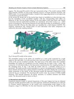
Fig. 1. Strategy for construction of a ∆epr mutant. (A) Construction of a chloramphenicol-
resistance (Cm
r
) plasmid, pUC118-Cm
r
∆epr. Fragment 3 was amplified with fragment a
(containing the repU promoter of pUB110) and fragment b (containing the chloramphenicol
resistance gene of pC194), and primers 7 and 8. Fragment 4 was amplified with fragments 1,
2, 3, and primers 5 and 6. The amplified fragment 4 was prepared by blunting and kination,
and then cloned into the SmaI site of pUC118 to generate pUC118-Cm
r
∆epr. (B) Construction
of the ∆epr mutant. B. subtilis 168 cells were transformed with pUC118-Cm
r
∆epr, followed
by selection for chloramphenicol resistance, obtaining Cm
r
∆epr. To obtain a ∆epr mutant
(deleted chloramphenicol-resistance cassette), the ampicillin concentration method was used
(Kodama et al., 2007a). The chloramphenicol-sensitive (Cm
s
) ∆epr mutant was confirmed by
PCR using primers 5 and 4.
Approaches for Improving Protein Production in
Multiple Protease-Deficient Bacillus Subtilis Host Strains
165
found in the culture supernatant of B. subtilis WB600 (Babe et al., 1998; Wu et al., 1991).
Whether it is present in the cell wall or in the culture medium is therefore a critical factor in
the degradation of heterologous proteins (Lee et al., 2000). Strains with deletion mutations in
multiple extracellular proteases have since been constructed with extracellular protease
activities of less than 0.5%, compared to the parental strain (Wu et al., 1991). It was recently
reported that an eight protease-deficient strain, WB800, was a useful host for the production
of various heterologous proteins (Murashima et al., 2002; L. Westers et al., 2006). However,
the use of B. subtilis as a host has remained limited to bulk industrial enzyme production.
Further optimization is necessary to develop production systems for heterologous proteins.
This chapter focuses on the inhibition of proteolysis of secreted proteins after the
translocation process by inactivation of multiple proteases which are extracellular (AprE,
Bpr, Epr, Mpr, NprB, NprE, Vpr, and WprA), leaked outside from intracellular (AprX), and
membrane-bound (HtrA and HtrB).
2.1 The intracellular protease, AprX is involved in degradation of a heterologous
protein
In B. subtilis, extracellular protease-deficient mutants have been used in attempts to increase
the productivity of heterologous proteins. We detected the protease activity of AprX using
protease zymography in the culture medium at the late stationary growth phase.
Construction of multiple-protease-deficient mutant without antibiotic-resistance markers
and the effect of AprX on the heterologous protein production are descrived in detail in the
following sections.
2.1.1 Construction of an eight-extracellular-protease-deficient mutant by marker-free
deletion in B. subtilis
Antibiotic-resistance marker genes were used to create new bacterial strains. However, the
number of markers available for use in B. subtilis and other bacteria is limited. We used the
“ampicillin concentration” method for the creation of eight-extracellular-protease-deficient
mutant with marker-free deletion (Fig. 1, Kodama et al., 2007a). Recently, several useful
methods were developed to produce unmarked mutations in B. subtilis (Liu et al., 2008;
Morimoto et al., 2008; Morimoto et al., 2009). These systems are more convenient for the
introduction of multiple mutations.
2.1.2 Detection of AprX activity in the culture supernatant with protease zymography
Zymography has been used to detect proteolytic enzymes after electrophoretic separation in
gels. Recently, the activities of some proteases, including Vpr have been detected by fibrin
zymography of the extracellular proteins of B. subtilis (Murashima et al., 2002; L. Westers et
al., 2006). The supernatant proteins of B. subtilis culture in modified 2xL broth (Kodama et
al., 2007b) at 8 h (exponential growth phase), 25 h (early stationary phase), 50 h (mid-
stationary phase), and 75 h (late stationary phase) (Fig. 2A) were analyzed by gelatin
zymography (Fig. 2B). The resulting zymogram shows the protease activities as clear bands
(or zones). In the exponential growth phase (8 h), no protease activity was detected (Fig. 2B,
lane 1). However, protease activity increased during the stationary phase, and was highest
Advances in Applied Biotechnology
166
at 75 h of the late stationary phase (Fig. 2B, lane 4). We examined the zymogram profile of
the supernatant from the eight-extracellular-protease-deficient mutant (Dpr8) at 75 h, and
found one clear band in the zymogram (Fig. 2C, lane 2). Protease activity disappeared in the
aprX mutant at 75 h (Fig. 2C, lane 3). All of the protease activities completely disappeared in
the KA8AX strain, in which nine genes (eight extracellular protease genes and aprX) were
disrupted (Fig. 2C, lane 4). These results support the idea that the protease is serine protease
AprX. To determine the serine and metal protease activities of this protease by zymography,
PMSF and EDTA (2 mM each) were added to the supernatant of the 75 h culture of Dpr8
(Fig. 2D). EDTA decreased the activity of the protease slightly, whereas 2 mM PMSF
completely inhibited the protease activity (Fig. 2D). These results suggest that the gelatin-
degrading protease from the supernatant of Dpr8 culture is AprX. To determine whether
AprX is the gelatin-degrading protease in the supernatant of the Dpr8 culture at 75 h, the
AprX-FLAG fusion protein was constructed. The fusion gene was expressed with the
original promoter and ribosomal binding site. On a zymographic gel, the activity bands
corresponding to AprX-FLAG from both 168/AprX-FLAG and Dpr8/AprX-FLAG strains
were located at slightly larger positions in size than those of AprX (Fig. 3). The size of the
band corresponded to the size of the FLAG peptide. These results indicate that the activity
of AprX is detectable as a single band by gelatin zymography of the supernatant of a 75 h
culture of B. subtilis strains.
Fig. 2. (A) Cells from the wild-type were cultured in modified 2xL broth at 30ºC. (B)
Protein samples were prepared from the supernatants of the wild-type, cultured for
various incubation times. Lane 1, 8 h culture; lane 2, 25 h; lane 3, 50 h; lane 4, 75 h. (C).
Protein samples were prepared from the supernatants of the protease-deficient mutants
after a 75 h culture. Lane 1, 168; lane 2, Dpr8; lane 3, AprXdd; lane 4, KA8AX. (D) PMSF or
EDTA (2 mM each) was added to the supernatants of Dpr8 after a 75 h culture. Lane 1,
Control (no addition); lane 2, addition of 2 mM PMSF; lane 3, addition of 2 mM EDTA.
The samples were analyzed on SDS-12% polyacrylamide gels with 0.1% (w/v) gelatin.
Proteins from the culture supernatants (equivalent to 0.3 µl) were applied to each lane for
panels B, C, and D.
Approaches for Improving Protein Production in
Multiple Protease-Deficient Bacillus Subtilis Host Strains
167
Fig. 3. Zymography of AprX-FLAG proteases. (A) Lane 1, 168; lane 2, 168/AprX-FLAG. (B)
Lane 1, Dpr8; lane 2, Dpr8/AprX-FLAG. Proteins from the supernatants of the 75-h cultures
(equivalent to 0.3 µl) were applied to the lanes for panels A and B. Arrowheads indicate the
positions of AprX (closed symbol) and AprX-FLAG (open symbol).
2.1.3 Intracellular AprX leaked to the culture medium during the late stationary phase
It has been supposed that AprX is a serine protease belonging to the subtilase superfamily,
and that it is an intracellular protease, because a canonical signal sequence for secretion has
not been found in this protease (Valbuzzi et al.; 1999). However, AprX was detected in the
culture medium by gelatin zymography (Fig. 3). aprX is transcribed during the stationary
phase, and the regulator of SinR exerts negative effect on its transcription directly or
indirectly (Valbuzzi et al.; 1999). However, aprX is not essential for either growth or
sporulation (Valbuzzi et al.; 1999). As a result, the function of AprX has remained poorly
understood. The Western blotting of AprX-FLAG from the intracellular fraction showed that
the expression of AprX-FLAG began at 25 h, and that the expression level markedly
increased after 50 h (Fig. 4). Our results agreed with a previous report that aprX is
transcribed during the stationary phase. In contrast, a weak AprX-FLAG expression was
detected in the supernatant only in the late stationary phase at 75 h (Fig. 4). This result
agreed with the zymogram pattern of wild-type AprX (Fig. 2B). A slight decrease in cell
density was observed after 50 h for the wild-type (Fig. 2A) and 168/AprX-FLAG strains
(data not shown). The bands corresponding to AprX-FLAG from both the intra- and
extracellular fractions were located at the same position. This result suggests that there is
insufficient secretion of AprX, due to the absence of an obvious signal sequence. These
observations also suggest that AprX is localized intracellularly by nature, and is leaked to
the culture medium during the late stationary phase due to cell lysis.
Fig. 4. Western blot analysis of AprX-FLAG. Western blot analysis was carried out to detect
AprX-FLAG in the 168/AprX-FLAG strain with the anti-FLAG antibody. Proteins of cells
(lanes 1-3) and supernatants (lanes 4-6) from 168/AprX-FLAG (0.02 OD600 units) were
prepared as described in Materials and Methods. The arrowhead indicates the position of
AprX-FLAG. The times of harvest of cells and supernatants are shown at the top.
Advances in Applied Biotechnology
168
2.1.4 AprX involved in degradation of the α-amylase-A522-PreS2 hybrid protein
AprX in the supernatant was able to degrade gelatin. Therefore, we considered that AprX
may affect the production of secreted proteins. pTUBE522-preS2 has already been
developed for the extracellular production of small peptides of the human hepatitis B virus
preS2 antigen (42 amino acids) fused with B. subtilis α-amylase (deleting the C-terminal
region to construct a peptide carrier) (Honda et al., 1993). To confirm the effect of AprX on
the degradation of heterologous proteins, we examined the production of α-amylase-A522-
PreS2 as a model of heterologous proteins, in multiple-protease-deficient B. subtilis strains.
Cells carrying pTUBE522-preS2 were cultured in modified 2xL broth for 25, 50, and 75 h. α-
amylase-A522-PreS2 in the supernatants from the cultures of Dpr7, Dpr8, and KA8AX
strains was analyzed by Western blotting with the anti-PreS2 antibody (Fig. 5A). The Dpr7
strain lacked seven extracellular proteases (AprE deficiency excluded). No positive band
corresponding to α-amylase-A522-PreS2 was detected at any phase for Dpr7 (pTUBE522-
preS2) (Fig. 5A, lanes 1-3). Dpr8 (pTUBE522-preS2) produced α-amylase-A522-PreS2 at
detectable levels, and production of the hybrid protein attained high levels after 50 h (Fig.
5A, lane 5). However, when AprX was produced in the supernatant of Dpr8 (pTUBE522-
preS2) at 75 h, the amount of α-amylase-A522-PreS2 decreased markedly (Fig. 6A, lane 6).
As expected, the degradation of α-amylase-A522-PreS2 was markedly inhibited in KA8AX
(pTUBE522-preS2) at 75 h, with the relative amount of the hybrid protein produced by this
strain being 1.8-times higher than that of Dpr8 at 50 h (Fig. 5A, lanes 5 and 9; Fig. 5B).
KA8AX produced α-amylase-A522-PreS2 up to 80 mg/L, which is at least eightfold higher
than the amount produced by the improved strain in a previous study (Lee et al., 2000; Fig.
5, lane 9). We also examined the degradation of α-amylase-A522-PreS2 by AprX protease.
First, we prepared AprX protease from KA8AX (pDG-AprX) that was grown in a medium
containing 1 mM IPTG for 4 h. The overexpression of AprX was confirmed by gelatin
zymography (Fig. 6A, lane 2). Afterwards, the α-amylase-A522-PreS2 protein prepared from
the supernatant of KA8AX (pTUBE522-preS2) at 75 h was mixed with AprX protease, and
the mixture was incubated at 37ºC for 60 min. The degradation of α-amylase-A522-PreS2
was analyzed by Western blotting using the anti-PreS2 antibody. H
2
O and intracellular
proteins extracted from KA8AX (pDG-AprX) cells cultured without IPTG did not decrease
the amount of the α-amylase-A522-PreS2 protein (Figs. 6B, C, lanes 2 and 4), but
intracellular proteins extracted from KA8AX (pDG-AprX) cells cultured with 1 mM IPTG
decreased the amount to 70% (Figs. 6B, C, lanes 5 and 6).
These results indicate that the AprX protease directly degraded the α-amylase-A522-PreS2
protein. One bottleneck of the production of α-amylase-A522-PreS2 was partially solved by
the disruption of eight extracellular proteases and AprX, as shown in this chapter. However,
the supernatant from the KA8AX culture at 75 h contained not only a small amount of α-
amylase-A522-PreS2, but also a large amount of α-amylase protein (determined by Western
blotting with the anti-α-amylase antibody; data not shown). On other hand, no PreS2
peptide was detected by Western blotting with the anti-PreS2 antibody (Fig. 5A). These
results indicate that the degradation of α-amylase-A522-PreS2 was not inhibited completely
in the KA8AX strain, and that there were as yet unidentified protease(s) involved in the
proteolysis of the PreS2 region. Therefore, there is still room for improving the inhibition of
hybrid protein degradation. It has been reported that IspA (Isp) was identified as a major
intracellular serine protease (Koide et al., 1986). We evaluated the inhibition of the