Bioluminescence Recent Advances in Oceanic Measurements and Laboratory Applications Part 5 ppt
Bạn đang xem bản rút gọn của tài liệu. Xem và tải ngay bản đầy đủ của tài liệu tại đây (1001.57 KB, 15 trang )
Bioluminescent Proteins: High Sensitive Optical Reporters
for Imaging Protein-Protein Interactions and Protein Foldings in Living Animals
51
cells. Protein–protein interactions are important determining factors in the regulation of
many cellular processes. Signaling pathways regulating cellular proliferation,
differentiation, and apoptosis are commonly mediated by protein-protein interactions as
well as reversible chemical modifications of proteins (e.g., phosphorylation, acetylation,
methylation, and sumoylation), which normally control sub-cellular trafficking and function
of proteins. To understand these modifications in proteins, and protein modification-
assisted or independent protein-protein interactions, several techniques have been
developed and studied in intact cells and in cell extracts. The yeast two-hybrid system is one
of the earliest techniques, which used enzyme beta-galactosidase as a reporter protein at the
beginning, and later was improved by adopting bioluminescent reporters for rapid
measurement. The latter is used extensively in screening for protein-protein interactions and
also for identifying small molecule drugs that alter (inhibit or enhance) protein-protein
interactions, which can be used as therapeutic agents for treating several cellular diseases
including cancer. The major limitation of this system is that it can only study the protein-
protein interactions occurring in the nucleus; otherwise it requires the study proteins to be
trafficked into the nucleus. The readout of yeast two-hybrid system is based on the amount
of reporter proteins produced during protein-protein interaction associated transcriptional
activation of reporter proteins (see more details in section 3.2). To circumvent this limitation,
other techniques have been developed, including the split ubiquitin system, Sos recruitment
system, dihydrofolate reductase complementation, -galactosidase complementation, -
lactamase complementation, the G protein fusion system, and, most recently, split-luciferase
(firefly luciferase, click-beetle luciferase, renilla luciferase, and Gaussia luciferase) and split-
fluorescent (GFP and RFP) complementation systems. Of these, the split-luciferase
complementation system provides significant advantage over other systems, particularly in
measuring protein-protein interactions in cell lysates, intact cells, and cell implants in living
animals by molecular imaging. The firefly luciferase complementation imaging is robust and
a broadly applicable bioluminescence approach with applications in both modification-
independent (phosphorylation, acetylation, methylation, and sumoylation) and dependent
protein-protein interactions.
1.3 Post genomic proteomic era
We are in a post-genomic proteomic era. The completion of the human genome project has
given us knowledge of the complete nucleotide sequences of human genome, their
arrangements in different chromosomes, and the number of functional genes that are
present in a human cell. The information collected from the human genome project along
with other bio-informatic tools have led to several major new directions in science, including
the characterization of RNAs (via transcriptional profiling), microRNAs, and proteins
(proteomes). The human genome project estimated the number of functional genes in a
human cell to range from 30,000 to 40,000. The concept of one protein, one function can
accommodate only a limited number of functions, and does not explain the vastly more
proteins needed by cells than those produced from the limited number of functional genes.
The management of additional cellular functions, including various house-keeping
functions and other specialized functions, mainly depends on the functional organ or tissue
types to which these cells are part of. It is logical and even necessary to postulate that
multifunctional proteins within the cell, and/or various collaborative interactions between
proteins, are needed as molecular machines to carry out the work within a cell. To illustrate,
the proteomes are much more dynamic and complex than the genome; it changes during
Bioluminescence – Recent Advances in Oceanic Measurements and Laboratory Applications
52
development in response to external stimuli, and form large interaction networks through
which they support and regulate each other. The genetic blueprint and the genome of
human cells are well known. However, the functions that genome encodes and program
through which the proteins are produced by the genetic blueprint are not well understood.
New research is only beginning to uncover the incredibly rich diversity of protein structure,
which is much more complex than that of DNA. One new direction has sought to isolate and
structurally characterize all the proteins that exist in the cell (Skolnick et al., 2000; Tucker et
al., 2001). Unlike DNA, proteins have a vast repertoire of structures to carry out the
diversity of functions. Once the proteins are identified and characterized, a second major
challenge to find out how they assemble into the molecular machines that perform the
cellular functions. Identifying all of the protein-protein interactions is fundamental for
understanding the cellular processes involved in virtually all biological interactions. The
collection of protein-protein interactions can be visualized as a map, in which proteins are
the nodes and the circuits are the interactions. A protein-protein interaction network or map
would then represent a search grid on which biological circuits are constructed (Tucker et
al., 2001; Wills 2001).
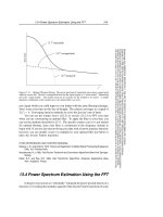
Fig. 1. Schematic illustration of current molecular imaging strategies, and their potential for
providing biological informations such as anatomical details, physiological data, and
metabolic status at the molecular level, for clinical applications in human. None of the
current strategies is uniquely superior in independently providing different informations
needed for making clinical decisions in diagnosis, staging and treatments especially in
oncology, diagnosis and treatment in several other diseases; each has its strengths and
weaknesses.
1.4 Complexity of protein interaction networks
There are thousands of different proteins active in a cell at any time. Many of these proteins
are working as enzymes that catalyze the chemical reactions of metabolism, while others
work as components of cellular machineries, such as ribosomes that read genetic
information and synthesize proteins. Still proteins are involved in the regulation of gene
Bioluminescent Proteins: High Sensitive Optical Reporters
for Imaging Protein-Protein Interactions and Protein Foldings in Living Animals
53
expression. Many proteins play their functional roles only in specific cellular compartments,
whereas others move from one compartment to another, acting as "signals". By directly
interacting with one another, proteins continually influence other functions (Wills 2001). In
addition, proteins are constantly produced and degraded in cells. The rates at which these
processes occur depend on how much of each protein is already present, how they interact
with each other, and with other macromolecules such as DNA and RNA, and regulate the
cellular mechanisms. One protein can speed up or slow down the rate of production of
another by interacting with DNA or RNA, which is needed for making that particular
protein. The interactions between different proteins that control different cellular functions
are therefore interdependent. When a mutation causes the loss of one of these essential
protein functions, then this can significantly affect the function of many other proteins, even
leading to cell death (Tucker et al., 2001). Clearly the interactions between different proteins
in a cell are much more complex than previously thought, and it is vital to understand their
fundamental interlinked networks. Protein–protein interactions are important determining
factors in the control of many cellular processes such as transcription, translation, cell
division, signal transduction, and oncogenic transformation. To modulate many of these
cellular events, it is essential to delineate which proteins are involved and how they interact
with one another, their precise roles in executing cellular functions, and techniques and
mechanisms needed to manipulate these interactions for novel drug development or
treatment strategies relevant to particular diseases. Biochemical pathways and networks
require many different systems of dynamic assembly and disassembly of proteins with other
proteins and nucleic acids (Michnick 2001). Much of modern biological research is
concerned with how, when, and where proteins interact with other proteins involved in
biological processes in the intact cellular context. The completion of the human genome
project has added a major impetus in research that can provide simple approaches to study
protein-protein interactions on a large scale in diseases, including cancer.
1.5 Cellular signaling pathways
The cellular regulatory mechanisms are interlinked. To understand the complex biological
processes, and disease states at a molecular level, a systematic approach is necessary to
illustrate signaling pathways. Efforts to elucidate the cellular mechanisms for different
pathological conditions have significantly increased after the Human Genome Project. Each
signaling pathway reacts to specific external stimuli that can be regulated by changes in
proteins and chemicals. Recent advances in large-scale and high-throughput techniques,
including functional genomics, proteomics, RNAi technology, and genomic-scale yeast two-
hybrid and protein complementation assays, have provided a tremendous amount of
information on signaling pathways. To extract the biological significance from the vast data,
it is necessary to develop an integrated environment for a formal and structured
organization of the available information, in a format suitable for analysis with
bioinformatics tools. To present a signaling pathway, a database must include information
on 1) the molecules involved in signaling in response to each external stimulus, 2) which
direction the signal is being conveyed, and 3) how the activities and sub-cellular
localizations of molecules are changed by protein modifications and/or protein-protein
interactions. Analyses of the first database containing such information should made it
possible to further expand the database to understand the signaling results in processes such
as proliferation, differentiation, and apoptosis, and to explicate how a network can be
Bioluminescence – Recent Advances in Oceanic Measurements and Laboratory Applications
54
composed of various signaling pathways in response to multiple external inputs. Signaling
entities ranging from small molecules and proteins-to-protein states and protein complexes
should be studied. It must be noted that these entities are not independent of one another.
For instance, protein complexes are composed of proteins, and a protein binding to a small
molecule can define a protein state. It is not surprising to find many gaps in the current
knowledge about any particular signaling pathway. In order to organize such diverse yet
incomplete information into a structured and coherent database, the use of a formal model
is indispensible. Differing levels of abstraction are inter-related so that essentially the same
signaling event can be described in detail at multiple levels. As model systems that
implement all the parameters become available, the sharing of models with integrated
biological data will be essential to fill in the gaps in our current knowledge base.
1.6 Complexity in studying protein interaction networks
There are no methods currently available to test protein-protein interaction networks, which
occur within a cell without introducing a constructed system that mimics the function of its
endogenous protein. It is to be expected that when a new protein of endogenous origin is
introduced in a cell in addition to the level of its counterpart expressed inside a cell, it will
have some direct physical effect on a number of other proteins. These new interactions may
cause some changes in the functional aspects of several other proteins. Such effects can be
felt right across the protein interaction network, most often becoming less significant as the
distance of the new protein from the other protein increases. It is also possible for
genetically modified cells to produce a new protein that will display completely new
patterns of protein interactions. This may not be evident until the cells find themselves in
some unusual circumstances. They may then respond in a very different way from wild-
type cells. Although the genetically engineered cells may appear to behave just like wild-
type cells, this cannot be guaranteed under all circumstances (Becker et al., 1990; Beeckmans
1999; Bode and Willmitzer 1975). However the techniques currently available for inserting
new DNA into the chromosomes of cells do not have any specific control mechanisms,
capable of directing the point of insertion in the organism's existing genome without
producing significant impact on the expression level of any of the endogenous proteins. Of
the gene delivery systems currently available, the adeno-associated virus is the only viral
mediated vector which can normally introduce and integrate a single copy of the transgene
specifically into human chromosome loci at 19 (19q13.3-qter). Otherwise, it is customary to
produce millions of cells with the new DNA inserted at essentially random positions in the
hope of producing at least some “hits.” Screening is then conducted to find those cells,
which must survive the engineering process and also express the newly inserted gene. These
survivors are then subjected to further screenings to find those that seem to behave most
like the wild-type, and yet possessing the new, desired, engineered properties. It is generally
assumed that any harm to an organism as a result of inserting a new gene will be observed
as a change in gross characteristics of the organism (Stopeck et al., 1998).
1.7 Biological importance in studying protein folding
As discussed in the previous sections, proteins are cellular macromolecules with complex
structural and functional properties. Dysfunctional protein folding represents the
Bioluminescent Proteins: High Sensitive Optical Reporters
for Imaging Protein-Protein Interactions and Protein Foldings in Living Animals
55
molecular foundation of a growing list of diseases in humans and animals. Proteins
undergo several levels of structural alterations executed by active chaperon complexes
(e.g., Hsp90, Hsp70) and indirectly by the inherent amino acid sequences, before they
become a biologically active functional entity of a cell. There is significant supporting
evidence that associates the misfolding of proteins with several cellular diseases,
including cancers (Table 2). Biologically representative in vitro and in vivo studies of these
abnormal events are best suited to the discovery of molecular mechanisms to prevent or
ameliorate such diseases. There is an active search for small molecules which assist
refolding of misfolded proteins into their biological functional forms, as equal or at near
equal levels of native forms, for the treatment of several biochemical disorders. However,
thus far no current technique can be optimally extended to imaging assays in intact living
subjects. The development of novel imaging techniques to quantitatively measure the
level of protein misfolding in cells and in living animals, and also of small molecule
mediated refolding, will be very useful for screening and pre-clinical evaluation of drugs
which rectify or cure these diseases. Normally, the conformational changes in protein
folding result in the close approximation of amino and carboxy termini in a great majority
of native proteins, at their functionally active forms. The ‘protein folding problem’ has
remained one of the more perplexing quandaries in fundamental biological research ever
since the classic work of Anfinsen some four decades ago on the hydrophobic-collapse
mechanism. How to predict the three-dimensional, biologically active, native structure of
a protein from its primary sequence, and how a protein reaches this native structure from
its denatured state are still unresolved questions. The intellectual conundrum of the
folding pathway of proteins, underscored by the Levinthal paradox, has been addressed
to some extent over the last twenty years by various proposed mechanisms for protein
folding, including the framework model (diffusion-collision and nucleation mechanisms)
(Anfinsen 1973; Levinthal 1969).
There is accumulating evidence that the conditions used for refolding proteins in vitro are
only distantly related to those found in vivo, where the physiological environment in living
cells exerts a profound influence on protein folding owing to the involvement of the
intracellular macromolecular background, which also contains folding catalysts and
molecular chaperones. Aside from the relevance of the protein-folding problem to
deciphering fundamental processes in cell biology, it is becoming clear that dysfunctional
protein folding represents the molecular foundation of a growing list of diseases in humans
and animals. There is mounting interest in such diseases arising from protein misfolding
and aggregation, including Alzheimer’s disease, amyloidosis, Creutzfeldt-Jakob disease,
cystic fibrosis and cancer, to name a few. Molecular chaperones are involved in the
protection of cells against protein damage through their ability to hold, disaggregate, and
refold damaged proteins or their ability to facilitate degradation of damaged proteins. Many
of the proteins implicated in the pathogenesis of misfolding diseases escape the diverse
chaperoning pathways that are in place to assist and assure the fidelity of correct protein
folding. More biologically representative in vivo structural and functional studies of these
abnormal events, carried out in the context of living cell environments, are likely best suited
to the discovery of molecular mechanisms to prevent or ameliorate such diseases
(Table 2)(Goetz et al., 2003).
Bioluminescence – Recent Advances in Oceanic Measurements and Laboratory Applications
56
Disease Mutant Protein/Protein
involved
Molecular Phenotype
Inability to fold
Cystic fibrosis CFTR Misfolding/altered Hsp70
and calnexin interactions
Marfan syndrome Fibrilin Misfolding
Amyotrophic sclerosis Superoxide dismutase Misfolding
Scurvy Collagen Misfolding
Maple syrup urine disease
-Ketoacid dehydrogenase
complex
Misassembly/Misfolding
Cancer p53 Misfolding/altered Hsp70
interaction
Osteogenesis imperfecta Type I procollagen pro a Misfolding/altered BiP
expression
Toxic folds
Scraple/Creutzfeldt-jakob/
familial isomnia
Prion protein Aggregation
Alzheimer’s disease
-Amyloid
Aggregation
Familial amyloidosis Transthyretin/lysozyme Aggregation
Cataracts Crystallins Aggregation
Mislocalization owing to
misfolding
Familial
hypercholesterolemia
LDL receptor Improper trafficking
1-Antitrypsin Deficiency 1-Antitrypsin
Improper trafficking
Tay-Sachs disease
-Hexosaminidase
Improper trafficking
Retinitis pigmentosa
Leprechunism
Rhodopsin
Insulin receptor
Improper trafficking
Improper trafficking
Table 2. Examples of some putative protein misfolding associated diseases and proteins
involved in these diseases
2. Molecular imaging
2.1 Role of molecular imaging in cancer research
Molecular imaging, a new field of pharmacology that exploits the multidimensional
approaches of light energy, has paved the way for easy understanding, interpretation, and
manipulation of biological events at the molecular level. Molecular imaging is instrumental
in the diagnostic aspects of various pathological conditions, and also in the evaluation of
drugs which target specific molecular and biochemical processes in living cells and in intact
living animals. This new field of research has flourished with the introduction of many
novel molecular imaging probes, and genes which emit light either by reacting with a
Bioluminescent Proteins: High Sensitive Optical Reporters
for Imaging Protein-Protein Interactions and Protein Foldings in Living Animals
57
substrate or with induction of light waves, as well as the advancement of sensitive imaging
instrumentations. Imaging techniques such as positron-emission tomography (PET), single-
photon emission tomography (SPECT) with the use of radioactive tracers, and magnetic
resonance imaging (MRI), are now widely used in the clinical observation of cancer
pathology. Recently, the use of combinatorial techniques like PET-CT and PET-MRI are
rapidly replacing conventional imaging methods. Positron Emission Tomography (PET) is
an imaging technique that produces three-dimensional images of the functional processes of
living subjects by capturing a pair of gamma rays emitted indirectly upon the injection of a
positron-emitting radionuclide into the living body with a biomolecule.
PET was introduced by David E. Kuhl and Roy Edwards of the University of Pennsylvania
in late 1950s, and has been continually updated and modified to correspond to the clinical
and research needs to work as an independent or a combinatorial device (Ter-Pogossian et
al., 1975). It has made invaluable contributions in cancer diagnosis, treatment, and research
by revealing tumor progression both in clinical and preclinical applications, especially in the
diagnosis and detection of tumor metastasis. Advances in PET scanner devices and the
introduction of novel radiotracers have fueled the progress of PET imaging.
Fluorodeoxyglucose (
18
F-FDG), an analogue of glucose, is the most common radiotracer
used for PET imaging, because it can reveal specific tissue metabolic activity. However,
18
F-
FDG is phosphorylated by hexokinase and the phosphate cannot be cleared in most tissues,
which can result in intense radiolabeling of tissues with high glucose uptake (Burt et al.,
2001). FDG-PET is widely used in clinical oncology for diagnosis, staging and follow-up
after treatment of tumors. Tissue and also molecule-specific radiotracers have been
introduced to monitor the expression level of structural and functional proteins (Torigian et
al., 2007). Steroid receptors have been associated with the growth of breast tumors, and thus
understanding the receptor status is essential for the treatment of breast cancer.
Radiolabeled ligands and their analogues are in preclinical application for receptor imaging.
18
F-fluro-17β-estradiol (FES) has been used in PET imaging to examine the estrogen receptor
status in different tissues of living subjects (Mintun et al., 1988). Bombesin, a peptide
isolated from the frog Bombinas bombina, binds with gastrin-releasing peptide (GRP) receptor
and has been implicated in breast cancer. This property has led the development of
radiolabeled bombesin for peptide receptor imaging in breast cancer diagnosis (Scopinaro et
al., 2002).
Single Photon Emission Computed Tomography (SPECT) is another imaging technique that
is similar to PET imaging. Unlike PET, however, the tracer used in SPECT emits gamma
rays that can be measured directly. In SPECT, the 2-D view of 3-dimensional images is
acquired by a gamma camera and eventually 3-D data set is generated with the use of
computer based tomographic reconstruction algorithm. Magnetic Resonance Imaging (MRI)
is another widely used imaging technique, but unlike PET and SPECT, MRI can be used to
view the anatomical nature of living subjects by generating data about the functional status
of tissues (MacDonald et al., 2010). Magnetic Resonance Imaging (MRI) is a well-established
diagnostic method to detect cancer. It has been widely used to detect breast cancer as it
produces the highest sensitivity in spatial resolution of all imaging modalities. Guinea et al.
(2010) investigated and analyzed the possible relationship between the magnetic resonance
imaging (MRI) features of breast cancer and its clinicopathological and biological factors
such as estrogen and progesterone receptor status, and expression of p53, HER2, ki67,
Bioluminescence – Recent Advances in Oceanic Measurements and Laboratory Applications
58
VEGFR-1, and VEGFR-2 (Fernandez-Guinea et al., 2010). Their results did not show a
significant association between the MRI parameters and any of the biological factors
included in the study. By contrast, other reports have shown that high spatial dynamic MRI
of morphological or kinetic analysis are associated with prognostic factors such as
expression of estrogen receptor (ER), expression of progesterone receptor (PR), and
expression of p53, c-erbB-2, and Ki-67 (Fernandez-Guinea et al., 2010, Szabo et al., 2003)
found that rim enhancement pattern, early maximal enhancement, and washout phenomena
are associated with the poor prognostic factors of histological differentiation, high Ki-67
index, and negative PR expression status (Szabo et al., 2003). It was suggested that these MR
images could be useful in the prognosis of breast cancer. The MR signals may also be used
to noninvasively identify highly aggressive breast carcinomas and help differentiate
between benign and malignant lesions. The difference in contrast enhancement has been
associated mainly with a higher vascular permeability in tumors, and the overexpression of
c-erbB2 in tumor cells is closely linked to increased expression of vascular endothelial
growth factor (VEGF) and Ki-67 in proliferation (Szabo et al., 2003).
Similar to nuclear and magnetic imaging modalities, optical imaging has also made a sizable
contribution in medical imaging (Weissleder and Pittet 2008). Fluorescence and
bioluminescence methods are used as a source of contrast in optical imaging, but a major
setback in optical imaging is the lack of penetration depth, which prevents its wide clinical
applications in humans. Near infrared (NIR) imaging has been identified as a useful optical
imaging technique because of the lower absorption coefficient of tissue to light in near
infrared region. Radiolabeled antibodies have been evaluated for breast cancer diagnosis
since 1978 with tumor associated antigens such as carcinoembryonic antigen (CEA) and the
polymorphic epithelial mucin antigen (MUC1) (Goldenberg et al., 1974). They were widely
used in studies of immunolocalization and radioscintigraphy. Clinically, affinity purified
131
I-labeled goat anti-CEA IgG was subjected in selective breast tumor targeting (Goldenberg
and Sharkey 2007). CEA-Scan with Arcitumomab, an US FDA approved anti-CEA antibody,
could detect breast cancer that has been missed by mammography. In addition, antibodies
against the HER2/neu receptor have also been investigated in detail either as therapeutic
and/or diagnostic agent. Biotinylated anti-HER2/neu antibodies have been used to increase
the contrast of MR images when they bind with avidin-gadolinium complexes (Artemov et
al., 2003). The
111
In labeled Trastuzumab (Herceptin) Fab has been identified as a selective
imaging agent to localize HER2/neu receptor in small BT-474 tumors (Tang et al., 2005).
2.2 Reporter gene imaging in living animals
Molecular imaging is a rapidly expanding field that attempts to visualize fundamental
molecular/cellular processes in living subjects (Gambhir 2002; Massoud and Gambhir 2003;
Weissleder 2002). Imaging molecular events in cells in their native environment within the
living subjects probably result in the least amount of perturbation of normal signaling
processes. However, this advantage of non-invasive imaging has a trade-off. For example,
most current techniques do not have single cell resolution at any significant depth within
the animal. Instead, bulk signals from large numbers of cells (hundreds to millions) are
needed. Newer methods that allow the observation of single cells within living subjects are
under active investigation, but are more invasive in nature (Jung and Schnitzer 2003; Mehta
et al., 2004). To produce a signal detectable outside the animal subject, the cells located
Bioluminescent Proteins: High Sensitive Optical Reporters
for Imaging Protein-Protein Interactions and Protein Foldings in Living Animals
59
inside the subject must produce a signal of sufficient intensity. The signal may come from a
fluorescent protein excited at the correct wavelength, the interaction of a bioluminescent
protein with its substrate (Figure 2), or from radiolabeled substrates that emit a signal in the
form of gamma rays. For optical signals, red light and near infrared light have the best
tissue penetration, and are therefore preferred. For radiation-based signals, the use of single
photon emitters and positron emitters generating gamma rays are favored. It is not possible
to use beta emitters (e.g.,
3
H), due to their minimal tissue penetration. The focus of this
chapter is primarily on optical technologies for imaging protein-protein interactions.
Although other approaches such as microPET can be used for imaging protein-protein
interactions (Luker et al., 2002a; Massoud et al., 2010), the much lower cost, higher
throughput, and greater sensitivity of optical imaging in small animals favor its use for
imaging protein-protein interactions. Additional discussions of other small animal imaging
technologies including microPET may be found elsewhere (Cherry and Gambhir 2001;
Massoud and Gambhir 2003).
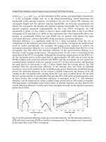
Fig. 2. Schematic illustration of the principle of optical bioluminescence imaging in cells. In
this strategy mammalian cells are labeled to express bioluminescent protein under a
constitutive (CMV, LTR, Ubiquitin, or CAG) or tissue-specific or an inducible promoter,
either by transfecting a plasmid with chemical agent (Liposome) or transduced with a viral
vector (Lentivirus, Adenovirus, Retrovirus, or Adeno-associated virus). The cells can be
allowed to express luciferase protein for a particular period of time and imaged by exposure
to substrate luciferin in intact cells, or can be measured by luminometer in cell lysates by
adding luciferin and other co-factors.
Bioluminescence – Recent Advances in Oceanic Measurements and Laboratory Applications
60
There are two primary types of optical imaging systems for living subjects: a) fluorescence
imaging, which use emitters such as green fluorescent protein (GFP), wavelength-shifted
GFP mutants, red fluorescent protein (RFP), “smart” near-infrared fluorescent (NIRF)
probes, and b) bioluminescence imaging, which utilizes a specific enzyme-substrate reaction
such as Firefly luciferase/D-Luciferin, Renilla luciferase/coelenterazine (Bhaumik and
Gambhir 2002a; Contag and Ross 2002; Tung et al., 1999) and several other bioluminescent
proteins with the respective substrates (Substrates and properties are shown in Table 1).
Emission of light from fluorescent markers requires external light excitation, while
bioluminescence systems generate light de novo after an injectable substrate is introduced. In
both cases, emitted light can be detected with a thermoelectrically cooled charge-couple
device camera (CCD), which can detect light in the visible light range (400 nm to 750 nm) to
near-infrared range (~800 nm) (Figure 3). Cooled to -120 to -150C, these cameras are
exquisitely sensitive to even weak luminescent sources within a light-tight “black-box”
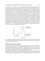
Fig. 3. Scheme of optical imaging (bioluminescence and fluorescence) in living animals. In
this strategy, mammalian cells stably expressing bioluminescent or fluorescent proteins are
implanted in animals (orthotopic or xenograft) and allowed to grow the tumors. The
animals were imaged by exciting with respective excitation wavelength of the protein used
for labeling in fluorescence imaging. The emitted light was captured by an optical cooled
charge coupled device camera, and quantitated by compatible software provided with the
system. Similarly, in bioluminescence imaging, the animals were injected with the respective
substrate of the bioluminescent reporter used for labeling the cells, and light emitted from
the tumor cells is collected without passing through filters.
Bioluminescent Proteins: High Sensitive Optical Reporters
for Imaging Protein-Protein Interactions and Protein Foldings in Living Animals
61
chamber, allowing for quantitative analysis of the data. The method of imaging
bioluminescence sources in living subjects with a CCD camera is relatively straightforward:
the animal is anesthetized, injected intravenously or intraperitoneally with the substrate,
and placed in the light-tight chamber for a few seconds to minutes. A standard light
photographic image of the animal is obtained, prior to a bioluminescence image captured by
the cooled CCD camera positioned above the subject within the confines of the dark
chamber. A computer subsequently superimposes the two images on one another, and
relative location of luciferase activity is inferred from the composite image. An adjacent
pseudocolor scale indicates relative or absolute number of photons detected. This scale does
not reflect the color (wavelength) of the emitted photons, but only the number of such
photons, measured in relative light units per minute (RLU/min). At present, the primary
disadvantage of fluorescence as compared to bioluminescence is the background level of
auto-fluorescence of tissues in the former approach. However, methods are being developed
to correct for auto-fluorescence and may allow greater use of fluorescence in the study of
protein-protein interactions in living subjects. To date, the initial validation of in vivo
imaging has been based primarily on bioluminescence. If auto-fluorescence issues can be
minimized, then it may be possible to image multiple interactions using different fluorescent
reporters. Comparison of optical-based imaging systems with the other imaging modalities,
such as the radionuclide-based or MRI-based systems, reveals important differences. One
major advantages of optical-based reporter systems is that they are at least an order of
magnitude more sensitive than the radionuclide-based techniques at limited depths (Ray et
al., 2003). Furthermore, the direct and indirect costs of optical systems are generally less
than radionuclide-based techniques or MRI. However, significantly less spatial information
is obtained from optical imaging, and the signal obtained from light-emitting reporter
systems is limited by the tissue depth from which it arises. Furthermore, while significant
progress has been made to localize fluorescent signals tomographically to obtain
distribution of fluorochromes in deep tissues (Ntziachristos et al., 2002), there are currently
only prototype instruments to obtain three-dimensional localization of the targeted optical
probes.
2.3 Optical reporter genes to image cellular process in small animal models
Luciferases are enzymes that emit light when they react with a specific substrate. A diverse
group of organisms make use of luciferase-mediated bioluminescence. Luciferases that
catalyze the light emitting reactions of fireflies, coelenterates, or bacteria show no nucleotide
homology to each other. The substrates (i.e., luciferins) of these reactions are also chemically
unrelated (Wilson and Hastings 1998). All these bioluminescent reactions combine
molecular oxygen with luciferin, to form a luciferase-bound peroxy-luciferin intermediate.
This, in turn, releases photons of visible light (Wilson and Hastings 1998) over an emission
spectrum range between 400 and 620 nm. Experimentally, the emitted light is used as a
"reporter" for the activity of any regulatory elements that control expression of luciferase.
Firefly luciferase (FLUC), cloned in 1985 from the firefly Photinus pyralis, is now emerging as
the gene of choice for in vivo and in vitro reporting of transcriptional activity in eukaryotic
cells (de Wet et al., 1985). FLUC emits
light from green to yellow in the presence of D-
Luciferin,
ATP, magnesium, and oxygen. The short half-life and fast rate of turnover of
FLUC (T
1/2
~ 3 h) in the presence of D-Luciferin
allows for real-time measurements, because
the enzyme does not
accumulate intracellularly to the extent of other reporters; thus, the
Bioluminescence – Recent Advances in Oceanic Measurements and Laboratory Applications
62
relationship between the enzyme concentration and the intensity of emitted light in vitro is
linear up to 7-8 orders of
magnitude. These properties potentially allow for sensitive
non-
invasive imaging of FLUC reporter gene expression in living
subjects (Massoud et al., 2004;
Wu et al., 2002). In recent years, considerable work with non-invasive imaging of firefly
luciferase has been carried out (Bhaumik and Gambhir 2002b; Contag et al., 2000; Contag et
al., 1997; Contag et al., 1998; Jawhara and Mordon 2004; Paulmurugan et al., 2002b; Ray et
al., 2002a; Wu et al., 2002; Wu et al., 2001).
The use of a second bioluminescent reporter [(Renilla Luciferase (RLUC)] with different
substrate utility than firefly luciferase, has allowed for monitoring of more than one process
at a time in mammalian cells. Renilla luciferase (RLUC), originally cloned and sequenced
from the sea pansy, Renilla reniformis, by Lorenz et al. (Lorenz et al., 1991), has been used as
a marker of gene expression in bacteria, yeast, plant and mammalian cells (Lorenz et al.,
1996). RLUC is widely distributed among coelenterates, fishes, squids and shrimps
(Hastings 1996). The enzyme RLUC catalyzes oxidation of its substrate, coelenterazine,
leading to bioluminescence. Coelenterazine has an imidazolopyrazine structure [2-(p-
hydroxybenzyl)-6-(p-hydroxyphenyl)-8-benzylimidazo [1, 2-a] pyrazin-3- (7H)-on], and
upon oxidation, releases blue light across a broad range, peaking at 480 nm (Wilson and
Hastings 1998). However, the native RLUC protein has some inherent limitations when used
in mammalian cells. Ten percent of its codons are associated with poor translation in
mammalian cells, limiting expression efficiency. Also, the presence of a large number of
potential transcription factor binding sites within RLUC sequences can cause anomalous
transcriptional behavior in mammalian cells. For many in vivo imaging applications,
researchers have utilized a synthetic Renilla luciferase reporter gene (hRLUC) that has been
codon optimized for efficient translation in mammalian cells. In addition, deletion of poly
(A) signals (AATAAA) and incorporation of a Kozak sequence at the beginning of the gene
contributed to a better expression. The resulting reporter gene has a higher transcriptional
efficiency, which enhances the detection of the reporter enzyme in cell culture and living
animals (Bhaumik et al., 2004).
After a long lag time, recently several other luciferases with different properties have been
isolated. Gaussia luciferase (GLUC) with its secretary property has been recently used for
secretary biomarker identification and validation in human cancers. As GLUC uses the same
substrate (coelenterazine) as RLUC and emits light in a similar wavelength (480nm), but
differs in its secretary property, its use has been limited in several applications. Similarly,
Metridia luciferase, with similar substrate utilization, emission wavelength and secretary
property as GLUC, has been identified, but it has not added much to the field of
bioluminescence imaging in living animals. Recent isolation of another luciferase (Vargula
luciferase) with similar physical properties (secretary, emits light at 480nm) as GLUC, but
uses a different substrate (Vargula luciferin), has the advantage of adding another
bioluminescent protein for multiplexing different bioluminescent reporters to enable
simultaneous monitoring of several biological processes in cells and in living animals.
3. Bioluminescent assays to study protein-protein interactions
As we discussed briefly in section 1.6 the different assays currently available for studying
protein-protein interactions, in this section we further explain how these assay systems
Bioluminescent Proteins: High Sensitive Optical Reporters
for Imaging Protein-Protein Interactions and Protein Foldings in Living Animals
63
work, and discuss the advantages most of these assays have over other complementary
systems, and their contributions to the field of molecular imaging in disease monitoring and
drug development processes.
3.1 Yeast two-hybrid system to study protein-protein interactions
Protein-protein interactions play vital part in almost all biological processes. Large networks
of interacting proteins control many important regulatory pathways. A full understanding
of any pathway or cellular processes will require a map of the binary interactions among the
proteins involved. One of the most widely used methods to detect biologically important
protein-protein interactions is the yeast two-hybrid system (Fields and Song 1989). In a two-
hybrid assay, the two proteins are expressed in yeast one fused to a DNA-binding domain
(BD) and the other fused to a transcription activation domain (AD). If the two proteins
interact, they activate transcription of one or more reporter genes that contain binding sites
of the BD. Investigated first in yeast, this classical two-hybrid system was later adopted for
mammalian cells with certain modifications (Luo et al., 1997). When expressed
simultaneously in the same cell, the interactions between the two mammalian proteins bring
the activation domain (VP16) and binding domain (Gal4) together that in turn bind to the
Gal4 domain binding sequences followed by a reporter gene. High levels of reporter gene
expression will indirectly indicate the physical interactions between the proteins of interest.
Most of the approaches used for high-throughput two-hybrid studies have been limited to
in vitro assays and cultured cells that do not represent the actual scenario in intact animal, as
well as being unable to predict the kinetics of the interaction. Thus, the development of
methods to non-invasively and repetitively image protein-protein interactions using the
mammalian two-hybrid system in living animal would significantly increase our knowledge
on the intricacy and complexity of different regulatory pathways.
To overcome these problems, we and others have developed a modified mammalian two-
hybrid system to detect protein-protein interactions in living mice using bioluminescence
and positron emission tomography (PET) imaging techniques (Luker et al., 2002b; Luker et
al., 2003; Ray et al., 2002b). To demonstrate the use of this system for imaging in living
animals, the two known interacting proteins, ID and MyoD, were used. These two proteins
strongly interact in vivo during muscle generation (Ray et al., 2002b). To modulate the
expression of these two fusion proteins (ID-GAL4 and MyoD-VP16), we used the NF-kB
promoter to drive expression of the Id-gal4 and/or myoD-vp16 fusion genes, and utilize
TNF- to induce the NF-kB promoter that controls the expression level of fusion proteins in
response to TNF- dose. The reporter construct comprised of five GAL4 binding sequences
followed by the firefly luciferase reporter gene. In cell culture, co-transfection with the
effector and reporter plasmids with variable levels of expression regulated by TNF- show
induction in FLUC activity. The FLUC activity directly correlate with the interaction
between the proteins used, which are Id and myoD in this case. By replacing these two
proteins, it is possible to look for other proteins that are currently not known for their
interactions (Figure 4).
A similar system was developed for a tetracycline inducible bi-directional vector carrying
two other proteins (the tumor suppressor p53 gene and SV40-T antigen) by other groups
(Luker et al., 2002b; Luker et al., 2003). Interactions of p53 and SV40-Tag proteins after
Bioluminescence – Recent Advances in Oceanic Measurements and Laboratory Applications
64
doxycycline induced expression resulted in formation of the VP16-Gal4 trans-activator
complex that binds to the Gal4 binding sequences driving expression of a HSV1-sr39TK-GFP
reporter fusion protein. Expression of GFP (fluorescence imaging) is detected at the level of
single cell, and expression of HSV1-sr39TK is imaged in living mice with microPET. Clones
of HeLa cells stably expressing both the reporter plasmid and the bi-directional effector
plasmid are isolated, and levels of interacting proteins are measured with increasing doses
of doxycycline and further confirmed by western blots, thymidine kinase radiotracer assays,
and fluorescence microscopy. Promising cell culture studies have led to further
investigations into the protein-protein interactions in living mice using different modalities
in cells and in vivo in living animals.
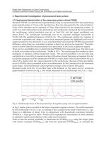
Fig. 4. Scheme of a single vector mammalian two hybrid system to study protein-protein
interactions. The vector is designed to have several components needed for measuring protein-
protein interaction. In this strategy, the vector expresses two fusion proteins under two
separate promoters. They can be either constitutive or inducible. One fusion protein contain
Gal4-DNA binding domain (Gal4-DBD) expresses with one of the study protein (Gal4-DBD-
Protein-X), and the other one expresses a small transactivating peptide derived from herpes
simplex virus thymidine kinase (HSV1) with another protein (Protein-Y-VP16). The same
vector has a specific DNA sequence repeated five times on which the Gal4-DBD can bind, and
a minimal promoter (TATA box), followed by a reporter gene (Luciferase). This minimal
promoter will not express any reporter protein until the Gal4-DBD binds to it and brings the
VP16 domain fused to another fusion protein by protein-protein interactions. The amount of
luciferase expression directly relate to the interaction which occurs between proteins X and Y.
Bioluminescent Proteins: High Sensitive Optical Reporters
for Imaging Protein-Protein Interactions and Protein Foldings in Living Animals
65
However, both the two-hybrid (TNF- and tetracycline inducible) approaches have several
limitations. Both use strong and constitutively (spontaneous) interacting proteins and are
unable to fully address weakly associated proteins or proteins with differential binding
affinity, and also to decipher the time-kinetics of protein-protein interactions. Moreover the
two-hybrid system could only detect the interacting proteins in the nucleus, not in the
cytoplasm where the largest pool of protein-protein interactions responsible for many
regulatory pathways occur. It is hoped that by combining non-invasive imaging approaches
involving detection of cytoplasmic protein-protein interaction (through protein
complementation study, split reporter strategy, FRET/BRET study etc.) with the two-hybrid
system, it will be possible to measure the complete spectrum of the pharmacokinetics and
pharmacodynamics of protein interactions in different regulatory pathways, as well as to
perform screening and pre-clinical evaluation of small molecule drugs for therapeutic
applications.
3.2 Bioluminescent reporter protein complementation assays to study protein-protein
interaction
Protein complementation assays with split luciferases (split Firefly, split Renilla, and split-
Gaussia luciferases) are highly useful techniques for studying protein-protein interactions.
Functional proteins can be assembled from one or more non-covalently attached
polypeptides, with the efficiency of assembly a measure of real time protein-protein
interactions, both in cells and in living animals. As discussed before, -galactosidase from
Escherichia coli is one of the first enzymes used extensively as an experimental reporter,
long before the discovery of several other reporter proteins such as Chloramphenicol
Acetyl Transferase (CAT), luciferases, and fluorescent proteins. Active -galactosidase is a
tetramer that hydrolyzes terminally non-reduced -galactose residues in sugars,
glycoproteins, and glycolipids (Hucho and Wallenfels 1972; Johnsson and Varshavsky
1994a; Johnsson and Varshavsky 1994b; Loontiens et al., 1970; Nichtl et al., 1998; Stagljar
et al., 1998). The identification of the -complementation process of -galactosidase
opened up the idea of using protein fragments coupled with an enzymatic assay to gauge
protein interactions (Hodges et al., 1992; Ullmann et al., 1968). Each monomer of the
tetramer can be cleaved into a small N- terminal -fragment (50-90 residues) and a large
(135 kDa fragment) -fragment. Addition of purified -fragment to dimers of
enzymatically inactive purified -fragments forms an active tetrameric enzyme, in a
process called -complementation (Hodges et al., 1992; Kippen and Fersht 1995; Smith
and Matthews 2001; Ullmann et al., 1968). This process suggests that synthetically
separated fragments of a single polypeptide might complement each other, and give rise
to an enzymatically active protein, particularly if the interaction is aided by fusion of the
halves to strongly interacting moieties. In the “split protein” strategy, a single reporter
protein/enzyme is cleaved into N-terminal and C-terminal halves; each half is fused to
one of two interacting proteins, X- and Y. Physical interactions between the two proteins
reconstitute the functional reporter protein, leading to enzymatic activities that can be
measured by in vitro or in vivo assays. This split protein strategy can work either through
protein-fragment complementation assays, or intein-mediated reconstitution assays
(Ozawa et al., 2001). To date, a number of different reporter proteins (-lactamase, -