Bioluminescence Recent Advances in Oceanic Measurements and Laboratory Applications Part 12 pot
Bạn đang xem bản rút gọn của tài liệu. Xem và tải ngay bản đầy đủ của tài liệu tại đây (323.2 KB, 15 trang )
Bioluminescence – Recent Advances in Oceanic Measurements and Laboratory Applications
156
the DEVD sites were cleaved, luciferase was able to fold appropriately and upon exposure
to luciferin, BL photons were produced. Therefore, apoptosis was successfully imaged non-
invasively using BLI (Laxman, Hall et al. 2002). Using another methodology, Niers et al
engineered the naturally secreted G-Luc so that it is separated by the DEVD sequence. They
showed that this fusion protein was retained in the cytoplasm of transfected cells in an
inactive form. Upon induction of apoptosis, the DEVD peptide was cleaved in response to
caspase-3 activation, freeing G-Luc, which then entered the secretory pathway where it was
folded properly and released from the cells. The G-Luc can be detected in the conditioned
medium in culture or in blood from live animals (Niers, Kerami et al. 2011). Scabini et al
2011 use a similar approach however in this case a formulated Z-DEVD-aminoluciferin is
delivered intraperiotneal to mice carrying human colon cancer or human glioblastoma cell
lines engineered to express luciferase. Upon induction of apoptosis Z-DEVD-aminoluciferin
is cleaved by caspase 3/7 releasing aminoluciferin that is now free to react with luciferase to
generate measurable BL. This group was able to show that after camptothecin and
temozolomide treatment of xenograft mouse models of colon cancer and glioblastoma
respectively, the treated mice showed higher induction of Z-DEVD-aminoluciferin
luminescent signal when compared to the untreated group. Combining D-luciferin that
measures the total tumor burden, with Z-DEVD-aminoluciferin that assesses apoptosis
induction via caspase activation, they were able to relate inhibition of tumor growth with
induction of apoptosis after treatment in the same animal over time (Scabini, Stellari et al.
2011). Hickson et al use the same methodology in a luciferase positive ovarian cancer and
breast cancer model. In these experiments, tumor cells were inoculated and allowed to
establish, subsequently animals were treated with docetaxel. Animals were injected with the
Z-DEVD-aminoluciferin before BL images were acquired. This group shows that more light
was detected in the docetaxel-treated group compared with the untreated group (Hickson,
Ackler et al. 2010).
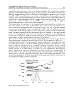
5.2.2 Imaging tumor hypoxia and angiogenesis
Oxygen is needed for proper cellular metabolism, thus hypoxia, which is common in
proliferating cancers, can significantly alter tumor biology on a molecular level. Monitoring
hypoxia in vivo can provide important information on tumor biology and response to
treatment. The transcription factor Hypoxia-inducing factor 1 (HIF1), is induced under
conditions of hypoxia and specifically binds to the hypoxia response element (HRE) to
promote transcriptional activation. Reporter vectors based on HRE elements driving
luciferase expression have been designed for longitudinal imaging of hypoxia. For example,
Viola et al inoculated mice with breast carcinoma cells transfected with an HIF-1α luciferase
reporter construct and treated these animals using cyclophosphamide or paclitaxel. They
showed that cyclophosphamide significantly inhibited tumor growth and caused an
increase in HIF-1α protein levels as quantified using BLI (Viola, Provenzale et al. 2008). As
discussed above, a transgenic mouse model was generated in which a chimeric protein
consisting of HIF-1α oxygen-dependent degradation domain (ODD) is fused to luciferase.
Hypoxic stress lead to the accumulation of ODD-luciferase which could then be identified
by non-invasive BL measurement (Goldman, Chen et al. 2011).
Hypoxia stimulates secretion of vascular endothelial growth factor (VEGF) which in turn
promotes angiogenesis. Transgenic mice have been engineered to express the VEGF receptor
Bioluminescence Applications in Preclinical Oncology Research
157
2 (VEGFR2) promoter that drives F-Luc expression. This mouse model can be used to
monitor angiogenesis induced by tumors. Angst et al sought to investigate pancreatic cancer
angiogenesis and thus employed the VEGFR2-Luc mouse. After orthotopic inoculation of
pancreatic cells, light emission corresponding to VEGFR activity began at day 4, which this
group suggests is likely due to wound healing, and continued throughout the experimental
period during tumor growth suggesting angiogenesis was occurring. The BL results were
confirmed using immunohistochemical staining for CD31 (Angst, Chen et al. 2010). In 2007,
Faley et al generated a transgenic reporter mouse, VEGF-GFP/Luc, in which an enhanced
green fluorescent protein-luciferase fusion protein is expressed under the control of a
human VEGF-A promoter. The VEGF-GFP/Luc animals exhibited intense BL throughout
the body at 1 week of age, but the signals declined as the mice grew so that the adult VEGF-
GFP/Luc mouse showed BL only in areas undergoing active wound healing. However, in
VEGF-GFP/Luc/MMTV mice, BL is observed in spontaneous tumors indicative of active
angiogenesis (Faley, Takahashi et al. 2007).
5.2.3 Imaging Protein – Protein interactions and cell signalling
In order to have a mechanistic understanding of tumor biology and response to therapy,
oncology research focuses on molecular alterations in the tumor or microenvironment.
Under many circumstances up-regulation of oncogenes results in changes in protein–protein
interactions, alterations in kinase activity and associated changes in important signalling
pathways that promote tumour cell survival and proliferation. Much work has been
accomplished to study these signalling cascades in vitro and in ex vivo tissue samples and
as a result many therapies have been developed to target these dysregulated pathways. For
these reasons there has been a great deal of interest in developing methods to visualize
molecular changes in live animals.
Three general methods are currently available for imaging protein-protein interactions in
living subjects using reporter genes: a modified mammalian two-hybrid system, a
bioluminescence resonance energy transfer (BRET) system, and split reporter protein
complementation and reconstitution strategies, these methods were reviewed by Massoud
et al in 2007 (Massoud, Paulmurugan et al. 2007). Paulmurgan developed the split reporter
system in vivo using very strongly interacting proteins MyoD and Id (Paulmurugan,
Umezawa et al. 2002). In 2004 this same group used split synthetic R-Luc protein to evaluate
heterodimerization of FRB and FKBP12 mediated by rapamycin. The rapamycin-mediated
dimerization of FRB and FKBP12 was studied in living mice by locating, quantifying, and
timing the R-Luc BL. Their work demonstrates that the split reporter system can be used to
screen small molecule drugs that impact protein-protein interactions in living animals
(Paulmurugan, Massoud et al. 2004).
It is also possible to use BLI for the evaluation of enzymatic activity such as kinase activity,
in vivo. Khan et al established a luciferase-based reporter to image EGFR kinase activity in
an in vivo model of squamous cell carcinoma (SCC). The EGFR Kinase reporter (EKR) is a
multidomain chimeric reporter where BL can be used as a marker for EGFR kinase activity.
The reporter is phosphorylated in the presence of active EGFR which interferes with
luciferase activity, if the substrate is not phosphorylated BL is available for imaging. This
reporter can therefore be used as an indicator for EGFR inhibition. Khan et al demonstrated
Bioluminescence – Recent Advances in Oceanic Measurements and Laboratory Applications
158
that a small molecule inhibitor of EGFR kinase activity (erlotinib) was able to inhibit kinase
activity in the SSC tumor model using BLI (Khan, Contessa et al. 2011).
BLI has also been used to monitor cell cycle signaling. In vivo BLI can be used to visualize
the accumulation of p27-Luc in human tumor cells after the administration of Cdk2
inhibitory drugs (Zhang and Kaelin 2005). Briat et al have generated luciferase-based p53-
reporter animals to monitor p53 activation. They showed that in response to doxorubicin
induced DNA damage, female animals had weak p53 luciferase activity in the oral cavity
while in males, the signal increased in the lower abdominal region (Briat and Vassaux 2008).
A reporter molecule has also been developed to measure Akt activity in animals via BLI.
The reporter comprises of an engineered luciferase molecule that undergoes a
conformational change and gains functionality in response to phosphorylation by Akt
(Zhang, Lee et al. 2007).
6. BLI in the study of gene activity, delivery and silencing
BLI provides a means to study gene delivery, activation using inducible systems, or
silencing of tumor promoting genes using RNA interference (RNAi). Delivery of genes can
be accomplished using multiple strategies, such as bacterial or viral vector delivery systems,
immune cell and stem cell based delivery systems or encapsulation using special
nanoparticle formulations such as liposomes or glucosylated polyethyleneimine. Monitoring
gene delivery using BLI has also been accomplished. For example Hu et al were able to
monitor TGF β receptor gene therapy efficacy in luciferase positive breast cancer metastases
simply by monitoring metastases development after gene delivery (Hu, Gerseny et al. 2011).
BLI also enables the evaluation of delivery itself. For example, Badr et al have made a
construct that comprises of 1) G-Luc, 2) the therapeutic gene cytosine deaminase and 3)
uracil phosphoribosyltransferase which converts the nontoxic compound 5-fluorocytosine
(5FC) into the drug 5-fluorouracil. A glioma cell line was engineered to express F-Luc. When
the constructed gene transfers into tumors, G-Luc allows monitoring of the duration and
magnitude of transgene expression while F-Luc imaging was used to monitor tumor growth
and response to therapy with the pro-drug 5FC (Badr, Niers et al. 2011). Ahn et al made an
adenoviral vector construct where the Survivin promoter (pSurv) amplifies the expression of
both the reporter gene F-Luc and therapeutic gene TRAIL. In an orthotopic hepatocellular
carcinoma (HCC) rat model, they showed that after systemic administration of the vector,
BLI revealed increased F-Luc activity within the tumor compared with the liver indicating
that the vector shows tumor-specific transgene expression (Ahn, Ronald et al. 2011). From a
gene silencing standpoint, use of luciferase-targeting siRNAs has been studied to define the
proof of principle that lipid based systemic administration of luciferase targeting siRNA is
able to silence luciferase gene expression in glioma (Ofek, Fischer et al. 2010) and bone
metastases (Takeshita, Hokaiwado et al. 2009).
7. Conclusion
BLI is a well-established tool in cancer research that can provide valuable insight into
biological processes in intact cells, excised tissues as well as in animal models of cancer. It
can facilitate medium-throughput assessments, it is very sensitive, and reasonably non-
invasive. The utility of BLI surpasses simple surveying of tumor growth. More specifically,
BLI can be used in the development of sophisticated animal models that examine minimal
or metastatic disease, therapeutic efficacy, disease relapse, mechanistic assessments of new
Bioluminescence Applications in Preclinical Oncology Research
159
treatment regimens, protein-protein interactions, and to gain a better understanding of basic
cancer biology. BLI facilitates visualization of processes such as metastasis, angiogenesis,
apoptosis and cell signaling in vivo. As noted by Badr et al, the sensitivity of BLI allows for
the early detection of tumors and therefore can be useful in the design of preclinical studies
assessing prevention strategies (Badr and Tannous 2011). As the BLI modality becomes
more popular, work is being done to improve the technology in order to optimize the
sensitivity and detection of BL photons. For example, IVIS by Caliper has introduced a
system where CT scans and BLI can be used simultaneously to generate three-dimensional
images of animals and their disease. Other groups are working on engineering novel
luciferases and luciferins to enhance their stability and pharmacokinetics in vivo. As
indicated, it is recognized that BLI faces some challenges (distribution and absorption of the
substrate as well as scattering issues effecting quantification), however continued use of BLI
and proper preclinical study design can overcome most of the problems associated with this
modality. BLI as a small animal imaging modality will be an integral part of the future of
pre-clinical oncology research and its applications are being refined to achieve an
understanding of disease development and response to therapy that was not previously
possible.
8. References
Ahmann, F. R., H. S. Garewal, et al. (1987). "Intracellular adenosine triphosphate as a
measure of human tumor cell viability and drug modulated growth." In Vitro Cell
Dev Biol 23(7): 474-480.
Ahn, B. C., J. A. Ronald, et al. (2011). "Potent, tumor-specific gene expression in an
orthotopic hepatoma rat model using a Survivin-targeted, amplifiable adenoviral
vector." Gene Ther 18(6): 606-612.
Angst, E., M. Chen, et al. (2010). "Bioluminescence imaging of angiogenesis in a murine
orthotopic pancreatic cancer model." Mol Imaging Biol 12(6): 570-575.
Badr, C. E., J. M. Niers, et al. (2011). "Suicidal gene therapy in an NF-kappaB-controlled
tumor environment as monitored by a secreted blood reporter." Gene Ther 18(5):
445-451.
Badr, C. E. and B. A. Tannous (2011). "Bioluminescence imaging: progress and applications."
Trends Biotechnol.
Baert, A. L. (2008). Encyclopedia of Diagnostic Imaging, Springer Reference.
Bevis, K. S., L. R. McNally, et al. (2011). "Anti-tumor activity of an anti-DR5 monoclonal
antibody, TRA-8, in combination with taxane/platinum-based chemotherapy in an
ovarian cancer model." Gynecol Oncol 121(1): 193-199.
Bhaumik, S. and S. S. Gambhir (2002). "Optical imaging of Renilla luciferase reporter gene
expression in living mice." Proc Natl Acad Sci U S A 99(1): 377-382.
Biron-Pain, K. and Y. St-Pierre (2011). "Monitoring mmp-9 gene expression in stromal cells
using a novel transgenic mouse model." Cell Mol Life Sci.
Briat, A. and G. Vassaux (2008). "A new transgenic mouse line to image chemically induced
p53 activation in vivo." Cancer Sci 99(4): 683-688.
Broggini-Tenzer, A., V. Vuong, et al. (2011). "Metabolism of tumors under treatment:
mapping of metabolites with quantitative bioluminescence." Radiother Oncol 99(3):
398-403.
Cecic, I., D. A. Chan, et al. (2007). "Oxygen sensitivity of reporter genes: implications for
preclinical imaging of tumor hypoxia." Mol Imaging 6(4): 219-228.
Bioluminescence – Recent Advances in Oceanic Measurements and Laboratory Applications
160
Cordero, A. B., Y. Kwon, et al. (2010). "In vivo imaging and therapeutic treatments in an
orthotopic mouse model of ovarian cancer." J Vis Exp(42).
Crouch, S. P., R. Kozlowski, et al. (1993). "The use of ATP bioluminescence as a measure of
cell proliferation and cytotoxicity." J Immunol Methods 160(1): 81-88.
Curtis, A., K. Calabro, et al. (2010). "Temporal Variations of Skin Pigmentation in C57Bl/6
Mice Affect Optical Bioluminescence Quantitation." Mol Imaging Biol.
Czupryna, J. and A. Tsourkas (2011). "Firefly luciferase and RLuc8 exhibit differential
sensitivity to oxidative stress in apoptotic cells." PLoS One 6(5): e20073.
de Wet, J. R., K. V. Wood, et al. (1987). "Firefly luciferase gene: structure and expression in
mammalian cells." Mol Cell Biol 7(2): 725-737.
Dickson, P. V., B. Hamner, et al. (2007). "In vivo bioluminescence imaging for early detection
and monitoring of disease progression in a murine model of neuroblastoma." J
Pediatr Surg 42(7): 1172-1179.
Dussmann, P., J. I. Pagel, et al. (2011). "Live in vivo imaging of Egr-1 promoter activity
during neonatal development, liver regeneration and wound healing." BMC Dev
Biol 11: 28.
Edinger, M., Y. A. Cao, et al. (2002). "Advancing animal models of neoplasia through in vivo
bioluminescence imaging." Eur J Cancer 38(16): 2128-2136.
Edinger, M., T. J. Sweeney, et al. (1999). "Noninvasive assessment of tumor cell proliferation
in animal models." Neoplasia 1(4): 303-310.
El-Deiry, W. S., C. C. Sigman, et al. (2006). "Imaging and oncologic drug development." J
Clin Oncol 24(20): 3261-3273.
Faley, S. L., K. Takahashi, et al. (2007). "Bioluminescence imaging of vascular endothelial
growth factor promoter activity in murine mammary tumorigenesis." Mol Imaging
6(5): 331-339.
Feng, M., J. Zhang, et al. (2011). "In vivo imaging of human malignant mesothelioma grown
orthotopically in the peritoneal cavity of nude mice." J Cancer 2: 123-131.
Frampas, E., C. Maurel, et al. (2011). "The intraportal injection model for liver metastasis:
advantages of associated bioluminescence to assess tumor growth and influences
on tumor uptake of radiolabeled anti-carcinoembryonic antigen antibody." Nucl
Med Commun 32(2): 147-154.
Garcia, T., A. Jackson, et al. (2008). "A convenient clinically relevant model of human breast
cancer bone metastasis." Clin Exp Metastasis 25(1): 33-42.
Garewal, H. S., F. R. Ahmann, et al. (1986). "ATP assay: ability to distinguish cytostatic from
cytocidal anticancer drug effects." J Natl Cancer Inst 77(5): 1039-1045.
Geusz, M. E., K. T. Blakely, et al. (2010). "Elevated mPer1 gene expression in tumor stroma
imaged through bioluminescence." Int J Cancer 126(3): 620-630.
Goldman, S. J., E. Chen, et al. (2011). "Use of the ODD-luciferase transgene for the non-
invasive imaging of spontaneous tumors in mice." PLoS One 6(3): e18269.
Graeser, R., C. Bornmann, et al. (2009). "Antimetastatic effects of liposomal gemcitabine and
empty liposomes in an orthotopic mouse model of pancreatic cancer." Pancreas
38(3): 330-337.
Hickson, J., S. Ackler, et al. (2010). "Noninvasive molecular imaging of apoptosis in vivo
using a modified firefly luciferase substrate, Z-DEVD-aminoluciferin." Cell Death
Differ 17(6): 1003-1010.
Hsieh, C. L., Z. Xie, et al. (2005). "A luciferase transgenic mouse model: visualization of
prostate development and its androgen responsiveness in live animals." J Mol
Endocrinol 35(2): 293-304.
Bioluminescence Applications in Preclinical Oncology Research
161
Hu, Z., H. Gerseny, et al. (2011). "Oncolytic Adenovirus Expressing Soluble TGFbeta
Receptor II-Fc-mediated Inhibition of Established Bone Metastases: A Safe and
Effective Systemic Therapeutic Approach for Breast Cancer." Mol Ther 19(9): 1609-
1618.
Huerta, S., X. Gao, et al. (2011). "Murine orthotopic model for the assessment of
chemoradiotherapeutic interventions in rectal cancer." Anticancer Drugs 22(4): 371-
376.
Iyer, M., F. B. Salazar, et al. (2004). "Noninvasive imaging of enhanced prostate-specific gene
expression using a two-step transcriptional amplification-based lentivirus vector."
Mol Ther 10(3): 545-552.
Iyer, M., F. B. Salazar, et al. (2005). "Non-invasive imaging of a transgenic mouse model
using a prostate-specific two-step transcriptional amplification strategy." Transgenic
Res 14(1): 47-55.
Jenkins, D. E., Y. Oei, et al. (2003). "Bioluminescent imaging (BLI) to improve and refine
traditional murine models of tumor growth and metastasis." Clin Exp Metastasis
20(8): 733-744.
Jia, W., S. Wang, et al. (2011). "A BAC transgenic reporter recapitulates in vivo regulation of
human telomerase reverse transcriptase in development and tumorigenesis."
FASEB J 25(3): 979-989.
Kalra, J., M. Anantha, et al. (2011). "Validating the use of a luciferase labeled breast cancer
cell line, MDA435LCC6, as a means to monitor tumor progression and to assess the
therapeutic activity of an established anticancer drug, docetaxel (Dt) alone or in
combination with the ILK inhibitor, QLT0267." Cancer Biol Ther 11(9): 826-838.
Kalra, J., C. Warburton, et al. (2009). "QLT0267, a small molecule inhibitor targeting integrin-
linked kinase (ILK), and docetaxel can combine to produce synergistic interactions
linked to enhanced cytotoxicity, reductions in P-AKT levels, altered F-actin
architecture and improved treatment outcomes in an orthotopic breast cancer
model." Breast Cancer Res 11(3): R25.
Karam, J. A., R. P. Mason, et al. (2003). "Molecular imaging in prostate cancer." J Cell Biochem
90(3): 473-483.
Khan, A. P., J. N. Contessa, et al. (2011). "Molecular imaging of epidermal growth factor
receptor kinase activity." Anal Biochem 417(1): 57-64.
Kheirolomoom, A., D. E. Kruse, et al. (2010). "Enhanced in vivo bioluminescence imaging
using liposomal luciferin delivery system." J Control Release 141(2): 128-136.
Kuzmits, R., P. Aiginger, et al. (1986). "Assessment of the sensitivity of leukaemic cells to
cytotoxic drugs by bioluminescence measurement of ATP in cultured cells." Clin Sci
(Lond) 71(1): 81-88.
Kuzmits, R., H. Rumpold, et al. (1986). "The use of bioluminescence to evaluate the influence
of chemotherapeutic drugs on ATP-levels of malignant cell lines." J Clin Chem Clin
Biochem 24(5): 293-298.
Laxman, B., D. E. Hall, et al. (2002). "Noninvasive real-time imaging of apoptosis." Proc Natl
Acad Sci U S A 99(26): 16551-16555.
Lee, Y. C., C. F. Huang, et al. (2010). "Src family kinase/abl inhibitor dasatinib suppresses
proliferation and enhances differentiation of osteoblasts." Oncogene 29(22): 3196-
3207.
Li, B., A. Torossian, et al. (2011). "A novel bioluminescence orthotopic mouse model for
advanced lung cancer." Radiat Res 176(4): 486-493.
Li, F., Q. Cheng, et al. (2010). "Generation of a novel transgenic mouse model for
bioluminescent monitoring of survivin gene activity in vivo at various
Bioluminescence – Recent Advances in Oceanic Measurements and Laboratory Applications
162
pathophysiological processes: survivin expression overlaps with stem cell
markers." Am J Pathol 176(4): 1629-1638.
Lin, A. H., J. Luo, et al. (2005). "Global analysis of Smad2/3-dependent TGF-beta signaling
in living mice reveals prominent tissue-specific responses to injury." J Immunol
175(1): 547-554.
Lipshutz, G. S., D. Titre, et al. (2003). "Comparison of gene expression after intraperitoneal
delivery of AAV2 or AAV5 in utero." Mol Ther 8(1): 90-98.
Luker, G. D., C. M. Pica, et al. (2003). "Imaging 26S proteasome activity and inhibition in
living mice." Nat Med 9(7): 969-973.
Luo, J. and T. Wyss-Coray (2009). "Bioluminescence analysis of Smad-dependent TGF-beta
signaling in live mice." Methods Mol Biol 574: 193-202.
Lyons, S. K., E. Lim, et al. (2006). "Noninvasive bioluminescence imaging of normal and
spontaneously transformed prostate tissue in mice." Cancer Res 66(9): 4701-4707.
Madero-Visbal, R. A., J. F. Colon, et al. (2010). "Bioluminescence imaging correlates with
tumor progression in an orthotopic mouse model of lung cancer." Surg Oncol.
Massoud, T. F., R. Paulmurugan, et al. (2007). "Reporter gene imaging of protein-protein
interactions in living subjects." Curr Opin Biotechnol 18(1): 31-37.
McNally, L. R., D. R. Welch, et al. (2010). "KISS1 over-expression suppresses metastasis of
pancreatic adenocarcinoma in a xenograft mouse model." Clin Exp Metastasis 27(8):
591-600.
Mishra, S., Y. Tang, et al. (2011). "Blockade of transforming growth factor-beta (TGFbeta)
signaling inhibits osteoblastic tumorigenesis by a novel human prostate cancer cell
line." Prostate 71(13): 1441-1454.
Momota, H. and E. C. Holland (2005). "Bioluminescence technology for imaging cell
proliferation." Curr Opin Biotechnol 16(6): 681-686.
Moriyama, E. H., M. J. Niedre, et al. (2008). "The influence of hypoxia on bioluminescence in
luciferase-transfected gliosarcoma tumor cells in vitro." Photochem Photobiol Sci 7(6):
675-680.
Moriyama, Y., E. H. Moriyama, et al. (2005). "In vivo study of the inflammatory modulating
effects of low-level laser therapy on iNOS expression using bioluminescence
imaging." Photochem Photobiol 81(6): 1351-1355.
Mueller-Klieser, W., M. Kroeger, et al. (1991). "Comparative imaging of structure and
metabolites in tumours." Int J Radiat Biol 60(1-2): 147-159.
Mueller-Klieser, W., S. Walenta, et al. (1988). "Metabolic imaging in microregions of tumors
and normal tissues with bioluminescence and photon counting." J Natl Cancer Inst
80(11): 842-848.
Mugabe, C., Y. Matsui, et al. (2011). "In vivo evaluation of mucoadhesive nanoparticulate
docetaxel for intravesical treatment of non-muscle-invasive bladder cancer." Clin
Cancer Res 17(9): 2788-2798.
Muniz, V. P., J. M. Barnes, et al. (2011). "The ARF tumor suppressor inhibits tumor cell
colonization independent of p53 in a novel mouse model of pancreatic ductal
adenocarcinoma metastasis." Mol Cancer Res 9(7): 867-877.
Niers, J. M., M. Kerami, et al. (2011). "Multimodal in vivo imaging and blood monitoring of
intrinsic and extrinsic apoptosis." Mol Ther 19(6): 1090-1096.
Nyati, M. K., Z. Symon, et al. (2002). "The potential of 5-fluorocytosine/cytosine deaminase
enzyme prodrug gene therapy in an intrahepatic colon cancer model." Gene Ther
9(13): 844-849.
O'Neill, K., S. K. Lyons, et al. (2010). "Bioluminescent imaging: a critical tool in pre-clinical
oncology research." J Pathol 220(3): 317-327.
Bioluminescence Applications in Preclinical Oncology Research
163
Ofek, P., W. Fischer, et al. (2010). "In vivo delivery of small interfering RNA to tumors and
their vasculature by novel dendritic nanocarriers." FASEB J 24(9): 3122-3134.
Paulmurugan, R., T. F. Massoud, et al. (2004). "Molecular imaging of drug-modulated
protein-protein interactions in living subjects." Cancer Res 64(6): 2113-2119.
Paulmurugan, R., Y. Umezawa, et al. (2002). "Noninvasive imaging of protein-protein
interactions in living subjects by using reporter protein complementation and
reconstitution strategies." Proc Natl Acad Sci U S A 99(24): 15608-15613.
Pesnel, S., Y. Guminski, et al. (2011). "(99m)Tc-HYNIC-spermine for imaging polyamine
transport system-positive tumours: preclinical evaluation." Eur J Nucl Med Mol
Imaging 38(10): 1832-1841.
Petru, E., B. U. Sevin, et al. (1990). "Comparative chemosensitivity profiles in four human
ovarian carcinoma cell lines measuring ATP bioluminescence." Gynecol Oncol 38(2):
155-160.
Prasad, G., T. Sottero, et al. (2011). "Inhibition of PI3K/mTOR pathways in glioblastoma and
implications for combination therapy with temozolomide." Neuro Oncol 13(4): 384-
392.
Ray, P. (2011). "Multimodality molecular imaging of disease progression in living subjects." J
Biosci 36(3): 499-504.
Rehemtulla, A., N. Taneja, et al. (2004). "Bioluminescence detection of cells having stabilized
p53 in response to a genotoxic event." Mol Imaging 3(1): 63-68.
Robbins, D. and Y. Zhao (2011). "Imaging NF-kappaB signaling in mice for screening
anticancer drugs." Methods Mol Biol 716: 169-177.
Runnels, J. M., A. L. Carlson, et al. (2011). "Optical techniques for tracking multiple
myeloma engraftment, growth, and response to therapy." J Biomed Opt 16(1):
011006.
Sano, D., F. Matsumoto, et al. (2011). "Vandetanib restores head and neck squamous cell
carcinoma cells' sensitivity to cisplatin and radiation in vivo and in vitro." Clin
Cancer Res 17(7): 1815-1827.
Scabini, M., F. Stellari, et al. (2011). "In vivo imaging of early stage apoptosis by measuring
real-time caspase-3/7 activation." Apoptosis 16(2): 198-207.
Schuetz, E., L. Lan, et al. (2002). "Development of a real-time in vivo transcription assay:
application reveals pregnane X receptor-mediated induction of CYP3A4 by cancer
chemotherapeutic agents." Mol Pharmacol 62(3): 439-445.
Sevin, B. U., Z. L. Peng, et al. (1988). "Application of an ATP-bioluminescence assay in
human tumor chemosensitivity testing." Gynecol Oncol 31(1): 191-204.
Shan, L., S. Wang, et al. (2008). "Bioluminescent animal models of human breast cancer for
tumor biomass evaluation and metastasis detection." Ethn Dis 18(2 Suppl 2): S2-65-
69.
Shimomura, O. (2006). Bioluminesence: Chemical Principles and Methods, World Scientific
Publishing
Spiotto, M. T., A. Banh, et al. (2010). "Imaging the unfolded protein response in primary
tumors reveals microenvironments with metabolic variations that predict tumor
growth." Cancer Res 70(1): 78-88.
Svensson, R. U., J. M. Haverkamp, et al. (2011). "Slow disease progression in a C57BL/6
pten-deficient mouse model of prostate cancer." Am J Pathol 179(1): 502-512.
Sweeney, T. J., V. Mailander, et al. (1999). "Visualizing the kinetics of tumor-cell clearance in
living animals." Proc Natl Acad Sci U S A 96(21): 12044-12049.
Takeshita, F., N. Hokaiwado, et al. (2009). "Local and systemic delivery of siRNAs for
oligonucleotide therapy." Methods Mol Biol 487: 83-92.
Bioluminescence – Recent Advances in Oceanic Measurements and Laboratory Applications
164
Taneja, P., D. P. Frazier, et al. (2009). "MMTV mouse models and the diagnostic values of
MMTV-like sequences in human breast cancer." Expert Rev Mol Diagn 9(5): 423-440.
Tang, Y., K. Shah, et al. (2003). "In vivo tracking of neural progenitor cell migration to
glioblastomas." Hum Gene Ther 14(13): 1247-1254.
Teitz, T., J. J. Stanke, et al. (2011). "Preclinical models for neuroblastoma: establishing a
baseline for treatment." PLoS One 6(4): e19133.
Tiffen, J. C., C. G. Bailey, et al. (2010). "Luciferase expression and bioluminescence does not
affect tumor cell growth in vitro or in vivo." Mol Cancer 9: 299.
Tivnan, A., L. Tracey, et al. (2011). "MicroRNA-34a is a potent tumor suppressor molecule in
vivo in neuroblastoma." BMC Cancer 11: 33.
van der Horst, G., J. J. van Asten, et al. (2011). "Real-time cancer cell tracking by
bioluminescence in a preclinical model of human bladder cancer growth and
metastasis." Eur Urol 60(2): 337-343.
Vikis, H. G., E. N. Jackson, et al. (2010). "Strain-specific susceptibility for pulmonary
metastasis of sarcoma 180 cells in inbred mice." Cancer Res 70(12): 4859-4867.
Viola, R. J., J. M. Provenzale, et al. (2008). "In vivo bioluminescence imaging monitoring of
hypoxia-inducible factor 1alpha, a promoter that protects cells, in response to
chemotherapy." AJR Am J Roentgenol 191(6): 1779-1784.
Vykhovanets, E. V., S. Shukla, et al. (2008). "Molecular imaging of NF-kappaB in prostate
tissue after systemic administration of IL-1 beta." Prostate 68(1): 34-41.
Walenta, S., M. Dellian, et al. (1992). "Pixel-to-pixel correlation between images of absolute
ATP concentrations and blood flow in tumours." Br J Cancer 66(6): 1099-1102.
Walenta, S., T. Schroeder, et al. (2002). "Metabolic mapping with bioluminescence: basic and
clinical relevance." Biomol Eng 18(6): 249-262.
Wang, H., F. Cao, et al. (2009). "Trafficking mesenchymal stem cell engraftment and
differentiation in tumor-bearing mice by bioluminescence imaging." Stem Cells
27(7): 1548-1558.
Woolfenden, S., H. Zhu, et al. (2009). "A Cre/LoxP conditional luciferase reporter transgenic
mouse for bioluminescence monitoring of tumorigenesis." Genesis 47(10): 659-666.
Wu, F., R. Xu, et al. (2008). "In vivo profiling of estrogen receptor/specificity protein-
dependent transactivation." Endocrinology 149(11): 5696-5705.
Yan, W., D. Xiao, et al. (2011). "Combined bioluminescence and fluorescence imaging
visualizing orthotopic lung adenocarcinoma xenograft in vivo." Acta Biochim
Biophys Sin (Shanghai) 43(8): 595-600.
Zhang, G. J. and W. G. Kaelin, Jr. (2005). "Bioluminescent imaging of ubiquitin ligase
activity: measuring Cdk2 activity in vivo through changes in p27 turnover."
Methods Enzymol 399: 530-549.
Zhang, L., K. C. Lee, et al. (2007). "Molecular imaging of Akt kinase activity." Nat Med 13(9):
1114-1119.
Zhang, N., S. Lyons, et al. (2009). "A spontaneous acinar cell carcinoma model for
monitoring progression of pancreatic lesions and response to treatment through
noninvasive bioluminescence imaging." Clin Cancer Res 15(15): 4915-4924.
Zhang, Q., A. A. Triplett, et al. (2010). "Temporally and spatially controlled expression of
transgenes in embryonic and adult tissues." Transgenic Res 19(3): 499-509.
Zumsteg, A., K. Strittmatter, et al. (2010). "A bioluminescent mouse model of pancreatic
{beta}-cell carcinogenesis." Carcinogenesis 31(8): 1465-1474.
Part 3
Bacterial Bioluminescence
8
Oscillation in Bacterial Bioluminescence
Satoshi Sasaki
Tokyo University of Technology
Japan
1. Introduction
Live cells show various dynamic characteristics, such as cell division or material production.
When we consider that a cell is a chemical reactor that contains an enzyme in its structure,
the rates of chemical reaction catalysed by them depend on the cell density. As the amount
of enzyme within the cell differs according to the rate of expression of a specific gene, the
rate of the reaction also depends on the condition of the cell. In short, chemical reactions
caused by cells are nonlinearly related to the cell density; the reaction rate is not
proportional to the cell density. This is one remarkable aspect of live cells. In the field of
chemical analysis, bacterial cell behaviour is often used. For example, changes in respiration
caused by chemical compounds that inhibit the respiratory chain (such as KCN) can be
quantified, theoretically, by measuring the changes in the dissolved oxygen concentration.
Biomaterial-based devices have been reported, such as biochips or biosensors. These are not
truly “bio” because they use an enzyme or antibody outside of the cell. Microbial sensors
(Melidis, P.; Georgiou, D.(2002).; Kang. KH.; Jang. JK.; Pham. TH.; Moon. H.; Chang. IS. &
Kim, BH. (2003).; Moon, H.; Chang, IS.; Kang, KH.; Jang, JK. & Kim, BH. (2004). ; Chang, IS.;
Moon, H.; Jang, JK. & Kim, BH. (2005).; Kogure, H.; Kawasaki, S.; Nakajima, K.; Sakai, N.;
Futase, K.; Inatsu, Y.; Bari, ML.; Isshiki, K. & Kawamoto, S. (2005).; Vaiopoulou, E.; Melidis,
P.; Kampragou, E. & Aivasidis, A. (2004).; Yano, Y.; Numata, M.; Hachiya, H.; Ito, S.;
Masadome, T.; Ohkubo, S.; Asano, Y. & Imato, T. (2001).; Kim, M.; Hyun, MS.; Gadd, GM.;
Kim, GT.; Lee, SJ. & Kim, HJ. (2009). Davila, D.; Esquivel, JP.; Sabate, N. & Mas, J. (2011).),
known as the analysis of devices that use live microbial cells as molecular-recognition
material, are the only exception. This sensor, however, is based on a shift from one
equilibrium to another. For example, a respiration inhibition-based microbial sensor
measures a certain toxic compound because the dissolved oxygen concentration near the
cells increases when the toxic compound exists. The main reason for the use of
microorganisms is that they are more cost-effective than purified enzymes or antibodies.
The dynamics of the bacterial cells are not at all used. The nonlinearity of cell behaviour has
recently been studied (Wu, BM.; Subbarao, KV. & Qin, QM. (2008).; Kenkre, V. M.; &,
Kumar, N. (2008).; Dobrescu, R. & Purcarea, VI. (2011)). A suitable bacterium model is,
therefore, needed to start a fundamental study on the nonlinearity of the cell. In our group
studies, bioluminescence characteristics have been identified (Sasaki S., Mori Y., Ogawa M.,
Funatsuka S.,(2010)). Bioluminescent bacteria are those that emit light autonomously
without the need of excitation light. The bioluminescence reaction is catalysed by bacterial
Bioluminescence – Recent Advances in Oceanic Measurements and Laboratory Applications
168
luciferase (Raushel, F. M. & T. O. Baldwin; (1989), Lee, J., Y. Y. Wang and B. G. Gibson;
(1991), Hastings, J. W. (1996), Shirazy, N. H., B. Ranjbar, S. Hosseinkhani, K. Khalifeh, A. R.
Madvar and H. Naderi-Manesh (2007)). The reaction requires a flavin mononucleotide
(FMNH2), a long-chain aliphatic aldehyde, and O
2
to produce light (Balny, C. and J. W.
Hastings (1975), Kurfurst, M., S. Ghisla and J. W. Hastings (1983), Tu, S. C., B. Lei, M. Liu, C.
K. Tang and C. Jeffers (2000)).
FMNH
2
+RCOOH+O
2
>FMN+RCOOH+H
2
O+hν
This reaction is catalysed by bacterial luciferase (Karatani, H.; Izuta, T. & Hirayama, S.
(2007)). This enzyme is synthesised by a process called quorum sensing, in which the
synthesis occurs only after the cells recognise each other to be above a threshold in density.
Two substrates, FMNH2 and RCHO (linear alkyl aldehyde), of the reaction are also
synthesised in the cell. The substrate with the least amount is, therefore, the rate-
determining factor. The intensity of the bioluminescence has been reported primarily in
connection with the oxygen concentration, but, theoretically, two other compounds might
be candidates. Bacterial luminescence that has been used for environmental monitoring has
been reviewed (Girott, S.; Ferri, E.N.; Fumo, M.G.; & Maiolini, E. (2008). Recently, an
oscillation in luminescence from a well-stirred bacterial suspension was reported (Sato, Y.
and S. Sasaki (2008)). Here, in this chapter, the relationship between the oxygen and
oscillation mode was investigated.
Changes in the luminescence spectra are also reported.
2. Experimentals
Bioluminescent bacteria, Photobacterium kishitanii, collected from the skin of a cuttlefish and
Todarodes pacificus (for sashimi), were purified and used. In a well-stirred solution, dissolved
oxygen is in equilibrium with the atmospheric oxygen. This may not be the case with a
bioluminescent bacterial suspension. As reported above, the luminescent reaction consumes
oxygen to produce light. Simultaneously, production of the substrate FMNH2 requires
energy that is produced by respiration. Karatani calculated the energy required to produce
light and concluded that the bacterial bioluminescence is an extremely oxygen-consuming
process. A bioluminescent bacterial suspension was, therefore, suspected to show a very low
dissolved oxygen (DO) concentration. In this study, we began with the measurement of both
DO and luminescent intensity through the period of oscillation.
As the luminescent reaction occurs inside the cell, the luminescent intensity is affected by
the [DO] inside the cell rather than that in the suspension. Because the dynamic
measurement of [DO] within a bacterium is considered to be difficult, we focused on any
change in cell density during the oscillation period. The colour of bacterial bioluminescence
is determined by the fluorescent protein (LumP) (Sato Y, Shimizu S, Ohtaki A, Noguchi K,
Miyatake H, Dohmae N, Sasaki S, Odaka M, Yohda M., Crystal structures of the lumazine
protein from Photobacterium kishitanii in complexes with the authentic chromophore, 6,7-
dimethyl- 8-(1'-D-ribityl) lumazine, and its analogues, riboflavin and flavin mononucleotide,
at high resolution., J Bacteriol. 2010 Jan;192(1):127-33.). We then, therefore, measured the
spectral change in luminescence through the oscillation period.
Oscillation in Bacterial Bioluminescence
169
2.1 Relationship between the bacterial bioluminescence and dissolved oxygen
concentration in a bacterial suspension
Photobacterium belongs to a family of Gram-negative, facultatively aerobic bacteria
(Urbanczyk, H.; Ast, JC. & Dunlap, PV. (2011)). We started by measuring the oxygen effect
on bioluminescence. The intensity of the bioluminescence was measured using a self-made
luminescence detector (five commercially available solar cells were connected in series) or
optical power meter (Model 3664, Hioki E.E. Co.). The output voltage generated by both
devices was measured and recorded with an A/D converting logger (NR 250, Keyence Co.).
An oscillation broth (Yeast extract 2.5 g L-1, Bacto peptone 5 g L-1, and NaCl 30 g L-1) or
marine broth (DifcoTM marine broth 2216, Becton, Dickinson, and Company) was prepared
and filtrated using a 0.22 µm filter (Nalgene disposable filter unit, Thermo Fisher Scientific,
Inc.). A glass cell with an inner diameter of 31 mm was placed over a magnetic stirrer. The
schematic illustration of the measurement system is shown in Fig. 1. All the equipment was
placed in an incubator (VS401, Versos Co., Ltd.) adjusted at 17˚C with 10, 20, 30, and 50 mL
of oscillation broth to determine the effects of the air-liquid interface area/volume. In
addition, the dilution effect of the marine broth on the oscillation mode was investigated by
diluting the broth 1.5 and 3 times. For the simultaneous measurement of luminescence and
dissolved oxygen concentration, an optical fibre-based DO sensor (FOXY R, Ocean Optics,
Inc.) was placed into the bacterial suspension (Fig. 2).
An aluminium foil cap was placed loosely on the glass tube to prevent contamination during the
measurement.
Fig. 1. Schematic illustration of the bioluminescence intensity measurement.
Bioluminescence – Recent Advances in Oceanic Measurements and Laboratory Applications
170
An optical fibre sensor tip was placed vertically in the middle of the bacterial suspension. An
aluminium foil cap was placed loosely on the glass tube to prevent contamination during the
measurement.
Fig. 2. Schematic illustration of the system for the simultaneous measurement of the
luminescence intensity and dissolved oxygen concentration.
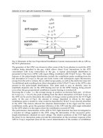
2.2 Simultaneous measurement of the luminescence and cell density during
oscillation
Continuous measurement of the optical density (OD) of the bacterial suspension was
performed using an OD meter (ODBox-A, TAITEC Co.). A 500 mL Erlenmeyer flask with
100 mL of bacterial suspension was set over a rotary shaker (NR-2, TAITEC Co.), and, on the
surface of the flask, five solar cells connected in a series were attached (Fig. 3). The
generated voltage was measured and recorded by the same logger as reported in 2.1. All the
equipment was placed in a self-made dark box, and measurements were performed at room
temperature ranging from 20 to 23˚C.