Proteomic Applications in Biology Part 11 potx
Bạn đang xem bản rút gọn của tài liệu. Xem và tải ngay bản đầy đủ của tài liệu tại đây (2.28 MB, 17 trang )
Assessment of Proteomics Strategies for Plant Cell Wall
Glycosyltransferasesin Wheat, a Non-Model Species: Glucurono(Arabino)Xylan as a Case Study
159
Gel area
covered MW
(kDa)
Slice # Total No of
peptide
queries
No of
sequences
with hits
No of hits
identified
Top hit
[No of peptides
matched]
GTs involved in
GAX biosynthesis
~70 - ~180
1 4,683 88 13 gi|6715512 None
2 4,845 141 11 gi|90025017 None
3 4,761 41 7 gi|90025017 None
4 5,262 137 23 gi|10720235 None
5 5,229 37 5 gi|10720235 None
6 4,971 17 5 gi|10720235 None
7 4,866 23 7 gi|6715512 None
8 5,009 23 6 gi|10720235 None
9 4,249 3 1 gi|10720235 None
10 4,804 12 1 gi|10720235 None
11 4,897 23 4 gi|10720235 None
12 4,623 160 15 gi|2493132 None
13 4,283 38 11 gi|10720235 None
16 3,937 10 3 gi|13375563 None
17 3,793 3 2 gi|129707 None
~30 to ~70
1 5,236 21 5 gi|10720235 None
2 4,722 16 4 gi|739292 None
3 4,552 13 4 gi|10720235 None
4 4,564 2 1 gi|10720235 None
5 4,623 49 4 gi|14017569 None
6 4,243 30 5 gi|6715512 None
7 3,950 4 2 gi|20322 None
8 4,165 4 2 gi|439586 None
9 4,596 40 8 gi|10720235 gi|159470791,
gi|159471277,
GT47 family
10 3,834 18 5 gi|57471704 None
11 3,481 5 2 gi|10720235 None
12 4,171 68 9 gi|90025017 None
13 4,852 17 3 gi|129708 None
14 3,880 21 5 gi|10720235 None
15 3,764 283 14 gi|2493131 None
16 1,007 26 4 gi|10720235 gi|2218152,
gi|4158232,
GT75 family
17 1,994 37 6 gi|10720235 None
18 2,212 40 8 gi|10720235 gi|2218152,
gi|4158232,
GT75 family
19 4,766 116 17 gi|10720235 None
20 4,790 122 21 gi|10720235 None
Total 37 149,614 1283 233
Table 5. Proteins involved in GAX biosynthesis identified in fraction #3 by Gel-LC-MS/MS
and LTQ strategy. The gel area between 30 and 180 kDa of SDS-PAGE was sliced into 20-40
slices (see Figure 3) and each slice was trypsin-digested and analyzed.
(among the 233 hits) that were unique to this strategy (not in previous analyses). Among the
unique hits identified by this strategy, two Chlamydomonas reinhardtii GTs (gi|159470791,
and gi|159471277) belonging to the GT47 family (both annotated as exostosin-like
glycosyltransferase) were identified by the following peptide RVAEADIPRL (score 56). This
Proteomic Applications in Biology
160
Accession
No
Annotation Score Peptides
matched
g
i|6715512 V-t
yp
e H+ ATPase B subunit [Nicotiana tabacum] 117 18
g
i|2493650 RuBisCO lar
g
e subuni
t
-bindin
g
protein subunit beta 77 2
g
i|475600 BiP isoform B [Gl
y
cine max] 102 5
g
i|123656 Heat shock-related protein [Spinacia oleracea] 94 7
g
i|115458184 Calreticulin famil
y
, Os04
g
0402100 62 2
g
i|42541152 Delta tonoplast intrinsic protein TIP2;2 [Triticum aestivum] 120 3
g
i|1709846 Photos
y
stem II 22 kDa protein [L
y
copersicon esculentum] 109 2
g
i|4099406 Camma-t
yp
e tono
p
last intrinsic
p
rotein [Triticum aestivum] 95 3
g
i|28569578 Allene oxide s
y
nthase [Triticum aestivum] 59 3
g
i|1709358 Nucleoside-triphosphatase [Pisum sativum] 81 2
g
i|904147 Adenosine triphosphatase [Sinofranchetia chinensis] 237 11
g
i|15010616 AT4
g
38510/F20M13_70 [Arabido
p
sis thaliana] 235 8
g
i|24496452 Actin [Hordeum vul
g
are] 172 5
g
i|115589744 S-adenos
y
lmethionine s
y
nthetase 1 [Triticum monococcum] 74 2
g
i|13375563 Li
p
id transfer
p
rotein
p
recursor [Triticum aestivum] 154 4
g
i|14017578 ATP s
y
nthase CF1 epsilon subunit [Triticum aestivum] 99 2
g
i|16225 Calmodulin [Arabidopsis thaliana] 69 2
g
i|75108545 Peroxiredoxin Q, chloro
p
lastic [Triticum aestivum] 95 2
g
i|464517 50S ribosomal protein L12-1 [Secale cereale] 67 4
g
i|115442509 Cy
t
-b5 family, Os01
g
0971500 [Oryza sativa (japonica cultivar-
g
roup)]
65 3
g
i|42565453 C
y
clo
p
hilin [H
y
acinthus orientalis] 59 2
g
i|68566191 C
y
tochrome b6-f complex [Triticum aestivum] 129 5
g
i|118104 Peptid
y
l-prol
y
l cis-trans isomerase [Zea ma
y
s] 89 2
g
i|154761388 C
y
clo
p
hilin [Triticum aestivum] 89 2
g
i|231496 Actin-58 [Solanum tuberosum] 166 8
g
i|115467154 Os06
g
0221200 annexin family [Oryza sativa (japonica cultivar-
g
roup)]
86 2
g
i|52548250 ADP-ribos
y
lation factor [Triticum aestivum] 162 6
g
i|74048999 Eukar
y
otic translation initiation factor 5A1 [Triticum aestivum] 71 2
g
i|57471704 Ribosomal protein L11 [Triticum aestivum] 186 8
g
i|432607 Ras-related GTP bindin
g
protein possessin
g
GTPase activity
[Or
y
za sativa]
119 4
g
i|115441299 Os01
g
0869800, PsbS subunit [Oryza sativa (japonica cultivar-
g
rou
p
)]
73 2
g
i|115444503 Os02
g
0171100 [Or
y
za sativa (
j
aponica cultivar-
g
roup)] 283 4
g
i|16304127 Gl
y
ceraldeh
y
de 3-phosphate deh
y
dro
g
enase 1 [Fra
g
aria x ananassa] 132 3
g
i|18071421 Putative deh
y
dro
g
enase [Or
y
za sativa (
j
aponica cultivar-
g
roup)] 125 4
g
i|166627 Nucleotide-bindin
g
subunit of vacuolar ATPase [Arabidopsis
thaliana]
115 2
g
i|8272480 Fructose 1,6-bisphosphate aldolase precursor [Avena sativa] 92 5
g
i|159470791 Exostosin-like
g
l
y
cos
y
ltransferase [Chlam
y
domonas reinhardtii] 56 2
g
i|159471277 Exostosin-like
g
l
y
cos
y
ltransferase [Chlam
y
domonas reinhardtii] 56 1
g
i|115451383 Os03
g
0200800 [Or
y
za sativa (
j
aponica cultivar-
g
roup)] 202 4
g
i|147858623 H
y
pothetical protein [Vitis vinifera] 65 2
Table 6. List of unique hits identified through Gel-LC-MS/MS and LTQ analysis of 20-40
slices covering 30-180 kDa area of the SDS-PAGE. Only hits with scores >55 and/or two
peptide matches are listed.
strategy, however, identified the exact wheat RGP protein (TaGT75-4, gi|4158232) with the
following peptides VPEGFDYELYNR and YVDAVLTIPK (both with score 59). Therefore,
this strategy successfully identified TaGT75-4 protein and homolog to TaGT47-13 but failed
to identify the exact TaGT47-13 protein or any homolog to TaGT43-4 protein.
Assessment of Proteomics Strategies for Plant Cell Wall
Glycosyltransferasesin Wheat, a Non-Model Species: Glucurono(Arabino)Xylan as a Case Study
161
4. Discussion
Hemicellulosic polymers such as GAX represent up to 40% (w/w) of grass cell walls (in
particular from growing tissues). In sharp contrast with the abundance of these polymers,
the GTs that synthesize these compounds are present in low amounts in Golgi membranes
of the plant cell. This observation suggests that these enzymes are highly active and may not
be required in large quantities in the plant cell. This low abundance of GTs has been the
main limiting factor in applying proteomics approaches to plant cell wall biosynthesis. To
further complicate the issue, isolation of GTs from Golgi membranes (or simply disrupting
these membranes) generally results in a drastic reduction or loss of transferase activity in
vitro. To detect this weak transferase activity in vitro, it is necessary to use very sensitive
biochemical assays (i.e., [
14
C]radiolabeled sugars-based assay). Since the loss of transfer
activity is GT-dependent, the biochemical assays are not the best way to estimate the
abundance of these enzymes in a particular protein preparation. Therefore, when working
with plant cell wall GTs, all these factors should be taken in consideration. In this work,
such in vitro assay was used to monitor the distribution of GAX synthase activity (from
Golgi-enriched membranes) on a linear sucrose density gradient supplemented with EDTA
as described earlier (Zeng et al., 2010). According to our in vitro assay, fraction #3 was
substantially enriched in GAX synthase activity (Figure 2), and it can be assumed that this
fraction is also enriched in TaGT43-4, TaGT47-13, and TaGT75-4 proteins. Therefore, fraction
#3 is an excellent starting material to evaluate proteomics strategies in identifying these
three GTs among a mixture of proteins. Furthermore, because genome and protein sequence
information from five grass species are currently publicly available (Figure 1), it can be
expected that proteomics analysis on wheat would be successful.
Our analyses indicated that gel-based proteomics approach (gel-LC-MS/MS) has a superior
result compared to gel-free approach (i.e., MudPIT). In the MudPIT strategy, LTQ and
Orbitrap analyses identified a total of 83 non-redundant proteins, but only 14 of these
proteins where in common (Figure 4). However, the Orbitrap gave higher scores and protein
identification rates. On the other hand, the Gel-LC-MS/MS strategy resulted in the
identification of a total of 180 non-redundant proteins, among which 83 proteins were in
common with MudPIT analyses (97 new proteins) (Figure 4).
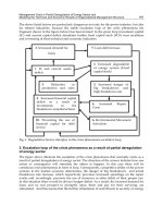
Fig. 4. Distribution of protein hits identified by Gel-LC-MS/MS and MudPIT strategies.
Proteomic Applications in Biology
162
Regarding the ability to identify GTs, the Gel-LC-MS/MS strategy identified most of the
GTs associated with GAX biosynthesis. Intriguingly, all the strategies used failed to identify
TaGT43-4 or any closest homolog from the NCBI database. Three possibilities could explain
this result: (i) the TaGT43-4 protein may be lost during the precipitation step (preparation of
the sample); (ii) TaGT43-4 is a very active enzyme and is present in only small amounts in
fraction #3, which may not be detectable by the LC-MS/MS methods used in this work, or
(iii) TaGT43-4 protein is somehow resistant to trypsin digestion.
Our hypothesis is that most of the TaGT43-4 protein was lost during the precipitation step,
as Golgi proteins are known to easily aggregated during precipitation and are very difficult
to re-solubilize in a buffer containing detergent. Although it has been shown that ASB-14
and SDS detergents are suited for solubilizing hydrophobic proteins (Herbert, 1999), their
use in this study may not be efficient in re-solubilizing freeze-dried or TCA/acetone
precipitated wheat Golgi proteins. In support of this hypothesis, fraction #3 should be
enriched in Golgi proteins (Zeng et al., 2010), but our analysis indicates that fraction #3 was
actually enriched in endoplasmic reticulum (14%), tonoplast (17%), and plastid (28%)
proteins, and Golgi proteins represented only 2% of the total hits (according to NCBI
annotation of possible subcellular localizations) (Figure 5). Therefore, a reliable
‘precipitation-re-solubilization’ strategy appears to present a crucial step that must be
optimized for minimal protein loss. Alternatively, improving enrichment strategies to
overcome protein loss during the `precipitation-re-solubilization` step should be developed.
Fig. 5. Classification of proteins identified in fraction #3 according to NCBI annotation of
their possible sub-localization.
Although all proteomics strategies employed here have failed to reveal the exact identity of
some GTs associated with GAX biosynthesis, proteomics is still a powerful tool, as many
low abundant GTs (among the 2% proteins from the Golgi) could be identified. Furthermore,
this work demonstrated that working with a non-model species without a fully sequenced
genome such as wheat did not seem to be an issue, as most (40-60%) of the proteins
identified were either from wheat sequences available in the databases, or were closest
homologs to the anticipated wheat proteins from grass species (rice, barley, maize, or
sorghum). The other limitation in applying proteomics to plant cell wall biosynthesis is the
capacity of a mass spectrometer analyzer to extract as many MS/MS spectra as possible to
increase the detection rate of proteins. To overcome all these issues and depending of the
Assessment of Proteomics Strategies for Plant Cell Wall
Glycosyltransferasesin Wheat, a Non-Model Species: Glucurono(Arabino)Xylan as a Case Study
163
complexity of the protein sample, we are proposing a workflow to carry out a successful
proteomics analysis (Figure 6). In this workflow, the first step is to assess the quality of the
sample by optimizing the precipitation step (removal of salts and contaminants) without
any protein loss. Our work demonstrated that “precipitation-re-solubilization” step is
crucial in a successful proteomics analysis of Golgi membrane proteins. Depending of the
complexity of the samples, the simplest proteomics strategy to try is the combination of
MudPIT fractionation with LTQ analysis. If the sample contains more than 500 proteins, the
use of high resolution mass spectrometry (e.g. Orbitrap) in combination with MudPIT could
Fig. 6. A proteomics workflow pipeline for efficient protein identification from unknown
samples.
be the easiest strategy to test. For more complex protein samples (more than 1000 proteins) it
may be necessary to combine 1-D SDS-PAGE fractionation, LC-MS/MS and the high
resolving power of the Orbitrap analyzer for optimal protein identification (Figure 6).
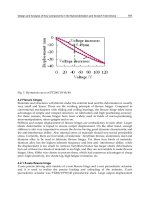
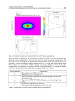
The distribution of GAX synthase over sucrose density gradient was intriguing. In the absence
of EDTA, all GAX synthase activity stabilized at the expected density of ~1.16g/mL (fractions
17 and 18 in Figure 2) along with the Golgi marker activity IDPase (Zeng et al., 2010). The
inclusion of EDTA in the sucrose gradient resulted in splitting of the activity into three density
areas, namely around density 1.09g/mL (fractions 2 and 3 in Figure 2), around density
1.14g/mL (fractions 12 and 13 in Figure 2), and around density 1.16g/mL (fractions 17 and 18
in Figure 2). Fraction #3 contained the highest GAX synthase activity. This shift in the density
Proteomic Applications in Biology
164
may suggest that GAX synthase activity is associated with various Golgi compartments. The
presence of such Golgi compartments was reported earlier by Mikami et al. (2001) in rice but
not in tobacco cells. They showed that rice Golgi complex fractionated into several
compartments by simple centrifugation on density gradient in presence of EDTA or MgCl
2
.
Recently, Asakura et al., (2006) used this strategy to isolate (and analyze by proteomics) rice
cis-Golgi membranes labeled with green fluorescent protein (GFP) fused to a cis-Golgi marker
SYP31 (which belongs to a family of SNARE proteins; soluble N-ethyl-melaeimide sensitive
factor attachment protein receptor). Interestingly, their proteomics results gave very similar
protein composition to our data, except that no members of the GT43, 47, and 75 were
identified in their study (Asakura et al., 2006). Taking together, these results suggest that the
cis-Golgi is less tightly attached to the medial and trans-Golgi compartments in grasses.
Therefore, we are tempted to propose a possible explanation of the effect of EDTA on the
dissociation of these Golgi compartments (Figure 7). Our hypothesis is that some ions (i.e.,
Ca
2+
) are involved in linking cis-Golgi (and probably the trans Golgi network [TGN]) to the
medial and trans-Golgi cisternae. In the absence of EDTA, the whole Golgi complex would
stabilize at an apparent high density (~1.16g/mL, fraction 17 and 18 in Figure 2). Upon
addition of EDTA into sucrose gradient, the ions are chelated leaving different Golgi
compartments stabilized at their corresponding densities (1.09, 1.12, and 1.16g/mL in Figure
2). In any case, the fractionation of the Golgi complex from grasses in the presence of EDTA is
an excellent tool for isolating different Golgi compartments.
Fig. 7. A diagrammatic representation of the possible effect of EDTA on the dissociation of
Golgi compartments, cis-, medial-, trans-Golgi, and trans Golgi network (TGN). EDTA
would chelate metal ions that mediate the attachment (red lines) of cis-Golgi compartment
to the medial and trans-Golgi cisternae.
5. Conclusion
We evaluated two proteomics strategies for MS/MS identification of GTs associated with
GAX synthase complex in wheat for which little genome sequence is available. The
evaluation of these strategies is based on their capacity to identify the exact wheat proteins
TaGT43-4, TaGT47-13, and TaGT75-4, or at least their closest homologous proteins from
other grass species such as rice, barley, maize, or sorghum. Therefore, these strategies are
MS/MS spectra quality-dependent and cross-species-dependent using error tolerant BLAST
search and de novo peptide sequences generated from the MS/MS spectra. Our data
indicated that the highest number of unique hits identified (180 proteins) was obtained
Assessment of Proteomics Strategies for Plant Cell Wall
Glycosyltransferasesin Wheat, a Non-Model Species: Glucurono(Arabino)Xylan as a Case Study
165
through Gel-LC-MS/MS strategy using the Orbitrap analyzer, but the fact that TaGT43-4
and/or its homologous proteins were not identified by this method underscores the
importance of optimizing sample preparation step (precipitation-re-solubilization). Based on
our results and interpretations, we have proposed a workflow chart that includes routinely
used proteomics methods and optimization steps to help increase the detection rate of
proteins of plant cell wall GTs from non-model plants.
6. Acknowledgment
The author would like to thank Dr. Green-Church for her valuable comments and editing of
the experimental procedures dealing with proteomics analysis. Thanks to Wei Zeng and
Nan Jiang for the excellent work preparing protein samples for proteomics. This work was
supported by the National Science Foundation (grant no. IOS–0724135 to A.F.)
7. References
Asakura, T., Hirose, S., Katamine, H., Kitajima, A., Hori, H., Sato, M.H., Fujiwara, M.,
Shimamoto, K. & Mitsui, T. (2006) Isolation and proteomic analysis of rice Golgi
membranes: cis-Golgi membranes labeled with GFP-SYP31. Plant Biotechnology 23:
475-485
Arabidopsis Genome Initiative (2000) Analysis of the genome sequence of the flowering
plant Arabidopsis thaliana. Nature 408: 796–815
Bodnar, W.M., Blackburn, R.K., Krise, J.M. & Moseley, M.A. (2003) Exploiting the
complementary nature of LC/MALDI/MS/MS and LC/ESI/MS/MS for increased
proteome coverage. J. Am. Soc. Mass Spectrom. 14: 971-979
Carpita, N.C. (2011) Update on Mechanisms of Plant Cell Wall Biosynthesis: How Plants
Make Cellulose and Other (1-4)-β-
D-Glycans. Plant Physiol., 155: 171-184
Chen, H.S., Rejtar, T., Andreev, V., Moskovets, E. & Karger, B.L. (2005) Enhanced
characterization of complex proteomic samples using LC-MALDI MS/MS:
exclusion of redundant peptides from MS/MS analysis in replicate runs. Anal.
Chem., 77: 7816-7825
Chen, Y.Y., Lin, S.Y., Yeh, Y.Y., Hsiao, H-H., Wu, C-Y., Chen, S-T. & Wang A. H-J. (2005) A
modified protein precipitation procedure for efficient removal of albumin from
serum. Electrophoresis, 26: 2117-27
Coutinho, P.M., Deleury, E., Davies, G.J. & Henrissat, B. (2003) An evolving hierarchical
family classification for glycosyltransferases. J. Mol. Biol. 328: 307-317
Ephritikhine, G., Ferro, M. and Rolland, N. (2004) Plant membranes proteomics: Plant
Physiol. Biochem., 42: 943-962
Faik, A. (2010) xylan biosynthesis: news from the grass. Plant Physiol., 153: 396-40
Faik, A., Bar-Peled, M., DeRocher, A.E., Zeng, W., Perrin, R.M., Wilkerson, C., Raikhel, N.V.
& Keegstra K. (2000) Biochemical characterization and molecular cloning of an
alpha-1,2-fucosyltransferase that catalyzes the last step of cell wall xyloglucan
biosynthesis in pea. J. Biol. Chem., 275: 15082-15089.
Ferro, M., Salvi, D., Brugiere, S., Miras, S., Kowalski, S., Louwagie, M., Garin, J., Joyard, J. &
Rolland, N. (2003) Proteomics of the chloroplast envelop membranes from
Arabidopsis thaliana. Mol. Cell Proteomics, 2: 325-345
Herbert, B. (1999) Advances in protein solubilization for two-dimenstional electrophoresis.
Electrophoresis, 20: 660-663
Proteomic Applications in Biology
166
Komatsu, S., Muhammad, A. & Rakwal, R. (1999) Separation and characterization of
proteins from green and etiolated shoots of rice (Ozyza sativa L.): towards a rice
proteome. Electrophoresis, 20: 630-636
Laemmli, U.K. (1970) Cleavage of structural proteins during the assembly of the head of
bacteriophage T4. Nature, 227: 680-685
McDonald, W.H., Ohi, R., Miyamoto, D.T., Mitchison, T.J. & Yates III, J.R. (2002)
Comparison of three directly coupled HPLC MS/MS, 2-phase MudPIT, and 3-
phase MudPIT. Int. J. Mass Spectrom. 219: 245-251
Mikami, S., Hori, H. & Mitsui, T. (2001) Separation of distinct compartments of rice Golgi
complex by sucrose density gradient centrifugation. Plant Sci., 161: 665-675
Newton, R.P., Breton, A.G., Smith, C.J. & Dudley, E. (2004) Plant proteome analysis by mass
spectrometry: principales, problems, pitfalls and recent developments.
Phytochemistry, 65: 1449-1485
Nesvizhskii, A.I., Vitek, O. & Aebersold, R. (2007) Analysis and validation of proteomic data
generated by tandem mass spectrometry. Nat. Methods 4: 787-797
O’Farrell, P.H. (1975) High resolution two-dimensional electrophoresis of proteins. J. Biol.
Chem., 250: 4007-4021
Patterson, S.D. (2003) Data analysis-the Achilles heel of proteomics. Nat. Biotechnol., 21: 221-222
Perrin, R.M., DeRocher, A.E., Bar-Peled, M., Zeng, W., Norambuena, L., Orellana, A.,
Raikhel, N.V. & Keegstra, K. (1999) Xyloglucan fucosyltransferase, an enzyme
involved in plant cell wall biosynthesis. Science 284: 1976–1979
Santoni, V., Kieffer, S., Desclaux, D., Masson, F. & Rabilloud, T. (2000) Membrane
proteomics: use of additive main effects with multiplicative interaction model to
classify plasma membrane proteins according to their solubility and electrophoretic
properties. Electrophoresis, 21: 3329-3344
Service, R.F. (2008) Proteomics. Proteomics ponders prime time. Science 321: 1758-1761
Shevchenko, A., Sunyaev, S., Loboda, A., Shevehenko, A., Bork, P., Ens, W. & Standing, K.G.
(2001) Charting the proteomes of organisms with unsequenced genomes by
MALDI-quadrupole time of flight mass spectrometry and Blast homology
searching. Anal. Chem., 73: 1917-1926
Simon, W.J., Maltman, D.J. & Slabas, A.R. (2008) Isolation and fractionation of the
endoplasmic reticulum from castor bean (Ricinus communis) endosperm for
proteomic analysis. Methods Mol. Biol., 425: 203-215
Washburn, M.P., Wolters, D. & Yates III, J.R. (2001) Large-scale analysis of the yeast proteome
by multidimensional protein identification technology. Nat. Biotechnol., 19: 242-247
Wolters, D.A., Washburn, M.P. & Yates III, J.R. (2001) An automated multidimentional
protein identification technology for shotgun proteomics. Anal Chem., 73: 5683-5690
Zellner, M., Winkler, W., Hayden, H., Diestinger. M., Eliasen, M., Gesslbauer, B., Miller, I.,
Chang, M., Kungl, A., Roth, E. & Oehler, R. (2005) Quantitative validation of
different protein precipitation methods in proteome analysis of blood platelets
Electrophoresis,26: 2481-2489
Zhu, W., Smith, J.W. & Huang C.M. (2010) Mass spectrometry-based label-free quantitative
proteomics. J. Biomed. Biotechnol., 840518. Doi:10.1155/2010/840518
Zeng, W., Jiang, N., Nadella, R., Killen, T.L., Nadella, V. & Faik, A. (2010) A
glucurono(arabino)xylan synthase complex from wheat contains members of the
GT43, GT47, and GT75 families and functions cooperatively. Plan
t Physiol., 154: 78-97
8
The Current State of the Golgi Proteomes
Harriet T. Parsons
1
, Jun Ito
1
,
Eunsook Park
2
, Andrew W. Carroll
1
, Hiren J. Joshi
1
,
Christopher J. Petzold
1
, Georgia Drakakaki
2
and Joshua L. Heazlewood
1
1
Joint BioEnergy Institute and
Physical Biosciences Division,
Lawrence Berkeley National Laboratory
2
Department of Plant Sciences,
University of California, Davis
USA
1. Introduction
The Golgi apparatus plays a central role in the eukaryotic secretory pathway shuttling
products between a variety of destinations throughout the cell. It is a major site for the post
translational modification and processing of proteins as well as having a significant role in
the synthesis of complex carbohydrates. The Golgi apparatus exists as a contiguous
component of the endomembrane system which encompasses the endoplasmic reticulum
(ER), plasma membrane, vacuoles, endosomes and lysozymes (Morre & Mollenhauer, 2009).
The Golgi apparatus is tightly linked to numerous signaling processes through membrane
and vesicular trafficking throughout the endomembrane. This interconnection provides
communication and recycling networks between the Golgi apparatus, the plasma
membrane, vacuoles and lysosomes. Thus, the Golgi apparatus represents significant
structure within the eukaryotic cell by regulating an array of complex biosynthetic
processes.
The Golgi apparatus was first described at the end of the 19
th
century by Camillo Golgi
using light microscopy on nerve tissue samples (Golgi, 1898; Dröscher, 1998). A half century
passed and with the development of the electron microscope a more detailed picture
emerged highlighting the extreme complexity and heterogeneity of the organelle in the
eukaryotic cell (Dalton & Felix, 1953). The classic structure of the Golgi apparatus is that of a
distinct membranous stack disassociated within the cytosol (Fig.1). This familiar image
conceals the underlying complexity and interconnected nature of this organelle within the
cell. With the development in the last decade of routine mass spectrometry-based
proteomics, applying these methods to functionally characterize biological systems has been
a major focus. The complexity and integrated structure of the Golgi apparatus and
associated membrane systems makes analysis of this organelle one of the most complicated
subcellular compartments to address with modern proteomics techniques. This chapter will
highlight recent advances in our knowledge about the Golgi apparatus in eukaryotic
systems that have been largely driven by the development of isolation procedures and
subsequent proteomic analysis.
Proteomic Applications in Biology
168
Fig. 1. Electron micrograph of a Golgi stack from the model plant Arabidopsis thaliana
highlighting the integrative structure and membrane organization. Scale bar = 200nm.
2. Differential density enrichment of Golgi
The basic technique of differential density enrichment of Golgi membranes represents the
most common isolation and enrichment process for downstream proteomic analyses. The
technique was well-established in most eukaryotic systems prior to the development of
mass spectrometry-based identification techniques. Consequently, analysis of the enriched
Golgi apparatus fraction using this method was a logical approach, although one limited by
contaminating organellar membranes and low gradient resolution. Yet, insights into
membrane systems associated with trafficking, post-translational modifications and
complex carbohydrate biosynthesis have been revealed.
Many of the initial approaches used to characterize Golgi associated proteins by differential
centrifugation employed SDS-PAGE arraying techniques prior to identification of proteins
by mass spectrometry. Nearly all of the early proteomic studies on enriched Golgi fractions
are from easily accessible samples such as rat livers, likely reflecting the need for the
development of purification techniques. The earliest ‘proteomic’ analyses of the Golgi
employed two-dimensional gel electrophoresis (2-DE) to array enriched stacked Golgi
fractions from rat livers (Taylor et al., 1997b). The study used cyclohexamide in an effort to
clear transitory proteins from the secretory pathway and reduce non-specific Golgi proteins.
While only a handful of proteins were identified by cross comparing reference maps and
immunoblotting the study demonstrated the validity of a proteomic approach to analyze the
Golgi and could discern resident proteins from cargo and cytosolic proteins (Taylor et al.,
1997b). Significantly, the study employed a recently developed sequential sucrose gradient
enrichment method (Taylor et al., 1997a) and enabled the reliable visualization of Golgi
proteins by 2-DE with reduced contaminants. The sequential sucrose enrichment of stacked
Golgi from liver samples involved initially loading a clarified homogenate (post-nuclear
supernatant) between 0.86 M and 0.25 M sucrose steps followed by centrifugation. The
resultant 0.5/0.86M interface (Int-2) was removed, adjusted to 1.15M sucrose and overlaid
with 1.0M, 0.86M and 0.25M sucrose and centrifuged. The resultant 0.25/0.86 interface
The Current State of the Golgi Proteomes
169
represented the enriched stacked Golgi fraction (Fig. 2). This fraction was estimated to
represent 200 to 400- fold enrichment over the post-nuclear fraction (Taylor et al., 1997a).
With the development of reliable protein identification through tandem mass spectrometry
this Golgi enrichment technique could be more readily exploited for functional proteomic
studies. This was undertaken through 2-DE arraying of Golgi samples using the sequential
sucrose technique on rat liver and mammary epithelial samples (Taylor et al., 2000; Wu et
al., 2000). Both studies employed early liquid chromatography-tandem mass spectrometry
(LC-MS/MS) methods using iontrap MS and resulted in the identification of 71 proteins
from 588 unique 2-DE spots of rat liver (Taylor et al., 2000) and over 30 distinct proteins
from mammary epithelial cells (Wu et al., 2000). This later work outlined a comparative
study of Golgi isolated from cells transiting from basal (steady-state) to maximal secretion.
Proteins identified by this study were upregulated during this transition and comprised a
series of Rab proteins, structural components such as microtubule motor proteins and
membrane fusion proteins e.g. Annexins (Wu et al., 2000). Another early attempt at
characterizing Golgi associated proteins by proteomic methods employed a previously
developed strategy to enrich WNG fractions (nuclear associated Golgi fractions) from rat
liver homogenates (Dominguez et al., 1999). The technique employs multiple rounds of low
speed centrifugation steps in conjunction with a 0.25 to 1.10 M sucrose gradient. For analysis
by mass spectrometry, proteins from the resultant rat liver WNG fraction were partitioned
into a detergent fraction using Triton X-114 to enrich for membrane proteins and arrayed by
SDS-PAGE or 1-DE (Bell et al., 2001). A total of 81 proteins were identified from 1-DE
arrayed samples using MALDI-MS, nano-MS/MS or Edman sequencing and included a
range of well-characterized Golgi proteins including lectins, KDEL receptor,
glycosyltransferases and trafficking proteins (Rabs, SNAREs, SCAMPs). While the results
demonstrated the applicability of the overall approach, a significant number of
contaminants were also identified in the samples (Bell et al., 2001).
Post-Nuclear
Sup ernatant
(PNS)
100,000g
0.86M
PNS-P
0.25M
1.3M
Load PNS
Pellet
Int-1
Int-2
Int-3
100,000g
1.0M
0.86M
0.25M
Int-2
1.15M
100,000g
Stacked
Golgi
Load Int-2
In 1.15M
First Gradient Second Grad ient
Fig. 2. Schematic outline of the sequential sucrose density gradient procedure developed for
the isolation and purification of Golgi stacks from rat liver (Taylor et al., 1997a). The values
within the tubes indicate concentration of sucrose (M: mol/L).
With the development of advanced gel-free approaches like multidimensional protein
identification technology (MudPIT) for the analysis of complex lysates by mass
spectrometry (Wu et al., 2003), a number of previously established enrichment strategies
were again analyzed. The two independent and sequential sucrose step gradient technique
(outlined above) was employed to isolate Golgi membranes from the livers of rats treated
with cyclohexamide (Wu et al., 2004). These fractions were analyzed directly by MudPIT,
Proteomic Applications in Biology
170
using a 12-step strong cation exchange (SCX) fractionation coupled to reverse phase LC-
MS/MS by iontrap MS. From this approach, 421 proteins were identified from Golgi
fractions isolated from rat liver homogenates (Wu et al., 2004). About 26% of the proteins
(110) could be confidently allocated to Golgi-associated functions with about 10% allocated
as unknowns. A large proportion of the identifications represented proteins from
contaminating organelles including ER (23%), plasma membrane (9%) and cytosol (9%).
Nonetheless, a large number of proteins were identified from major Golgi functional
processes including transporters, SNARES, glycosyltransferases, G-proteins and clathrin
proteins (Wu et al., 2004). Importantly, the gel-free approach enabled the detection of
protein modifications with a total of 10 Golgi proteins identified with arginine
dimethylation, a potential regulator of protein function (Wu et al., 2004). Stacked Golgi
membranes isolated from rat liver homogenates were again analyzed by MudPIT (using a
Q-TOF) in a separate study. After initially enriching the membranes by a discontinuous
sucrose gradient, repeated centrifugations through 1.3M sucrose were used to clarify and
purify the Golgi stacks (Takatalo et al., 2006). Replicate experiments were fractionated
offline by SCX prior to analysis by LC-MS/MS. A total of 1125 proteins were identified in
the two experiments by combining results from two separate MS/MS search algorithms
(Takatalo et al., 2006). Analysis of these protein identifications found that 35% contained at
least one predicted transmembrane domain and 201 proteins could be assigned as Golgi or
transport related based on functional annotations. When compared to the similar approach
of Wu et al., (2004) a total of 399 proteins were identified by both studies. The multiple
isolation and analysis strategies enabled the number of unknown proteins from the rat liver
Golgi fraction to be significantly extended by 89 proteins. Overall, proteins associated with
glycosylation, Golgi matrix complexes, enzymes involved in isomerase and transferase
activities and protein trafficking components were all identified (Takatalo et al., 2006).
Nonetheless, limitations in the purification procedure are highlighted by positive allocations
of this proteome to the cytosol (15%), contaminating membranes (20%) and the cryptically
named ‘undetermined location’ (20%) (Takatalo et al., 2006).
In an effort to address contamination issues and to better tease apart and define the ER,
quantitative proteomic strategies were subsequently employed on rat liver Golgi enriched
by sucrose density centrifugation (Gilchrist et al., 2006). Previous methods used to isolate rat
Golgi fractions (Bell et al., 2001) were used as well as rough and smooth microsomal
preparations (Paiement et al., 2005). This was followed by 1-DE, band isolation and analysis
by nanoLC-MS/MS (Q-TOF MS). Based on annotation information, Golgi fractions were
estimated to comprise around 10% mitochondrial protein, although there was a distinction
in proteins identified between the Golgi and microsomal fractions. Multiple biological
replicates for the Golgi and microsomes were undertaken and proteins quantified by
redundant spectral counting techniques (Blondeau et al., 2004). Hierarchical clustering with
Pearson correlation was used to visualize protein abundance measures and this analysis
identified four major groups which co-clustered with organelle markers sec61 (rough
microsome), calnexin (ER), p97 (smooth microsomes) and mannosidase II (Golgi). This co-
clustering with defined markers indicated that quantifying distinct fractions could be used
to tease apart the secretory pathway (Gilchrist et al., 2006). Nonetheless only 43 additional
proteins could be confidently assigned to the Golgi compared to a previous analysis that
had identified 81 proteins (Bell et al., 2001). Consequently COP1 vesicle enrichment from the
Golgi fraction was also undertaken to extend the proteome. Overall 1430 proteins were
identified from the rat liver secretory system with quantitative proteomics enabling the
The Current State of the Golgi Proteomes
171
confident assignment of 193 proteins to Golgi/COP1 vesicles, with 405 proteins shared
between the Golgi and ER fractions. The work identified a host of secretory proteins
including Rabs, SNARES, Sec proteins and unknowns (Gilchrist et al., 2006). A similar
MudPIT approach in mouse was undertaken by employing spectral counting for
quantification of proteins from multiple subcellular fractions (cytosol, microsomes,
mitochondria and nuclei) to determine distinct proteomes (Kislinger et al., 2006). This study
also isolated compartments from different organs and undertook organ specific transcript
profiles to develop machine-learning techniques to classify subcellular localizations. The
enrichment method for the microsomal fraction (Golgi) resulted from high speed
centrifugation of a post-nuclear supernatant and thus represents a fairly crude membrane
preparation. Nonetheless, by employing samples from multiple organs, transcript profiling
and modeling techniques a significant number of secretory proteins were identified and
expressions profiled (Kislinger et al., 2006).
The focus on cellular secretory system as an important component in agricultural milk
production was explored with the analysis of microsomal fractions from bovine mammary
glands (Peng et al., 2008). The study used a 100,000g microsomal pellet from homogenized
mammary tissue clarified at 10,000g to isolate enriched microsomal fractions. Samples were
arrayed by 1-DE and analyzed by nanoLC-MS/MS, by linear iontrap (LTQ). Of the 703
proteins identified from these fractions nearly half were designated as likely secretory
components with a total of 48 (~ 7%) allocated as originating from the Golgi apparatus.
These included ADP-ribosylation factors, coatomer protein complex components,
transporters, Rab and Sec proteins (Peng et al., 2008).
Surprisingly, only one study has used basic density centrifugation techniques and
proteomics to examine the Golgi apparatus of yeast, the most widely studied eukaryotic
system (Forsmark et al., 2011). The sec6-4 yeast mutant was identified several decades ago as
a temperature sensitive mutant that accumulates post-Golgi secretory vesicles (Walworth &
Novick, 1987) although until recently no proteomic analysis had been undertaken. Recently
the sec6-4 mutant was used to undertake comparative proteomics against the sec23-1, a yeast
mutant that is depleted of vesicles (Kaiser & Schekman, 1990). Comparative proteomics was
undertaken between these two yeast mutants using iTRAQ on fractionated vesicles purified
on 9-step linear sorbitol gradients. After density centrifugation, fractions showing the
highest signal for SNARE proteins from the two mutant lines were pooled and used for
analysis by nanoLC-MS/MS by Orbitrap MS. A total of 242 proteins were identified from
the pooled fractions with 91 having greater abundance in the sec6-4 mutant lines. Many of
the most highly differentially expressed proteins were cell wall associated and plasma
membrane transporters all of which were likely cargo proteins. The analysis also identified
many vesicle proteins which included SNAREs, GTPases, protein glycosylation components
and V-ATPase complex components (Forsmark et al., 2011).
While there has been significant interest in plant cell wall biosynthesis for the past decade,
only two studies have used proteomics to explore density enriched Golgi fractions from
plants. The absence of many studies likely reflects the reported difficulties in isolating Golgi
membranes from plants. With this problem in mind, one of the first attempts employed a
rice suspension cell culture expressing a cis-Golgi marker (35S::GFP-SYP31 construct) in an
attempt to characterize enriched fractions from discontinuous sucrose density gradient
(Asakura et al., 2006). The enrichment procedure utilized sequential discontinuous sucrose
gradients on a microsomal fraction to greatly enrich membranes containing the 35S::GFP-
SYP31 construct. The technique also employed the addition of MgCl
2
which had previously
Proteomic Applications in Biology
172
been shown to separate distinct Golgi compartments (Mikami et al., 2001). Twice purified
Golgi membranes were arrayed by both 1-DE and 2-DE, but the 2-DE arrays yielded few
identifications. Protein bands from 1-DE were excised and analyzed by MALDI-TOF MS. A
total of 63 proteins were identified with 70% containing predicted transmembrane domains.
A variety of Golgi proteins were identified including COP complex components, GTPases,
Rab proteins, an EMP70 and phospholipase D (Asakura et al., 2006). Very little is known
about the role of the Golgi apparatus in secondary cell wall formation in plants due to
considerable technical issues in sample preparation. Recently, an analysis of membrane
enriched fractions from developing compression wood of Pinus radiata has attempted to
address this shortage in knowledge (Mast et al., 2010). Microsomal fractions were enriched
from compression wood homogenates using discontinuous gradients with maximal Golgi
marker activity found at the 8/27% interface. Fractions were further extracted using a TX-
114 Triton phase separation technique in order to remove contaminating proteins (e.g.,
actin). Samples were analyzed using nanoLC-MS/MS by linear iontrap (LTQ). A total of 175
proteins were identified, 66 in the aqueous phase and 103 in the detergent phase. Only a
handful of proteins were confidently allocated to the Golgi apparatus after functional
annotations, further highlighting the inherent difficulties in working with this tissue. These
included laccases, cellulose synthases and a xylosyltransferase (Mast et al., 2010).
3. Immunoaffinity purification of Golgi
The immunoaffinity purification (IP) of organelles has been a widely used technique for
decades. Since the approach relies on the presence of differential epitopes rather than
differences in physical parameters it is highly amenable for isolating functionally distinct
subcellular organelles (Richardson & Luzio, 1986). This approach is often combined with
sucrose gradient fractionation. The use of IP and proteomics to isolate and characterize
subcellular compartments of the secretory system has been most effectively applied in
animal systems. Early approaches investigating Golgi immunoisolation employed baby
hamster kidney (BHK) cells infected with mutant vesicular stomatitis virus (VSV). After
temperature induction the viral G protein accumulates at the trans-Golgi network (TGN)
serving as bait for the immunoisolation of the corresponding compartment. Immunoisolated
TGN was visualized with TEM confirming its structural integrity. Several polypeptides
were enriched in the isolated fraction (de Curtis et al., 1988).
An early study which highlighted future approaches, Hobman et al., (1998) successfully
used IP to identify ER exit sites in BHK cells. This study used the Rubella virus E1
glycoprotein, which normally localizes in a subset of Smooth ER (SER) and does not overlap
with COPI coated ER. Tubular networks of SER were separated by cell fractionation,
followed by immunoisolation using Dynabeads coated with an antibody against epitope
tagged E1. Using electron microscopy, the authors demonstrated that the isolated
membrane structures were distinguished from COPI coated ER. Western blot analysis
against known markers identified the presence of proteins in this compartment and
included ER-Golgi intermediate and transitional ER markers. These results indicated that
this tubular distinct subdomain of SER provides the site for COPII vesicle biogenesis. A
further early study demonstrating the viability of the approach used antibody-conjugated
magnetic beads facilitating the isolation of peroxisomes from rat liver (Kikuchi et al., 2004).
Peroxisomes separated by cell fractionation were further isolated with an antibody against
PMP70 (70-kDa peroxisomal membrane protein). Proteomic analysis was carried out, using
The Current State of the Golgi Proteomes
173
in gel digestion and LC-MS/MS was accommodated in a hybrid type-Q-TOF mass
spectrometer. The proteome contained 34 known peroxisomal proteins and a minor number
of mitochondrial proteins. Two unknown proteins were added to the peroxisomal proteome
by this study; a Lon protease and a bi-functional protein consisting of an aminoglycoside
phosphotransferase-domain.
The application of IP followed by protein identification through mass spectrometry
addressing the proteome of the secretory components was first undertaken to examine the
ER-Golgi intermediate compartments or ERGIC of humans (Breuza et al., 2004). The
approach used the drug Brefeldin A to cause an over accumulation of ERGIC clusters in
human HepG2 cells. The post nuclear supernatant was loaded onto a linear 13-29%
Nycodenz gradient and fractions enriched for the ERGIC-53 marker pooled. Dynabeads
coupled to KDEL receptor monoclonal antibodies were used to IP the ERGIC membranes.
Samples were arrayed by 1-DE and protein bands analyzed by MALDI-TOF MS. A total of
19 proteins were identified including ERGIC-53, the KDEL receptor, SEC22b, cargo
receptors and membrane trafficking components.
The use of IP on recombinant lines expressing specific epitopes was first undertaken in yeast
(Inadome et al., 2005). Strains expressing recombinant SNARE proteins Myc
6
-sed5 and
Myc
6
-Tlg2 were employed to isolate vesicles associated with early (sed5) and late (Tlg2)
Golgi compartments. Since recombinant Myc tagged proteins are employed the IP does not
require protein specific antibodies. The approach utilized the supernatant from a 100,000g
centrifugation step to purify both Myc
6
-sed5 and Myc
6
-Tlg2 vesicles using an anti-Myc
monoclonal antibody. Vesicles were affinity purified from each strain using Protein A-
Sepharaose beads and resultant proteins arrayed by 1-DE. Protein bands were excised and
analyzed by MALDI-TOF MS. A total of 29 proteins were identified from the Sed5 vesicles
and 32 proteins from the Tlg2 vesicles. Both proteomes contained large proportions of
known Golgi proteins including Rab GTPases, SNAREs, mannosyltransferases, V-ATPase
proteins (Inadome et al., 2005). Interestingly a number of proteins were identified
exclusively in the either the early Sed5 vesicles, namely COPII components involved in
transport from ER to Golgi and mannosyltransferases involved in protein glycosylation.
This fraction included a protein of unknown function (svp26) that was shown to be involved
in retention of membrane proteins in early Golgi compartments. Similarly, in the late vesicle
Tgl2 proteome, a significant number of proteins with no known function were identified.
Isolation of synaptic vesicles by immunopurification from mammalian systems is a well-
developed procedure (Morciano et al., 2005; Burre et al., 2007). Due to the dynamic nature of
synaptic vesicle trafficking, it has been challenging to isolate synaptic vesicles from their
corresponding plasma membrane with conventional vesicle isolation techniques.
Consequently an IP approach was employed on samples from rat brains and enabled the
identification of several proteins that might be involved in the regulation of
neurotransmitter release and the structural dynamics of the nerve terminal (Morciano et al.,
2005). Synaptic vesicles were isolated from two synaptosomal enriched sucrose fractions,
followed by IP with Dynabeads covered with antibodies against synaptic vesicle protein 2
(SV2). The isolated proteins were separated by BAC/SDS-PAGE and identified by MALDI-
TOF MS. The free vesicle fraction contained 72 proteins while 81 were identified in the
plasma membrane containing, denser fraction. Although many proteins, involved in vesicle
trafficking and tethering were identified in both fractions, several proteins were specific to
one fraction. For example, several isoforms of Rab2, Rab3, Rab11, Rab14 were only found in
the free vesicles. The PM associated fractions contained several proteins that could modulate
Proteomic Applications in Biology
174
presynaptic function, including GTPases and Na+/K+-ATPases, which are potentially
associated with the plasma membrane. Several physicochemical conditions were evaluated
to enrich low abundant proteins (Burre et al., 2007), among these the addition of SB/DTT or
NP40 showed that it can improve the efficacy of protein elution from the magnetic beads.
Moreover, the use of phase separation can divide proteins according to their
hydrophobicity, resulting in the isolation of more integral membrane proteins.
The selection of bait proteins might be a crucial prerequisite for specific vesicle
immunoisolation. Motor proteins could be excellent baits for dissecting a subset of post-Golgi
membrane compartments. Recently, a kinesin motor, calsyntenin-1, has been successfully used
to identify its cargo vesicles from neuronal axon in mouse brain (Steuble et al., 2010). This
study involved IP from two distinct subfractionated organelle populations, using Dynabeads
coupled with anti-calsyntenin antibodies. Solubilized proteins were analyzed by LTQ-ICR-FT
MS. This approach allowed the identification of endosomes that contained calsyntenin-1 and
identification of their specific resident proteins. A combination of biochemical analysis and
immunocytochemistry lead to a model of two distinct, non-overlapping endosomal
populations of calsyntenin-1. An early endosome population, containing β-amyloid precursor
protein (APP) and second, a APP negative, recycling endosomal population.
In contrast with the studies in animal and microbial systems only one pioneering study has
been undertaken in plants (Drakakaki et al., 2011). The SYP61 TGN and early endosome
compartment was isolated, employing an IP approach from SYP61-CFP transgenic
Arabidopsis plants (Fig. 3). This involved a two-step procedure, comprising sucrose
gradient fractionation followed by IP using agarose beads coupled with antibodies against
GFP, facilitating the SYP61 compartment isolation. In total, 145 proteins were identified by
MudPIT nano-LC MS/MS. These include the SYP61 SNARE complex and its regulatory
proteins (SYP41, VTI12, and VPS45), GTPases and proteins involved in vesicle trafficking.
Other SYP4 members such as SYP43 were also found in the SYP61 vesicle proteome,
establishing new protein association with SYP61. Several proteins of unknown function,
Fig. 3. Schematic representation of vesicle immunoisolation. A). Subcellular localization of
SYP61-CFP in transgenic Arabidopsis root cells. Arrows indicate SYP61-CFP vesicles. Scale
bar = 10µm. B). Diagram of vesicle IP. Vesicles or compartments are isolated from cell lysate
with the aid of an antibody against a target protein. Compartments are not to scale. B:
Protein A-Agarose bead; A: GFP antibody; G: Golgi apparatus; S: SYP61 containing vesicles;
L: cell lysates, V:vacuole.
The Current State of the Golgi Proteomes
175
such as ECHIDNA were included providing the opportunity of new cargo identification.
Plasma membrane associated proteins such as the SYP121-complex and members of the
cellulose synthase family were also present. These findings suggested a role of SYP61 in
exocytic trafficking and possibly in transporting of cell wall components.
Immunoisolation of TGN vesicles can not only facilitate the identification of its proteome
but also other components. A recent study in yeast has analyzed the lipid profile of TGN
vesicles transporting a transmembrane raft protein. It demonstrated that the isolated
compartment was selectively enriched with ergosterol and sphingolipid, thus providing
evidence that TGN can sort membrane lipids (Klemm et al., 2009). As more different
subcompartments of Golgi are purified, we will gain greater insights into the nature of the
sorted components and their transported cargo.
4. Subcellular correlation analysis of Golgi from complex lysates
For proteomic analysis of an isolated organelle, it is critical that the sample is of high purity
to minimize the inclusion of contaminant proteins. If the organelle sample is only partially
enriched, far greater scrutiny is required to confidently differentiate between proteins that
genuinely reside in the organelle and contaminants. Highly purified samples are relatively
straightforward to obtain for proteomic analyses of mitochondria (Sickmann et al., 2003;
Taylor et al., 2003; Heazlewood et al., 2004), chloroplasts (Friso et al., 2004; Kleffmann et al.,
2004) and nuclei (Bae et al., 2003; Turck et al., 2004; Mosley et al., 2009), mainly through the
use of differential centrifugation techniques. However, obtaining pure organelles of the
endomembrane system such as the Golgi, endoplasmic reticulum (ER) and plasma
membrane is difficult as they are highly interconnected and are of similar sizes and densities
(Wu et al., 2004; Hanton et al., 2005). To avoid the task of having to isolate pure
endomembrane system organelles, several groups have instead developed techniques to
analyze multiple subcellular compartments simultaneously with crude organelle samples.
One such technique is the localization of organelle proteins by isotope tagging (LOPIT),
where organelles are partially separated by equilibration centrifugation in a self-forming
iodixanol density gradient (Dunkley et al., 2004; Sadowski et al., 2006; Lilley & Dunkley,
2008). Protein distribution patterns are quantified by differential isotope tagging of proteins
across organelle fractions with LC-MS/MS on a Q-TOF instrument. Finally, multivariate
analysis of isotopically-tagged peptide fragment ion data is used to correlate proteins with
similar density gradient ion distributions with that of known organelle marker proteins to
determine their subcellular location.
The first LOPIT study was performed on twelve crude membrane fractions from
Arabidopsis callus culture that were selectively tagged with light or heavy isotope-coded
affinity tags (ICATs) (Dunkley et al., 2004). ICATs are chemical probes containing 1) a
reactive group targeting free cysteine residues, 2) an isotopically coded linker to distinguish
between heavy (deuterium or
13
C) and light tags (protons or
12
C) and 3) an affinity tag such
as biotin or avidin to capture the labeled peptide or protein (Gygi et al., 1999). The twelve
membrane fractions were organized into six pair-wise comparisons of ICAT light- and
heavy-tagged fractions. The six ICAT protein sample pairs were pooled, digested with
trypsin and the ICAT-labeled peptides were avidin-affinity purified and analyzed by LC-
MS/MS (Q-TOF). Multivariate analysis of LC-MS/MS data determined the relative
abundances of 170 identified Arabidopsis proteins. Of these, a subset of 28 known or
predicted Arabidopsis organelle marker proteins were used by multivariate analysis to