Báo cáo hóa học: " Research Article Multiadaptive Bionic Wavelet Transform: Application to ECG Denoising and Baseline Wandering Reduction" doc
Bạn đang xem bản rút gọn của tài liệu. Xem và tải ngay bản đầy đủ của tài liệu tại đây (1.54 MB, 11 trang )
Hindawi Publishing Corporation
EURASIP Journal on Advances in Signal Processing
Volume 2007, Article ID 41274, 11 pages
doi:10.1155/2007/41274
Research Article
Multiadaptive Bionic Wavelet Transform: Application to
ECG D enoising and Baseline Wandering Reduction
Omid Sayadi and Mohammad B. Shamsollahi
Biomedical Signal and Image Processing Laboratory (BiSIPL), School of Electrical Engineering,
Sharif University of Te chnology, P.O. Box 11365-9363, Tehran, Iran
Received 7 May 2006; Revised 22 October 2006; Accepted 11 January 2007
Recommended by Maurice Cohen
We present a new modified wavelet transform, called the multiadaptive bionic wavelet transform (MABWT), that can be applied
to ECG signals in order to remove noise from them under a wide range of variations for noise. By using the definition of bionic
wavelet transform and adaptively determining both the center frequency of each scale together with the T-function, the problem
of desired signal decomposition is solved. Applying a new proposed thresholding rule works successfully in denoising the ECG.
Moreover by using the multiadaptation scheme, lowpass noisy interference effects on the baseline of ECG will be removed as a
direct task. The method was extensively clinically tested with real and simulated ECG signals which showed high performance of
noise reduction, comparable to those of wavelet transform (WT). Quantitative evaluation of the proposed algorithm shows that
the average SNR improvement of MABWT is 1.82 dB more than the WT-based results, for the best case. Also the procedure has
largely proved advantageous over wavelet-based methods for baseline wandering cancellation, including both DC components and
baseline drifts.
Copyright © 2007 O. Sayadi and M. B. Shamsollahi. This is an open access article distributed under the Creative Commons
Attribution License, which permits unrestricted use, distribution, and reproduction in any medium, provided the original work is
properly cited.
1. INTRODUCTION
The heart is a hollow muscular organ which through a co-
ordinated muscle contraction generates the force to circulate
blood throughout the body. Each beat of our heart is trig-
gered by an electrical impulse from special sinus node cells
in the atrium. T he electrical impulse travels to other parts of
the heart and causes the heart to contract. An electrocardio-
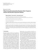
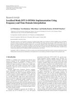
gram (ECG) records these electrical signals. A normal ECG
describes the electrical activity in the heart, and can be de-
composed in characteristic components, named the P, Q, R,
S, and T waves. Each of these components has its own typi-
cal form and behavior and each heart beat traces the familiar
morpholog y labeled by these peaks and t roughs as shown in
Figure 1.
When an elect rocardiogram is recorded, it would be con-
taminated with many kinds of noise [1], such as the follow-
ing.
(i) Baseline wandering, which can be modeled by low pass
noise.
(ii) 50 or 60 Hz power-line interference.
(iii) Electromyogram (EMG), which is an electric signal
caused by the muscle motion during effort test.
(iv) Motion artifact, which comes from the variation of
electrode-skin contact impedance produced by elec-
trode movement during effort test.
Since ECG is mostly contaminated with noise, extraction
of pure cardiological indices from noisy measurements has
been one of the major concerns of biomedical signal process-
ing and needs reliable signal processing techniques to pre-
serve the diagnostic information of the recorded signal. For
example, the S-T segment in the ECG signal is used for di-
agnosing ischemia, myocardial infarction and indicating an
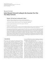
imbalance of myocardial oxygen supply. The aim of this pa-
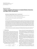
per is to remove common noise caused by motion artifact,
Figure 2(b), and also baseline wandering, Figure 2(c).
Techniques have been proposed to extract the ECG com-
ponents contaminated with the background noise and allow
the measurement of subtle features in the ECG signal. One
of the common approaches is the adaptive filters architecture
which has been used for the noise cancellation of ECGs con-
taining baseline wandering, power line interference, EMG
2 EURASIP Journal on Advances in Signal Processing
0 100 200 300 400 500 600 700 800
−1
−0.5
0
0.5
1
1.5
2
2.5
Samples
Amplitude (mV)
Q
S
P
R
T
ST
segment
Figure 1: A typical human’s ECG signal.
noise, and motion artifacts [2, 3]. Statistical techniques such
as principal component analysis [4], indep endent compo-
nent analysis [5, 6],andneuralnetworks[7] have also been
used to extract a noise-free signal from the noisy ECG. Over
the past several years, wavelet transform (WT) methods have
also received great deal of attention for denoising of signals
having multiresolution characteristics such as the electrocar-
diogram [8–12].
Besides the above algorithms, baseline wandering re-
moval has been addressed in the literature individually. Base-
line estimation using cubic spline [13], baseline construction
by linearly inter polating between preknown isoelectric levels
estimated from PR intervals [14], linear filtering [15], and
the use of wavelet packets [16] are major approaches in this
field.
Among transform-based methods, bionic wavelet trans-
form (BWT), introduced by Yao and Zhang [17], is mainly
developed and being optimized by the human biosystem
and has showed promising results in speech processing. The
term “bionic” goes back to the fac t that BWT was origi-
nally inspired by a biological mechanism which is related
to the human’s biosystem. This idea motivated us to apply
the BWT for processing the electrocardiograms. In this pa-
per we attempted to apply BWT with new modifications to
be properly adjusted for ECG processing, especially for de-
noising applications. The new proposed algorithm employs a
multiadaptation scheme and leaves the multiadaptive bionic
wavelet transform as a novel ECG analyzer.
The paper is organized as follows. Section 2 provides the-
oretical background on the definition of the bionic wavelet
transform. In Section 3 BWT is optimized for ECG sig-
nal analysis with modifying the multiadaptation scheme.
Section 4 focuses on denoising and investigates the multi-
adaptive bionic wavelet transform to be applied to ECG for
denoising and baseline wandering cancellation. Finally, the
simulation results are provided in Section 5 followed by dis-
cussion and conclusions coming in Section 6.
0 500 1000 1500 2000
−1
−0.5
0
0.5
1
1.5
2
2.5
Samples
Amplitude (mV)
(a)
0 500 1000 1500 2000
−1
−0.5
0
0.5
1
1.5
2
2.5
Samples
Amplitude (mV)
(b)
0 500 1000 1500 2000
−2
−1.5
−1
−0.5
0
0.5
1
1.5
2
2.5
Samples
Amplitude (mV)
(c)
Figure 2: (a) Normal ECG, (b) noisy ECG, (c) baseline wandered
ECG with its baseline trace.
2. DEFINITION OF BIONIC WAVELET TRANSFORM
The wavelet transform comprises the coefficients of the ex-
pansion of the original signal x(t) with respect to a basis
h
a,τ
(t), each element of which is a dilated and translated ver-
sion of a function h(t), called the mother wavelet, according
to
h
τ,a
(t) =
1
|a|
h
t − τ
a
. (1)
Depending on the choice of the mother wavelet appro-
priately, the basis can be orthogonal or biorthogonal. The
wavelet transform coefficients, given by the inner product of
x( t) and the basis functions,
WT
x
(τ, a) =<x(t), h
τ,a
(t) >=
1
|a|
x( t)h
∗
t − τ
a
dt
(2)
comprise the time-frequency representation of the origi-
nal signal. The wavelet transform has good localization in
O. Sayadi and M. B. Shamsollahi 3
0 200 400 600 800 1000 1200 1400 1600
−1
0
1
2
3
Samples
Amplitude
(mV)
(a)
0 200 400 600 800 1000 1200 1400 1600
30
20
10
Samples
Scale
(b)
0 200 400 600 800 1000 1200 1400 1600
30
20
10
Samples
Scale
(c)
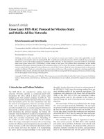
Figure 3: (a) MIT-BIH ECG record number 106, (b) time-
frequency representation with WT, (c) the same representation with
BWT.
both frequency and time domains, having fine frequency
resolution and coarse time resolution at lower frequency,
and coarse frequency resolution and fine time resolution at
higher frequency. Since this matches the characteristic of
most signals, it makes the wavelet transform suitable for
time-frequency analysis.
The concept of multiresolution analysis can be extended
from the fixed wavelet mother function to the varying in the
time spread case. This new approach is known as the adaptive
bionic wavelet tra nsform and would be addressed next.
The idea behind the BWT is to replace the constant qual-
ity factor of the wavelet transform with a variable adaptive
quality factor [17]. To do this, one can make changes in the
mother func tion of the wavelet transform. Satisfy ing the ad-
missible condition for the mother wavelet, the oscillating h(t)
can be represented as
h(t)
=
h(t)exp
j2πf
0
t
,(3)
where f
0
is the center frequency of h(t)and
h(t) is its enve-
lope function. Using a T-value, the BWT mother function
would be stated as follows:
h
T
(t) =
1
T
h
t
T
exp
j2πf
0
t
. (4)
The BWT is now defined by the following equation [17]:
BWT
x
(τ, a) =
1
|a|
x( t)h
∗
T
t − τ
a
dt
=
1
T
|a|
x( t)
h
∗
t − τ
aT
×
exp
−
j2πf
0
t − τ
a
dt.
(5)
As can be seen, in contrast to the wavelet transfor m, both
the amplitude and the time spread of BWT mother function
depend on the T value. For evaluating the T parameter, Yao
and Zhang adopted a general nonlinear form based on a pre-
viously introduced auditory model. This results in the fol-
lowing formula for a function, namely, the T-function:
T(τ + Δτ)
=
1 −
G
1
BWT
s
BWT
s
+
BWT
x
(τ, a)
−1
×
1+
G
2
∂ BWT
x
(τ, a)/∂t
−1
,
(6)
where
G
1
,
G
2
,andBWT
s
are constants, and BWT
x
(τ, a; h)is
the BWT coefficientattimeτ and scale a,andΔτ is the cal-
culation step. Clearly, it is the T-function that brings adap-
tation to the BWT. For detailed information about how the
T-function is derived and the underlying mechanism, the
reader is referred to [18, 19].
3. BIONIC WAVELET TRANSFORM OPTIMIZATION
FOR ECG ANALYSIS
According to the definition of BWT, there is a major differ-
ence in resolution of time-frequency span of analyzing win-
dows. In fact, in the WT, for a fixed mother function, all the
windows in a certain scale along the t-axis are fixed and the
window size of the WT varies with the change of analyz-
ing frequency. However, both the time and frequency reso-
lutions can be different in the BWT even in a certain scale.
The adjustment of the BWT resolution in the same scale is
controlled by T-function, which is related to the signal in-
stantaneous amplitude and its first-order differential [17].
Figure 3 shows the time-frequency representation for an
ECG signal with both WT and BWT. Notice the smoothing in
the BWT representation which is the direct result of windows
changes over certain scales.
It only remains to set the BWT parameters efficiently
so that it c an decompose the signal into finite number of
scales and afterwards, determine the most energetic ones,
and choose a global or local threshold. In order to optimize
the BWT parameters we have used a semioptimal method
considering both analytic and morphological aspects of the
analyzed signal. As we are considering ECG signal, we should
be aware of its var iability.
Presumably the most important feature for an ECG sig-
nal is the frequency range in which its main components
occur. Although there are some other components like ven-
tricular late potentials (VLPs), we have restricted our interest
on P, Q, R, S, and T. The resulting frequency range is up to
100 Hz.
4 EURASIP Journal on Advances in Signal Processing
Let f
0
be the initial center frequency of the mother
wavelet. In [17] it has a value equal to 15165.4 Hz and as the
scale goes higher and higher, the center frequency will de-
crease in the following way:
f
m
=
f
0
q
m
, q>1. (7)
For ECG we do not need such high f
0
, so we optimized
it simply by running the program for different values of f
0
and then minimizing the gradient of error variance by com-
paring the results—numerically and morphologically—with
each other. It has been found that if the center frequency lies
in the range of 360 to 500 Hz there would be no much distor-
tion on the analyzed ECG. The reason b ehind this choice lies
in the fact that to have no aliasing it is preferred to choose
the center frequency of the first scale a value more than ECG
sampling frequency. Here we have chosen f
0
= 400 which
yields satisfac tory results.
Unlike [17], in our method q is not a global constant,
but for each signal and scale of decomposition it takes a fixed
value which should obey an adaptation procedure. Besides
for every m, that is, in each distinct scale, it is adapted for
different time-frequency windows. More explanation on how
q is determined according to every analyzing window is to
be given in the next section. Other parameters are exactly
the same as what was stated in [20, 21] which used BWT
for speech enhancement and denoising. These constants are
G
1
= 0.87,
G
2
= 45, and BWT
s
= 0.8.
Finally, the calculation step is determined due to the sam-
pling frequency. If we let f
s
be the sampling frequency, then
the step will be Δτ
= 1/f
s
.
4. APPLICATION TO DENOISING AND BASELINE
WANDERING REDUCTION
For investigating the applications of MABWT to ECG sig-
nal, we have restricted our interest on denoising and baseline
wandering reduction. In each of the following sections, we
have proposed modifications to BWT so as to be optimized
for the field of analysis. For baseline wander ing elimination,
the goal is optimizing BWT by adopting a multiadaptation
scheme in which the low passed wandering is automatically
removed and there is no need for extra processing on distinct
subbands.
4.1. Denoising
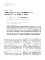
After BWT optimization, the denoising technique illustrated
in Figure 4 is used to reduce the amount of noise contamina-
tion in the ECG signal. In implementation, BWT coefficients
can be easily calculated based on corresponding WT coeffi-
cients by
BWT
x
(τ, a; T) = K × WT
x
(τ, a), (8)
where K is a factor depending on T [18]. Especially, for the
real Morlet function h(t)
= e
−(t/T
0
)
2
, which is used as the
Denoised
ECG
Noisy
ECG
WT
BWT coefficients
K factor
Thresholding
1/K
Inverse WT
Figure 4: Block diagram of the bionic wavelet transform denoising
technique.
mother function in our experiments, K is equal to [21]:
+∞
−∞
e
−t
2
dt
T/T
0
2
+1
≈
1.7725
T/T
0
2
+1
. (9)
Here we have used Donoho’s proposed approach for de-
noising including two major categories, hard thresholding
and soft thresholding [10, 22]. Choosing the threshold value
can be determined in many ways. Donoho derived the fol-
lowing formula based on white Gaussian noise assumption:
thr
= σ
2log
2
N, (10)
where thr is the threshold value, N is the length of the noisy
signal, and σ
= AMFS /0.6745, with AMFS denoting the
absolute median estimated on the first scale of the bionic
wavelet coefficients.
Knowing the fact that it is the T-function (or the K fac-
tor, equivalently) which results in the adaptive characteristic
of the BWT, it may be a good idea to use the values of the
T-function to come to a new thresholding rule. On the other
hand, it is expected for the BWT coefficients to take small val-
ues in the higher scales of decomposition with regard to (8),
the same as the WT coefficients. Thus it seems to be logic to
use the values of the T-functioninlowerscalesofdecom-
position. Combining the above idea with Donoho’s formula
yields the following threshold which is a new modification of
(10). Let T
fs
(i) be the value of T-function in the first step of
computing. The threshold is formulated as
thr
=
σ
i
α
i
T
fs
(i)
2log
2
N. (11)
In fact a weighted average of T-function values in the first
scale of decomposition with the bionic wavelet transform is
added to (10) to get better results. The α
i
-weights are chosen
with t ry and error for the algorithm to have the highest per-
formance. But an experimentally right choice is to let the α
i
coefficients be a decreasing function.
Yao showed that if the sig nal and its first-order differen-
tial are continuous, BWT can reconstruct the original signal
without distortion [17]. In the current approach and after
thresholding, the coefficients of BWT are divided by K-factor
followed by an inverse WT transform which reconstructs the
denoised version of the signal.
O. Sayadi and M. B. Shamsollahi 5
4.2. Baseline wandering reduction
Among various methods used for baseline wandering reduc-
tion, an efficient technique was proposed by Park et al. [23].
They presented a wavelet adaptive filter (WAF) which con-
sists of two parts, in the first part the signal is decomposed
into seven subbands and in the second part the seventh low-
est band subsignal is adaptively filtered. But the method suf-
fers from preserving the signal quality, specially in the S-T
segment.
Another approach which has utilized the wavelet pack-
ets to eliminate the ECG baseline wandering was introduced,
which removes the components that are not correlated to
ECG and have such characteristics that are somehow added
to it [16]. But the method does not fully take the advantage
of interbeat correlation of the ECG signal. Hence, baseline
drifts that occur occasionally and cannot be considered to
be an added source to the ECG would not be removed effi-
ciently.
As for the case of BWT, the resolution in the time-
frequency domain can be adaptively adjusted not only by
the signal frequency but also by the signal instantaneous am-
plitude and its first-order differential. Hence, analyzing the
ECG signal in the time-frequency plane of BWT not only has
a good chance to remove the baseline wanderings but also
has promising results preserving the clinical information of
the ECG record.
Since the spectrum of the baseline is below the spectrum
of the ECG signal, therefore its energy concentration in cor-
responding time-scale plane does not change much as the
scale is changed in the binary decomposition tree, but the
energy of the ECG signal decreases as the scale is changed.
Therefore, in the binary tree search we reach a point that the
energy of the ECG signal almost vanishes ( no details in that
scale) but we still have considerable energy for baseline wan-
dering.
Using the multiadaptive bionic wavelet transfor m makes
possible the detection and reduction of ECG baseline wan-
dering in low frequency subsignals. As we mentioned be-
fore the central frequency of each scale is a daptively chosen.
For adaptation purpose, we have used the following criteria
based on the time-frequency representation of the analyzed
signal under the adaptive BWT.
If we suppose that the baseline wandering can be esti-
mated by a sinusoidal function (see Figure 2(c)), the fre-
quency of the sine func tion can be approximately estimated
using the spectrum of the signal, in which we should seek
for a peak. In most cases the second distinguished peak in
the Fourier transform of the signal corresponds to the wan-
dering frequency. For ECG signals with the baseline wander-
ings which cannot be considered as sinusoidal, the f
w
esti-
mation is obtained from the time-frequency representation
of the signal. In this case a frequency exists all over the time
which deals with the baseline trace. After determining an es-
timation of the baseline wandering frequency, f
w
,wemayuse
the thresholding rule of (11 ) for the three consecutive scales,
the one that contains f
w
, the previous and the next scales. To
evaluate the coefficients of the multiadaptive bionic wavelet
transform we need the value of q. To determine the value of
q we remember the fact that the mean of the baseline wan-
dered ECG and the mean of the baseline corrected ECG have
the maximum distance. Thus we use the maximum distance
(MD) criteria to verify the optimal q for which the distance
between the MABWT coefficients of original signal and the
baseline corrected signal is maximized. We define the MD
criteria as
q
opt
= arg max
q
MABWT
x
(τ, a)
− MABWT
x
(τ, a)
2
(τ,a)∈
m
,
(12)
where x and
x are the original and processed signals, m is
the scale number (a
= 2
−m
), and
m
denotes the analyzing
window of the mth scale which is centered at f
w
. Solving the
above optimization problem, we will have q for the next scale.
Although q is optimally selected, the number of scales m re-
mains to be determined according to the q value of the previ-
ous scale. In other words, we begin with m
= 1. For the first
scale, that is, m
= 1, MABWT is computed using an initial
value for q.Then(12) gives the optimum value of q for the
next scale, m
= 2. The procedure proceeds iteratively until
the center frequency of the analyzing window, which is de-
fined according to both m and q (refer to (7)), goes beyond
the desired predefined ECG frequency range.
The step by step multiadaptation together with the
adapted T-function (or K-factor) of BWT can cope with the
problem of ECG baseline wandering reduction better than
WT.Furthermoreaswehaveanestimationof f
w
, the adap-
tation can be used only in three successive scales in which
the mid-scale has the closest center frequency to f
w
. So the
implementation is possibly time consuming.
5. SIMULATION RESULTS
To show that MABWT is appropriate for ECG denoising we
have used two types of ECG signals, both simulated and real
ones. We used the MIT-BIH arrhythmia database [24] as the
reference for our real signals, all with sampling frequencies,
f
s
, equal to 360 Hz. For the simulated ECGs we have used
the dynamical model which was introduced for generation
of synthetic ECG cardiac signals [25, 26].
The signals were decomposed using MABWT up to 40
scales depending on the different values of q that was opti-
mally selected using (12). We have used Morlet wavelet as
mother wavelet and its support length is chosen as [
−4, 4],
and 2.5π is chosen as its oscillatory frequency [21]. For sim-
ulation, we have chosen f
0
= 400 Hz ( f
0
>f
s
). In addition
we have considered the simplest case for our new threshold-
ing rule, (11). Hence, we have set α
i
= 1/i.
5.1. Denoising
In order to investigate the performance of various methods,
artificial white Gaussian noises with different variances were
generated and added to the test signals. As mentioned before
two kinds of thresholding methods, hard and soft, were ap-
plied for denoising based on the modified thresholding rule
introduced by (11). Besides, for easier comparison we have
6 EURASIP Journal on Advances in Signal Processing
applied the wavelet-based denoising with Daubechies (db4)
wavelets to each signal and we have shown the results for
both hard and soft thresholdings. Figures 5–7 show some
typical results, considering real and simulated ECG signals
of two general categories, that is, nor mal and abnormal ones.
One can see that in many cases, hard thresholded signal is
much similar to the soft thresholded ECG, which is due to
the intrinsic smoothness in BWT.
Simulation results provide supportive evidence to claim
that MABWT has some a dvantages over the traditional WT
for ECG denoising. First, it has higher sensitivity so it is more
probable for WT to have little single noise samples (speckles)
remained. Second, MABWT has a smoothing property with
respect to its resolution variation over the time-frequency
plane, and this is exactly what we are seeking in many denois-
ing techniques. This is particularly true for real ECGs (see
Figure 6). Furthermore, the effect of the adaptation is clearly
obvious in the first samples of the reconstructed ECGs with
MABWT (refer to Figures 5(e) and 5(f)). But as the adapta-
tion proceeds, the reconstructed signal follows the clean ECG
morphology.
Another field of interest for the proposed multiadap-
tive method is to denoise and reconstruct abnor mal signals.
Figure 7(a) shows an ectopic beat among a normal ECG cy-
cle. We have applied the MABWT to the signal and have
shown the results corresponding to hard MABWT (HBWT).
It is clear that the denoised signal is noise-free but cannot fol-
low the ST segment of the ectopic beats, reasonably because
any abnormality corresponds to an abrupt change in the sig-
nal rhythm and consequently the adaptation needs time to
follow this behavior (Figure 7(b)). But fortunately this does
not affect the diagnostic features of the ECG signal such as
the QT or the ST intervals, and the QRS complex.
Since we have used the adaptation in a limited number
of scales, abrupt changes may be tracked not efficiently, but
it can be shown that if the adaptation includes all decom-
position subbands and also the abnormality can be consid-
ered semicyclic, but not necessarily stationary, the MABWT
technique would come over the above problem (refer to
Figure 7(c)).
For evaluating the performance of the proposed BWT we
have used the SNR improvement measure by the means of
the expression:
imp[dB]
= SNR
output
− SNR
input
= 10 log
i
x
d
(i) − x(i)
2
i
x
n
(i) − x(i)
2
,
(13)
where x denotes the original ECG, x
d
is the denoised sig-
nal, and x
n
represents the noisy ECG signal. Figure 8 com-
pares the improvement values between WT and MABWT.
For evaluation, we have chosen first 4096 samples of the
noise free MIT-BIH record number 100 as our reference ECG
signal. As mentioned before, both hard and soft threshold-
ings have been considered for denoising. One can see that in
lower input SNRs, soft MABWT (SBWT) has a better perfor-
mance and as the input SNR is increased SBWT remains the
best choice with improvements much more than that of WT.
0 200 400 600 800 1000
−0.5
0
0.5
1
Samples
Amplitude (mV)
(a)
0 200 400 600 800 1000
−0.5
0
0.5
1
Samples
Amplitude (mV)
(b)
0 200 400 600 800 1000
−0.5
0
0.5
1
Samples
Amplitude (mV)
(c)
0 200 400 600 800 1000
−0.5
0
0.5
1
Samples
Amplitude (mV)
(d)
0 200 400 600 800 1000
−0.5
0
0.5
1
Samples
Amplitude (mV)
(e)
0 200 400 600 800 1000
−0.5
0
0.5
1
Samples
Amplitude (mV)
(f)
Figure 5: Typical results of different methods for an input simu-
lated signal of 6 dB. (a) Clean ECG, (b) noisy input signal, (c) WT
(hard), (d) WT (soft), (e) MABWT (hard), and (f) MABWT (soft).
O. Sayadi and M. B. Shamsollahi 7
0 500 1000 1500 2000
−1
−0.5
0
0.5
1
Samples
Amplitude (mV)
(a)
0 500 1000 1500 2000
−1
−0.5
0
0.5
1
Samples
Amplitude (mV)
(b)
0 500 1000 1500 2000
−1
−0.5
0
0.5
1
Samples
Amplitude (mV)
(c)
0 500 1000 1500 2000
−1
−0.5
0
0.5
1
Samples
Amplitude (mV)
(d)
0 500 1000 1500 2000
−1
−0.5
0
0.5
1
Samples
Amplitude (mV)
(e)
0 500 1000 1500 2000
−1
−0.5
0
0.5
1
Samples
Amplitude (mV)
(f)
Figure 6: Typical results of different methods for the MIT-BIH
record 117 with an input SNR of 4 dB. (a) Clean ECG, (b) noisy in-
put signal, (c) WT (hard), (d) WT (soft), (e) MABWT (hard), and
(f) MABWT (soft).
0 500 1000 1500 2000 2500 3000 3500 4000
−1.5
−1
−0.5
0
0.5
1
1.5
2
2.5
3
Samples
Amplitude (mV)
(a)
0 500 1000 1500 2000 2500 3000 3500 4000
−1.5
−1
−0.5
0
0.5
1
1.5
2
2.5
3
Samples
Amplitude (mV)
(b)
0 500 1000 1500 2000 2500 3000 3500 4000
−1.5
−1
−0.5
0
0.5
1
1.5
2
2.5
3
Samples
Amplitude (mV)
(c)
Figure 7: Denoising results of HBWT applied to an ECG (MIT-BIH
record 119) with ectopic beats. (a) Clean and noisy ECG, (b) HBWT
with 3 levels of adaptation, (c) HBWT with the adaptation applied
to all decomposition levels. The surrounding circles indicate the ST
segment of the ectopic beats.
Moreover, HBWT acts better than hard WT and for SNRs
higher than 8 dB, it passes the soft WT performance. Figure 8
clarifies that for a wide range of input SNRs, the MABWT
improvement (hard or/and soft) has a noticeable difference
to that of WT, especially outstanding for lower SNRs for
which having nearly 2 dB improvement in MABWT is of ma-
jor importance.
In order to have a comparison between the performance
of the proposed method and conventional wavelet-based
ECG denoising schemes, WT and the MABWT were tested
on the database. The results of the SNR improvement for the
input SNR of 5 dB are listed in Table 1 .
According to these results the MABWT performance is
always better than the corresponding WT for hard and soft
thresholdings, as was already expected. Although WT and
8 EURASIP Journal on Advances in Signal Processing
−10 −50 5101520
−2
−1
0
1
2
3
4
5
6
7
8
9
Input SNR (dB)
Improvement (dB)
WT soft
WT hard
BWT soft
BWT hard
Figure 8: WT and MABWT filter output SNR improvement ver-
sus different input SNRs for the first 4096 samples of the MIT-BIH
record number 100.
MABWT always improve the input SNR, MABWT with soft
thresholding has the best overall performance with a max-
imum SNR improvement of 8.2 dB, while the best result of
WT-based approach goes back to 6.6 dB.
5.2. Baseline wandering reduction
We have divided the baseline wanderings into the following
two general categories: DC components affecting the base-
line not to be at zero level, and baseline drift which is a con-
sequence of low frequency interferences. For both cases with
the appropriate choices for the level of decomposition, that
is, number of scales m, and the center frequency f
0
,remov-
ing baseline wandering would be a direct task. Remember
that we have assigned a fixed value to f
0
, experimentally. On
the other hand there is an interdependency between m and
q (7) but with the help of the iterative algorithm discussed
in Section 4 , there would be only one degree of freedom to
determine the accurate values of the mentioned parameters.
Again it only remains to find q with respect to the optimiza-
tion problem of (12)form
= 2 and continue with the com-
puted value(s) for the next scale(s). To clarify the effective-
ness of the proposed technique, we have chosen real ECG
signals including two classes of baseline wandering simulta-
neously, and we have run the baseline wandering cancellation
algorithm with both WT and MABWT. The results are pro-
vided in Figures 9–11.
Results show that although for the case of wavelet trans-
form the denoised signal is somehow free of the baseline
interference, but the drifts still exist unless applying a low-
pass filtration to the properly selected subband (Figure 9(b)).
Moreover, the thresholding not only affects the baseline but
Table 1: WT and MABWT denoising performance on the MIT-BIH
arrythmia database.
Record
no.
SNR improvement (dB)
WT WT MAB WT MABWT
(hard) (soft) (hard) (soft)
100 5.1 6.5 6.4 7.8
101
4.2 5.5 5.3 6.9
103
5.0 6.1 5.8 7.7
105
5.1 6.0 5.8 8.1
112
5.2 6.4 6.1 8.2
113
5.0 6.2 5.9 7.9
115
5.1 6.6 6.3 7.8
116
5.0 6.5 6.4 8.0
117
4.8 6.0 5.8 7.9
119
4.7 5.8 5.6 7.6
122
4.4 5.6 5.2 6.9
123
5.1 6.5 6.4 7.8
200
4.2 5.4 5.3 6.9
201
5.0 5.4 5.5 7.5
202
4.9 5.8 5.7 7.8
205
5.0 5.4 5.5 7.4
209
5.2 6.0 5.9 8.1
210
4.3 5.4 5.3 6.9
212
4.0 5.0 4.8 6.7
213
5.2 6.6 6.4 8.2
219
5.2 6.5 6.3 8.0
220
5.1 6.5 6.4 8.1
221
4.9 6.0 5.9 7.9
230
5.0 5.9 5.6 7.9
233
4.8 5.8 5.5 7.8
Average 4.86 5.98 5.80 7.67
also has destructive effects on the signal morphology, espe-
cially on the ST segment, which is extremely sensitive to noise
and is of great clinical significance (Figure 10(b)). But in the
denoised signal with MABWT, baseline wandering is com-
pletely removed and also a noise-free version of the signal
is obtained without the need for any extra filtering. There
are more with the advantages of MABWT over other base-
line correction techniques. It is capable of eliminating not
only those drifts which could be modeled as additive sources
[16], but also the ones that have no correlation with a pure
ECG. In addition, the method has no destructive effects on
the morphology of the signal and can cope with both normal
and abnormal beats, while previously introduced methods
have problems with these cases [8].
The results are also presented for simulated baseline per-
turbations, as depicted in Figure 12. We have chosen the
MIT-BIH record number 207, which includes bundle branch
blocks, together with ventricular flutter wave. A random
noise of 6.5 dB was added to evaluate the noise reduction
performance of the algorithm, while reducing the baseline
drift. The difference between processed and original signal
is shown for soft thresholding. It shows that WT processed
O. Sayadi and M. B. Shamsollahi 9
0 500 1000 1500 2000 2500 3000 3500 4000
−1
0
1
2
Samples
Amplitude
(mV)
(a)
0 500 1000 1500 2000 2500 3000 3500 4000
−1
0
1
2
Samples
Amplitude
(mV)
(b)
0 500 1000 1500 2000 2500 3000 3500 4000
−1
0
1
2
Samples
Amplitude
(mV)
(c)
Figure 9: Typical results of different methods for baseline correc-
tion in presence of DC components and lowpass interference. (a)
MIT-BIH record 203, (b) result of applying WT, (c) result of apply-
ing MABWT.
signal has its maximum difference on the QRS complexes
and the T waves. In contrast to WT, the processed signal with
BWT, when adapted to the signal, follows its morphology,
leading the difference to be ignored. Consequently, WT can-
not handle precise baseline wander removal, and there is a
significant difference in the amplitude of the fiducial points
of the signal, compared to the original ECG. Hence, there
may be distortions in the locations of the ECG points and
interval features.
6. DISCUSSION AND CONCLUSION
We have presented and validated a new multiadaptive version
of the bionic wavelet transform and its applications to noise
and baseline wandering suppression in elect rocardiograms
combined with modifications of the traditional threshold-
ing rules. The MABWT aims at integrating into the standard
BWT a mechanism that adjusts the center frequency of every
analyzing scale in a signal-adaptive fashion. Moreover, the
value of the T-function, which controls the mother wavelet,
is directly influenced on the threshold to come to an appro-
priate criterion for the denoising approach which uses the
transform coefficients’ information.
To show that BWT improvement is really effective in clin-
ical situations, the method has been validated using several
ECG recordings with a w ide variety of wave morphologies
from MIT-BIH arrhythmia database, and also simulated sig-
nals. For denoising purpose, using the MABWT representa-
0 200 400 600 800 1000 1200 1400 1600 1800
−0.5
0
0.5
1
Samples
Amplitude
(mV)
(a)
0 200 400 600 800 1000 1200 1400 1600 1800
−0.5
0
0.5
1
Samples
Amplitude
(mV)
(b)
0 200 400 600 800 1000 1200 1400 1600 1800
−0.5
0
0.5
1
Samples
Amplitude
(mV)
(c)
Figure 10: Typical results of different methods for baseline wander-
ing cancellation in presence of sinusoidal drifts. (a) MIT-BIH record
228, (b) result of applying WT, (c) result of applying MABWT.
0 500 1000 1500 2000 2500 3000 3500 4000
−0.5
0
0.5
1
1.5
Samples
Amplitude
(mV)
(a)
0 500 1000 1500 2000 2500 3000 3500 4000
−0.5
0
0.5
1
1.5
Samples
Amplitude
(mV)
(b)
0 500 1000 1500 2000 2500 3000 3500 4000
−0.5
0
0.5
1
1.5
Samples
Amplitude
(mV)
(c)
Figure 11: Typical results of different methods for baseline wan-
dering elimination for a non-correlated baseline artifact. (a) MIT-
BIH record 222, (b) result of applying WT, (c) result of applying
MABWT.
10 EURASIP Journal on Advances in Signal Processing
1000 2000 3000 4000 5000 6000 7000 8000
−2
−1.5
−1
−0.5
0
0.5
1
1.5
Samples
Amplitude (mV)
(a)
1000 2000 3000 4000 5000 6000 7000 8000
−0.6
−0.4
−0.2
0
0.2
0.4
0.6
Samples
Amplitude (mV)
(b)
Figure 12: Simulated baseline perturbation removal and noise re-
duction for an abnormal ECG. (a) MIT-BIH record 207, and the
added baseline trace, (b) differences between WT (dashed line) and
MABWT (solid line) processed signals and the original ECG.
tion of the ECG signal and with the help of the modified
threshold (11) there was an average SNR improvement of
5.80 dB for h ard thresholding and 7.67 dB for soft thresh-
olding, corresponding to 0.94 dB and 1.69 dB improvement,
respectively, compared to the WT-based methods, on the as-
sumption of additive white Gaussian noise sources. None of
the more complex test cases result in an improvement less
than 4.8 dB. The MABWT-based denoising, in contrast to
most denosing methods found in the literature, allows taking
the advantage of characteristics of adaptive time-scale anal-
ysis. Hence, there are advantages with the suggested tech-
nique compared to conventional other denoising methods;
there are fewer speckles remaining in the denoised signal,
and the reconstructed signal follows a smoothing behavior.
In contrast to other efficient denoising techniques, abrupt
changes that occur in most abnormal cases would not be
tracked unless the adaptation includes all subbands. Con-
sequently, MABWT is able to outperform other algorithms
and the modified threshold selection rule, which uses the in-
formation of the transform coefficients in the first scale of
decomposition, has a noise reduction r a tio well within the
acceptable range.
Also, the proposed MABWT algorithm removes baseline
wandering, while preserving the clinical information and the
morphology of the ECG record. Unlike other baseline correc-
tion schemes, it removes the components of all wandering
classes that are not correlated to ECG and have such char-
acteristics that are somehow added to it, DC components,
and low frequency interfering drifts. Moreover, the proposed
algorithm is also capable of improving signal-to-noise ratio
while eliminating the baseline wandering.
The superior performance of the proposed technique is
a direct result of the multiadaptive scheme, which gives the
opportunity to study different types of noise through the in-
tuitive modification of BWT parameters. Moreover MABWT
can be calculated using WT by adding a K-factor which re-
sults in a fast implementation of the proposed algorithm.
Finally, it should be mentioned that the importance of
the multiadaptive technique goes beyond the single problem
of ECG denoising since it is applicable to every type of data
considering a case-dependent optimization. In fact, once the
time-frequency features of the data are extracted, the prob-
lem obtains an abstract representation on which it is possible
to apply whichever experience matured in MABWT.
ACKNOWLEDGMENTS
The authors wish to thank Mr. Reza Sameni for his assistance
and deep review of the paper. This work has been supported,
financially and intellectually, by the Iran Telecommunication
Research Center (ITRC), under Grant no. T/500/15023.
REFERENCES
[1] Z. Frankiewicz and E. Pi
ˆ
etka, “Komputerowe eliminacja linii
izoelektrycznej z sygnau EKG,” Problemy Techniki Medycznej,
vol. 16, no. 1, 1985.
[2] N. V. Thakor and Y S. Zhu, “Applications of adaptive filtering
to ECG analysis: noise cancellation and arrhythmia detection,”
IEEE Transactions on Biomedical Engineering,vol.38,no.8,pp.
785–794, 1991.
[3] P. Laguna, R. Jane, O. Meste, et al., “Adaptive filter for event-
related bioelectric signals using an impulse correlated ref-
erence input: comparison with signal averaging techniques,”
IEEE Transactions on Biomedical Engineering, vol. 39, no. 10,
pp. 1032–1044, 1992.
[4]G.B.MoodyandR.G.Mark,“QRSmorphologyrepresen-
tation and noise estimation using the Karhunen-Lo
`
eve trans-
form,” in Proceedings of Computers in Cardiology, pp. 269–272,
Jerusalem, Israel, September 1989.
[5] A. K. Barros, A. Mansour, and N. Ohnishi, “Removing ar-
tifacts from electrocardiographic signals using independent
components analysis,” Neurocomputing,vol.22,no.1–3,pp.
173–186, 1998.
[6] T. He, G. D. Clifford, and L. Tarassenko, “Application of ICA
in removing artefacts from the ECG,” to appear in Neural Pro-
cessing Letters.
[7] G. D. Clifford and L. Tarassenko, “One-pass training of op-
timal architecture auto-associative neural network for detect-
ing ectopic beats,” Electronics Letters, vol. 37, no. 18, pp. 1126–
1127, 2001.
[8] K. Daqrouq, “ECG baseline wander reduction using discrete
wavelet transform,” Asian Journal of Information Technology,
vol. 4, no. 11, pp. 989–995, 2005.
[9]H.A.Kestler,M.Haschka,W.Kratz,etal.,“De-noisingof
high-resolution ECG signals by combining the discrete wavelet
transform with the Wiener filter,” in Proceedings of Computers
in Cardiology, pp. 233–236, Cleveland, Ohio, USA, September
1998.
O. Sayadi and M. B. Shamsollahi 11
[10] D. L. Donoho, “De-noising by soft-thresholding,” IEEE Trans-
actions on Information Theory, vol. 41, no. 3, pp. 613–627,
1995.
[11] M. Popescu, P. Cristea, and A. Bezerianos, “High resolution
ECG filtering using adaptive BSayesian wavelet shrinkage,” in
Proceedings of Computers in Cardiology, pp. 401–404, Cleve-
land, Ohio, USA, September 1998.
[12] P. M. G. Agante Da Silva and J. P. Marques De S
´
a, “ECG
noise filtering using wavelets with soft-thresholding methods,”
in Proceedings of Computers in Cardiology, pp. 535–538, Han-
nover, Germany, September 1999.
[13] C. R. Meyer and H. N. Keiser, “Electrocardiogram baseline
noise estimation and removal using cubic splines and state-
space computation techniques,” Computers and Biomedical Re-
search, vol. 10, no. 5, pp. 459–470, 1977.
[14] P. W. MacFarlane, J. Peden, J. Lennox, M. P. Watts, and T.
D. Lawrie, “The Glasgow system,” in Trends in Computer-
Processed Electrocardiograms: Proceedings of the IFIP Work-
ing Conference on Trends in Computer-Processed Elect rocar-
diograms, pp. 143–150, North-Holland, New York, NY, USA,
1977.
[15] J. A. Van Alste and T. S. Schilder, “Removal of base-line wander
and power-line interference from the ECG by an efficient FIR
filter with a reduced number of taps,” IEEE Transactions on
Biomedical Engineering, vol. 32, no. 12, pp. 1052–1060, 1985.
[16] B. Mozaffary and M. A. Tinati, “ECG baseline wander elimina-
tion using wavelet packets,” Transactions on Engineering, Com-
puting and Technology, vol. 3, pp. 22–24, 2004.
[17] J. Yao and Y. T. Zhang, “Bionic wavelet transform: a new time-
frequency method based on an auditory model,” IEEE Trans-
actions on Biomedical Engineering, vol. 48, no. 8, pp. 856–863,
2001.
[18] J. Yao and Y. T. Zhang, “Cochlear is an inhomogeneous, active
and nonlinear model,” in Proceedings of the 1st Joint Meeting
of BMES & IEEE/EMBS, p. 1031, Atlanta, Ga, USA, October
1999.
[19] J. Yao and Y. T. Zhang, “From otoacoustic emission modeling
to bionic wavelet transform,” in Proceedings of the 22nd Annual
International Conference of the IEEE Engineering in Medicine
and Biology Society, vol. 1, pp. 314–316, Chicago, Ill, USA, July
2000.
[20] J. Yao and Y. T. Zhang, “The application of bionic wavelet
transform to speech signal processing in cochlear implants
using neural network simulations,” IEEE Transactions on
Biomedical Engineering, vol. 49, no. 11, pp. 1299–1309, 2002.
[21] X. Yuan, “Auditory model-based bionic wavelet transform for
speech enhancement,” M.S. thesis, Speech and Signal Process-
ing Laboratory, Marquette University, Milwaukee, Wis, USA,
2003.
[22] D. L. Donoho and I. M. Johnstone, “Adapting to unknown
smoothness via wavelet shrinkage,” Journal of the American
Statistical Association, vol. 90, no. 432, pp. 1200–1224, 1995.
[23] K. L. Park, K. J. Lee, and H. R. Yoon, “Application of a wavelet
adaptive filter to minimise distortion of the ST-segment,”
Medical and Biological Engineering and Computing, vol. 36,
no. 5, pp. 581–586, 1998.
[24] “The MIT-BIH Arrhythmia Database,” .
biu.ac.il/physiobank/database/mitdb/.
[25] P.E.McSharry,G.D.Clifford, L. Tarassenko, and L. A. Smith,
“A dynamical model for generating synthetic electrocardio-
gram signals,” IEEE Transactions on Biomedical Engineering,
vol. 50, no. 3, pp. 289–294, 2003.
[26] P.E.McSharryandG.D.Clifford, “ECGSYN—a realistic ECG
waveform generat o r,” />ecgsyn/
.
Omid Sayadi was born in Shiraz, Iran, in
1983. He majored in biomedical engineer-
ing and received the B.S. degree from Sha-
hed University of Tehran, Iran, in 2005.
He was a Research Scientist with BehsazTeb
Co., Tehran, from 2003 to 2005. He is
currently an M.S. student of biomedical
engineering at the Electrical Engineering
Department of Sharif University of Tech-
nology, Tehran, Iran, and a member of
Biomedical Signal and Image Processing Laboratory (BiSIPL). His
research deals with dynamical models for E CG gener ation, model-
based ECG processing, the application of wavelet concepts, and es-
pecially multiadaptive bionic wavelet t ransform to biomedical sig-
nal processing solutions, signature verification, and efficient heart
modeling.
Mohammad B. Shamsollahi was born in
Qom, Iran, in 1965. He received the B.S. de-
gree in electrical engineering from Tehran
University, Tehran, Iran, in 1988, and
the M.S. degree in electrical engineering,
Telecommunications, from the Sharif Uni-
versity of Technology, Tehran, Iran, in 1991.
He received the Ph.D. degree in electrical
engineering, biomedical signal processing,
from the University of Rennes 1, Rennes,
France, in 1997. Currently, he is an Assistant Professor with the
Department of Electrical Engineering, Sharif University of Tech-
nology, Tehran, Iran. His research interests include biomedical
signal processing, brain computer interface, time-scale and time-
frequency signal processing.