Sinh ly truyen dich tro ng hs
Bạn đang xem bản rút gọn của tài liệu. Xem và tải ngay bản đầy đủ của tài liệu tại đây (1.23 MB, 15 trang )
Messina et al.
Intensive Care Medicine Experimental
(2022) 10:46
/>
Intensive Care Medicine
Experimental
Open Access
REVIEWS
Pathophysiology of fluid administration
in critically ill patients
Antonio Messina1,2* , Jan Bakker3,4, Michelle Chew5, Daniel De Backer6, Olfa Hamzaoui7, Glenn Hernandez8,
Sheila Nainan Myatra9, Xavier Monnet10, Marlies Ostermann11, Michael Pinsky12, Jean‑Louis Teboul10 and
Maurizio Cecconi1,2
*Correspondence:
1
IRCCS Humanitas Research
Hospital, Via Alessandro Manzoni
56, Rozzano, 20089 Milan, Italy
2
Department of Biomedical
Sciences, Humanitas University,
Pieve Emanuele, Milan, Italy
3
NYU Langone Health
and Columbia University Irving
Medical Center, New York, USA
4
Erasmus MC University
Medical Center, Rotterdam, The
Netherlands
5
Department of Anaesthesia
and Intensive Care, Biomedical
and Clinical Sciences, Linköping
University, Linköping, Sweden
6
Department of Intensive Care,
CHIREC Hospitals, Université
Libre de Bruxelles, Brussels,
Belgium
7
Service de Reanimation
PolyvalenteHopital Antoine
Béclère, Hopitaux Universitaires
Paris-Saclay, Clamart, France
8
Departamento de Medicina
Intensiva, Facultad de Medicina,
Pontificia Universidad Católica de
Chile, Santiago, Chile
9
Department
of Anaesthesiology, Critical Care
and Pain, Tata Memorial Hospital,
Homi Bhabha National Institute,
Mumbai, India
10
Hôpitaux Universitaires ParisSud, Hôpital de Bicêtre, Medical
Intensive Care Unit, Le KremlinBicêtre, Paris, France
11
Department of Intensive Care,
King’s College London, Guy’s & St
Thomas’ Hospital, London, UK
12
Department of Critical
Care Medicine, University
of Pittsburgh, Pittsburgh, PA, USA
Abstract
Fluid administration is a cornerstone of treatment of critically ill patients. The aim of this
review is to reappraise the pathophysiology of fluid therapy, considering the mecha‑
nisms related to the interplay of flow and pressure variables, the systemic response to
the shock syndrome, the effects of different types of fluids administered and the con‑
cept of preload dependency responsiveness. In this context, the relationship between
preload, stroke volume (SV) and fluid administration is that the volume infused has to
be large enough to increase the driving pressure for venous return, and that the result‑
ing increase in end-diastolic volume produces an increase in SV only if both ventricles
are operating on the steep part of the curve. As a consequence, fluids should be given
as drugs and, accordingly, the dose and the rate of administration impact on the final
outcome. Titrating fluid therapy in terms of overall volume infused but also considering
the type of fluid used is a key component of fluid resuscitation. A single, reliable, and
feasible physiological or biochemical parameter to define the balance between the
changes in SV and oxygen delivery (i.e., coupling “macro” and “micro” circulation) is still
not available, making the diagnosis of acute circulatory dysfunction primarily clinical.
Take‑home messages
– Fluids are drugs used in patients with shock to increase the cardiac output with
the aim to improve oxygen delivery to the cells. The response to fluid administration is determined by the physiological interaction of cardiac function and venous
return. In septic shock, the beneficial clinical response of fluid administration is
rapidly reduced after few hours and fluid titration is crucial to avoid detrimental
fluid overload. The fluid challenge is a fluid bolus given at a defined quantity and
rate to assess fluid responsiveness.
– The ideal fluid for critically ill patients does not exist; however, crystalloids should
be used as first choice. Balanced crystalloid solutions may be associated with better outcomes but the evidence is still low. Albumin infusion may have a role in
already fluid resuscitated patients at risk of fluid overload.
© The Author(s) 2022. Open Access This article is licensed under a Creative Commons Attribution 4.0 International License, which permits
use, sharing, adaptation, distribution and reproduction in any medium or format, as long as you give appropriate credit to the original
author(s) and the source, provide a link to the Creative Commons licence, and indicate if changes were made. The images or other third
party material in this article are included in the article’s Creative Commons licence, unless indicated otherwise in a credit line to the mate‑
rial. If material is not included in the article’s Creative Commons licence and your intended use is not permitted by statutory regulation or
exceeds the permitted use, you will need to obtain permission directly from the copyright holder. To view a copy of this licence, visit http://
creativecommons.org/licenses/by/4.0/.
Messina et al. Intensive Care Medicine Experimental
(2022) 10:46
– Fluid administration is integrated into the complex management of pressure and
flow “macro” hemodynamic variables, coupled to the “micro” local tissue flow distribution and regional metabolism. Macro-variables are managed by measuring
systemic blood pressure and evaluating the global cardiac function. The critical
threshold of oxygen delivery to the cells is difficult to estimate, however, several
indexes and clinical signs may be considered as surrogate of that, and integrated
in a decision-making process at the bedside.
Background
Fluid administration is one of the most common but also one of the most disputed interventions in the treatment of critically ill patients. Even more debated is the way how
to appraise and manage the response (in terms of flow and pressure variables) to fluid
administration, which ranges from a prosaic “just give fluids” to the fluid challenge,
to the evaluation of fluid responsiveness before fluid administration to, finally, recent
approaches based on machine learning and Artificial Intelligence aimed at personalizing
its use [1–3].
Shock occurs in many intensive care unit (ICU) patients, representing a life-threatening condition that needs both prompt recognition and treatment to provide adequate
tissue perfusion and thus oxygen delivery to the cells [4]. A large trial in more than 1600
patients admitted to ICU with shock and requiring vasopressors demonstrated that
septic shock was the most frequent type of shock, occurring in 62% of patients, while
cardiogenic shock (16%), hypovolemic shock (16%) and other types of distributive (4%)
or obstructive (2%) shock were less frequent. The progression of this syndrome is associated with mitochondrial dysfunction and deregulated cell-signaling pathways, which
can lead to multiple organ damage and failure and, eventually, untreatable hemodynamic
instability and death [5].
Optimal treatment of shock is time-dependent and requires prompt and adequate
combined support with fluids and/or vasopressors [4, 6–8]. The rationale, supported
by robust evidence from several physiological and clinical studies, is to improve oxygen
delivery (DO2), so that systemic oxygen requirements can be met [4, 6]. Oxygen delivery
is defined as the product of oxygen content and cardiac output (CO). Pathological cellular oxygen utilization results from a tissue oxygen request exceeding the D
O2 or the cellular inability to use O2. Our understanding of the mechanisms of shock has improved in
the last decades, shifting the clinical practice from a “one size fits all” policy to individualized management [4, 9, 10].
Fluids are the first line of treatment in critically ill patients with acute circulatory failure aiming to increase venous return, stroke volume (SV) and, consequently, CO and
DO2 [4]. The effect of the increase in CO following fluid resuscitation on blood pressure
is not linear and related to baseline conditions (see "Fluids and ICU outcomes: does the
type of fluid matter?") [4, 11–15].
Dr Thomas Latta first described the technique of fluid resuscitation to treat an episode
of shock in 1832 in a letter to the editor of The Lancet [16]. He injected repeated small
boluses of a crystalloid solution to an elderly woman and observed that the first bolus
did not produce a clinically relevant effect; however, after multiple boluses (overall 2.8
L) ‘soon the sharpened features, sunken eye, and fallen jaw, pale and cold, bearing the
Page 2 of 15
Messina et al. Intensive Care Medicine Experimental
(2022) 10:46
manifest imprint of death’s signet, began to glow with returning animation; the pulse
returned to the wrist’. This lady was ultimately the first fluid responder reported in the
literature.
This meaningful witness from the past addresses several physiological and clinical
issues, which are still valid after almost 200 years:
CO is the dependent variable of the physiological interaction of cardiac function
(described by the observations of Otto Frank and Ernest Starling more than 100 years
ago) [17] and venous return (based on Guyton’s relationship between the elastic recoil
of venous capacitance vessels, the volume stretching the veins, the compliance of the
veins and the resistance of the venous system) [18, 19]. Fluid responsiveness indicates
that the heart of the patient is operating in the steep part of Frank–Starling’s curve of
heart function, while fluid non-responsiveness is observed on the flat part of the curve
where an increase in preload doesn’t increase CO further [20–22]. The lady treated by
Dr. Latta probably did not respond to the first fluid bolus because the volume infused
was insufficient to increase venous return (i.e., induce a change in the stressed volume
related to venous compliance) [18, 19]. Thus, the volume infused is a crucial factor. The
most recent Surviving Sepsis Campaign guidelines again recommend to administer an
initial fluid volume of at least 30 ml/kg in patients with sepsis, which is considered, on
average, a safe and effective target [6]. However, as the goal of fluid therapy is to increase
SV and then CO, fluids should only be given if the plateau of cardiac function has not
been reached in the individual patient. In fact, and probably even before reaching this
point, fluid administration that does not increase CO can be considered futile. The fluid
challenge (FC) is a hemodynamic diagnostic test consisting of the administration of a
fixed volume of fluids with the purpose of identifying fluid responsive patients who will
increase CO in response to fluid infusion [12, 23, 24]. This approach allows the individual titration of fluids and reduces the risk of fluid overload, which affects patients’
clinical outcome and mortality [9, 25–27].
In clinical practice, the likelihood of a beneficial clinical response to an FC is rapidly
reduced after a few hours following the onset of septic shock resuscitation which renders
the optimization of fluid therapy quite complex without adopting hemodynamic monitoring and resuscitation targets (i.e., CO increase above predefined thresholds) [28].
Fluid administration in responsive shock patients is associated with clinically evident
signs of restored organ perfusion. Hence, administering fluids during shock and observing the patient’s clinical improvement at the bedside has proven to be reasonable since
1832. Does the target matter? There is no single clinical or laboratory variable that unequivocally represents tissue perfusion status. Therefore, a multimodal assessment is recommended [4]. Several aspects should be taken into account when identifying a variable
as a potential trigger or target for fluid resuscitation, but most importantly the variable
has to be flow-sensitive [29]. This means choosing a variable that exhibits an almost realtime response to increases in systemic blood flow and/or perfusion pressure and may be
suitable to assess the effect of a fast-acting therapy such as a fluid bolus over a very short
period of time (e.g., 15 min) [30]. Persistent hyperlactatemia may not be an adequate
trigger since it has multiple aetiologies, including some that are non-perfusion related
in many patients [e.g., hyperadrenergism or liver dysfunction], and pursuing lactate
normalization may thus increase the risk of fluid overload [31]. Indeed, a recent study
Page 3 of 15
Messina et al. Intensive Care Medicine Experimental
(2022) 10:46
showed that systemic lactate levels remained elevated in 50% of a cohort of ultimately
surviving septic shock patients. In contrast, flow-sensitive variables such as peripheral
CO2 gradients were norperfusion, central venous O
2 saturation, and venous–arterial p
mal in almost 80% of patients at two hours [32]. Peripheral perfusion, as represented
by capillary refill time (CRT), appears to be a physiologically sound variable to be used
as a trigger and a target for fluid resuscitation. A robust body of evidence confirms that
abnormal peripheral perfusion after early [33] or late [34–36] resuscitation is associated
with increased morbidity and mortality. A cold, clammy skin, mottling and prolonged
CRT have been suggested as triggers for fluid resuscitation in patients with septic shock.
Moreover, the excellent prognosis associated with a normal CRT or its recovery, its rapid
response time to fluid loading, relative simplicity, availability in resource-limited settings, and its capacity to change in parallel with perfusion of physiologically relevant territories such as the hepatosplanchnic region [37], are strong reasons to consider CRT as
a target for fluid resuscitation in septic shock patients. A recent major randomized controlled trial (RCT) demonstrated that CRT-targeted resuscitation was associated with
lower mortality, less organ dysfunction, and lower treatment intensity than a lactatetargeted one, including less resuscitation fluids [38, 39]. Septic shock is characterized
by a combination of a decrease in vascular tone, affecting both arterioles and venules,
myocardial depression, alteration in regional blood flow distribution and microvascular
perfusion and increased vascular permeability. Moreover, macro- and micro-circulation
are physiologically regulated to maintain the mean arterial pressure (MAP) by adapting
the CO to the local tissue flow distribution, which is associated with regional metabolism. From a clinical perspective, once normal organ perfusion is achieved, the rationale
for augmenting macro hemodynamic variables (MAP and CO) by giving fluids or vasopressors, is quite low.
The lady’s pulse “returned to the wrist”, implying that flow and pressure responses in
that patient were linked. In daily practice, hypotension is frequently used to trigger fluid
administration. The MAP target is also used by many ICU physicians as an indicator
to stop fluid infusion [40]. This assumption is flawed in many aspects. First, restoring
MAP above predetermined targets does not necessarily mean reversing shock; similarly, a MAP value below predefined thresholds does not necessarily indicate shock [4].
Second, and more importantly, the physiological relationship between changes in SV
and changes in MAP is not straightforward and depends on vascular tone and arterial
elastance. In patients with high vasomotor tone, an increase in SV after fluid administration will be associated with an increase in MAP. This is typically the case in patients
with pure hypovolemia, such as hemorrhagic shock, in whom the physiologic response
to hemorrhage includes severe venous and arterial vasoconstriction. In patients with low
vasomotor tone, such as in sepsis but also during deep anesthesia, MAP hardly changes
after fluid administration even though SV may markedly increase. The lack of a significant relationship between MAP and SV has been demonstrated in many ICU patients,
especially during septic shock [41–43]. Interestingly, dynamic arterial elastance (computed as respiratory changes in pulse pressure divided by changes in SV) can be used to
identify patients who are likely to increase their MAP in response to fluids [44, 45], but
this requires specific monitoring tools. Finally, one should recall that the main purpose
of fluid administration is to increase tissue perfusion, and hence changes in MAP should
Page 4 of 15
Messina et al. Intensive Care Medicine Experimental
(2022) 10:46
be considered as beneficial but not regarded as the main target for fluid administration
[46].
Since human physiology has remained consistent over the centuries, the mechanisms
in the interplay of flow and pressure, the systemic response of these variables to the
shock syndrome, the effects of fluid administration and the concept of preload dependency and preload responsiveness are still valid. This paper aims to integrate these physiological concepts with recent advances related to three main pathophysiological aspects
of fluid administration in ICU patients.
The fluid challenge, fluid bolus and fluid infusion: does the rate of administration matter?
According to the Frank–Starling law, there is a curvilinear relationship between preload
(the end-diastolic transmural pressure) and the generated SV, which is affected by the
inotropic condition of the heart muscle (for a given preload, increased inotropy would
enhance the response and, hence, the SV, and vice versa). The curve is classically subdivided into two zones that can be distinguished: (1) a steep part where small preload
changes produce a marked increase in SV (preload dependent zone) and (2) a flat part
where the SV is minimally or not affected by preload changes (preload independence
zone).
The physiological link behind the described relationship between preload, SV and fluid
administration is that the volume infused has to be large enough to increase the driving pressure for venous return, and that the resulting increase in end-diastolic volume
produces an increase in SV only if both ventricles are operating on the steep part of the
curve. Accordingly, the FC may be defined as the smallest volume required to efficiently
challenge the system. Thus, the only reason to give fluids during resuscitation of circulatory shock is to increase the mean systemic pressure with the aim to increase the driving
pressure for venous return (defined as mean systemic pressure minus right atrial pressure), as shown in a recent prospective study exploring the cardiovascular determinants
of the response to resuscitation efforts in septic patients [47]. Most FC will increase
mean systemic pressure, if given in large enough volumes and at a fast enough rate as
described below. However, a simultaneous increase in right atrial pressure suggests that
the subject is not volume responsive, and their preload responsiveness status needs to be
reassessed.
Considering the FC as a drug (e.g., study the response by applying a pharmacodynamic
methodology) has been the topic of only a few studies. The first small-sized study conducted by Aya et al. in postoperative patients, demonstrated that the minimum volume
required to perform an effective FC was 4 ml/kg [48]. However, in the literature, most of
the studies in the field of fluid responsiveness and FC response in ICU patients adopt a
volume of 500 ml (on average) [49], which is largely above 4 ml/kg for the vast majority
of ICU patients. Interestingly, 500 mL was also the median volume administered in clinical practice in the FENICE study (an observational study including 311 centers across 46
countries) [40], whereas a lower mean volume (250 ml) is usually used in high-risk surgical patients undergoing goal-directed therapy optimization [50]. This difference may
imply that a larger fluid bolus is often adopted not just to assess fluid responsiveness but
also to treat an episode of hemodynamic instability, implying a therapeutic effect of fluid
administration. Since, the use of repetitive fluid boluses may increase the risk of fluid
Page 5 of 15
Messina et al. Intensive Care Medicine Experimental
(2022) 10:46
overload, the prediction of fluid responsiveness prior to FC administration is a key point
which, unfortunately, remains challenging [4, 51–54]. In fact, several bedside clinical
signs, systemic pressures and static volumetric variables adopted in the clinical practice
at the bedside are poorly predictive of the effect of FC infusion [53–55]. To overcome
these limitations, bedside functional hemodynamic assessment has gained in popularity,
consisting of a maneuver that affects cardiac function and/or heart–lung interactions,
with a subsequent hemodynamic response, the extent of which varies between fluid
responders and non-responders [53–56].
Recently, all aspects related to FC administration were investigated, showing that the
amount of fluid given, the rate of administration and the threshold adopted to define
fluid responsiveness impact on the final outcome of an FC [57–61]. A RCT showed that
the duration of the administration of an FC affected the rate of fluid responsiveness,
shifting from 51.0% after a 4 ml/kg FC completed in 10 min to 28.5% after an FC completed in 20 min [57]. However, this study was conducted in a limited sample of neurosurgical patients during a period of hemodynamic stability, which limits the external
validity of the results in different surgical settings or in critically ill patients.
What would be the best rate of infusion when boluses of fluid are given without using
the FC technique? It has been postulated that slower rates may limit vascular leakage
due to a less abrupt increase in hydrostatic pressure. Recently, a large multicentric trial
randomized 10,520 patients to receive fluids at an infusion rate reflecting current standard of care [a fluid bolus of 500 ml over approximately 30 min, i.e., the upper limit of
infusion rate for infusion pumps (999 ml/h; 16 ml/min)] versus a slower infusion rate
(333 ml/h; 5.5 ml/min), which reflects less than the 25% percentile in FENICE cohort
study [62]. Importantly, the rates adopted in this trial were overall slower than those
adopted in clinical studies where the FC is used to correct hemodynamic instability
(i.e., 500 ml in 10 min = 50 ml/min; 500 ml in 20 min = 25 ml/min), suggesting that the
authors applied a fluid bolus, just not at the “correct” rate. Neither the primary outcome
(90-day mortality), nor all of the secondary clinical outcomes during the ICU stay were
different between the two groups, suggesting that the infusion rate of continuous fluid
administration for fluid expansion does not affect clinical outcomes [62]. This was not
unexpected as only the administration rate differed, while the total amount of fluids was
identical and the proportion of volume responsive patients was probably also similar in
the two groups (even though not measured, this proportion is assumed to be identical as
per the effects of randomization in large groups).
Fluids and ICU outcomes: does the type of fluid matter?
The ideal fluid for patients in shock should have a composition similar to plasma to support cellular metabolism and avoid organ dysfunction and should be able to achieve a
sustained increase in intravascular volume to optimize CO. Unfortunately, no ideal fluid
exists. The available fluid options are broadly divided into three groups: crystalloids, colloids, and blood products. The latter have few very specific indications, including shock
in trauma patients and hemorrhagic shock, and will not be discussed in this review.
Colloids are composed of large molecules designed to remain in the intravascular space for several hours, increasing plasma osmotic pressure and reducing the need
for further fluids. Despite their theoretical advantages, patients with sepsis often have
Page 6 of 15
Messina et al. Intensive Care Medicine Experimental
(2022) 10:46
alterations in glycocalyx and increased endothelial permeability, which may lead to
extravasation of colloids’ large molecules [63, 64], increases the risk of global increased
permeability syndrome and abolishes the primary advantage [65]. Colloids are further
divided into semisynthetic colloids and albumin. Semisynthetic colloids include hydroxyethyl starches, dextrans and gelatins, which have demonstrated either no effect [66] or
detrimental consequences in critically ill patients, increasing the risk of acute kidney
injury (AKI) [67, 68]. Thus, the use of semisynthetic colloids in shock patients should be
abandoned.
Albumin is distributed in intravascular and extravascular fluid. In health, up to 5% of
intravascular albumin leaks per hour into the extravascular space [transcapillary escape
rate (TER)] giving a distribution half-time of about 15 h. This rate may increase up to
20% or more in septic shock. Accordingly, the measured TER of albumin to the tissues
(the so-called “TCERA”) is said to be an index of ‘vascular permeability [69].
The role of albumin for fluid therapy is still debated (reference 64). Although theoretically promising for its anti-inflammatory and anti-oxidant proprieties [70], and for its
supposedly longer intravascular confinement due to the interaction between its surface
negative charges and the endovascular glycocalyx [70], clinical data have been conflicting [30, 71]. While the use of albumin was associated with improved MAP, the relative
risk of mortality was similar to crystalloid infusion [71]. A predefined subgroup analysis
of the ‘Comparison of Albumin and Saline for Fluid Resuscitation in the Intensive Care
Unit’ (SAFE) study suggested that albumin should be avoided in patients with traumatic
brain injury. In contrast, albumin is recommended for patients with chronic liver disease
and in combination with terlipressin for patients with hepatorenal syndrome [72, 73].
The most recent Surviving Sepsis Guidelines also suggest using albumin in patients with
sepsis who have received large volume crystalloid resuscitation [6].
On the other waterside of fluid therapy, crystalloids are composed of water and electrolytes [74]. Saline 0.9% was the first crystalloid solution to be utilized in humans. Its
drawbacks are an unphysiological concentration of chloride and sodium and high osmolarity, which have been associated with nephrotoxicity and hyperchloremic acidosis [75].
Extracellular chloride influences the tone of the afferent glomerular arterioles, directly
impacting the glomerular filtration rate (GFR). Several balanced solutions have since
been introduced, such as Ringer’s lactate (Hartmann’s solution), Ringer’s acetate and
Plasmalyte. These solutions have a lower chloride concentration and lower osmolarity
(between 280 and 294mosm/l) and are buffered with lactate or acetate to maintain electroneutrality. In healthy adult human volunteers, infusion of 2 l of saline 0.9% versus a
balanced crystalloid solution decreased urinary excretion of water and sodium [76].
Several recent RCTs assessed the effect of balanced solutions vs saline 0.9% in critically ill patients (Table 1). The SPLIT trial, conducted in 4 ICUs, showed no advantage in either group [77]. The SMART trial was a monocentric study (5 ICUs in 1
academic center) comparing Plasmalyte versus saline 0.9% in critically ill patients
admitted to ICU [78]. A significant difference in favor of Plasmalyte was found in the
composite outcome MAKE30 consisting of death from any cause, new renal replacement therapy or persistent renal dysfunction within 30 days [78]. The Plasma-Lyte
148® versus Saline (PLUS) study was a blinded RCT in 5037 adult patients expected
to stay in the ICU for at least 72 h and needing fluid resuscitation [79]. Patients with
Page 7 of 15
Messina et al. Intensive Care Medicine Experimental
(2022) 10:46
Page 8 of 15
Table 1 Recent randomized controlled trials comparing saline 0.9% versus balanced crystalloids
Study
SPLIT [77]
SMART [78]
BaSICS [62]
PLUS [79]
Setting
4 ICUs in New
Zealand
5 ICUs in single
center in USA
75 ICUs in Brazil
53 ICUs in Australia
and New Zealand
Study design
Double-blind,
cluster-randomized,
double-crossover
trial
Open-label, clustercrossover trial
Double-blind, facto‑ Double-blind rand‑
rial, randomized
omized controlled
clinical trial
trial
Number of partici‑
pants
2,278
15,802
11,052
5,037
Population
Critically ill adults
(mainly surgical)
Critically ill adults
Critically ill adults
(~ 50% elective
surgery)
Critically ill adult
patients (expected to
stay in the ICU for at
least 72 h)
Intervention
Plasmalyte
RLS/Plasmalyte
Plasmalyte
Balanced multielec‑
trolyte solution
Control
0.9% NaCl
0.9% NaCl
0.9% NaCl
0.9% NaCl
Primary outcome
(intervention vs
control)
AKI (9.6% vs 9.2%;
p = 0.77)
MAKE30 (14.3% vs
15.4%; p = 0.04)
90-day mortality
(26.4% vs 27.2%;
p = 0.47)
90-day mortal‑
ity (21.8% vs 22%;
p = 0.90)
Secondary out‑
comes (intervention
vs control)
In-hospital mortality In-hospital mortality AKI with RRT (0.88%
(7.6% vs 8.6%)
(25.2% vs 29.4%)
vs 0.93%)
RRT (3.3% vs 3.4%)
RRT (2.5% vs 2.9%)
NeuroSOFA > 2
(32.1% vs 26%)
New RRT (12.7% vs
12.9%)
No significant differ‑
ence in maximum
increase in serum
creatinine
ICU intensive care unit, RLS ringer-lactate solution, AKI acute kidney injury, MAKE30 clinical outcome consisting of death
from any cause, new renal replacement therapy or persistent renal dysfunction within 30 days, NaCl saline solution, RRT
renal replace therapy, SOFA sequential organ failure assessment score
traumatic brain injury or at risk of cerebral edema were excluded. There was no significant difference in 90-day mortality or AKI between both groups. Similarly, the
‘Balanced Solutions in Intensive Care Study’ (BaSICS), a multi-center double-blind
RCT comparing the same fluid solutions in 11,052 patients in 75 ICUs across Brazil, found no significant difference in mortality or renal outcomes [62]. An updated
meta-analysis of 13 high-quality RCTs, including the PLUS and BaSICS trials, concluded that the balanced crystalloid effect ranged from a 9% relative reduction to a
1% relative increase in mortality with a similar decrease in risk of AKI [80].
A possible cofounding factor of trials investigating the effect of different types of
crystalloids on the final outcome could be related to the volume and type of fluid
administration prior to enrollment. In fact, a secondary post hoc analysis of the
BaSICS trial categorized the enrolled patients according to fluid use in the 24 h
before enrollment and according to admission type, showing a high probability that
90-day mortality was reduced in patients who exclusively received balanced fluids
[81].
Overall, considering that balanced solutions in sepsis may be associated with
improved outcomes compared with chloride-rich solutions and the lack of costeffectiveness studies comparing balanced and chloride-rich crystalloid solutions,
balanced crystalloids are recommended (weak recommendation) as first-line fluid
type in patients with septic shock [6, 78].
Messina et al. Intensive Care Medicine Experimental
(2022) 10:46
Fluid administration response during acute circulatory failure
Prompt fluid resuscitation in the early phase of acute circulatory failure is a key recommended intervention [11, 82]. On the other hand, the hemodynamic targets and the
safety limits indicating whether to stop this treatment in already resuscitated patients
are relatively undefined and poorly titratable to the specific patient response [9, 82].
However, targeted fluid management is of pivotal importance to improve the outcome of
hemodynamically unstable ICU patients since both hypovolemia and hypervolemia are
harmful [4]. Acute circulatory dysfunction is often approached by using a fluid resuscitation, with the purpose of optimizing the CO to improve the D
O2. However, a single, reliable, and feasible physiological or biochemical parameter to define the balance between
the changes in CO and in DO2 (i.e., coupling “macro” and “micro” circulation) is still not
available, making the diagnosis of acute circulatory dysfunction primarily clinical [3].
However, recognizing the value of the CO itself or tracking its changes after fluid
administration, is poorly associated with the variables usually evaluated at the bedside.
In fact, the ability of ICU physicians to estimate the exact CO value based on clinical examination is rather low (i.e., 42–62% of the cases), often leading to incongruent
evaluations (meaning that the CO was estimated as increased, whereas the real CO was
decreased, or vice versa) [83].
The role of echocardiography in ICU has changed in the last decades with more focus
on the characteristics of the individual patient. “Critical care echocardiography” (CCE)
is performed and interpreted by the intensivists 24/7 at the bedside, to help diagnosing
the type of shock, to guide therapy according to the type of shock and, finally, to customize the therapy at the bedside by re-evaluating the strategies adopted [84, 85]. The
global adoption of CCE has been hampered by technical problems (portability and availability of the machines), and by a lack of formal training programs for CCE. These gaps
have been recently filled by technical progress providing high-quality images at the bedside, and by new guidelines for skills certification, developed by the European Society of
Intensive Care Medicine [86], the American [87] and the Canadian Society of Echocardiography [88], and training standards [89]. CCE should now be considered as a part of
the routine assessment of ICU patients with hemodynamic instability since assessment
of cardiac function plays a central role in therapy.
The CCE-enhanced clinical evaluation of hemodynamically unstable patients should
be coupled with clinical variables evaluating the relationship between DO2 and oxygen
consumption. In fact, although the exact value of “critical” D
O2 is difficult to estimate,
the systemic effects of overcoming this threshold can be recognized.
The CRT measures the time required to recolor the tip of a finger after pressure is
applied to cause blanching. Since this maneuver depends on the applied pressure, AitOufella et al. recommended to use just enough pressure to remove the blood at the tip
of the physician’s nail illustrated by appearance of a thin white distal crescent under the
nail, for 15 s [36]. CRT at 6 h after initial resuscitation was strongly predictive of 14-day
mortality (area under the curve of 84% [IQR: 75–94]). Hernandez et al. reported that
CRT < 4 s, 6 h after resuscitation was associated with resuscitation success, with normalization of lactate levels 24 h after the occurrence of severe sepsis/septic shock [90].
A prospective cohort study of 1320 adult patients with hypotension in the emergency
room, showed an association between CRT and in-hospital mortality [91].
Page 9 of 15
Messina et al. Intensive Care Medicine Experimental
(2022) 10:46
Serum lactate is a more objective metabolic surrogate to guide fluid resuscitation. Irrespectively of the source, increased lactate levels are associated with worse
outcomes [92], and lactate-guided resuscitation significantly reduced mortality as
compared to resuscitation without lactate monitoring [93]. Since serum lactate is
not a direct measure of tissue perfusion [94], a single value is less informative than
the trend of lactate clearance. However, serum lactate normalization is indicative of
shock reversal whereas severe hyperlactatemia is associated with very poor outcomes.
Recent published data showed that lactate levels > 4 mmol/l combined with hypotension are associated with a mortality rate of 44.5% in ICU patients with severe sepsis or
septic shock [92]. For instance, a large retrospective study showed that a subgroup of
ICU patients with severe hyperlactatemia (lactate > 10 mmol/l) had a 78.2% mortality,
which increased up to 95% if hyperlactatemia persisted for more than 24 h [95].
ScvO2 reflects the balance between oxygen delivery and consumption, being a surrogate value of mixed venous oxygen saturation (normally the ScvO2 is 2–3% lower
than SvO2) [96]. It was previously considered as a therapeutic target in the management of early phases of septic shock [14, 97, 98] but this approach has been challenged by the negative results of three subsequent large multicentric RCTs [99–101]
and is no longer recommended [82]. However, since the ARISE, PROMISE and the
PROCESS trials probably included populations of less severe critically ill patients
compared to the study by Rivers et al. [97] (i.e., lower baseline lactate levels, S
cvO2 at
or above the target value at the admission, and lower mortality in the control group)
[99–101], the normalization of low S
cvO2 in the early phase of septic shock can be
still considered a good goal of successful resuscitation. While the incidence of low
ScvO2 in current practice is low [102], the persistence of high values of ScvO2 is associated with mortality in septic shock patients, probably indicating an irreversible
impairment of oxygen extraction by the cells [69].
The venous-to-arterial CO2 tension difference (ΔPCO2) and central venous oxygen
saturation (ScVO2) provide adjunctive relevant clinical information. It is obtained
by measuring central venous P
CO2 sampled from a central vein catheter and arterial P CO2 and strongly correlates with the venous-to-arterial C
O2 tension difference
CO2 in mixed venous blood (PvCO2
[P (v-a) C
O2], which is the gradient between P
measured with pulmonary artery catheter) and PCO2 in arterial blood (PaCO2):
P(v-a)CO2 = P vCO2-PaCO2 [103]. This point is crucial because the results of several
studies in the past analyzing the changes of P(v-a) C
O2 during shock emphasize that
it is still useful to measure central venous P
CO2 instead of mixed venous blood. In
health, ΔPCO2 ranges between 2 and 6 mmHg.
The pathophysiological background of the determinants of this index is rather complex but ΔPCO2 changes in a shock state are coupled with other indices of tissue perfusion. First of all, according to a modified Fick equation, ΔPCO2 is linearly linked to
CO2 generation and inversely related to CO [104]. Several clinical studies confirmed
both the strong association between CO and P (v-a) C
O2 and between impairment in
microcirculatory perfusion and tissue P
CO2 [105]. Accordingly, an elevated ΔPCO2
may be due to either a low CO state or to an insufficient microcirculation to remove
the additional CO2 in hypoperfused tissues despite an adequate CO.
Page 10 of 15
Messina et al. Intensive Care Medicine Experimental
(2022) 10:46
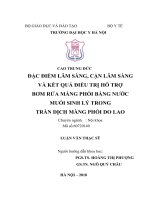
Fig. 1 Decision-making process at the bedside to guide and titrate fluid administration during an episode
of acute circulatory failure. CCE critical care echocardiography, CO cardiac output, CRT capillary refill time, FC
fluid challenge, ΔPCO2 venous-to-arterial CO2 tension difference, ScVO2 central venous oxygen saturation
These conditions may be further investigated by coupling the information obtained
cVO2. In fact, an increased ΔPCO2 associated with a decreased
by ΔPCO2 and S
cVO2 associated with an
ScVO2 is suggestive of a low CO, whereas a normal/high S
increased ΔPCO2 indicates impaired tissue perfusion. Pragmatically, a normal ∆PCO2
(< 6 mmHg) value in a shocked patient should defer from increasing the CO as first
step; instead, regional blood flow may be impaired even in presence of a normal/high
CO.
All these aspects may be integrated into a decision-making algorithm where the
clinical signs of hypoperfusion are coupled with the CCE evaluation (Fig. 1). The clinical recognition of signs of systemic hypoperfusion should trigger the use of fluids
with the purpose of optimizing the CO and improving the D
O2. This choice recognizes fluids as drugs that should only be used as long as the effect on CO is likely.
Monitoring fluid responsiveness during the resuscitation phase of an episode of acute
circulatory failure may be achieved by applying a “closed-loop” operative strategy,
where the signs of tissue hypoperfusion and the findings of CCE are re-evaluated
after each fluid bolus. More sophisticated tools are useful when the cardiovascular
system reaches the plateau of clinical response or, earlier, when the CCE shows acute
or acute-on-chronic cardiac dysfunction at the baseline examination of the patient.
Conclusions
The physiology of fluid administration in critically ill patients is of major importance in ICU. With a solid basis in the dynamic and complex balance between cardiovascular function and systemic response, fluids should be considered as drugs and
intensivists should consider their pharmacodynamic and biochemical properties to
optimize the therapy. A multimodal approach is required since single physiological
or biochemical measurements able to adequately assess the balance between the CO
and tissue perfusion pressure are still lacking. The assessment of response to fluid
Page 11 of 15
Messina et al. Intensive Care Medicine Experimental
(2022) 10:46
administration may be obtained by coupling the changes of different signs of tissue
hypoperfusion using clinical and invasive hemodynamic monitoring, with the evaluation of cardiac function based on critical care echocardiography.
Acknowledgements
None to declare.
Author contributions
AM and MC drafted the manuscript; JB, MC, DDB, OH, GH, SM, XM, MO, MP and JLT substantially contributed to manu‑
script preparation. All the authors read and approved the final version of the paper.
Funding
None to declare.
Availability of data and materials
Not applicable.
Declarations
Ethics approval and consent to participate
Not applicable.
Consent for publication
Not applicable.
Competing interests
The authors declare no competing interests for this paper.
Received: 22 June 2022 Accepted: 17 October 2022
References
1. Bataille B, de Selle J, Moussot PE, Marty P, Silva S, Cocquet P (2021) Machine learning methods to improve bedside
fluid responsiveness prediction in severe sepsis or septic shock: an observational study. Br J Anaesth 126:826–834
2. Komorowski M, Celi LA, Badawi O, Gordon AC, Faisal AA (2018) The artificial intelligence clinician learns optimal
treatment strategies for sepsis in intensive care. Nat Med 24:1716–1720
3. De Backer D, Aissaoui N, Cecconi M, Chew MS, Denault A, Hajjar L, et al (2022) How can assessing hemodynamics
help to assess volume status? Intensive Care Med
4. Cecconi M, De Backer D, Antonelli M, Beale R, Bakker J, Hofer C et al (2014) Consensus on circulatory shock and
hemodynamic monitoring. Task force of the European society of intensive care medicine. Intensive Care Med
40:1795–1815
5. Singer M (2017) Critical illness and flat batteries. Crit Care 21:309
6. Evans L, Rhodes A, Alhazzani W, Antonelli M, Coopersmith CM, French C et al (2021) Surviving sepsis campaign:
International guidelines for management of sepsis and septic shock 2021. Intensive Care Med 47:1181–1247
7. Lat I, Coopersmith CM, De Backer D, Coopersmith CM, Research Committee of the Surviving Sepsis C (2021) The
surviving sepsis campaign: fluid resuscitation and vasopressor therapy research priorities in adult patients. Inten‑
sive Care Med Exp 9:10
8. Bakker J, Kattan E, Annane D, Castro R, Cecconi M, De Backer D et al (2022) Current practice and evolving concepts
in septic shock resuscitation. Intensive Care Med 48:148–163
9. Hjortrup PB, Haase N, Bundgaard H, Thomsen SL, Winding R, Pettila V et al (2016) Restricting volumes of resuscita‑
tion fluid in adults with septic shock after initial management: the classic randomised, parallel-group, multicentre
feasibility trial. Intensive Care Med 42:1695–1705
10. Ince C (2017) Personalized physiological medicine. Crit Care 21:308
11. Myburgh JA, Mythen MG (2013) Resuscitation fluids. N Engl J Med 369:1243–1251
12. Cecconi M, Parsons AK, Rhodes A (2011) What is a fluid challenge? Curr Opin Crit Care 17:290–295
13. Magder S (2010) Fluid status and fluid responsiveness. Curr Opin Crit Care 16:289–296
14. Dellinger RP, Levy MM, Rhodes A, Annane D, Gerlach H, Opal SM et al (2013) Surviving sepsis campaign: Interna‑
tional guidelines for management of severe sepsis and septic shock, 2012. Intensive Care Med 39:165–228
15. Sanfilippo F, Messina A, Cecconi M, Astuto M (2020) Ten answers to key questions for fluid management in inten‑
sive care. Med Intensive
16. Latta T (1832) Relative to the treatment of cholera by the copious injection of aqueous and saline fluid into the
veins. Lancet 2:274–277
17. Katz AM (2002) Ernest henry starling, his predecessors, and the “law of the heart.” Circulation 106:2986–2992
18. Guyton AC, Jones CE (1973) Central venous pressure: physiological significance and clinical implications. Am Heart
J 86:431–437
19. Guyton AC, Richardson TQ, Langston JB (1964) Regulation of cardiac output and venous return. Clin Anesth 3:1–34
20. Feihl F, Broccard AF (2009) Interactions between respiration and systemic hemodynamics. Part i: basic concepts.
Intensive Care Med 35:45–54
Page 12 of 15
Messina et al. Intensive Care Medicine Experimental
(2022) 10:46
21. Feihl F, Broccard AF (2009) Interactions between respiration and systemic hemodynamics. Part ii: practical implica‑
tions in critical care. Intensive Care Med 35:198–205
22. Vincent JL, De Backer D (2013) Circulatory shock. N Engl J Med 369:1726–1734
23. Marik PE, Monnet X, Teboul JL (2011) Hemodynamic parameters to guide fluid therapy. Ann Intensive Care 1:1
24. Monnet X, Marik PE, Teboul JL (2016) Prediction of fluid responsiveness: an update. Ann Intensive Care 6:111
25. Boyd JH, Forbes J, Nakada TA, Walley KR, Russell JA (2011) Fluid resuscitation in septic shock: a positive fluid bal‑
ance and elevated central venous pressure are associated with increased mortality. Crit Care Med 39:259–265
26. Marik PE, Linde-Zwirble WT, Bittner EA, Sahatjian J, Hansell D (2017) Fluid administration in severe sepsis and
septic shock, patterns and outcomes: an analysis of a large national database. Intensive Care Med 43:625–632
27. Marik PE (2016) Fluid responsiveness and the six guiding principles of fluid resuscitation. Crit Care Med
44:1920–1922
28. Kattan E, Ospina-Tascon GA, Teboul JL, Castro R, Cecconi M, Ferri G et al (2020) Systematic assessment of fluid
responsiveness during early septic shock resuscitation: secondary analysis of the andromeda-shock trial. Crit Care
24:23
29. Kattan E, Castro R, Vera M, Hernandez G (2020) Optimal target in septic shock resuscitation. Ann Transl Med 8:789
30. Finfer S, Bellomo R, Boyce N, French J, Myburgh J, Norton R et al (2004) A comparison of albumin and saline for
fluid resuscitation in the intensive care unit. N Engl J Med 350:2247–2256
31. Hernandez G, Bellomo R, Bakker J (2019) The ten pitfalls of lactate clearance in sepsis. Intensive Care Med 45:82–85
32. Hernandez G, Luengo C, Bruhn A, Kattan E, Friedman G, Ospina-Tascon GA et al (2014) When to stop septic shock
resuscitation: clues from a dynamic perfusion monitoring. Ann Intensive Care 4:30
33. Lara B, Enberg L, Ortega M, Leon P, Kripper C, Aguilera P et al (2017) Capillary refill time during fluid resuscitation
in patients with sepsis-related hyperlactatemia at the emergency department is related to mortality. PLoS ONE
12:e0188548
34. Lima A, Jansen TC, van Bommel J, Ince C, Bakker J (2009) The prognostic value of the subjective assessment of
peripheral perfusion in critically ill patients. Crit Care Med 37:934–938
35. Ait-Oufella H, Lemoinne S, Boelle PY, Galbois A, Baudel JL, Lemant J et al (2011) Mottling score predicts survival in
septic shock. Intensive Care Med 37:801–807
36. Ait-Oufella H, Bige N, Boelle PY, Pichereau C, Alves M, Bertinchamp R et al (2014) Capillary refill time exploration
during septic shock. Intensive Care Med 40:958–964
37. Brunauer A, Kokofer A, Bataar O, Gradwohl-Matis I, Dankl D, Bakker J et al (2016) Changes in peripheral perfusion
relate to visceral organ perfusion in early septic shock: a pilot study. J Crit Care 35:105–109
38. Hernandez G, Ospina-Tascon GA, Damiani LP, Estenssoro E, Dubin A, Hurtado J et al (2019) Effect of a resuscitation
strategy targeting peripheral perfusion status vs serum lactate levels on 28-day mortality among patients with
septic shock: the andromeda-shock randomized clinical trial. JAMA 321:654–664
39. Zampieri FG, Damiani LP, Bakker J, Ospina-Tascon GA, Castro R, Cavalcanti AB et al (2020) Effects of a resuscitation
strategy targeting peripheral perfusion status versus serum lactate levels among patients with septic shock. A
Bayesian reanalysis of the andromeda-shock trial. Am J Respir Crit Care Med 201:423–429
40. Cecconi M, Hofer C, Teboul JL, Pettila V, Wilkman E, Molnar Z et al (2015) Fluid challenges in intensive care: the
Fenice study: a global inception cohort study. Intensive Care Med 41:1529–1537
41. Lakhal K, Ehrmann S, Perrotin D, Wolff M, Boulain T (2013) Fluid challenge: tracking changes in cardiac output with
blood pressure monitoring (invasive or non-invasive). Intensive Care Med 39:1953–1962
42. Dufour N, Chemla D, Teboul JL, Monnet X, Richard C, Osman D (2011) Changes in pulse pressure following fluid
loading: a comparison between aortic root (non-invasive tonometry) and femoral artery (invasive recordings).
Intensive Care Med 37:942–949
43. Pierrakos C, Velissaris D, Scolletta S, Heenen S, De Backer D, Vincent JL (2012) Can changes in arterial pressure be
used to detect changes in cardiac index during fluid challenge in patients with septic shock? Intensive Care Med
38:422–428
44. Garcia MI, Romero MG, Cano AG, Aya HD, Rhodes A, Grounds RM et al (2014) Dynamic arterial elastance as a
predictor of arterial pressure response to fluid administration: a validation study. Crit Care 18:626
45. Cecconi M, Monge Garcia MI, Gracia Romero M, Mellinghoff J, Caliandro F, Grounds RM et al (2015) The use of
pulse pressure variation and stroke volume variation in spontaneously breathing patients to assess dynamic arte‑
rial elastance and to predict arterial pressure response to fluid administration. Anesth Analg 120:76–84
46. van der Ven WH, Schuurmans J, Schenk J, Roerhorst S, Cherpanath TGV, Lagrand WK et al (2022) Monitoring, man‑
agement, and outcome of hypotension in intensive care unit patients, an international survey of the European
society of intensive care medicine. J Crit Care 67:118–125
47. Guarracino F, Bertini P, Pinsky MR (2019) Cardiovascular determinants of resuscitation from sepsis and septic shock.
Crit Care 23:118
48. Aya HD, Ster IC, Fletcher N, Grounds RM, Rhodes A, Cecconi M (2015) Pharmacodynamic analysis of a fluid chal‑
lenge. Crit Care Med
49. Messina A, Longhini F, Coppo C, Pagni A, Lungu R, Ronco C et al (2017) Use of the fluid challenge in critically ill
adult patients: a systematic review. Anesth Analg 125:1532–1543
50. Messina A, Pelaia C, Bruni A, Garofalo E, Bonicolini E, Longhini F et al (2018) Fluid challenge during anesthesia: a
systematic review and meta-analysis. Anesth Analg
51. Vincent JL (2011) Let’s give some fluid and see what happens “versus the” mini-fluid challenge. Anesthesiology
115:455–456
52. Vincent JL, Weil MH (2006) Fluid challenge revisited. Crit Care Med 34:1333–1337
53. Pinsky MR (2015) Functional hemodynamic monitoring. Crit Care Clin 31:89–111
54. Pinsky MR, Payen D (2005) Functional hemodynamic monitoring. Crit Care 9:566–572
55. Hadian M, Pinsky MR (2007) Functional hemodynamic monitoring. Curr Opin Crit Care 13:318–323
Page 13 of 15
Messina et al. Intensive Care Medicine Experimental
(2022) 10:46
56. Messina A, Dell’Anna A, Baggiani M, Torrini F, Maresca GM, Bennett V et al (2019) Functional hemodynamic tests:
a systematic review and a metanalysis on the reliability of the end-expiratory occlusion test and of the mini-fluid
challenge in predicting fluid responsiveness. Crit Care 23:264
57. Messina A, Palandri C, De Rosa S, Danzi V, Bonaldi E, Montagnini C et al (2021) Pharmacodynamic analysis of a
fluid challenge with 4 ml kg(− 1) over 10 or 20 min: a multicenter cross-over randomized clinical trial. J Clin Monit
Comput
58. Aya HD, Rhodes A, Chis Ster I, Fletcher N, Grounds RM, Cecconi M (2017) Hemodynamic effect of different doses of
fluids for a fluid challenge: a quasi-randomized controlled study. Crit Care Med 45:e161–e168
59. Toscani L, Aya HD, Antonakaki D, Bastoni D, Watson X, Arulkumaran N et al (2017) What is the impact of the fluid
challenge technique on diagnosis of fluid responsiveness? A systematic review and meta-analysis. Crit Care 21:207
60. Ayala A, M FL, Machado A (1986) Malic enzyme levels are increased by the activation of NADPH-consuming path‑
ways: detoxification processes. FEBS Lett 1986;202:102–106
61. Messina A, Sotgiu G, Saderi L, Cammarota G, Capuano L, Colombo D, et al.: Does the definition of fluid responsive‑
ness affect passive leg raising reliability? A methodological ancillary analysis from a multicentric study. Minerva
Anestesiol 2021
62. Zampieri FG, Machado FR, Biondi RS, Freitas FGR, Veiga VC, Figueiredo RC et al (2021) Effect of slower vs faster
intravenous fluid bolus rates on mortality in critically ill patients: the basics randomized clinical trial. JAMA
326:830–838
63. Brunkhorst FM, Engel C, Bloos F, Meier-Hellmann A, Ragaller M, Weiler N et al (2008) Intensive insulin therapy and
pentastarch resuscitation in severe sepsis. N Engl J Med 358:125–139
64. Hahn RG, Dull RO (2021) Interstitial washdown and vascular albumin refill during fluid infusion: novel kinetic
analysis from three clinical trials. Intensive Care Med Exp 9:44
65. Woodcock TE, Woodcock TM (2012) Revised starling equation and the glycocalyx model of transvascular fluid
exchange: an improved paradigm for prescribing intravenous fluid therapy. Br J Anaesth 108:384–394
66. Annane D, Siami S, Jaber S, Martin C, Elatrous S, Declere AD et al (2013) Effects of fluid resuscitation with colloids
vs crystalloids on mortality in critically ill patients presenting with hypovolemic shock: the cristal randomized trial.
JAMA 310:1809–1817
67. Myburgh JA, Finfer S, Bellomo R, Billot L, Cass A, Gattas D et al (2012) Hydroxyethyl starch or saline for fluid resusci‑
tation in intensive care. N Engl J Med 367:1901–1911
68. Perner A, Haase N, Guttormsen AB, Tenhunen J, Klemenzson G, Aneman A et al (2012) Hydroxyethyl starch
130/0.42 versus ringer’s acetate in severe sepsis. N Engl J Med 367:124–134
69. Textoris J, Fouche L, Wiramus S, Antonini F, Tho S, Martin C et al (2011) High central venous oxygen saturation in
the latter stages of septic shock is associated with increased mortality. Crit Care 15:R176
70. Vincent JL (2009) Relevance of albumin in modern critical care medicine. Best Pract Res Clin Anaesthesiol
23:183–191
71. Caironi P, Tognoni G, Masson S, Fumagalli R, Pesenti A, Romero M et al (2014) Albumin replacement in patients
with severe sepsis or septic shock. N Engl J Med 370:1412–1421
72. Salerno F, Navickis RJ, Wilkes MM (2013) Albumin infusion improves outcomes of patients with spontaneous
bacterial peritonitis: a meta-analysis of randomized trials. Clin Gastroenterol Hepatol 11(123–130):e121
73. Joannidis M, Wiedermann CJ, Ostermann M (2022) Ten myths about albumin. Intensive Care Med 48:602–605
74. Varrier M, Ostermann M (2015) Fluid composition and clinical effects. Crit Care Clin 31:823–837
75. Yunos NM, Bellomo R, Glassford N, Sutcliffe H, Lam Q, Bailey M (2015) Chloride-liberal vs. chloride-restrictive intra‑
venous fluid administration and acute kidney injury: an extended analysis. Intensive Care Med 41:257–264
76. Chowdhury AH, Cox EF, Francis ST, Lobo DN (2012) A randomized, controlled, double-blind crossover study on the
effects of 2-l infusions of 0.9% saline and plasma-lyte(r) 148 on renal blood flow velocity and renal cortical tissue
perfusion in healthy volunteers. Ann Surg 256:18–24
77. Young P, Bailey M, Beasley R, Henderson S, Mackle D, McArthur C et al (2015) Effect of a buffered crystalloid solu‑
tion vs saline on acute kidney injury among patients in the intensive care unit: the split randomized clinical trial.
JAMA 314:1701–1710
78. Semler MW, Self WH, Wanderer JP, Ehrenfeld JM, Wang L, Byrne DW et al (2018) Balanced crystalloids versus saline
in critically ill adults. N Engl J Med 378:829–839
79. Finfer S, Micallef S, Hammond N, Navarra L, Bellomo R, Billot L et al (2022) Balanced multielectrolyte solution
versus saline in critically ill adults. N Engl J Med 386:815–826
80. Hammond DA, Lam SW, Rech MA, Smith MN, Westrick J, Trivedi AP et al (2020) Balanced crystalloids versus saline
in critically ill adults: a systematic review and meta-analysis. Ann Pharmacother 54:5–13
81. Zampieri FG, Machado FR, Biondi RS, Freitas FGR, Veiga VC, Figueiredo RC et al (2022) Association between type
of fluid received prior to enrollment, type of admission, and effect of balanced crystalloid in critically ill adults: a
secondary exploratory analysis of the basics clinical trial. Am J Respir Crit Care Med 205:1419–1428
82. Rhodes A, Evans LE, Alhazzani W, Levy MM, Antonelli M, Ferrer R et al (2017) Surviving sepsis campaign: interna‑
tional guidelines for management of sepsis and septic shock: 2016. Intensive Care Med 43:304–377
83. Hiemstra B, Eck RJ, Keus F, van der Horst ICC (2017) Clinical examination for diagnosing circulatory shock. Curr
Opin Crit Care 23:293–301
84. Vignon P, Begot E, Mari A, Silva S, Chimot L, Delour P et al (2018) Hemodynamic assessment of patients with septic
shock using transpulmonary thermodilution and critical care echocardiography: a comparative study. Chest
153:55–64
85. Messina A, Greco M, Cecconi M (2019) What should i use next if clinical evaluation and echocardiographic haemo‑
dynamic assessment is not enough? Curr Opin Crit Care 25:259–265
86. Expert Round Table on Echocardiography in ICU (2014) International consensus statement on training standards
for advanced critical care echocardiography. Intensive Care Med 40:654–666
87. Spencer KT, Kimura BJ, Korcarz CE, Pellikka PA, Rahko PS, Siegel RJ (2013) Focused cardiac ultrasound: recommen‑
dations from the american society of echocardiography. J Am Soc Echocardiogr 26:567–581
Page 14 of 15
Messina et al. Intensive Care Medicine Experimental
(2022) 10:46
88. Burwash IG, Basmadjian A, Bewick D, Choy JB, Cujec B, Jassal DS et al (2011) 2010 Canadian cardiovascular society/
Canadian society of echocardiography guidelines for training and maintenance of competency in adult echocar‑
diography. Can J Cardiol 27:862–864
89. Robba C, Wong A, Poole D, Al Tayar A, Arntfield RT, Chew MS et al (2021) Basic ultrasound head-to-toe skills for
intensivists in the general and neuro intensive care unit population: consensus and expert recommendations of
the European society of intensive care medicine. Intensive Care Med 47:1347–1367
90. Hernandez G, Pedreros C, Veas E, Bruhn A, Romero C, Rovegno M et al (2012) Evolution of peripheral vs metabolic
perfusion parameters during septic shock resuscitation. A clinical-physiologic study. J Crit Care 27:283–288
91. Londono J, Nino C, Diaz J, Morales C, Leon J, Bernal E et al (2018) Association of clinical hypoperfusion variables
with lactate clearance and hospital mortality. Shock 50:286–292
92. Casserly B, Phillips GS, Schorr C, Dellinger RP, Townsend SR, Osborn TM et al (2015) Lactate measurements in sep‑
sis-induced tissue hypoperfusion: results from the surviving sepsis campaign database. Crit Care Med 43:567–573
93. Jansen TC, van Bommel J, Schoonderbeek FJ, Sleeswijk Visser SJ, van der Klooster JM, Lima AP et al (2010) Early
lactate-guided therapy in intensive care unit patients: a multicenter, open-label, randomized controlled trial. Am J
Respir Crit Care Med 182:752–761
94. Levy B (2006) Lactate and shock state: the metabolic view. Curr Opin Crit Care 12:315–321
95. Haas SA, Lange T, Saugel B, Petzoldt M, Fuhrmann V, Metschke M et al (2016) Severe hyperlactatemia, lactate clear‑
ance and mortality in unselected critically ill patients. Intensive Care Med 42:202–210
96. Bloos F, Reinhart K (2005) Venous oximetry. Intensive Care Med 31:911–913
97. Rivers E, Nguyen B, Havstad S, Ressler J, Muzzin A, Knoblich B et al (2001) Early goal-directed therapy in the treat‑
ment of severe sepsis and septic shock. N Engl J Med 345:1368–1377
98. Dellinger RP, Levy MM, Carlet JM, Bion J, Parker MM, Jaeschke R et al (2008) Surviving sepsis campaign: interna‑
tional guidelines for management of severe sepsis and septic shock: 2008. Intensive Care Med 34:17–60
99. Investigators A, Group ACT, Peake SL, Delaney A, Bailey M, Bellomo R et al (2014) Goal-directed resuscitation for
patients with early septic shock. N Engl J Med 371:1496–1506
100. Pro CI, Yealy DM, Kellum JA, Huang DT, Barnato AE, Weissfeld LA et al (2014) A randomized trial of protocol-based
care for early septic shock. N Engl J Med 370:1683–1693
101. Mouncey PR, Osborn TM, Power GS, Harrison DA, Sadique MZ, Grieve RD et al (2015) Trial of early, goal-directed
resuscitation for septic shock. N Engl J Med 372:1301–1311
102. van Beest PA, Hofstra JJ, Schultz MJ, Boerma EC, Spronk PE, Kuiper MA (2008) The incidence of low venous oxygen
saturation on admission to the intensive care unit: a multi-center observational study in the Netherlands. Crit Care
12:R33
103. Cuschieri J, Rivers EP, Donnino MW, Katilius M, Jacobsen G, Nguyen HB et al (2005) Central venous-arterial carbon
dioxide difference as an indicator of cardiac index. Intensive Care Med 31:818–822
104. Lamia B, Monnet X, Teboul JL (2006) Meaning of arterio-venous pco2 difference in circulatory shock. Minerva
Anestesiol 72:597–604
105. Mallat J, Lemyze M, Tronchon L, Vallet B, Thevenin D (2016) Use of venous-to-arterial carbon dioxide tension differ‑
ence to guide resuscitation therapy in septic shock. World J Crit Care Med 5:47–56
Publisher’s Note
Springer Nature remains neutral with regard to jurisdictional claims in published maps and institutional affiliations.
Page 15 of 15