Chapter 058. Anemia and Polycythemia (Part 8) docx
Bạn đang xem bản rút gọn của tài liệu. Xem và tải ngay bản đầy đủ của tài liệu tại đây (104.6 KB, 5 trang )
Chapter 058. Anemia and
Polycythemia
(Part 8)
Bone Marrow Examination
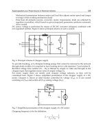
A bone marrow aspirate and smear or a needle biopsy may be useful in the
evaluation of some patients with anemia. In patients with hypoproliferative anemia
and normal iron status, a bone marrow is indicated. Marrow examination can
diagnose primary marrow disorders such as myelofibrosis, a red cell maturation
defect, or an infiltrative disease (Figs. 58-14, 58-15, and 58-16). The increase or
decrease of one cell lineage (myeloid vs. erythroid) compared to another is
obtained by a differential count of nucleated cells in a bone marrow smear [the
myeloid/erythroid (M/E) ratio]. A patient with a hypoproliferative anemia (see
below) and a reticulocyte production index < 2 will demonstrate an M/E ratio of 2
or 3:1. In contrast, patients with hemolytic disease and a production index > 3 will
have an M/E ratio of at least 1:1. Maturation disorders are identified from the
discrepancy between the M/E ratio and the reticulocyte production index (see
below). Either the marrow smear or biopsy can be stained for the presence of iron
stores or iron in developing red cells. The storage iron is in the form of ferritin or
hemosiderin . On carefully prepared bone marrow smears, small ferritin granules
can normally be seen under oil immersion in 20–40% of developing erythroblasts.
Such cells are called sideroblasts.
Figure 58-14
Normal bone marrow. This is a low-power view of a section of a normal
bone marrow biopsy stained with hematoxylin and eosin (H&E). Note that the
nucleated cellular elements account for ~40–50% and the fat (clear areas) accounts
for ~50–60% of the area. (From Hillman et al.)
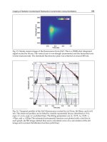
Figure 58-15
Erythroid hyperplasia. This marrow shows an increase in the fraction of
cells in the erythroid lineage as might be seen when a normal marrow compensates
for acute blood loss or hemolysis. The M/E ratio is about 1:1. (From Hillman et
al.)
Figure 58-16
Myeloid hyperplasia. This marrow shows an increase in the fraction of
cells in the myeloid or granulocytic lineage as might be seen in a normal marrow
responding to infection. The M/E ratio is >3:1. (From Hillman et al.)[newpage]
Other Laboratory Measurements
Additional laboratory tests may be of value in confirming specific
diagnoses. For details of these tests and how they are applied in individual
disorders, see Chaps. 98, 99, 100, 101, and 102.
Definition and Classification of Anemia
Initial Classification of Anemia
The functional classification of anemia has three major categories. These
are: (1) marrow production defects (hypoproliferation), (2) red cell maturation
defects (ineffective erythropoiesis ), and (3) decreased red cell survival (blood
loss/hemolysis). The classification is shown in Fig. 58-17. A hypoproliferative
anemia is typically seen with a low reticulocyte production index together with
little or no change in red cell morphology (a normocytic, normochromic anemia)
(Chap. 98). Maturation disorders typically have a slight to moderately elevated
reticulocyte production index that is accompanied by either macrocytic (Chap.
100) or microcytic (Chaps. 98, 99) red cell indices. Increased red blood cell
destruction secondary to hemolysis results in an increase in the reticulocyte
production index to at least three times normal (Chap. 101), provided sufficient
iron is available. Hemorrhagic anemia does not typically result in production
indices of more than 2.0–2.5 times normal because of the limitations placed on
expansion of the erythroid marrow by iron availability.