Báo cáo khoa học: "Can prophylactic breast irradiation contribute to cardiac toxicity in patients with prostate cancer receiving androgen suppressing drugs?" ppt
Bạn đang xem bản rút gọn của tài liệu. Xem và tải ngay bản đầy đủ của tài liệu tại đây (606.1 KB, 6 trang )
BioMed Central
Page 1 of 6
(page number not for citation purposes)
Radiation Oncology
Open Access
Research
Can prophylactic breast irradiation contribute to cardiac toxicity in
patients with prostate cancer receiving androgen suppressing
drugs?
Carsten Nieder*
1
, Adam Pawinski
1
, Nicolaus H Andratschke
2
and
Michael Molls
2
Address:
1
Radiation Oncology Unit, Nordlandssykehuset HF, 8092 Bodø, Norway and
2
Department of Radiation Oncology, Klinikum rechts der
Isar der Technischen Universität München, 81675 Munich, Germany
Email: Carsten Nieder* - ; Adam Pawinski - ; Nicolaus H Andratschke - ;
Michael Molls -
* Corresponding author
Abstract
Background: Androgen suppression treatment (AST) might increase the risk of cardiac morbidity
in prostate cancer patients. Possible explanations were provided, however, they disregard the
potential contribution of prophylactic radiotherapy to the mamillary regions (PMRT, prescribed to
avoid gynecomastia).
Methods: We studied the exposure of the heart in a typical electron beam PMRT setting by
evaluating computed tomography (CT) scans in 40 non-cancer patients (age 65 and 75 years in 50%
each) and 17 prostate cancer patients. Five of the younger, 7 of the older and 4 of the cancer
patients had significant cardiac disease.
Results: The median distance between skin and outer heart contour decreased with age. In all
three groups, patients with cardiac morbidity had smaller distances. When using the CT-
determined PMRT beam energy, 10% of the younger, 15% of the older and none of the prostate
cancer patients would receive approximately 50% of the prescription dose to a part of the heart
(2 had no history of cardiac disease). When using the clinically rather than CT-determined beam
energy, as often done in daily practice, an additional 12.5% of the non-cancer and 12% of the
prostate cancer patients would be exposed to comparably high doses.
Conclusion: The present data provide preliminary evidence that PMRT might be a factor that
contributes to cardiac side effects. Previous studies that established a relationship between AST
and cardiac morbidity did not include information on delivery of PMRT.
Background
Androgen suppression including temporary suppression
in patients receiving curative radiation therapy represents
an important treatment option for patients with prostate
cancer [1]. One of the disadvantages and side effects of
androgen suppression is the increased risk of cardiac tox-
icity, another one the risk of gynecomastia development
[2-4], e.g., during treatment with goserelin acetate and
Published: 10 January 2008
Radiation Oncology 2008, 3:2 doi:10.1186/1748-717X-3-2
Received: 12 October 2007
Accepted: 10 January 2008
This article is available from: />© 2008 Nieder et al; licensee BioMed Central Ltd.
This is an Open Access article distributed under the terms of the Creative Commons Attribution License ( />),
which permits unrestricted use, distribution, and reproduction in any medium, provided the original work is properly cited.
Radiation Oncology 2008, 3:2 />Page 2 of 6
(page number not for citation purposes)
flutamide [5] or with bicalutamide [6-8]. Prophylactic
radiation therapy to both mamillar regions (PMRT)
before the start of androgen suppression might decrease
the likelihood of gynecomastia [7,9,10]. However,
depending on the anatomical situation, left-sided PMRT
might lead to a certain exposure of the heart to ionizing
radiation.
Typically, single electron beams with a sharp dose gradi-
ent are used, having the advantage of limited tissue pene-
tration. In contrast to most other situations in
contemporary radiation oncology, no 3-dimensional
computed tomography (CT)-based treatment planning is
used. Therefore, the exact dose distribution is unknown
for the individual patient, leaving room for accidental
dose exposure of the heart. In the health region of North-
ern-Norway for example, where one of the authors' insti-
tutions is located, a standard clinical set-up for PMRT is
used. It consists of a single dose of 15 Gy delivered via cir-
cular fields, diameter 7 cm, electron energy 9 MeV (6 and
12 MeV in slim and obese patients, respectively). Both the
left and right perimamillar regions are treated with one
such field. Using similar techniques, the authors from
Munich, Germany, administer 3 fractions of 4 Gy each.
Both regimens are among those previously studied by dif-
ferent groups, where PMRT was found to prevent gyneco-
mastia development [7,9,10].
Recent articles provide possible explanations for the ele-
vated risk of cardiac diseases in patients treated with
androgen suppression, e.g., changes in lipid metabolism
[11]. However, we hypothesised that administration of
PMRT might further contribute to long-term toxicity in a
multifactorial scenario. Therefore, the present study exam-
ined potential radiation doses to the heart in a group of 40
individuals who underwent thoracic imaging for various
medical reasons and 17 patients with prostate cancer.
Methods
We first analysed 40 male patients who received contrast-
enhanced CT scans of the thorax for various medical rea-
sons (unrelated to cancer treatment) after appropriate
institutional informed consent. Twenty patients were 65
years old and 20 were 75 years old. They were selected
from the radiology departments database (Nord-
landssykehuset, Bodø, Norway) based on their date of
birth. The search was started with patients born 01. June
1942 and 1932, respectively, and continued towards the
end of the year until 20 patients were identified in each
group. They were not allowed to have significant lung
abnormalities such as previous surgery, tumors or pleural
effusions. All medical records were also available in the
hospital's data system. They were reviewed to identify
those patients with a history of serious heart disease such
as myocardial infarction, aortocoronar bypass surgery and
other coronary artery interventions. Asymptomatic coro-
nary artery disease, elevated blood pressure or mild types
of cardiac dysfunction were not considered for the pur-
pose of this study.
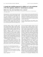
In each patient, the left mamilla (center of the PMRT
field) was identified on the CT scans and the distance
between the skin and anterior border of the pectoral mus-
culature was measured (Figure 1). This value was used to
calculate the electron beam energy needed for PMRT. Pre-
viously published electron depth-dose distribution data
(Table 1) were used. The therapeutic depth of the elec-
trons was to match the anterior border of the pectoral
musculature, which corresponds to the posterior border
of the target volume, as closely as possible. Then, both the
optimal CT-based electron beam and the clinically used
standard 9 MeV beam were chosen for further evaluation.
At a caudal distance of 3 cm from the mamilla, i.e. close
to the inferior field border, the dose to the heart was esti-
mated. As evident from the CT scans, only the distal parts
of the field might cause relevant doses to the heart. We
measured the distance between the skin and the outer
contour of the heart and used the data from Table 1 to
estimate the heart dose. The same methods were used to
examine the first 17 patients with prostate cancer who
were treated since the opening of the Radiation Oncology
Unit at Nordlandssykehuset in June 2007. Not all of these
patients actually received PMRT, some were treated for
metastatic disease. Finally, the CT scans of the prostate
cancer patients, which were available in our treatment
planning system (Varian Eclipse), were used to calculate
Axial contrast-enhanced computed tomography scan at the level of the left mamilla displaying both the distance between the skin surface and the pectoral musculature (2.4 cm) and the field size of 7 cmFigure 1
Axial contrast-enhanced computed tomography scan at the
level of the left mamilla displaying both the distance between
the skin surface and the pectoral musculature (2.4 cm) and
the field size of 7 cm. Note that only very low heart expo-
sure results from electron beam irradiation at this level, i.e.
the center of the field.
Radiation Oncology 2008, 3:2 />Page 3 of 6
(page number not for citation purposes)
actual 3-D dose distributions and dose-volume histo-
grams in representative cases, i.e. those patients where the
left anterior descending coronary artery (LAD) could be
identified. Varian Eclipse uses the Generalized Gaussian
Pencil Beam algorithm for calculating electron dose distri-
butions. The plans were calculated for a Varian Clinac
treatment unit.
Results
Out of 20 65-years-old patients, 5 had a history of signifi-
cant cardiac disease. In the 75-years-old group, 7 patients
belonged to this subset. Among the prostate cancer
patients, 4/17 had significant cardiac disease. The latter
group had a median age of 72 years, range 58–83 years.
The required beam energy for PMRT was different from 9
MeV in the majority of patients. While 6 patients in both
non-cancer-groups actually were best treated with 9 MeV,
11 and 13 patients in the 65-years and 75-years group
would have benefited from choosing 6 MeV. In 3 and 1
individuals, 12 MeV were necessary to cover the pre-pec-
toral region adequately. In the prostate cancer patients, 9
MeV was appropriate in 6 cases, 6 MeV in 8 cases, 12 MeV
in 2 cases and 15 MeV in 1 case.
The median distance between skin and outer heart con-
tour decreased with age from 6.25 cm in the 65-years
group to 5.35 cm in the 75-years group (range 3.1–8.7 cm
and 2.6–8.7 cm, respectively). In prostate cancer patients,
5.5 cm were measured (range 3.8–8.1 cm). In all three
groups, patients with cardiac morbidity had smaller dis-
tances. In the 65-years-old patients, the median values
were 5.1 vs. 6.7 cm for patients with/without serious heart
disease. In the older patients these figures were 4.2 vs. 5.6
cm. In the prostate cancer patients, 4.8 vs. 5.7 cm were cal-
culated. For all groups combined, 5.0 vs. 6.4 cm were cal-
culated. When using the CT-based beam energy, two of
the younger non-cancer patients (10%) would receive
≥50% of the prescription dose to a relatively small part of
the anterior myocardial wall of the left ventricle and the
small vessels in this region. Both patients had a history of
cardiac disease (Table 2). Among the older patients, one
would receive ≥50% to a small heart volume, while two
would receive ≥50% to a more extended part of the heart
(total 5/40 patients, 12.5%). Only one of these three 75-
years-old patients had a history of cardiac disease (Figure
2). None of the prostate cancer patients would receive
comparably high doses to the heart when CT-based beams
were used. When using the inappropriate 9 MeV beam
rather than the optimal 6 MeV beam, one additional
younger non-cancer patient plus four additional older
patients would receive an unnecessary heart exposure. In
the absence of CT information, two of the prostate cancer
patients (12%) would belong to the group with unneces-
sary heart exposure when using the 9 MeV beam rather
then the optimal 6 MeV beam (Figure 3). The use of the
12 or 15 MeV beam, where appropriate in obese patients
would be possible without concerns.
The 3-D dose distributions were first evaluated in prostate
cancer patients for the 9 MeV beam, even though this
energy would not be appropriate if CT information was
available for treatment planning. The examples revealed
that the mean dose to the heart is in the range of 2 to 5%
of the prescription dose. Five percent corresponds to 0.75
Gy if one uses a single fraction of 15 Gy. The proximal
Table 2: Individual data of patients with heart exposure from prophylactic breast radiation therapy.
Patientnr. Age (years) Heart disease CT-based beam energy Skin-heart distance Exposure
1 65 yes 12 MeV 5,1 cm Moderate
2 65 yes 9 MeV 4,2 cm Moderate
3 75 yes 9 MeV 4,0 cm Distinct
4 75 no 6 MeV 2,6 cm Moderate
5 75 no 9 MeV 3,7 cm Distinct
6 65 no 6 MeV 3,1 cm Moderate**
7 75 no 6 MeV 3,8 cm Moderate**
8 75 yes 6 MeV 4,0 cm Moderate**
9 75 yes 6 MeV 3,9 cm Moderate**
10 75 yes 6 MeV 3,3 cm Distinct**
11* 64 no 6 MeV 3,9 cm Moderate**
12* 83 no 6 MeV 3,9 cm Moderate**
* prostate cancer patient
**when using the standard 9 MeV beam in the absence of CT scan information
Table 1: Electron beam dose distribution (values might vary, e.g.,
with field size, source-skin-distance and tissue homogeneity),
adapted from [22].
Beam energy Surface dose Therapeutic depth Depth of 50%
isodose
6 MeV 72% 20 mm 24 mm
9 MeV 78% 30 mm 38 mm
12 MeV 83% 40 mm 50 mm
Radiation Oncology 2008, 3:2 />Page 4 of 6
(page number not for citation purposes)
parts of the LAD received up to 14% of the prescription
dose, i.e. 2.1 Gy. The distal parts were indistinguishable
from the myocardium of the left ventricle with the CT pro-
tocols used in these patients. In general, the highest doses
to the heart were seen in the anterior part of the left ven-
tricle and the interventricular septum (Figure 3). Up to
80% of the prescription dose was observed in very small
volumes (<3%) of these areas. Even the volume of the left
ventricle receiving 50% of the dose, i.e. 7.5 Gy, was com-
parably small (maximum 5%). Up to 10% of the left ven-
tricle received 25% of the dose, i.e. 3.75 Gy, and up to
18% received 10% of the dose, i.e. 1.5 Gy. If one takes the
patients' individual anatomy into account and selects the
6 MeV beam in such cases, the doses to the left ventricle
decrease drastically. The same small volumes that would
receive 50–80% of the dose with the 9 MeV beam, would
so receive 10–20% and the mean dose to the left ventricle
would not exceed 5% of the prescription dose, i.e. 0.75
Gy.
Discussion
The present analysis is to our knowledge the first one that
addresses the role of PMRT as a potential cause of cardiac
morbidity in prostate cancer patients receiving androgen
suppression therapy. It was performed both in cancer
patients and randomly selected individuals having had CT
examinations for other medical reasons. The results in
these groups were largely comparable. We used 3-D treat-
ment planning with display of isodose distributions and
dose-volume histograms only in those patients whose CT
scans already were entered into the treatment planning
system, i.e. prostate cancer patients, and only if the LAD
could be identified. Data from these patients suggest that
parts of the left ventricle might be exposed to 50–80% of
the prescription dose, even if the mean doses in general
are low. Studies in electron boost treatment for breast can-
cer have also shown that the heart might be exposed to
unexpected radiation doses in a proportion of these
patients [12]. The present data suggest that standard non-
CT-based approaches often are unsatisfactory and that
individual 3-D treatment planning might benefit a con-
siderable number of patients because it can reduce the
radiation dose to the heart. This benefit appears to
increase with patient age and pre-existing cardiac morbid-
ity. Even among those treated with the appropriate beam
energy, up to 12.5% of the patients might be at risk for
exposure of the heart to unnecessary radiation doses. This
figure increases when the beam energy is determined just
on the basis of a clinical examination without exact ana-
tomical information.
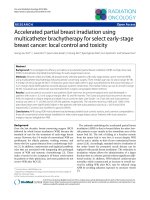
We arbitrarily decided to depict in Figure 2 the depth
where approximately 50% of the prescription dose is
administered. At first glance, 50% of a prescription dose
of 15 Gy (single fraction) or 12 Gy (in 3 fractions) appears
relatively low compared to the heart doses reported from
radiation treatment in a variety of mediastinal tumors
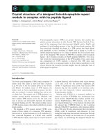
Axial contrast-enhanced computed tomography scan 3 cm caudal from the mamilla displaying on the lower image the approximate depth of the 50% isodose from a standard 9 MeV electron beam (6 MeV would have been appropriate)Figure 3
Axial contrast-enhanced computed tomography scan 3 cm
caudal from the mamilla displaying on the lower image the
approximate depth of the 50% isodose from a standard 9
MeV electron beam (6 MeV would have been appropriate).
3-D planning illustrates that the actual dose to the heart is
even higher. The left ventricle (contoured in yellow) is the
part of the heart that receives the highest dose (maximum
80%). The blue isodose wash refers to 33% of the prescrip-
tion dose, i.e. 5 Gy.
Axial contrast-enhanced computed tomography (CT) scan 3 cm caudal from the mamilla displaying the approximate depth of the 50% isodose from the CT-determined 9 MeV electron beamFigure 2
Axial contrast-enhanced computed tomography (CT) scan 3
cm caudal from the mamilla displaying the approximate depth
of the 50% isodose from the CT-determined 9 MeV electron
beam. In this 65-years-old non-cancer patient with previous
heart disease, parts of the left ventricle would be exposed to
unexpected doses of ionizing radiation.
Radiation Oncology 2008, 3:2 />Page 5 of 6
(page number not for citation purposes)
[13]. Several data sets suggest, however, that doses as low
as 4–5 Gy might contribute to cardiac toxicity [14-16].
These epidemiologic findings are largely compatible with
radiobiologic data on the pathogenesis of radiation-
induced heart disease, as comprehensively reviewed by
Schultz-Hector and Trott [17]. The endothelial lining of
blood vessels might be particularly vulnerable, resulting
in slowly progressive functional and structural alterations.
On the basis of these findings, even partial heart expo-
sures might contribute to long-term damage, which typi-
cally becomes manifest after several years [18]. In reality,
the 50% isodose might reach even further into the heart
than displayed in Figure 2, because the air-containing
lungs allow for deeper penetration of the electron beam
than soft tissues. Figure 3 confirms that the 50% isodose
depth taken from the values in Table 1 might underesti-
mate the actual dose distribution in a patient.
Is it possible to relate or fit our preliminary findings to the
published cardiac toxicity data? An observational study of
a population-based cohort of 73,196 Medicare enrollees
age 66 years or older who were diagnosed with locore-
gional prostate cancer during 1992 to 1999 and observed
through 2001 was recently published [2]. The authors
analysed in this Surveillance, Epidemiology and End
Results database whether treatment with GnRH agonists
was associated with coronary heart disease, myocardial
infarction, and sudden cardiac death. Men with prevalent
diabetes and coronary heart disease were excluded. The
mean age at diagnosis was 74 years. More than one third
of men received a GnRH agonist during follow-up. GnRH
agonist use was associated with increased risk of coronary
heart disease (adjusted HR, 1.16; P < .001), myocardial
infarction (adjusted HR, 1.11; P = .03), and sudden car-
diac death (adjusted HR, 1.16; P = .004). Therapy for as
few as 1–4 months was associated with an increased risk
of coronary artery disease. Unfortunately, the database
did not include information about use of oral antiandro-
gens, combined androgen blockade and PMRT in this
cohort.
Another group evaluated whether the timing of fatal myo-
cardial infarction was influenced by the administration of
androgen suppression therapy [3]. The study cohort com-
prised 1,372 men who were enrolled onto three rand-
omized trials between 1995 and 2001. In the three trials,
the men were randomly assigned to receive radiation ther-
apy with 0 versus 3 versus 6, 3 versus 8, or 0 versus 6
months of androgen suppression (goserelin plus fluta-
mide or a GnRH agonist only). The median age was
68–72.5 years in the three trials. Men age 65 years or older
who received 6 months of androgen suppression experi-
enced shorter times to fatal infarction compared with men
in this age group who did not receive such medication (P
= .017). Even three months of treatment might shorten
the time to fatal myocardial infarction, but additional evi-
dence is needed to strengthen this hypothesis. As commu-
nicated by the principal investigators, PMRT was not
offered in two of the trials, while the exact proportion of
patients that received this treatment is unknown from the
Canadian trial (personal communication, July 2007). It is
therefore not possible to compare the available clinical
results with the percentage of patients that might receive
relevant radiation doses to the heart in our present study.
Importantly, other data from patients treated with radia-
tion therapy plus androgen suppression also suggest that
hormonal manipulation might result in greater non-can-
cer mortality [19].
Despite the fact that a causal relationship between the rel-
atively low radiation doses from PMRT and cardiac mor-
bidity or mortality can not be proven at this time, it
appears prudent to minimize all factors that might con-
tribute to non-cancer mortality in these patients. Even if
PMRT should be considered as just one of the potential
factors contributing to cardiac morbidity in patients
receiving androgen suppression therapy, the question
arises whether the use of non-3-dimensional planning
and treatment techniques should continue in an era
where advanced technology that reduces the dose to the
heart and takes, e.g., advantage of breathing control,
which might help to increase the distance between tho-
racic wall and heart, is available [20] and where the occa-
sional patients with still unacceptable radiation treatment
plans can switch to alternative treatments such as
tamoxifen [7]. In addition, androgen suppression regi-
mens with lower rates of symptomatic gynecomastia
might be considered [21]. Future epidemiologic studies
on cardiac side effects of androgen suppression should try
to include data on the use of PMRT [Additional file 1].
Conclusion
The present data provide preliminary evidence that PMRT
might be a factor that contributes to the cardiac side
effects of androgen suppression therapy in certain patients
where the distance between the PMRT target volume and
the outer heart contour is small. Previous studies that
established a relationship between androgen suppression
and cardiac morbidity did not include information on
delivery of PMRT in their patient cohorts.
Competing interests
The author(s) declare that they have no competing inter-
ests.
Authors' contributions
CN and AP carried out the data acquisition and analysis.
CN and NHA drafted the manuscript. CN, NHA and MM
participated in the design of the study. All authors read
and approved the final manuscript.
Publish with Bio Med Central and every
scientist can read your work free of charge
"BioMed Central will be the most significant development for
disseminating the results of biomedical research in our lifetime."
Sir Paul Nurse, Cancer Research UK
Your research papers will be:
available free of charge to the entire biomedical community
peer reviewed and published immediately upon acceptance
cited in PubMed and archived on PubMed Central
yours — you keep the copyright
Submit your manuscript here:
/>BioMedcentral
Radiation Oncology 2008, 3:2 />Page 6 of 6
(page number not for citation purposes)
Additional material
Acknowledgements
None
References
1. Kumar S, Shelley M, Harrison C, Coles B, Wilt TJ, Mason MD: Neo-
adjuvant and adjuvant hormone therapy for localised and
locally advanced prostate cancer. Cochrane Database Syst Rev
2006:CD006019.
2. Keating NL, O'Malley AJ, Smith MR: Diabetes and cardiovascular
disease during androgen deprivation therapy for prostate
cancer. J Clin Oncol 2006, 24:4448-4456.
3. D'Amico AV, Denham JW, Crook J, Chen MH, Goldhaber SZ, Lamb
DS, Joseph D, Tai KH, Malone S, Ludgate C, Steigler A, Kantoff PW:
Influence of androgen suppression therapy for prostate can-
cer on the frequency and timing of fatal myocardial infarc-
tions. J Clin Oncol 2007, 25:2420-2425.
4. Higano CS: Side effects of androgen deprivation therapy:
monitoring and minimizing toxicity. Urology 2003, 61(2 Suppl
1):32-38.
5. Denis LJ, Keuppens F, Smith PH, Whelan P, de Moura JL, Newling D,
Bono A, Sylvester R: Maximal androgen blockade: final analysis
of EORTC phase III trial 30853. EORTC Genito-Urinary
Tract Cancer Cooperative Group and the EORTC Data
Center. Eur Urol 1998, 33:144-151.
6. Tyrrell CJ, Payne H, See WA, McLeod DG, Wirth MP, Iversen P, Arm-
strong J, Morris C, 'Casodex' Early Prostate Cancer Trialist Group:
Bicalutamide ('Casodex') 150 mg as adjuvant to radiother-
apy in patients with localised or locally advanced prostate
cancer: results from the randomised Early Prostate Cancer
Programme. Radiother Oncol 2005, 76:4-10.
7. Perdona S, Autorino R, De Placido S, D'Armiento M, Gallo A, Dami-
ano R, Pingitore D, Gallo L, De Sio M, Bianco AR, Di Lorenzo G: Effi-
cacy of tamoxifen and radiotherapy for prevention and
treatment of gynaecomastia and breast pain caused by bical-
utamide in prostate cancer: a randomised controlled trial.
Lancet Oncol 2005, 6:295-300.
8. Van Poppel H, Tyrrell CJ, Haustermans K, Cangh PV, Keuppens F,
Colombeau P, Morris T, Garside L: Efficacy and tolerability of
radiotherapy as treatment for bicalutamide-induced gynae-
comastia and breast pain in prostate cancer. Eur Urol 2005,
47:587-592.
9. Tyrrell CJ, Payne H, Tammela TL, Bakke A, Lodding P, Goedhals L,
Van Erps T, Boon T, Van De Beek C, Andersson SO, Morris T, Carroll
K: Prophylactic breast irradiation with a single dose of elec-
tron beam radiotherapy (10 Gy) significantly reduces the
incidence of bicalutamide-induced gynecomastia. Int J Radiat
Oncol Biol Phys 2004, 60:476-483.
10. Widmark A, Fosså SD, Lundmo P, Damber JE, Vaage S, Damber L,
Wiklund F, Klepp O: Does prophylactic breast irradiation pre-
vent antiandrogen-induced gynecomastia? Evaluation of 253
patients in the randomized Scandinavian trial SPCG-7/
SFUO-3. Urology 2003, 61:145-151.
11. Chen KC, Peng CC, Hsieh HM, Peng CH, Hsieh CL, Huang CN,
Chyau CC, Wang HE, Peng RY: Antiandrogenic therapy can
cause coronary arterial disease. Int J Urol 2005, 12:886-891.
12. Coleman J, Park C, Villarreal-Barajas JE, Petti P, Faddegon B: A com-
parison of Monte Carlo and Fermi-Eyges-Hogstrom esti-
mates of heart and lung dose from breast electron boost
treatment. Int J Radiat Oncol Biol Phys 2005, 61:621-628.
13. Hancock SL, Tucker MA, Hoppe RT: Factors affecting late mor-
tality from heart disease after treatment of Hodgkin's dis-
ease. JAMA 1993, 270:1949-1955.
14. Shimizu Y, Pierce DA, Preston DL, Mabuchi K: Studies of the mor-
tality of atomic bomb survivors. Report 12, part II. Noncan-
cer mortality: 1950–1990. Radiat Res 1999, 152:374-389.
15. Carr ZA, Land CE, Kleinerman RA, Weinstock RW, Stovall M, Griem
ML, Mabuchi K: Coronary heart disease after radiotherapy for
peptic ulcer disease. Int J Radiat Oncol Biol Phys 2005, 61:842-850.
16. Darby SC, Doll R, Gill SK, Smith PG: Long term mortality after a
single treatment course with X-rays in patients treated for
ankylosing spondylitis. Br J Cancer 1987, 55:179-190.
17. Schultz-Hector S, Trott KR: Radiation-induced cardiovascular
diseases: is the epidemiologic evidence compatible with the
radiobiologic data? Int J Radiat Oncol Biol Phys 2007, 67:10-18.
18. Hooning MJ, Botma A, Aleman BM, Baaijens MH, Bartelink H, Klijn JG,
Taylor CW, van Leeuwen FJ: Long-term risk of cardiovascular
disease in 10-year survivors of breast cancer. J Natl Cancer Inst
2007, 99:365-375.
19. Hanks GE, Pajak TF, Porter A, Grignon D, Brereton H, Venkatesan V,
Horwitz EM, Lawton C, Rosenthal SA, Sandler HM, Shipley WU, Radi-
ation therapy Oncology Group: Phase III trial of long-term adju-
vant androgen deprivation after neoadjuvant hormonal
cytoreduction and radiotherapy in locally advanced carci-
noma of the prostate: the Radiation Therapy Oncology
Group protocol 92-02. J Clin Oncol
2003, 21:3972-3978.
20. Nieder C, Schill S, Kneschaurek P, Molls M: Influence of different
treatment techniques on radiation dose to the LAD coro-
nary artery. Radiat Oncol 2007, 2:20.
21. Shahinian VB, Kuo YF, Freeman JL, Goodwin JS: Determinants of
androgen deprivation therapy use for prostate cancer: role
of the urologist. J Natl Cancer Inst 2006, 98:839-845.
22. Hogstrom KR: Electron-beam therapy: Dosimetry, planning,
and techniques. In Principles and Practice of Radiation Oncology 4th
edition. Edited by: Perez CA, Brady LW, Halperin EC, Schmidt-Ullrich
RK. Philadelphia (PA): Lippincott, Williams and Wilkins;
2004:252-282.
Additional file 1
Correspondence published in the Journal of the National Cancer Institute.
The text provided represents a recent publication on the same topic.
Click here for file
[ />717X-3-2-S1.pdf]