POCKET GUIDE FOR CUTANEOUS MEDICINE AND SURGERY - PART 4 pot
Bạn đang xem bản rút gọn của tài liệu. Xem và tải ngay bản đầy đủ của tài liệu tại đây (133.37 KB, 26 trang )
P1: JPJ
0521618134c05 CB1006/Lane 0 521 61813 4 November 28, 2005 14:16
Microbiology and Immunology 59
DNA Viruses
Virus Disease DNA Envelope
Herpes- ds yes
Alpha
Simplex HSV-1 and 2
Varicella VZV (HSV-3)
Beta
CMV CMV (HSV-5)
Roseola HSV-6
Gamma
Lympho EBV (HSV-4)
Hepadna- HBV ds yes
Adeno- pharyngitis, ARD ds no
Papo- no
Papilloma- papilloma ds
Polyoma- BK, JC ss
Parvo- B-19 ss no
Pox- molluscum, orf ds yes
variola, vaccinia
P1: JPJ
0521618134c05 CB1006/Lane 0 521 61813 4 November 28, 2005 14:16
60 Pocket Guide for Cutaneous Medicine and Surgery
RNA Viruses
Virus Disease RNA Env
Toga- ss yes
Alpha- E/W/V encephalitis
Rubivi- rubella virus (German measles)
Corono- ss yes
Retro- HIV, HTLV (+ sense) ss yes
Picorna- ss no
Entero- polio, coxsackie
Rhino- rhinovirus
Hepato- HAV
Calici- ss no
Flavi- HCV, Jap/SL encephalitis ss yes
Dengue, Yellow fever, W Nile
Reo- ds no
Orthomyxo- influenza virus ss yes
Paramyxo- ss yes
Paramyxo- parainfluenza 1 and 3
Rubula- parainfluenza 2 and 4
Morbilli- measles
Pneumo- RSV
Rhabdo- rabies ss yes
Bunya- CA encephalitis, Hantavirus ss yes
Arena- LCM, Lassa virus ss yes
Filo- Ebola-Marburg virus ss yes
P1: JPJ
0521618134c05 CB1006/Lane 0 521 61813 4 November 28, 2005 14:16
Microbiology and Immunology 61
HPV Types and Disease
Disease HPV Types
Actinic keratosis 36
Bowen’s disease 34
Bowenoid papulosis 16, 18, 33, 34, 42
Butcher’s warts 7
Cervical cancer 16, 18, 31, 33, 35, 39
Condyloma acuminata 6, 11, 16, 18
Condyloma, flat 42
Epidermodysplasia verruciformis 5, 8–10, 12, 14, 15, 17,
19–29, 36, 47, 50
Genital papilloma 42
Giant condyloma accuminata of
Buschke and Lowenstein
6, 11
Laryngeal papilloma 6, 11, 16, 18
Laryngeal carcinoma 6, 11, 30, 40
Keratoacanthoma 36, 37
Malignant melanoma 38
Oral focal epithelial hyperplasia
(Heck’s disease)
13, 32
Stucco keratoses 9, 16, 23b
Verrucous carcinoma of the foot 2
Verruca, filiform 2
Verruca, mosaic (plantar) 2 (4, 60, 63, 65)
Verruca, palatal 2
Verruca, plana 3, 10, 28, 41, 49
Verruca, plantar/palmar/myrmecia 1
Verruca, vulgaris 2, 7
P1: JPJ
0521618134c05 CB1006/Lane 0 521 61813 4 November 28, 2005 14:16
62 Pocket Guide for Cutaneous Medicine and Surgery
Smallpox
Caused by variola (poxvirus)
Major Criteria:
r
Febrile prodrome 1–4 days before rash: fever ≥101
◦
F and at
least one of following: prostration, headache, backache,
chills, vomiting, severe abdominal pain, prodromal
eruption in “swimming trunk” distribution
r
Classic lesions: deep-seated, firm/hard, round
well-circumscribed, vesicles/pustules (may become
umbilicated/confluent)
r
Lesions in same stage of development (unlike chickenpox)
Minor Criteria:
r
Centrifugal distribution
r
First lesions on oral mucosa/palate, face, forearms
r
Appears toxic/moribund
r
Slow evolution
r
Lesions on palms/soles
Vaccination Timeline
Day Cutaneous finding
3–4 Papule
5–6 Vesicle with surrounding erythema → vesicle with
center
8–9 Well-formed pustule
12+ Pustule crusts over → scab
17–21 Scab detaches → scar
P1: JPJ
0521618134c05 CB1006/Lane 0 521 61813 4 November 28, 2005 14:16
Microbiology and Immunology 63
Complications of smallpox vaccination
r
eczema vaccinatum (seen with eczematous patients)
r
generalized vaccinia (children with IgM deficiency prone)
r
vaccinia necrosum (usually infants <6mowith immune
deficiency)
r
roseola vaccinia (symmetrical eruption macules, papules)
r
congenital vaccinia (following vaccination in pregnancy)
P1: JPJ
0521618134c05 CB1006/Lane 0 521 61813 4 November 28, 2005 14:16
64 Pocket Guide for Cutaneous Medicine and Surgery
Candida Antigen Therapy for Verrucae
r
0.1 ml Candida test Ag intradermal
r
assess reaction at 48 hour (positive >5 mm)
r
Candida antisera for injection (based on initial reaction):
induration (mm) injection (ml)
5–20 0.3
21–40 0.2
>40 0.1
Exclusion criteria:
r
prior allergy to Candida antisera
r
pregnancy
r
HIV type I
r
iatrogenic immunosuppression
r
primary immunosuppression
r
generalized dermatitis
Notes:
r
treat largest of multiple verrucae
r
maximum of 3 treatments
r
non-responders S/P 3 treatments → cryotherapy
Johnson SM et al. Intralesional injection of mumps or Candida skin test
antigens: a novel immunotherapy for warts. Arch Dermatol 2001; 137: 451–
455.
P1: JPJ
0521618134c05 CB1006/Lane 0 521 61813 4 November 28, 2005 14:16
Microbiology and Immunology 65
Squaric Acid Sensitization Therapy
r
sensitize with 1–2% squaric acid dibutylester under
occlusion to ∼2cm
2
area of normal skin on upper arm
overnight
r
patient may wash after 24 hour period
r
may re-sensitize in 7–10 days if needed
r
apply squaric acid to verruca after sensitized q 2 weeks
Exclusion criteria:
r
intolerance to squaric acid
r
pregnancy
r
chronic allergic contact dermatitis
r
systemic immunosuppression
Silverberg NB, Lim JK, Paller AS, Mancini AJ. Squaric acid immunotherapy
for warts in children. JAmAcad Dermatol 2000; 42: 803–808.
Lee AN, Mallory SB. Contact immunotherapy with squaric acid dibutylester
for the treatment of recalcitrant warts. JAmAcad Dermatol 1999; 41: 595–
599.
P1: JPJ
0521618134c05 CB1006/Lane 0 521 61813 4 November 28, 2005 14:16
66 Pocket Guide for Cutaneous Medicine and Surgery
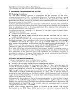
Immunology
Naive T-cell
IL-12
IL-2
IL-2, IFN-gamma
Macrophage activation,
DTH response
IL-4, IL-5, IL-10
Ab response, IgE
Eosinophils
IL-4
Th1
Th2
Examples:
Tuberculoid leprosy Lepromatous leprosy
Cutaneous T-cell lymphoma S
´
ezary syndrome
Psoriasis Atopic dermatitis
Note: Diseases classified as either Th1 or Th2 often have some components
of both but can be classified based on the predominant cytokine profiles
P1: JPJ
0521618134c05 CB1006/Lane 0 521 61813 4 November 28, 2005 14:16
Microbiology and Immunology 67
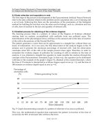
Complement System
Ab complex (lgG or lgM)
activated C1
CLASSICAL
ALTERNATIVE
C4 + C2
C3
C3b
C5
C5b
C5-9 (MAC)
C6-9
C3bBb3b
C3bBbC3b
C3
microbial surface
polysaccharides
factor B
classical C3 convertase
alternative C3 convertase
alternative C5 convertase
classical C5 convertase
factor D
C4b2a
C4b2a3ab
C1
r
classical pathway:
r
activated by IgG or IgM (Ag bound, not soluble)
r
IgM > IgG3 > IgG1 = IgG2 (IgG4 does not bind C1q)
r
alternative pathway: activated without Ab
r
MB lectin pathway: (structural similarity to C1q)
r
binds various pathogens: Candida, Listeria, Neisseria,
Cryptococcus, Salmonella
r
functions of complement proteins:
• cell lysis: C5-C9
• opsonization/phagocytosis: C3bi
• vascular response: C5a > C3a > C4a
• PMN activation: C5a
• immune complex removal: classical pathway, C3b
• B cell activation: C3bi
Deficiencies of early complement associated with autoimmune
disease; late components associated with infection.
P1: JPJ
0521618134c05 CB1006/Lane 0 521 61813 4 November 28, 2005 14:16
68
P1: JZZ
0521618134c06 CB1006/Lane 0 521 61813 4 November 28, 2005 14:26
Clinical Pearls
69
P1: JZZ
0521618134c06 CB1006/Lane 0 521 61813 4 November 28, 2005 14:26
70
P1: JZZ
0521618134c06 CB1006/Lane 0 521 61813 4 November 28, 2005 14:26
Angioedema
C1 INH
Quantitative
C1 INH
Qualitative CH50 C1 C4 C2 C3
HAE
type 1
↓↓ ↓↓ nl/↓ nl ↓↓ ↓ nl
HAE
type 2
nl / ↑↓↓ nl/↓ nl ↓↓ ↓ nl
AAE
type 1
↓↓ ↓↓ ↓ ↓↓
AAE
type 2
nl / ↓↓↓ ↓↓↓↓↓↓↓↓nl
Legend: HAE (hereditary angioedema); AAE (acquired angioedema)
r
subcutaneous edema, upper respiratory/GI tract
involvement
r
no pruritus and no urticaria
r
screening test of choice is C4 (↓from continuous
consumption)
r
HAE type 1 – AD inheritance; ↓ production of normal
C1-INH
r
AAE type 1 associated with lymphoproliferative
diseases
r
AAE type 1 2
◦
anti-idiotypic Ab to monoclonal Ig
synthesized by B lymphocytes; treat with attentuated
androgens
71
P1: JZZ
0521618134c06 CB1006/Lane 0 521 61813 4 November 28, 2005 14:26
72 Pocket Guide for Cutaneous Medicine and Surgery
r
AAE type 2 2
◦
IgG
1
autoantibodies that bind to and
interfere with function of C1-INH; treat with
glucocorticoids
Gigli I and Rosen FS. Angioedema associated with Complement Abnor-
malities. In: Freedberg IM et al., eds. Fitzpatrick’s Dermatology in General
Medicine, 6 ed. New York, NY: McGraw-Hill. 2003:1139–1143.
Odom RB, James WD, Berger TG. Andrews’ Diseases of the Skin, 9th ed.,
Philadelphia: W.B. Saunders, Co. 2000: p. 166.
P1: JZZ
0521618134c06 CB1006/Lane 0 521 61813 4 November 28, 2005 14:26
Clinical Pearls 73
Terminology of Skin Lesions
Primary Skin Lesions
Macule small, flat discoloration
Papule small (<1 cm)circumscribed, solid elevation
Nodule large (1–2 cm)circumscribed, elevation
Tumor large nodule (>2 cm)
Plaque large (>1 cm) flat-topped elevation, often
formed by confluence of papules
Pustule small circumscribed elevation containing
purulent material
Vesicle small (<5 mm) collection of clear fluid
Bulla large (>5 mm) collection of clear fluid
Telangiectasis dilated superficial blood vessel
Wheal irregular edematous plaque
Patch macule with texture change
Secondary Skin Lesions
Scale residual epidermal cells
Crust scab
Erosion focal loss of epidermis
Ulcer focal loss of epidermis and dermis
Fissure linear ulcer/erosion
Excoriation traumatized area (often linear)
(2
◦
scratching)
Lichenification thickening with accentuation of skin lines
(2
◦
rubbing)
P1: JZZ
0521618134c06 CB1006/Lane 0 521 61813 4 November 28, 2005 14:26
74 Pocket Guide for Cutaneous Medicine and Surgery
Nail Terminology
Onycholysis separation of distal nail plate from nail bed
Onychomadesis separation of entire nail plate beginning
proximally
Onychogryphosis overgrowth of nail (“ram’s horn”
appearance)
Onychocryptosis ingrown nail
Onychauxis thick nail
Onychoschizia splitting of nails into layers parallel to
surface
Onychorrhexis longitudinal ridging of nails
Onychomalacia softening of nails
Brachyonychia short, wide nails (“raquet nails” in
Rubenstein-Taybi)
Koilonychia spoon nails (iron deficiency;
Plummer-Vinson; hyperthyroidism;
hemochromotosis)
Platonychia flattened nails
Hapalonychia thinning of nail plate
Trachyonychia rough nails
Beau’s lines horizontal ridges in nail plate (slow matrix
proliferation during acute illness)
Mee’s lines associated with heavy metals and some
chemotherapy
Half and Half nails (Lindsay’s nails) transverse white lines
associated with renal disease (transverse
white line in nail bed)
P1: JZZ
0521618134c06 CB1006/Lane 0 521 61813 4 November 28, 2005 14:26
Clinical Pearls 75
Terry’s nails transverse lines associated with liver
disease (whitening of nail bed)
Muehrckes’ nails pale bands on nailbed associated with
hypoalbuminemia
“Shoreline” nails drug-induced exfoliative dermatitis
(alternating bands of nail plate
discontinuity and leukonychia)
Bilobed nails only few reported cases
Yellow nails: yellow nail syndrome, Candida,
carotenemia, MTX, AZT
Blue nails: Wilson’s disease, argyria, AZT, HIV,
antimalarials, busulfan
Red lunulae: carbon monoxide, CV disease, lupus,
alopecia areata
Nail pitting: psoriasis vulgaris, alopecia areata
Psoriasis: nail pitting, oil spots, onycholysis
P1: JZZ
0521618134c06 CB1006/Lane 0 521 61813 4 November 28, 2005 14:26
76 Pocket Guide for Cutaneous Medicine and Surgery
Pediatric Dermatology
Rubeola (Measles)
r
paramyxovirus
r
8–12 days post-exposure (no signs)
r
prodrome: malaise, fever, cough, coryza, conjunctivitis;
Koplik spots in 2–3 days after onset of symptoms
r
erythematous maculopapular rash ∼5days after onset of
symptoms (cephalocaudal progression)
r
atypical measles (individual vaccinated with killed
vaccine)
Rubella (German measles)
r
rubella virus (RNA togavirus)
r
no prodrome during incubation (14–21 days)
r
erythematous, maculopapular, discrete rash (starts on face
and spreads to body over 24
◦
;resolves by day 3)
r
lymphadenopathy (posterior cervical and suboccipital)
r
ocular pain with upward and lateral gaze characteristic
r
fever may accompany onset of erythema
r
Forscheimer’s spots – pinpoint rose-colored
macules/petechia on soft palate
Roseola
r
HHV6(>HHV7)
r
abrupt fever days 3–5 (appears well)
r
maculopapular rash on 3rd day (centrifugal) as fever
deferresces; leukocytosis
r
rash evolves in 12
◦
and lasts 1–2 days
r
95% are 6 months to 2 years of age
r
Berliner’s sign – palpebral edema
r
spread via oropharyngeal secretion
P1: JZZ
0521618134c06 CB1006/Lane 0 521 61813 4 November 28, 2005 14:26
Clinical Pearls 77
Erythema infectiosum (Fifth’s disease)
r
parvovirus B-19 (ssDNA)
r
prodrome consists of fever, HA, pharyngitis, malaise
r
slapped cheek appearance
r
erythematous, reticulated, pruritic, macular rash
(arms → trunk, legs) (reticulated hyperpigmentation)
r
aplastic crisis in patients with hemoglobinopathies
r
acute arthropathy in adults (and papular gloves and
stockings)
r
risk of hydrops fetalis and spontaneous abortion
Hand-foot-mouth disease
r
coxsackie A16 virus; enterovirus 71
r
prodrome of fever, anorexia, oral pain followed by oral
mucosal ulcers and erythematous patches and vesicles on
hands, feet, and buttocks
Varicella Zoster Virus (VZV)
r
incubation: 10–21 days
r
absent or mild prodrome
r
vesicles in varying stages of development (cephalocaudal)
r
immunocompromised children with VZV are given VZIG
within 96
◦
of exposure
r
acyclovir reserved for immunocompromised with
disseminated varicella
r
contagious from 24
◦
before onset of rash until all lesions
are crusted over
P1: JZZ
0521618134c06 CB1006/Lane 0 521 61813 4 November 28, 2005 14:26
78 Pocket Guide for Cutaneous Medicine and Surgery
Kawasaki disease (Mucocutaneous lymph node
syndrome)
r
systemic vasculitis of unknown etiology
r
characteristic features:
r
fever of unknown origin for >5days
r
acral/perineal erythema/desquamation
r
cervical nonsuppurative lymphadenopathy
r
edema/desquamation of hands and feet
r
conjunctivitis
r
strawberry tongue
r
3 phases:
r
acute: lasts 1–2 weeks
r
subacute: begins when fever, rash, LAD resolve;
marked by desquamation and thrombocytosis;
risk of arthritis, coronary aneurysms
r
convalescent: 6–8 weeks after onset; ESR normal
r
treat with aspirin and IVIG
Scarlet fever
r
usually associated with streptococcal pharyngitis
r
erythrogenic toxins B and C most commonly seen
r
highest incidence in children 2–10 (can occur in adults)
r
fever, malaise, pharyngitis → exanthem 48
◦
later (neck
spreading down) → pinpoint papules (sandpaper feel;
often spares palms and soles); circumoral pallor;
accentutation in skin folds (Pastia’s lines) → lasts
∼5days → desquamates (often in sheets)
r
enanthem: pharyngitis, palatal petechia, white strawberry
tongue → red strawberry tongue
P1: JZZ
0521618134c06 CB1006/Lane 0 521 61813 4 November 28, 2005 14:26
Clinical Pearls 79
Varicella Zoster Virus and Pregnancy
r
maternal VZV infection within first 20 weeks gestation
may result in congenital varicella syndrome
r
VZIG should not be given once mother has developed
varicella
r
VZIG should be given for significant exposures within first
72–96 hours (use limited to seronegative women)
r
if mother develops varicella 5 days before or 2 days after
delivery → administration of VZIG is warrranted
(consider iv acyclovir therapy)
P1: JZZ
0521618134c06 CB1006/Lane 0 521 61813 4 November 28, 2005 14:26
80 Pocket Guide for Cutaneous Medicine and Surgery
Diagnosis of Systemic Lupus Erythematosus
Requires 4 of 11 for diagnosis:
Malar erythema (tends to spare nasolabial folds)
Discoid lupus erythematosus
Photosensitivity (patient history or examination)
Oral ulcers (oral/nasopharyngeal ulceration; usually painless)
Arthritis (nonerosive) involving ≥2 peripheral joints
(characterized by tenderness, swelling or effusion)
Serositis (pericarditis or pleuritis)
Nephropathy
persistent proteinuria >0.5 g/d or 3+ (or)
cellular casts (red cell, hemoglobin, granular, tubular, mixed)
Neurologic disorder(seizures/psychosis in absence of drugs or
metabolic derangements)
Hematologic disorder
r
hemolytic anemia with reticulocytosis or
r
leukopenia <4000/mm
3
on 2 occasions or
r
lymphopenia <1500/mm
3
on 2 occasions or
r
thrombocytopenia <100,000/mm
3
Immunologic disorder (+LE-prep; anti-DNA Ab or Sm Ag or
false + for syphilis known to be + for ≥6 months)
Antinuclear antibody
Ta n EM, Cohen AS, Fries JF, Masi AT, McShane DJ, Rothfield NF et al. The
1982 revised criteria for the classification of systemic lupus erythematosus.
Arthritis and Rheumatism 1982; 25:1271–1277.
P1: JZZ
0521618134c06 CB1006/Lane 0 521 61813 4 November 28, 2005 14:26
Clinical Pearls 81
Useful Laboratory Tests in Evaluation of SLE
Complete blood count anemia, leukopenia, thrombocytopenia
Differential check for lymphopenia
ESR usually elevated (but nonspecific)
Creatinine ± elevated with renal involvement
Urinalysis check for proteinuria, hematuria, casts
RPR/VDRL false-positive test may occur with SLE
ANA 95% with SLE (use Hep-2 cell line)
dsDNA increased risk of renal disease
ssDNA sensitive but not specific
Sm highest specificity for SLE
nRNP decreased risk of renal disease
C3/C4 decreased with active disease
antiphospholipid Ab may occur with SLE
anti-histone Ab drug-induced lupus
Koopman WJ, Boulware DW, Heudebert GR. Clinical Primer of Rheumatol-
ogy. Philadelphia: Lippincott Williams and Wilkins. 2003: p. 167.
P1: JZZ
0521618134c06 CB1006/Lane 0 521 61813 4 November 28, 2005 14:26
82 Pocket Guide for Cutaneous Medicine and Surgery
Antinuclear antibodies
Pattern Target Antibody Disorder
Homogeneous Chromatin anti-dsDNA SLE
anti-dsDNA Drug-induced LE
anti-histone
Peripheral Chromatin anti-DNA SLE
Nuclear mem anti-laminin
Speckled/ fine Nuclear RNP anti-Sm SLE (nephritis)
anti-Ro/SSA SCLE,Sjögren’s
anti-La/SSB Sjögren’s
anti-U1RNP SLE, MCTD
Chromatin anti-Ku SLE, scleroderma
anti-SCl-70 Scleroderma
Speckle/discrete Chromatin anti-centromere CREST
Nucleolar Nuclear RNP anti-U3RNP Scleroderma
Nucleolar comp anti-RNA Pol I
anti-Pm-SCl
Jaworsky C. Connective tissue diseases. In: Elder D et al. Lever’s Histopathol-
ogy of the Skin. Philadelphia: Lippincott-Raven. 1997: p. 267.
P1: JZZ
0521618134c06 CB1006/Lane 0 521 61813 4 November 28, 2005 14:26
Clinical Pearls 83
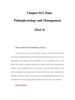
Porphyrias
PORPHYRIAS
Glycine + Succinyl CoA
Pyridoxal phosphate and ALA synthase
Aminolevulinic acid
Hydroxymethylbilane
CEP
Spontaneous
Uroporphyrinogen III
Co Synthase
Uroporphyrinogen III
Uroporphyrinogen I
Urorporphyrinogen
decardoxylase
Coprophyrinogen III
Coprophyrinogen
oxidase
oxidase
Coprophyrinogen
Harderoporphyrinogen
Harderoporphyria
Protoporphyrinogen
Protoporphyrinogen
Protoporphyrin
Ferrochelatase
Heme
oxidase
VP
EPP
Coprophyrinogen I
PCT/HEP
HCP
ALA dehydratase deficiency
ALA dehydratase
Porphobilinogen
Porphobilinogen deaminase
AIP