Clinical Pancreatology for Practising Gastroenterologists and Surgeons - part 8 potx
Bạn đang xem bản rút gọn của tài liệu. Xem và tải ngay bản đầy đủ của tài liệu tại đây (618.51 KB, 56 trang )
anaplastic cancers parallels the advanced loss of cellu-
lar differentiation in cultured islets, where the cells lose
every known islet-cell marker.
Clinical studies supporting the role of
islets in pancreatic carcinogenesis
Although it has been known for almost a century that
nearly 80% of patients with pancreatic cancer have im-
paired glucose metabolism, either frank diabetes or
impaired glucose tolerance (IGT), the reason has re-
mained a mystery. Remarkably, the degree of IGT and
diabetes in these patients has been known to vary. Some
patients require insulin treatment whereas others do
not. Fasting serum glucose levels may be in the normal
range, but an oral glucose tolerance test may yield a di-
agnosis of IGT. On the other hand, a small subset of pa-
tients shows normal glucose metabolism. It is possible
that differences in the patient population are the reason
that morphologic and molecular biological approaches
have not provided clues for understanding the biology
of this dismal disease.
The association between diabetes and pancreatic
cancer has remained a matter of controversy. Accord-
ing to recent studies, IGT or diabetes mellitus develops
shortly before the clinical manifestations of the disease
or is diagnosed at the first clinical admission. There are,
however, a few who believe that diabetes is a predispos-
ing factor, especially in cases where diabetes is present
for more than 5 years before the diagnosis of cancer.
Since the latency of pancreatic cancer is unclear, and the
development of some cancers seems to take as long as
10 years, the role of diabetes as a predisposing factor
remains questionable. Consequently, it appears that
the development of pancreatic cancer is associated with
the abnormality in islet cell function. Some suggested
mechanisms include the primary alteration of islet cells
by the carcinogen or secondary damage by cancer cells,
either directly or via the production of substances that
affect islet cell function.
Experimental studies in the hamster model described
above and anecdotal observations indicate that islet
cells may also play a role in pancreatic carcinogenesis in
humans. This is highlighted by the development of
altered glucose metabolism in small tumors that are
located in the periphery of the pancreas but which do
not cause chronic pancreatitis and in the very early
stages of cancer development. The involvement of islet
cells in pancreatic carcinogenesis explains at least some
of the clinical observations.
Based on the published data and our experience,
glucose intolerance is a pancreatic cancer-associated
symptom as well as the result of the primary alteration
of islet-cell function and differentiation in response
to causative carcinogens. Experimental studies have
shown that glucose intolerance coincides with the first
appearance of microscopic pancreatic tumors. Studies
have also shown that changes in islet hormones accom-
PART III
370
Figure 44.5 An enlarged islet in a patient with pancreatic
cancer. Malignant glands have replaced most islet cells
without any signs of islet cell destruction, including invasion
of the islet cells and the surrounding tissue. In some areas
intact islet cells are present within the malignant epithelium
(e.g., lower right area). Anti-insulin antibody, ABC
(avidin–biotin complex) method, ¥ 120.
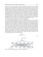
Figure 44.6 Papillary projection of a well-differentiated
pancreatic adenocarcinoma. Numerous insulin-producing
cells are seen at the base and within the papillary fold. Anti-
insulin, ABC (avidin–biotin complex) method, ¥ 120.
pany the early development of pancreatic cancers.
These hormone changes and insulin resistance resem-
ble the metabolic changes in pancreatic cancer patients.
In humans, IGT or diabetes has been noticed in small,
localized, and early pancreatic cancer. In Japanese
patients, IGT was the only abnormality in patients with
small pancreatic cancer.
The overwhelming opinion that pancreatic cancer
develops from ductal epithelium might be the reason
why islet-cell alterations in pancreatic cancer patients
have not been the focus of research. When we studied
the pattern of islet cells immunohistochemically in 14
pancreatic cancer specimens, 14 chronic pancreatitis
samples, and 10 normal pancreata as controls, we
found that 10 of 14 cancer specimens showed a signifi-
cant loss of islet b cells. Of the 10 cases, IGT was con-
firmed in four but no information was available about
glucose metabolism in the remaining cases. The inci-
dence of islet-cell alterations in our material (72%)
correlates with the frequency of abnormal glucose
metabolism in pancreatic cancer patients. Remarkably,
most altered islet cells were in the vicinity of the cancer.
In only one case was the abnormality also found in
an area remote from the cancer. Since tumor-free pan-
creatic tissues were available in only five cases, the
frequency of islet cell alteration in the teletumoral area
could not be determined. Other noteworthy findings
associated with this abnormality were the signs of al-
tered islet cell differentiation, including the formation
of intrainsular ductular structures and the expression
of tumor-associated antigens CA-19-9, TAG-72, and/
or DU-PAN-2 in islet cells and intrainsular ductular
cells (see Fig. 44.3). This finding indicates that in these
patients islet cells have the ability to form an abnormal
cell population.
Possible mechanism of altered glucose
metabolism in pancreatic cancer
It has been proposed that amylin, a peptide with a
molecular weight of 2030, or other yet unknown sub-
stances released from cancer cells are responsible for
the development of IGT. Because we believe that most
cancers arise from altered islet cells, the production of
these substances from cancer cells is self-explanatory.
Cancer cells are known to inherit some of the biological
properties of the cells from which they are derived. In-
deed, several studies show the expression of neuroen-
docrine markers in pancreatic cancer cells. From a
pathophysiologic point of view, the production of dia-
betogenic material from islet cells appears more plausi-
ble, as it is well known that islet cells have the potential
to produce many different pancreatic and extrapancre-
atic peptides simultaneously. They also have the ability
to shift from synthesis of one hormone to synthesis of
another. A good example is the coproduction and core-
lease of insulin and amylin, the synchrony of which is
altered in pancreatic cancer. The improvement in IGT
and diabetes after tumor resection (70% pancreatecto-
my or curative resection) by no means indicates that it
was the tumor that produced the diabetogenic sub-
stances, because removal of cancer tissue also removes
the altered islet cells that may actually have produced
the diabetogenic material. Moreover, we must be aware
that nearly all well-differentiated pancreatic cancers
contain endocrine cells, sometimes in remarkably high
numbers (Fig. 44.7), which could also be the source of
altered hormone production and which are also re-
moved with the cancer. For example, although tumor
extracts from diabetic patients with pancreatic cancer
showed a marked reduction of glycogen synthesis in
skeletal muscles, examination of the tumor revealed
that tumor tissue contained islet hormones. Although
from a clinical standpoint the issue of whether the
diabetogenic material is produced by cancer cells or al-
tered islet cells is trivial, elucidation of the mechanism is
CHAPTER 44
371
(a)
(b)
Figure 44.7 Presence of a large number of islet cells within
the malignant epithelium. (a) Many b cells are incorporated
within the glandular structures. Anti-insulin antibody, ABC
method, ¥ 25. (b) Malignant glandular structures containing
more endocrine than cancer cells. Multilabeling technique,
¥ 120.
crucial to understanding the biology of the disease
and in planning future diagnostic and therapeutic
modalities.
Differences in the clinical expression of
pancreatic cancer
Although it appears that alteration of glucose metabo-
lism can provide a diagnostic marker, some observa-
tions complicate the issue. According to clinical
observations, only 60–70% of patients develop IGT
or diabetes and the minority (30–40%) do not.
Although IGT improves after surgery in many patients,
in some it does not or it gets even worse. There are
conflicting reports and inadequate information on the
incidence of peripheral insulin resistance, IGT, and
diabetes before and after surgery. According to one
study, 59% of pancreatic cancer patients with either
diabetes (45%) or IGT (14%) show improvement
after curative surgery, whereas studies by Permert
et al., using a hyperglycemic clamp method, show
normalization of IGT and improvement of diabetes in
around 60% of patients. Consequently, it can be as-
sumed that 10–40% of pancreatic cancer patients ei-
ther do not show any improvement of the abnormality
after surgery or IGT becomes worse. The latter figures
could be even higher if one considers that postoperative
improvement of IGT and diabetes could be due to the
postoperative physical condition and dietary regimens
of the patients rather than the consequence of tumor re-
moval. It is unclear whether the observed improvement
is just temporary or if the abnormality reappears at the
time of tumor recurrence. Although many reasons
could be responsible for the lack of postoperative
improvement of glucose metabolism in the subset of
patients, it is highly possible that altered islet cells
producing diabetogenic substances exist in a teletu-
moral area not removed by surgery or some hidden
(metastatic) tumors are left behind, for example in
the liver.
Since in a follow-up study glucose homeostasis in-
creasingly worsened in patients who did not have cura-
tive surgery, the extent of the tumor and/or altered islets
seems to be responsible for glucose metabolism. There
are, as yet, no studies examining the extent of islet cell
alteration within, around, and remote from cancer.
Also, there are limited follow-up studies of patients
after surgery.
Possible etiologic factors for islet-cell
alteration in pancreatic cancer
The results of our 30 years of experience in human and
experimental pancreatic cancer has led us to believe
that islet cells are the primary targets of carcinogens. In
our view, all pancreatic tumors, endocrine or exocrine,
are derived from islets. The structure of the carcinogen
determines the phenotypic expression of the ensuing
tumors. Streptozotocin, a nitrosamide, produces
islet-cell tumors, whereas BOP, a nitrosamine, induces
a ductal type of tumor. In hamsters and humans,
cultured islet cells transdifferentiate into ductal cells. In
hamsters, BOP treatment of isolated purified islets
leads to tumor cells that grow in vivo as ductal adeno-
carcinoma. When we treat cultured human ductal and
islet cells with BOP, only the treated islet cells are
able to grow in a serum-free medium and show K-ras
mutation, a marker for pancreatic cancer (unpublished
results). In an ongoing study we are following the
characteristics of these cells and expect their malignant
transformation.
The most convincing support for our view is the
finding that all drug-metabolizing enzymes, which
are believed to be involved in the metabolism of
environmental carcinogens, including tobacco-specific
carcinogen, nitrosamines, polycyclic aromatic com-
pounds, and aromatic amines, are primarily or
exclusively expressed in the islet cells of humans and
laboratory species. Considering the anatomy of the
blood supply of the pancreas, where a major portion of
the arterial blood goes to islets before nourishing the
exocrine pancreas, the presence of drug-metabolizing
enzymes in islet cells is understandable. Hence, islet
cells seem to play the role of pancreatic filters. The
availability of these enzymes makes islet cells the pri-
mary target of blood-borne carcinogens. Because most
of these enzymes are present in a higher concentration
or exclusively in islet cells in the head of the pancreas,
the frequent occurrence of pancreatic cancer in the
head may be explained. Carcinogen-induced alter-
ations in the islets in teletumoral regions of the pan-
creas could be the reason for the altered production of
hormones and, hence, the maintenance of IGT after
tumor removal. This explanation, however, is not
conclusive because not all pancreatic cancer patients
develop a glucose metabolic abnormality. Is this related
to the different biology of cancer, as has been suggested
by a study where a correlation was found between the
PART III
372
degree of IGT severity and the histologic type of can-
cer? Is this because tumors develop from islets in pa-
tients with IGT or diabetes and, in a minority of the
patients, from other cells? Or could this be related to
the severity and extent of islet-cell damage? Neverthe-
less, the data suggest that, with regard to the glucose
metabolic alteration, there are at least three subsets of
pancreatic cancer patients, possibly with tumors of
different biology. The published data and our own
experience suggest the following subsets (Fig. 44.4):
1 pancreatic cancer patients without IGT or diabetes
(IGT–, about 20–30%);
2 pancreatic cancer patients with IGT or diabetes
(IGT+, about 70–80%), whose glucose intolerance or
diabetes improves postoperatively (IGT+/–);
3 patients in whom the abnormality does not or only
slightly improves (IGT+/+) after tumor resection.
Possible mechanism of differing clinical
presentation of pancreatic cancer
Reasons for the glucose metabolic abnormality in pan-
creatic cancer are not well understood. The suggestion
that islet-cell destruction by cancer cells is the principal
cause has been refuted, mainly because even small and
localized tumors in the head of the pancreas are associ-
ated with abnormal glucose tolerance.
A few studies dealing with the alteration of islet hor-
mones at the tissue level have found a reduction in
the number of b cells in pancreatic cancer patients. No
information is available on the frequency and extent of
the process, and its specificity for pancreatic cancer.
The question of specificity is important because about
45–75% of patients with chronic pancreatitis also
develop abnormal glucose tolerance or frank diabetes
mellitus. Consequently, it is reasonable to assume that
damage to the islets by scar tissue and inflammation,
which are also associated with pancreatic cancer, could
be the underlying mechanism.
Clearly, disturbance of the subtle balance between
exocrine and endocrine tissue by cancer, with asso-
ciated inflammation and sclerosis, is expected to lead to
deregulation of hormone secretion. However, because
even localized and small tumors in the head of the pan-
creas not affecting islet-rich areas of the organ cause the
glucose metabolic abnormality, it is likely that a factor
or factors produced by cancer cells play a role. This
view is supported by the finding that surgical removal
of tumor by 85–90% pancreatectomy improves dia-
betes and normalizes glucose metabolism.
Several clinical studies have shown significant
changes in the serum levels of islet hormones in pancre-
atic cancer patients, but little is known about the pat-
terns of islets at the tissue level. In one study a reduction
in b cells has been reported but the extent of the alter-
ations and their specificity for pancreatic cancer have
not been investigated. The latter issue deserves par-
ticular attention, because glucose-abnormality and
diabetes also occurs in chronic pancreatitis. Therefore,
we systematically examined the patterns of islets in
pancreatic cancer in comparison with chronic pancre-
atitis and the normal pancreas. We selected archival
pancreatic cancer specimens that had tumor-free areas
close to and remote from the cancer because, as stated
earlier, it is believed that factors released by cancer cells
affect the islets directly via a paracrine pathway.
In 10 pancreatic cancer specimens, a significant
reduction in b cells was found. Also, in eight of them, a
significant increase in a cells was found as well. This re-
sult thus correlates with the incidence of pathologic
serologic hormone levels in pancreatic cancer patients.
Reasons for the lack of similar alterations in the four
other patients are obscure. We could not find any corre-
lation between islet alterations, sex, age, smoking
habit, alcohol consumption, stage of the disease, and
tumor morphology. We also did not find any significant
changes in the islet-cell distribution in the chronic pan-
creatitis specimens, even within sclerotic and fibrotic
tissue. Therefore, the suggestion that fibrosis or sclero-
sis associated with pancreatic cancer may have caused
the b-cell loss by obstructing blood vessels could be
excluded. Consequently, the described islet alteration
appears to be specific for pancreatic cancer. Because
pathologic islet hormone serum levels also occur in
chronic pancreatitis patients, it seems that the mecha-
nism of altered glucose metabolism in the two diseases
differs. In chronic pancreatitis the abnormality seems
to be due to altered insulin secretion, whereas in pan-
creatic cancer the defect appears to be in the machinery
of insulin synthesis as evidenced by the reduced levels of
insulin and C-peptide as well as of amylin, which is nor-
mally costored and cosecreted with insulin. Endocrine
cells were found in the malignant epithelium in nine of
our ten cases with b-cell alteration. Similar findings
have been reported in up to 80% of cases. Also, the
presence of nesidioblastosis in four of our ten cases
with decreased b-cell number could reflect a compen-
CHAPTER 44
373
satory process against b-cell loss. The question of why
b cells in pancreatic cancer are exclusively affected
remains to be investigated.
The increase in a cells, which was more pronounced
in cancer tissue from diabetics than in tissue from dia-
betics without cancer, coincides with the serologic
findings. The abnormality also differs from hormonal
changes in chronic pancreatitis, where serum concen-
trations of glucagon have been found to be reduced or
normal. Nevertheless, in our chronic pancreatitis sam-
ples we could not detect any alteration in the number
of glucagon cells, possibly because it is the secretion of
glucagon that is affected not the number of a cells.
Also, contrary to clinical observations of increased so-
matostatin levels in pancreatic cancer patients, we
could not find any significant changes in the number of
somatostatin cells. Whether the source of increased
serum somatostatin is derived from pancreatic or ex-
trapancreatic somatostatin cells remains to be seen.
The greater alteration of islets within or immediately
around cancer supports the hypothesis that factors re-
leased by cancer cells play a role in this process. Because
alterations like hydropic swelling were also found in
tissues remote from cancer, although in lesser degree, a
humoral pathway also seems to exist. Examination of
pancreatic tissue further away from the cancer would
clarify this. If this is found to be the case, the identifica-
tion of the causative factor(s) released from cancer cells
could present an early pancreatic cancer marker, espe-
cially in view of the findings that an abnormality in glu-
cose metabolism also occurs in small localized cancers.
One of the reasons for the differing results in the pub-
lished data and pancreatic hormone levels in pancreat-
ic cancer patients could be the inclusion of different
subsets of patients with pancreatic cancer in these
studies. It may be that the anatomic location of the
tumor plays a role in these differences. Tumors in the
head region obstructing the main pancreatic duct can
cause severe (secondary) chronic pancreatitis and,
hence, diabetes. However, there are differences in dia-
betes induced by chronic pancreatitis and pancreatic
cancer. For example, diabetes improves after a 70%
pancreatectomy in pancreatic cancer but not after sur-
gical intervention in chronic pancreatitis. According to
our recent studies, the size and cell constitution of islets
are significantly different between primary pancreatitis
and pancreatitis caused by cancer. Contrary to the islets
in pancreatic cancer patients, which are of normal size
or enlarged, about 95% of the islets in primary chronic
pancreatitis measure less than 100 mm in diameter.
Moreover, tumors developing in the upper and dorsal
half of the head of the pancreas do not affect the pan-
creatic duct very much and hence are not accompanied
by a significant chronic pancreatitis. Whether these
tumors cause diabetes is unclear. Another argument
against the role of secondary pancreatitis in the induc-
tion of diabetes derives from the experience that even
patients with small tumors in the periphery of the pan-
creas not causing chronic pancreatitis show abnormal
glucose tolerance.
Another major shortcoming of past studies is the lack
of adequate control groups. There is not a single study
that correlates the morphologic findings of both cancer
and islet cells with plasma hormone levels of the pa-
tients. To our knowledge, there is only one limited
study that compares the hormone levels in pancreatic
cancer patients with that of healthy and noninsulin-
requiring diabetic persons: a low level of plasma amylin
was found in patients with type II diabetes and in those
with pancreatic cancer and diabetes, and an increased
level of amylin in pancreatic cancer patients without
diabetes. Such correlative studies could provide impor-
tant data for an understanding of the disease. For
example, a low insulin level in a patient with IGT and
altered islet cells could reflect impaired insulin release
or synthesis in the altered islets. In fact, an inverse rela-
tionship has been found between the number of insulin
cells in islets and the fasting plasma glucose level, sug-
gesting that the alteration of islet cells is the primary
cause of the glucose abnormality in these patients.
Other studies also point to the primary alterations of
islet cells, including reduced insulin and C-peptide re-
sponse after glucose load and an increase in proinsulin
secretion. The Sproinsulin/SC-peptide ratio, which has
been found to be increased in pancreatic cancer with
IGT but decreased after tumor removal, further sub-
stantiates the functional alteration of islet cells.
From a therapeutic point of view, the identification
of a different subpopulation of pancreatic cancer
patients is important because these patients could
respond differently to therapeutic modalities. For
example, does the genetic constitution of the tumors
from the different pancreatic cancer subpopulations
differ? Can the pattern of IGT help to better distinguish
between sporadic and familial pancreatic cancer? If
IGT is due to the substances released from islet cells or
cancer cells, then is it expected that IGT+/– patients
become IGT+/+ after tumor recurrence? If that is the
PART III
374
case, the recurrence of the abnormality, and possibly its
severity, could have predictive value. The existing re-
sults indicate that the occurrence of diabetes in pancre-
atic cancer cannot be explained by a single mechanism.
The increase in peripheral insulin resistance, suppres-
sion of insulin secretion, impaired proinsulin con-
version, altered fat and carbohydrate metabolism,
presence of acute or chronic pancreatitis, medications
for underlying disease, altered nutritional habits,
weight loss, and many other factors seem to play im-
portant roles in the development and course of pancre-
atic cancer.
Conclusion
Past attempts to develop early diagnostic modalities
have proven useless. The expression of tumor-associat-
ed antigens may have value in monitoring the disease
but are unable to detect the cancer in early developmen-
tal stages. Despite the promising molecular biological
approach, the method lacks specificity as the K-ras mu-
tation is not specific for pancreatic cancer and can be
found in patients with chronic pancreatitis as well as in
individuals without pancreatic diseases. The most so-
phisticated imaging techniques are still unable to detect
tumors less than 5 cm with accuracy. The frequent asso-
ciation between pancreatic cancer and IGT offers the
most logical approach for detecting small tumors in
most patients. This method could be applied readily
in individuals prone to pancreatic cancer, including
members of pancreatic cancer and hereditary chronic
pancreatitis. Our studies pointing to the role of islets in
pancreatic cancer and in the development of altered
glucose metabolism should be further investigated. The
development of a multidisciplinary program involving
researchers in various fields of medicine, toxicology,
nutrition, cellular and molecular biology, and epidemi-
ology is a necessary step in revealing the true nature of
this deadly disease.
Recommended reading
Ahren B, Andren-Sandberg A. Glucose tolerance and insulin
secretion in experimental pancreatic cancer in the Syrian
hamster. Res Exp Med 1993;193:21–26.
Bonner-Weir S, Baxter LA, Schuppin GT, Smith FE. A second
pathway for regeneration of adult exocrine and endocrine
pancreas. A possible recapitulation of embryonic develop-
ment. Diabetes 1993;42:1715–1720.
Bouwens L. Transdifferentiation versus stem cell hypothesis
for the regeneration of islet beta-cells in the pancreas.
Microsc Res Tech 1998;43:332–336.
Bouwens L, Kloppel G. Islet cell neogenesis in the pancreas.
Virchows Arch 1996;427:553–560.
Cersosimo E, Pisters PW, Pesola G, McDermott K, Bajorunas
D, Brennan MF. Insulin secretion and action in patients with
pancreatic cancer. Cancer 1991;67:486–493.
Gittes GK, Galante PE, Hanahan D, Rutter WJ, Debase HT.
Lineage-specific morphogenesis in the developing pancreas:
role of mesenchymal factors. Development 1996;122:439–
447.
Gullo L, Ancona D, Pezzilli R, Casadei R, Campione O. Glu-
cose tolerance and insulin secretion in pancreatic cancer.
Ital J Gastroenterol 1993;25:487–489.
Jonsson J, Carlsson L, Edlund T, Edlund H. Insulin-promoter-
factor 1 is required for pancreas development in mice.
Nature 1994;371:606–609.
Kimura W, Morikane K, Esaki Y, Chan WC, Pour PM. Histo-
logical and biological patterns of microscopic ductal
adenocarcinomas detected incidentally at autopsy. Cancer
1998;82:1839–1849.
Muscarella P, Knobloch TJ, Ulrich AB et al. Identification and
sequencing of the Syrian golden hamster (Mesocricetus
auratus) p16(INK4a) and p15(INK4b) cDNAs and their
homozygous gene deletion in cheek pouch and pancreatic
tumor cells. Gene 2001;278:235–243.
Ordonez NG, Balsaver AM, Mackay B. Mucinous islet cell
(amphicrine) carcinoma of the pancreas associated with
watery diarrhea and hypokalemia syndrome. Hum Pathol
1988;19:1458–1461.
Permert J, Ihse I, Jorfeldt L, von Schenck H, Arnquist HJ,
Larsson J. Improved glucose metabolism after subtotal
pancreatectomy for pancreatic cancer. Br J Surg 1993;80:
1047–1050.
Permert J, Ihse I, Jorfeldt L, von Schenck H, Arnqvist HJ,
Larsson J. Pancreatic cancer is associated with impaired
glucose metabolism. Eur J Surg 1993;159:101–107.
Permert J, Larsson J, Westermark GT et al. Islet amyloid
polypeptide in patients with pancreatic cancer and diabetes.
N Engl J Med 1994;330:313–318.
Pour PM, Kazakoff K, Carlson K. Inhibition of
streptozotocin-induced islet cell tumors and N-
nitrosobis(2-oxopropyl)amine-induced pancreatic ex-
ocrine tumors in Syrian hamsters by exogenous insulin.
Cancer Res 1990;50:1634–1639.
Pour PM, Weide L, Liu G et al. Experimental evidence for the
origin of ductal-type adenocarcinoma from the islets of
Langerhans. Am J Pathol 1997;150:2167–2180.
Pour PM, Schmied BM, Ulrich AB, Friess H, Andren-
Sandberg A, Buchler MW. Abnormal differentiation of
CHAPTER 44
375
islet cells in pancreatic cancer. Pancreatology 2000;1:110–
116.
Pour PM, Standop J, Batra SK. Are islet cells the gatekeepers of
the pancreas? Pancreatology 2002;2:440–448.
Rosenberg L, Rafaeloff R, Clas D et al. Induction of islet cell
differentiation and new islet formation in the hamster:
further support for a ductular origin. Pancreas1996;13:38–
46.
Schmied B, Liu G, Moyer MP et al. Induction of adenocarci-
noma from hamster pancreatic islet cells treated with N-
nitrosobis(2-oxopropyl)amine in vitro. Carcinogenesis
1999;20:317–324.
Schmied BM, Liu G, Matsuzaki H et al. Differentiation of
islet cells in long-term culture. Pancreas 2000;20:337–
347.
Standop J, Schneider MB, Ulrich A et al. The pattern of
xenobiotic-metabolising enzymes in the human pancreas.
J Toxicol Environ Health 2002, in press.
Ulrich AB, Schmied BM, Matsuzaki H et al. Increased expres-
sion of glutathione S-transferase-pi in the islets of patients
with primary chronic pancreatitis but not secondary chron-
ic pancreatitis. Pancreas 2001;22:388–394.
Yuan S, Rosenberg L, Paraskevas S, Agapitos D, Duguid WP.
Transdifferentiation of human islets to pancreatic ductal
cells in collagen matrix culture. Differentiation 1996;61:
67–75.
PART III
376
The dismal prognosis of pancreatic cancer is mostly due
to the fact that this tumor is usually diagnosed at a late
stage. There are no specific early symptoms and diag-
nostic imaging has limitations. As a result, the disease
often eludes detection during its formative stages.
Therefore, accurate tools for early diagnosis and
screening are particularly important for this tumor. We
also need markers that allow estimation of prognosis,
disease progression, and treatment response and which
help us to select the optimum therapeutic strategy for a
patient.
Alterations in gene sequences, expression levels,
and protein structure or function are used as tumor
markers. This field is fast-moving and expanding, but
also littered with numerous examples of might-have-
beens. Very few markers have passed successfully from
the bench to the bedside. In this chapter we highlight
the present state of tumor markers in pancreatic cancer
and point out developments that may lead to a diagnos-
tic breakthrough in the near future. Because 80–90% of
tumors of the exocrine pancreas are adenocarcinomas
of ductal cell origin, we focus on markers for ductal
pancreatic adenocarcinoma.
CA-19-9
CA-19-9 is the most frequently used serum-based
marker for pancreatic cancer. The protein is a
carbohydrate cell-surface antigen (sialylated lacto-N-
fucopentose) related to the Lewis blood group sub-
stance. It was originally isolated in 1979 as a colorectal
cancer-specific antigen and it is found in the normal ep-
ithelial cells of the gallbladder, biliary ducts, pancreas,
and stomach. The elevation of CA-19-9 in pancreatic
and other malignancies is thought to be due to in-
creased production and secretion of the antigen from
malignant cells. Multiple studies have shown that while
elevations in serum CA-19-9 appear useful in the diag-
nosis of adenocarcinoma of the upper gastrointestinal
tract and in the surveillance of colon cancer, its greatest
sensitivity is in the detection of pancreatic adenocarci-
noma. To date, CA-19-9 is considered one of the most
useful tumor markers for pancreatic malignancies.
However, up to 30% of patients with pancreatic cancer
do not exhibit elevated serum CA-19-9 levels. The sen-
sitivity of the CA-19-9 serum assay ranges between 69
and 93%, the specificity between 46 and 98%. The
higher the level of CA-19-9, the greater the sensitivity
and specificity of the assay. Elevations in CA-19-9 cor-
relate with the degree of tumor differentiation and with
the extent of disease. Consequently, CA-19-9 levels are
lower in patients with localized disease and this marker
is therefore of little use as a screening marker to detect
early pancreatic cancers. It has been suggested that very
high levels of CA-19-9 indicate unresectable tumors
and that the pretreatment CA-19-9 level is a strong pre-
dictor of survival. There are conflicting results about
whether the response of CA-19-9 to chemotherapy
and/or radiotherapy is useful for predicting survival.
In addition, CA-19-9 is a useful marker for detecting
recurrent disease and can therefore be used for the
surveillance of patients after surgery for pancreatic
cancer.
A general clinical problem is to determine whether a
pancreatic mass is due to malignancy or chronic pan-
creatitis. Furthermore, if chronic pancreatitis is estab-
lished, it is important to know whether there is any sign
377
45
What can be expected from tumor
markers in pancreatic cancer?
Thomas Seufferlein and Guido Adler
of malignant transformation. CA-19-9 is of very
limited value in solving this problem, since elevated
CA-19-9 levels are also found in benign processes such
as acute and chronic pancreatitis, chronic liver disease,
and biliary tract disease. Consequently, in patients with
suspected pancreatic cancer due to chronic pancreati-
tis, the sensitivity and specificity of serum CA-19-9 in
the detection of pancreatic cancer were only 44% and
80% respectively. Marked elevations of CA-19-9 are
essentially limited to cirrhosis and acute obstructive
cholangitis. Biliary obstruction in the absence of
cholangitis does not usually produce significant eleva-
tions of CA-19-9. The elevated CA-19-9 levels seen
with obstructive cholangitis may be due to increased
production from the inflamed epithelial cells, along
with leakage into the serum due to elevated biliary tract
pressure. In the setting of acute inflammatory process-
es, serum CA-19-9 values generally return to normal
when biliary drainage is achieved and infection re-
solves. Thus, an elevated serum CA-19-9 as a marker
for malignancy must be interpreted with caution when
a pancreatic mass is associated with an inflammatory
hepatobiliary process.
Genetic markers for the detection of
pancreatic cancer in tissues
Because of a better understanding of the genetic pro-
gression of many common neoplasms, DNA mutations
in oncogenes or tumor-suppressor genes are increasing-
ly used as genetic markers. Studies in pancreatic cancers
and preneoplastic lesions, the so-called pancreatic in-
traepithelial neoplasia (PanIn), led to the discovery of
specific genetic modifications that occur at early stages
of pancreatic carcinogenesis. For example, overexpres-
sion of p21
WAF/CIP1
is an early event in precursor
lesions, whereas p53 alterations and the loss of
DPC4/Smad4 are late events in PanIn development.
Ki-ras mutations
Activating Ki-ras mutations are the first genetic
changes detected in the progression to pancreatic can-
cer. They occur in about 30% of lesions that show the
earliest stages of histologic disturbance. Therefore, the
analysis of Ki-ras mutations has been regarded as a
milestone in the early detection of pancreatic cancer.
Ki-ras point mutations at codon 12 are also detectable
in 75–100% of pancreatic cancer tissues. However,
ductal lesions in patients with chronic pancreatitis, and
in the normal pancreas also, exhibit Ki-ras mutations
without additional indications of neoplastic transfor-
mation such as severe dysplasia or mutated P53 pro-
tein. Furthermore, Ki-ras mutations are found in
benign pancreatic tumors. Thus, Ki-ras as a single
marker is not sufficient to establish the diagnosis of
pancreatic cancer in a tissue sample.
p53
Alterations in p53 are late events in PanIn develop-
ment. Overexpression of p53 is almost exclusively
found in pancreatic cancers and not in benign pan-
creatic tumors. However, only about half of pancreatic
cancers exhibit p53 mutations, which limits the
value of p53 analysis for the diagnosis of pancreatic
cancer.
Telomerase
Telomerase is a ribonucleoprotein that is involved in
telomere maintenance. The enzyme is required for im-
mortalization of cells and is expressed by almost every
cancer. Telomerase activity has been found in up to
90% of malignant pancreatic tumors but is virtually
absent from benign tumors, suggesting that telomerase
is activated concomitantly with carcinogenesis. Telom-
erase activity could therefore be an interesting marker
for pancreatic cancer. However, telomerase assays that
determine the precise level of enzyme activity should be
used, since low levels of telomerase can be detected in
noncancerous tissues leading to false-positive results in
less accurate assays.
KOC
The KOC (KH domain containing protein overex-
pressed in cancer) gene is highly overexpressed in pan-
creatic cancer. Recent data suggest that KOC is a highly
specific and sensitive marker for pancreatic cancer in
tissue samples.
Mucin family
Mucins are heavily glycosylated, high-molecular-
weight glycoproteins that play a protective role for ep-
ithelial tissues and are possibly involved in the renewal
PART III
378
and differentiation of the epithelium, cell adhesion,
and cellular signaling. An aberrant expression pattern
of mucins can be detected in various malignancies.
Mucins may promote the invasive and metastatic po-
tential of tumors by contributing to the cell-surface
adhesion properties and through morphogenetic signal
transduction. MUC-1 has been shown to be overex-
pressed in pancreatic adenocarcinomas and PanIns by
immunohistochemistry. Other groups have reported
that MUC-4 is the only mucin that is differentially
expressed at the mRNA level in pancreatic cancers.
Expression of MUC-4 is found in up to 89% of pancre-
atic cancers and in all PanIn grades, particularly PanIn
3 lesions. However, a few nonneoplastic lesions, in-
cluding reactive ducts in chronic pancreatitis, are also
MUC-4 positive in immunohistochemistry.
Pancreatic cancer markers in serum
A highly sensitive and specific marker that is detectable
in the serum of patients at risk of developing pancreatic
cancer would be ideal for screening. Apart from CA-
19-9, only few such markers have been described. As
described above, CA-19-9 is not suitable as an early
marker and is not elevated in up to 30% of patients
with pancreatic cancer.
Apart from proteins, DNA mutations can be
detected in serum or plasma samples. The mechanism
by which this DNA is released is poorly understood.
Ki-ras mutations were found in the plasma of 27%
of patients with pancreatic cancer, particularly when
distant metastases were present. Such mutations are
also detectable in about 5% of patients with chronic
pancreatitis. Thus, Ki-ras mutation analysis in serum is
specific but has low sensitivity.
The epidermal growth factor receptor (EGFR) is
overexpressed in the majority of pancreatic cancers.
EGFR mRNA is detectable in the peripheral blood of
18% of patients with pancreatic cancer and not in
healthy controls. Thus, this marker may be very specific
but is not sensitive enough for screening.
MUC-4 mRNA can also be detected in peripheral
blood mononuclear cells of pancreatic cancer patients,
but is undetectable in peripheral blood mononuclear
cells of healthy volunteers or patients with chronic pan-
creatitis or other cancers. MUC-4 may indeed be useful
in differentiating between chronic pancreatitis and
pancreatic cancer in patients with a pancreatic mass.
Pancreatic juice: the best screening
material for pancreatic cancer?
Because of the difficulties in obtaining biopsy speci-
mens from patients with suspected pancreatic cancer
and the low sensitivity of serum-based approaches,
much hope has been placed in the analysis of pancreatic
juice.
Unfortunately, Ki-ras polymerase chain reaction
(PCR) of pancreatic juice or bile has a low sensitivity for
diagnosing pancreatic cancer. In a prospective trial,
codon 12 mutations of the Ki-ras gene were detected in
pancreatic juice and bile of 38% of patients with pan-
creatic cancer, 8% of patients with chronic pancreati-
tis, 18.7% of patients with other malignancies, and
7.3% of patients with benign diseases or normal
findings. In different studies, Ki-ras mutations were
detected in pancreatic juice of up to 30% of noncancer-
ous patients and in more than 60% of patients with
benign mucous cell hyperplasia of pancreatic ductal
epithelium with chronic inflammation. However,
more sensitive and/or quantitative PCR tests may allow
differentiation of pancreatic cancer from chronic pan-
creatitis. Using quantitative assays such as restriction
fragment length polymorphism (RFLP) or hybridiza-
tion protection assays, Ki-ras mutations can be
detected in up to 84% and 65% of pancreatic cancers
respectively.
Mutations of p53 in pancreatic juice were detected
in 42% of pancreatic cancers. However, no muta-
tions were detectable in mucin-producing adenomas
or in chronic pancreatitis or normal tissue, making
p53 a specific but not very sensitive marker.
Combined analysis of Ki-ras and p53 mutations may
therefore enhance the genetic diagnosis of pancreatic
cancer.
Assessing prognosis
The most relevant prognostic factors in pancreatic
cancer to date are tumor grade, tumor size greater
than 45 mm, resection margin involvement, and
perineural invasion. Interestingly, in one study loss of
DPC4/Smad4 expression in pancreatic cancer corre-
lated with resectability and was associated with
improved survival after resection, whereas resection
did not improve survival in patients whose tumor ex-
pressed DPC4/Smad4. Aberrant expression p21
WAF1
,
CHAPTER 45
379
cyclin D1, p53, or p16
INK4a
was not associated with a
difference in survival.
Various other markers have been associated with
poor prognosis in pancreatic cancer. The presence
of anti-p53 antibodies in serum is likely to predict a
poor prognosis for patients after surgery for pancreatic
cancer. Similarly, the detection of plasma Ki-ras
mutations correlates with shorter survival of patients
with pancreatic cancer. Patients with pancreatic tumors
that reexpress the pancreatic duodenal homeobox
gene (PDX1), which is normally expressed in pan-
creatic duct cells during pancreatic development,
have a significantly worse prognosis than those with
PDX1-negative tumors. In addition, overexpression
of the pancreatitis-associated protein correlates
with short survival. The detection of disseminated
tumor cells in the peritoneal cavity and bone marrow
using antibodies against CA-19-9, 17-1A tumor-
associated antigen, and cytokeratins correlates
inversely with survival of patients after surgery for
pancreatic cancer.
Gene expression analysis in
pancreatic cancer
Microarray analysis is widely used to detect changes in
gene expression in cancer. This technique allows rapid
assessment of the expression of thousands of genes in
one experiment and can be used to identify differences
in gene expression pattern in different samples. One
major aim of gene expression analysis in pancreatic
cancer is the identification of novel genes that are
differentially expressed in pancreatic cancer as
compared with normal pancreas or pancreatitis tissue
and may hence be classified as “candidate disease
genes.” Indeed, using this technique, multiple candi-
date disease genes for pancreatic cancer have been iden-
tified. Genes overexpressed in pancreatic cancer are
associated with processes such as cell–cell and cell–
matrix interactions, cytoskeletal remodeling, prote-
olytic activity, calcium homeostasis, cell proliferation,
and host desmoplastic response. An alternative ap-
proach is the serial analysis of gene expression (SAGE),
a comprehensive cloning and sequencing method that
is used to identify and quantify gene expression, partic-
ularly of low-copy-number genes. Using a SAGE ap-
proach, mesothelin has been identified as a new marker
for pancreatic cancer.
Microchip-based approaches have also been devel-
oped for high-throughput analysis, such as screening a
large number of samples for mutations in oncogenes.
For diagnostic purposes it is ultimately desirable to
select a small number of candidate genes that can be
spotted on a diagnostic chip in order to detect tumors in
samples such as pancreatic juice or fine-needle aspirates
with high sensitivity and specificity.
Expression profiling also furthers our understanding
of the molecular pathology of pancreatic cancer. Vari-
ous programs are underway to characterize the differ-
ent PanIn stages at the level of gene expression. Using
state of the art bioinformatics, the specific gene expres-
sion pattern of a tumor can be linked to prognosis or
drug activity patterns. This will be used in future to se-
lect patients for treatment and to predict responsive-
ness of the patient’s tumor to chemotherapeutic agents
or targeted therapies.
The future of all markers:
proteomic pattern analysis?
Until recently, the search for cancer-related proteins for
early disease detection has been conducted on a case
by case basis. Proteins have been identified that are
overexpressed as a consequence of the disease process
and are shed into body fluids. This approach, as shown
above, is laborious and time-consuming. The emerging
field of clinical proteomics is especially well suited to
the discovery and implementation of novel biomarkers,
including their posttranslational modifications. Re-
cently, using mass spectrometry-driven proteomic
analysis, a proteomic profile was derived from sera of
patients with prostate and ovarian cancer as well as
from sera of unaffected patients. Using this informa-
tion the investigators established a unique discrimina-
tory pattern of peptides in the serum of patients with
prostate and ovarian cancer. This algorithm enabled
them to correctly identify patients with prostate and
ovarian cancer in a blinded set of samples. Surprisingly,
even patients with Stage I ovarian disease were cor-
rectly identified. Similar data in pancreatic cancer are
not yet available. However, proteomic pattern diagno-
sis might be a real step forward in the early diagnosis of
pancreatic cancer in serum samples.
PART III
380
What can be expected from tumor
markers in pancreatic cancer?
In the foreseeable future CA-19-9 will continue to be
the most widely used marker for monitoring response
or disease progression. The conclusion to be drawn
from the multiple studies examining single markers is
that there is no single marker that allows early detec-
tion of pancreatic cancer with high sensitivity and
specificity in a compartment that is readily accessible
such as serum. Given the complex genetic profile of
pancreatic cancer, it is also predictable that such a
marker is unlikely to be found. However, microarray
analysis on diagnostic chips containing a set of distinc-
tive candidate genes may in future enable us to differen-
tiate between normal pancreas, chronic pancreatitis,
and pancreatic cancer and to detect the formative
stages of this disease in tissue samples or pancreatic
juice. Ultimately, proteomic pattern analysis in serum
samples could be the revolution in the early diagnosis of
pancreatic cancer.
References
Andrianifahanana M, Moniaux N, Schmied BM et al. Mucin
(MUC) gene expression in human pancreatic adenocarcino-
ma and chronic pancreatitis: a potential role of MUC4 as a
tumor marker of diagnostic significance. Clin Cancer Res
2001;7:4033–4040.
Argani P, Iacobuzio-Donahue C, Ryu B et al. Mesothelin is
overexpressed in the vast majority of ductal adenocarcino-
mas of the pancreas: identification of a new pancreatic
cancer marker by serial analysis of gene expression (SAGE).
Clin Cancer Res 2001;7:3862–3868.
Biankin AV, Morey AL, Lee CS et al. DPC4/Smad4 expression
and outcome in pancreatic ductal adenocarcinoma. J Clin
Oncol 2002;20:4531–4542.
Buchholz M, Boeck W, Fensterer H. Use of DNA arrays/
microarrays in pancreatic research. Pancreatology 2001;1:
581–586.
Caldas C, Kern SE. Related K-ras mutation and pancreatic
adenocarcinoma. Int J Pancreatol 1995;18:1–6.
Castells A, Puig P, Mora J et al. K-ras mutations in DNA
extracted from the plasma of patients with pancreatic carci-
noma: diagnostic utility and prognostic significance. J Clin
Oncol 1999;17:578–584.
Eskelinen M, Haglund U. Developments in serologic detection
of human pancreatic adenocarcinoma. Scand J Gastroen-
terol 1999;34:833–844.
Gress TM, Muller-Pillasch F, Geng M et al. A pancreatic can-
cer-specific expression profile. Oncogene 1996;13:1819–
1830.
Halm U, Schumann T, Schiefke I et al. Decrease of CA 19–9
during chemotherapy with gemcitabine predicts survival
time in patients with advanced pancreatic cancer. Br J
Cancer 2000;82:1013–1016.
Iacobuzio-Donahue CA, Maitra A, Olsen M et al. Exploration
of global gene expression patterns in pancreatic adenocarci-
noma using cDNA microarrays. Am J Pathol 2003;162:
1151–1162.
Koizumi M, Doi R, Toyoda E et al. Increased PDX-1 expres-
sion is associated with outcome in patients with pancreatic
cancer. Surgery 2003;134:260–266.
Logsdon CD, Simeone DM, Binkley C et al. Molecular profil-
ing of pancreatic adenocarcinoma and chronic pancreatitis
identifies multiple genes differentially regulated in pancre-
atic cancer. Cancer Res 2003;63:2649–2657.
Luttges J, Diederichs A, Menke MA et al. Ductal lesions in pa-
tients with chronic pancreatitis show K-ras mutations in a
frequency similar to that in the normal pancreas and lack
nuclear immunoreactivity for p53. Cancer 2000;88:2495–
2504.
Maitra A, Adsay NV, Argani P et al. Multicomponent analysis
of the pancreatic adenocarcinoma progression model using
a pancreatic intraepithelial neoplasia tissue microarray.
Mod Pathol 2003;16:902–912.
Micke O, Bruns F, Schafer U et al. CA 19–9 in the therapy
monitoring and follow-up of locally advanced cancer of the
exocrine pancreas treated with radiochemotherapy. Anti-
cancer Res 2003;23:835–840.
Mu DQ, Wang GF, Peng SY. p53 protein expression and
CA19.9 values in differential cytological diagnosis of pan-
creatic cancer complicated with chronic pancreatitis. World
J Gastroenterol 2003;9:1815–1818.
Mueller F, Bommer M, Lacher U et al. KOC is a novel mole-
cular indicator of malignancy. Br J Cancer
2003;8:699–
701.
Ringel J, Faulmann FG, Brandt R et al. MUC4 mRNA in pe-
ripheral blood mononuclear cells (PBMC) as a potential
tumor marker for pancreatic cancer. Proc ASCO 2001;
42:A616.
Saad ED, Machado MC, Wajsbrot D et al. Pretreatment CA
19–9 level as a prognostic factor in patients with advanced
pancreatic cancer treated with gemcitabine. Int J Gastroin-
test Cancer 2002;32:35–41.
Schlieman, Ho HS, Bold RJ. Utility of tumor markers in
determining resectability of pancreatic cancer. Arch Surg
2003;138:951–955.
Slesak B, Harlozinska-Szmyrka A, Knast W et al. Tissue
polypeptide specific antigen (TPS), a marker for differentia-
tion between pancreatic carcinoma and chronic pancreati-
tis. A comparative study with CA 19–9. Cancer 2000;89:
83–88.
CHAPTER 45
381
Trumper L, Menges M, Daus H et al. Low sensitivity of the
ki-ras polymerase chain reaction for diagnosing pancreatic
cancer from pancreatic juice and bile: a multicenter
prospective trial. J Clin Oncol 2002;20:4331–4337.
Uemura K, Hiyama E, Murakami Y et al. Comparative
analysis of K-ras point mutation, telomerase activity, and
p53 overexpression in pancreatic tumours. Oncol Rep
2003;10:277–283.
Xie MJ, Motoo Y, Iovanna JL et al. Overexpression of pancre-
atitis-associated protein (PAP) in human pancreatic ductal
adenocarcinoma. Dig Dis Sci 2003;48:459–464.
Yamaguchi Y, Watanabe H, Yrdiran S. Detection of mutations
of p53 tumor suppressor gene in pancreatic juice and
its application to diagnosis of patients with pancreatic
cancer: comparison with K-ras mutation. Clin Cancer Res
1999;5:1147–1153.
PART III
382
Introduction
The long-term prognosis of patients with pancreatic
cancer remains dismal. Surgical resection is the only po-
tential curative therapy but this is frequently unfeasible
due to locally advanced disease or to its extrapancreatic
extension at diagnosis. Preoperative evaluation and
staging of the disease must be carried out to assess
tumor resectability, exclude the presence of extrapan-
creatic invasion, and prevent unnecessary operative
exploration. If localized disease is confirmed, the
anatomic relationship with peripancreatic major ves-
sels must be evaluated. Encasement or invasion of the
celiac axis or common hepatic or superior mesenteric
arteries is an absolute contraindication to surgery. In
the case of the venous system, if nonobstructive venous
involvement is observed, surgical resection and recon-
struction should be evaluated by an experienced team
of surgeons who will determine when the tumor should
be amenable to complete extirpation. In the presence
of distant metastatic disease, palliative therapy only
should be considered.
Stage classification
Stage classification of cancer is based on the observa-
tion that survival rates are higher when the disease is lo-
calized than when it has spread beyond the organ of
origin. Staging of cancer is used to analyze and compare
groups of patients. In order to create a global system of
cancer staging and to ensure a common language is
used, in 1987 the Union Internationale Contre le
Cancer (UICC) and the American Joint Committee
on Cancer (AJCC) agreed to simultaneously publish a
classification based on the TNM system, an expression
of the anatomic extent of the disease. New editions of
the TNM Classification of Malignant Tumors (UICC)
and the Cancer Stage Manual (AJCC) are developed
periodically. Stage classification of exocrine pancreatic
cancer is also included in this system.
In the stage classification of exocrine pancreatic can-
cer (Table 46.1), the TNM classification describes the
anatomic extent of tumor at the primary site (T), the
presence or absence of tumor in regional lymph nodes
(N), and the presence or absence of metastasis (M). T is
divided into four major categories (T1–T4), depending
on the size or spread of the primary tumor. N and M are
divided into two categories (0–1) depending on the ab-
sence or presence of regional lymph-node metastasis
and the absence or presence of distant metastasis, re-
spectively. Isolated tumor cells or small clusters of
tumor cells less than 0.2 mm that can be detected by im-
munohistochemistry or molecular methods in lymph
nodes or at distant sites should be classified as N0 or
M0 respectively, as their significance from a biological
point of view is not yet established.
The TNM system classifies the individual TNM ele-
ments separately and then groups them into stages.
Stage grouping in the case of exocrine pancreatic cancer
is basically distributed in five stages (0–IV).
Stage 0 corresponds to carcinoma in situ, N0, M0.
Stage I is subdivided into Stage IA, which includes only
T1, and Stage IB, which includes only T2.
Stage II is differentiated into Stage IIA (T3, N0, M0)
and Stage IIB (T1–3, N1, M0);
Stage III includes T4, any N and M0.
Stage IV includes any T, any N, and M1.
383
46
Stage classification of
pancreatic cancer
Antonio Farré
Despite discrepancies between different surgical teams,
most surgeons apply the following criteria of nonre-
sectability: (i) celiac axis or hepatic artery origin inva-
sion; (ii) superior mesenteric artery invasion; (iii) portal
vein or superior mesenteric vein invasion (semicircular
encasement or extension superior to 15 mm); and (iv)
distant metastasis. Therefore, according to the
UICC/AJCC stage classification, only certain T1 tu-
mors, corresponding to Stage IA, are resectable and
very few of the tumors classified as Stage IB and IIA
(T2–3, N0, M0) are candidates for resection.
The pathologic classification (pTNM) and stage
grouping is based on examination of a surgically resec-
ted specimen and can be used as a guide to the need for
adjuvant therapy and prognosis. Since only a minority
of patients with pancreatic cancer undergo surgical re-
section of the pancreas, a single TNM classification
must apply to both clinical and pathologic staging.
In 1993, the Japanese Pancreatic Society (JPS) pro-
posed a more complex stage classification, with more
detailed evaluation of the degree of invasion. In con-
trast with the UICC/AJCC system, in the JPS TNM
classification T refers only to tumor size and is recorded
as T1 when tumor is 2.0 cm or less, T2 when
2.1–4.0 cm, T3 when 4.1–6.0 cm, and T4 when over
6.1 cm. Local extent of tumor is expressed as S, indicat-
ing serosal invasion (S 0, 1, 2, 3); RP, indicating
retroperitoneal invasion (RP 0, 1, 2, 3); and PV, indicat-
ing portal vein invasion (PV 0, 1, 2, 3), where 0 is
absence of invasion, 1 suspected invasion, 2 definite
invasion, and 3 severe invasion. The N factor is divided
into four grades: N0, no metastasis; N1, primary
lymph-node group metastasis; N2, secondary lymph-
node group metastasis; and N3, tertiary lymph-node
group metastasis. The M factor, distant metastasis, is
divided into M0, no distant metastasis, and M1, distant
metastasis.
Major differences between the UICC/AJCC and JPS
classifications concern local tumor spread and the ex-
tent of lymph-node involvement. The JPS classification
is a more accurate reflection of disease prognosis but is
complex and difficult to use. Taking into account all the
pros and cons in the application of both systems
(UICC/AJCC and JPS), a new pancreatic cancer TNM
classification and stage grouping system has been pro-
posed by Japanese authors based on a combination of
the two systems, and draws on the merits of both.
The TNM system (UICC/AJCC and JPS) does not in-
clude other nonanatomic prognostic factors currently
in use or under study, such as cytologic and histologic
observations, serum measurements of tumor expres-
sion, and enzyme and genetic measurements, that may
influence outcome predictions and treatment decisions.
Once they are correctly identified and validated, such
data might become candidates for future incorporation
in the TNM staging system.
In this way, the extent of surgical resection, when
possible, has recently promoted the increasing use of
the R classification based on histopathologic analysis
of the resected material, where R0 is complete resection
PART III
384
Table 46.1 TNM classification and stage grouping of
exocrine pancreatic cancer. (From Greene et al. 2002.)
Primary tumor (T)
TX Primary tumor cannot be assessed
T0 No evidence of primary tumor
Tis Carcinoma in situ
T1 Tumor limited to the pancreas, 2 cm or less in greatest
dimension
T2 Tumor limited to the pancreas, more than 2 cm in
greatest dimension
T3 Tumor extends beyond the pancreas but without
involvement of the celiac axis or the superior
mesenteric artery
T4 Tumor involves the celiac axis or the superior
mesenteric artery (unresectable primary tumor)
Regional lymph nodes (N)
NX Regional lymph nodes cannot be assessed
N0 No regional lymph node metastasis
N1 Regional lymph node metastasis
Distant metastasis (M)
MX Distant metastasis cannot be assessed
M0 No distant metastasis
M1 Distant metastasis
Stage grouping
Stage 0 Tis N0 M0
Stage IA T1 N0 M0
Stage IB T2 N0 M0
Stage IIA T3 N0 M0
Stage IIB T1 N1 M0
T2 N1 M0
T3 N1 M0
Stage III T4 Any N M0
Stage IV Any T Any N M1
with clear margins, R1 complete resection with positive
resection margins, and R2 incomplete resection of
macroscopic tumor. The R classification is not part
of the TNM system but is prognostically of great
significance.
Morphologic tumor staging
Early diagnosis of pancreatic cancer is extremely diffi-
cult. In Western countries, usually only 15–20% of
patients with pancreatic cancer are candidates for
resection compared with about 80% who will receive
palliative treatment only. The late presentation of the
disease is due to a combination of the silent behavior of
the tumor in its early development and delay in diagno-
sis as a result of late presentation of the patient and lack
of sufficiently sensitive diagnostic tools to identify
localized disease. New multimodality treatment can be
achieved with an improvement in preoperative staging
and a better selection of patients for surgical therapy.
As previously mentioned, the aim of staging is to detect
those patients who have potentially resectable localized
cancer from those with locally unresectable or metasta-
tic disease.
Preoperative and perioperative staging can be based
on the results obtained with currently available imag-
ing techniques used in the diagnosis of pancreatic can-
cer. The diagnostic procedures generally used in the
work-up of patients with suspected pancreatic cancer
are highly sensitive and accurate in predicting those
patients who will not be candidates for resection, thus
avoiding unnecessary laparotomy. The prediction of
unresectability may be as high as 90% when correlated
with intraoperative findings. However, at present there
are major difficulties in accurately predicting
resectability, as almost 30% of patients considered to
be eligible for resection are found to have either small
hepatic or peritoneal metastasis or invasion of vascular
structures.
Computed tomography
Helical computed tomography (CT) is now considered
one of the most accurate methods in the diagnosis and
staging of pancreatic cancer. Furthermore, CT-guided
biopsy by fine-needle aspiration (FNA) can be per-
formed at the initial scan to obtain histologic samples
to confirm the diagnosis and/or the presence of meta-
stasis. The accuracy of helical CT for staging unre-
sectable carcinomas is virtually 100%. However, CT
has proven far less reliable in predicting resectability,
with an accuracy of 70–80%. Dual-phase helical CT,
performed with contrast enhancement, provides infor-
mation on tumor location and its resectability, afford-
ing excellent evaluation of its relationship to the celiac
axis, superior mesenteric artery, and portal and
superior mesenteric veins, as well as to the presence
of enlarged lymph nodes, hepatic metastases, and/or
ascites (Fig. 46.1). The presence of locally advanced
tumor extension into the soft-tissue planes surrounding
the celiac axis, mesenteric artery, superior mesenteric
and portal veins, or retroperitoneum is assessed. The
criteria of lymph-node involvement are often impos-
sible to evaluate from size alone. Normal-size lymph
nodes may contain tumor, and enlarged lymph nodes
may be the result of inflammatory or reactive hyperpla-
sia. Helical CT also appears to be limited in the detec-
tion of small hepatic and peritoneal metastasis. Despite
the improved accuracy of CT, approximately 15–20%
of patients may have unsuspected metastasis. Liver and
peritoneal metastasis smaller than 1.0 cm are difficult
to detect by CT.
Multislice CT has recently been introduced. It pro-
vides an improvement in anatomic volume coverage
and also separates arterial from venous image acquisi-
tion phases. Reconstruction by three-dimensional
imaging of peripancreatic vasculature in an attempt to
determine vascular involvement by pancreatic cancer is
controversial.
CHAPTER 46
385
Figure 46.1 Unresectable pancreatic cancer:
contrast-enhanced helical CT showing a 3-cm hypodense
mass in the body of the pancreas (encircled area) that invades
and occludes celiac axis.
Endoscopic ultrasonography
Endoscopic ultrasonography (EUS) is a reliable method
for pancreatic tumor detection. The close proximity of
the transducer to the pancreatic area ameliorates the
resolution of this procedure in comparison with tran-
scutaneous techniques. EUS can diagnose tumors as
small as 5 mm in diameter and characterize their exten-
sion, assessing resectability by its capacity to determine
vascular invasion and lymph-node enlargement.
Incipient venous infiltration may be detected by EUS
and may therefore be helpful in predicting resectability.
The application of color Doppler ultrasound technolo-
gy has enhanced the assessment of vascular involve-
ment. EUS-guided FNA of suspicious tissues is safe and
can provide reliable and useful information for the
management of these patients. FNA performed during
an EUS procedure also helps to establish a diagnosis of
malignancy, with an accuracy of 90%. The inability to
completely evaluate the liver for metastasis is a dis-
advantage of this technique.
Magnetic resonance imaging
Although the results from magnetic resonance imaging
(MRI) rival those obtained by helical CT, this is not a
standard procedure in the work-up of patients with
pancreatic cancer and their preoperative staging. MRI
is comparable to CT in detection of pancreatic cancer,
enlarged lymph nodes, and hepatic metastasis. High-
resolution and functional imaging have notoriously im-
proved the arterial and venous definitions necessary for
evaluation of resectability. Magnetic resonance cholan-
giopancreatography (MRCP) is excellent and provides
information about the site and extent of hepatic biliary
obstruction and periampullary strictures, often making
endoscopic retrograde cholangiopancreatography un-
necessary. The combination of MRCP with pancreatic
MRI accurately predicts resectability. Altered renal
function and allergies to iodine-based contrast agents
that impede the use of CT are probably the main indica-
tions for the use of MRI in staging of pancreatic cancer.
Positron emission tomography
Positron emission tomography (PET) with
18
F-
fluorodeoxyglucose (
18
F-FDG) is a nonaggressive tech-
nique based on the greater incorporation of glucose
and its analog
18
F-FDG in malignant cells compared
with mostly healthy tissues.
18
F-FDG does not undergo
further metabolism in tumor cells and remains trapped
for a sufficient time to allow for imaging. It is not a first-
line diagnostic method in pancreatic cancer and its
utility in this tumor has not yet been well established.
As the hypermetabolism of glucose is not restricted to
malignancy but can also occur in certain inflammatory
and infective conditions, PET images may be falsely
positive in the presence of pancreatitis. Highly differen-
tiated tumors and those with blood glucose levels above
130 mg/dL may not metabolize the PET marker and
there may be difficulties in interpreting false-negative
results. The main strength of PET lies in its ability to de-
tect distant metastasis not imaged by CT. PET shows
the best detection rate for the staging of lymphatic
metastases. PET images can be fused with CT and/or
MRI images, aiding accuracy in the detection of tumor
spread.
Laparoscopy
Laparoscopy, the only nonimaging technique in the
work-up, has been proposed for staging pancreatic
cancer and thereby avoiding an unnecessary laparo-
tomy. Superficial liver metastasis and peritoneal dis-
semination are frequently found in pancreatic cancer
patients. These implants commonly measure no more
than a few millimeters and can only be detected by di-
rect visualization via laparoscopy or at laparotomy.
Several series have demonstrated that the routine use of
laparoscopy identifies occult metastatic disease in up to
40% of cases deemed resectable by other studies. In
10–20% of patients with apparently resectable tumor
in the head of the pancreas according to CT, la-
paroscopy will reveal either distant metastasis or local
invasion that contraindicates pancreaticoduodenec-
tomy. In the case of carcinoma of the body or tail of the
pancreas, this prevalence almost doubles. However, the
role of routine preoperative staging laparoscopy is con-
troversial, especially in tumors located in the head
of the pancreas. It seems reasonable to perform
laparoscopy, prior to laparotomy, in patients with a
tumor considered potentially resectable, especially if it
is located in the body or tail of the pancreas.
Conclusions
Data obtained during preoperative staging of pancrea-
PART III
386
tic cancer allows classification of the tumor with regard
to its potential resectability and thus ensures more ap-
propriate treatment. Patients with a pancreatic cancer
considered to be resectable have a long-term survival
rate of 20% and a median survival of 15–20 months,
whereas patients with locally advanced nonmetastatic
disease have a median survival of 6–10 months. Pa-
tients with metastatic disease have the shortest median
survival that varies between 3 and 6 months.
Contrast-enhanced multislice CT is the standard
imaging procedure for classifying patients as having
localized resectable, locally advanced, or metastatic
pancreatic cancer. Additional procedures such as EUS,
MRI, MRCP, and PET are best reserved for those cases
in which CT yields equivocal findings or in patients
who cannot undergo contrast-enhanced CT. La-
paroscopy prior to laparotomy should be reserved for
patients deemed resectable because of a 10–20% inci-
dence of occult liver or peritoneal metastasis not detec-
ted by imaging methods.
Recommended reading
Alazraki N. Imaging of pancreatic cancer using fluorine-18
fluorodeoxyglucose positron emission tomography. J
Gastrointest Surg 2002;6:136–138.
DiMagno EP, Reber HA, Tempero MA. AGA technical review
on the epidemiology, diagnosis, and treatment of pancreatic
ductal adenocarcinoma. American Gastroenterological
Association. Gastroenterology 1999;117:1464–1484.
Greene FL, Page DL, Fleming ID et al. (eds) AJCC Cancer
Staging Manual, 6th edn. Berlin: Springer-Verlag, 2002:
157–164.
Habr F, Akerman P. Role of endoscopic ultrasound in the diag-
nosis and staging of pancreatic cancer. Front Biosci
2000;5:30–35.
Horton KM. Multidetector CT and three-dimensional imag-
ing of the pancreas: state of the art. J Gastrointest Surg
2002;6:126–128.
Japan Pancreas Society. Classification of Pancreatic
Carcinoma. Tokyo: Kanehara & Co., 1996: 1–65.
Pisters PWT, Lee JE, Vauthey JN et al. Laparoscopy in the stag-
ing of pancreatic cancer. Br J Surg 2001;88:325–337.
Sheridan MB, Ward J, Guthrie JA et al. Dynamic contrast-
enhanced MR imaging and dual-phase helical CT in the
pre-operative assessment of suspected pancreatic cancer: a
comparative study with receiver operating characteristic
analysis. Am J Roentgenol 1999;173:583–590.
Sobin LH, Wittekind C (eds) International Union Against
Cancer (UICC), TNM Classification of Malignant
Tumours, 6th edn. New York: John Wiley & Sons, 2002:
93–96.
Tsunoda T, Eto T, Tsuchiya R. Staging of pancreatic cancer: a
new Japanese stage classification based on TNM factors. In:
H Beger, AL Warshaw, MW Büchler, DL Carr-Locke, JP
Neoptolemos, C Russel, MG Sarr (eds) The Pancreas.
Oxford: Blackwell Science, 1998: 943–949.
CHAPTER 46
387
388
Introduction
The radiologic diagnosis and staging of pancreatic
cancer depends predominantly on high-quality cross-
sectional imaging. With advancements in computer
and scanner technology, three-dimensional software
programs, and the increasing availability of multidetec-
tor scanners, computed tomography (CT) emerges as
the imaging modality of choice for the detection, stag-
ing, and follow-up of patients with pancreatic cancer.
Although other imaging modalities, such as magnetic
resonance imaging (MRI) and ultrasound perform an
adjunctive role, CT is considered the primary imaging
method for pancreatic imaging.
Evolution of CT scanning
The introduction of CT into clinical practice in the late
1970s finally made possible direct noninvasive imaging
of the pancreas. Prior to this time, pancreatic imaging
was limited to ultrasound, nuclear medicine, and
barium studies. Such evaluations only inferred the pre-
sence of pancreatic disease by visualizing secondary
signs such as displacement of bowel and widening of
the duodenal C-loop. In fact, when these early exami-
nations were positive, patients typically exhibited large
tumors, which were not amenable to curative surgical
intervention. Similarly, angiography was limited to the
creation of vascular maps since this method is not able
to visualize the pancreas directly. In contrast with these
other imaging modalities, CT is able to directly and
non-invasively visualize the pancreas and peripancre-
atic structures including the mesenteric and portal ves-
sels. In addition, potential sites of metastases, including
the liver and peripancreatic lymph nodes, can be
imaged during a single examination.
Today, over 20 years later, CT remains the primary
study for evaluating the pancreas. However, significant
advancements in CT technology, along with the intro-
duction of three-dimensional imaging techniques, have
substantially improved pancreatic imaging. Under-
standing these changes is essential to fully appreciate
the current role of CT in imaging pancreatic pathology.
Advanced CT imaging of pancreatic cancer can im-
prove patient outcome by correctly detecting tumor
earlier as well as accurately evaluating the extent of
disease in order to identify patients who could poten-
tially benefit from aggressive surgical management.
Multidetector CT and three-dimensional imaging
CT plays an essential role in evaluation of pancreatic
cancer. It has emerged as the gold standard in pancrea-
tic imaging due to two major innovations: the develop-
ment of multidetector CT (MDCT) scanners and
improvements in three-dimensional imaging software
and hardware.
The introduction of MDCT scanners in the late
1990s revolutionized CT scanning. MDCT offers the
latest advances in CT technology by combining multi-
ple rows of detectors and faster gantry rotation speeds.
Early CT scanners acquired data in 10-mm slices at a
rate of 1 slice per minute, producing limited visualiza-
tion of small tumors and adjacent vasculature. The in-
troduction of single detector spiral scanners in the late
1980s was definitely an improvement over conven-
tional dynamic scanners, but these scanners still had
47
Imaging diagnosis and staging of
pancreatic cancer: which methods
are essential?
Marchelle J. Bean, Karen M. Horton, and Elliot K. Fishman
limited speed and resolution. In contrast, the new 16-
slice MDCT scanners allow pancreatic imaging with
0.75-mm slices, at a rate of 32 slices per second. In fact,
the entire abdomen can be scanned in less than 10 s.
This results in high-resolution datasets and has
greatly improved three-dimensional imaging and CT
angiography (CTA).
Three-dimensional CT imaging is essential when
evaluating a patient with suspected pancreatic
pathology. In the past, a CT examination was acquired
and reviewed in the axial plane only, which is not
adequate when evaluating complex anatomy. MDCT
scanners still acquire the data in the axial plane, but
instead of a series of slices the data are actually a
volume. This volume can then be viewed with three-
dimensional software to better display the anatomy
and/or pathology. The three-dimensional CT dataset is
manipulated using different orientations and cut planes
in order to best demonstrate the pancreas and peripan-
creatic vasculature. Also, the radiologist is able to ad-
just the window level, center, brightness, and opacity in
order to accentuate certain structures such as soft tissue
or vessels. CTA information obtained from the same
dataset can be reconstructed using volume rendering
and maximum-intensity projection (MIP) techniques.
This noninvasive method allows faster and more accu-
rate visualization of vascular encasement/invasion of
the celiac axis, superior mesenteric artery, superior
mesenteric vein, and portal vein.
CT technique
Oral and intravenous contrast
For detection of pancreatic lesions, proper patient
preparation and technique is essential. Patient prepara-
tion includes optimal contrast administration (oral and
intravenous) as well as the acquisition of thin high-
resolution slices. Oral contrast (1 L of water) is admin-
istered over 20 min to ensure adequate delineation and
distension of small bowel. Water as an oral contrast
agent also optimizes visualization of potential small pe-
riampullary masses and allows performance of CTA
of the peripancreatic vessels, without complicated
editing.
Fast bolus injection of 100–120 mL intravenous
contrast at a rate of 3–5 mL/s through a 19–20 gauge
catheter placed in an antecubital vein produces the
parenchymal enhancement and vascular opacification
necessary for the visualization and staging of pancrea-
tic lesions.
Scan protocol
For detailed imaging of the pancreas, we utilize our 16-
slice MDCT (Siemens Sensation 16, Siemens, Malvern,
PA) to obtain 0.75-mm slices through the liver and pan-
creas. These thin slices greatly improve the three-
dimensional dataset.
With these faster scanning capabilities, dual-phase
imaging of the pancreas is now possible, with the acqui-
sition of both arterial- and venous-phase datasets using
a single bolus injection of contrast. Arterial-phase
images are acquired 25 s after the start of the injection,
while portal venous-phase images are obtained at 60 s.
Therefore high-resolution arterial and venous vascular
maps can be created that are comparable to conven-
tional angiography and serve as an essential anatomic
map for staging and presurgical evaluation.
CT evaluation of pancreatic cancer
The overall 5-year survival rate for pancreatic adeno-
carcinoma is les than 5%, with the majority of patients
presenting with advanced disease at the time of diagno-
sis (70–90%). The goal of CT is to detect the tumor and
to determine which patients are candidates for surgical
resection. The criteria for pancreatic cancer resectabi-
lity include the absence of hepatic metastasis, peri-
toneal metastasis or lymph-node involvement, as well
as the lack of vascular encasement.
Imaging features
Normal pancreas
The detection of pancreatic pathology relies upon an
appreciation of the normal appearance of the pancreas.
The pancreas is classically divided into head, neck,
body, and tail. The pancreatic head lies within the duo-
denal sweep, anterior to the junction of the left renal
vein and inferior vena. An extension of the left and cau-
dal portion of the head is the uncinate process, which
extends adjacent to the superior mesenteric vessels and
has a tongue-like pointed contour. Immediately to the
left of the head and resting ventrally to superior mesen-
teric vessels is the pancreatic neck. The pancreatic body
rests behind the lesser sac and stomach with its dorsal
CHAPTER 47
389
surface abutting the splenic vein. The pancreatic tail is
often at the same level or cephalad to the pancreatic
body and follows the splenic vessels into the splenic
hilum.
The normal upper limit for width of the pancreatic
head is 2.5–3 cm. The normal anterior–posterior width
of the body of the pancreas varies, but 2.0 cm is
generally accepted as the upper limit of normal. The
maximum normal width for the pancreatic tail is
approximately 1.5 cm. These are only rough guidelines
for judging the size of the pancreas.
The normal pancreas demonstrates homogeneous
enhancement on both the arterial- and venous-phase
images. The pancreatic duct can be seen in normal pa-
tients, but should measure less than 2 mm in diameter.
In addition the peripancreatic fat should be homoge-
neous and a good fat plane should be visible between
the pancreas and adjacent structures.
Tumor detection
The majority of pancreatic cancers occur as a focal
mass in the head/neck (65%), with only 20% located in
the body and 10% in the tail (Fig. 47.1). However,
approximately 5–10% of tumors will result in diffuse
enlargement of the entire gland and can be a diagnostic
challenge. The entire pancreas can be enlarged as a
result of diffuse tumor infiltration of the gland or as a
result of pancreatitis caused by a focal mass. It is
sometimes difficult to distinguish the two on a single
CT scan.
In the past, the CT diagnosis of pancreatic cancer re-
lied heavily on the visualization of a discrete mass, a
3–6 cm mass being the classic description. This is still
true, but today’s CT technology also allows visualiza-
tion of smaller masses as well as subtle density and tex-
tural differences as clues to an underlying malignancy.
On noncontrast examinations, a tumor may appear
as a region of subtle hypodensity often due to edema
and necrosis. However, most pancreatic tumors are dif-
ficult to visualize without intravenous contrast. With
faster scanners, dual-phase (arterial and late phase)
imaging is of paramount importance to ensure optimal
gland enhancement, allowing detection of subtle and
small tumors (< 2 cm) by alteration in density of the
pancreas, as well as evaluation of vascular involve-
ment. Arterial-phase imaging is superior for detection
of some pancreatic tumors (islet cell), while venous-
phase imaging is better in other instances when the
tumor and gland have maximal differentiation of en-
hancement (adenocarcinoma). Therefore it is essential
to always acquire images during both the arterial and
venous phases.
In contrast-enhanced studies, most adenocarcino-
mas will enhance less than adjacent parenchyma, ap-
pearing as a low-density mass. However, small tumors,
which do not distort the shape of the gland, may be de-
tected only during arterial-phase scanning, appearing
as a subtle region of low attenuation. On venous-phase
imaging, these small tumors often appear isodense with
normal pancreas and are therefore undetectable. Other
small tumors may not be apparent on arterial images
and are only detected on late-phase scanning.
Arterial-phase imaging is also key for less common
vascular pancreatic tumors including islet-cell tumors,
and hypervascular metastasis such as renal cell carci-
noma or carcinoid. With arterial-phase imaging,
hypervascular lesions enhance more than the normal
pancreas, appearing hyperdense to normal pancreatic
tissue. These lesions often wash out rapidly, becoming
isodense with the pancreas on venous-phase sequences.
In some patients, it may not be possible to visualize
small pancreatic masses directly. In these cases, the
presence of secondary signs, including duct dilatation
or gland atrophy, are clues to the presence of an under-
lying mass. For example, pancreatic and common bile
duct dilation in the absence of a stone is very suspicious
of a small pancreatic or periampullary mass, even if the
actual tumor is not visible. Similarly, an abrupt transi-
tion in pancreatic duct caliber is highly suspicious for
tumor at the transition point, even if no discrete mass is
visible. Atrophy of the pancreas distal to a mass is also a
common secondary finding.
Pancreatic cancer versus pancreatitis
Difficulty can occasionally arise when trying to distin-
guish pancreatic cancer from pancreatitis since the CT
findings can overlap. Common CT features in patients
with chronic pancreatitis include gland atrophy, dilata-
tion of the pancreatic and bile ducts, pancreatic calcifi-
cations, and pseudocyst formation. However, focal
pancreatic enlargement can occur that can simulate
pancreatic tumor. An abrupt change in caliber of the
pancreatic duct favors pancreatic cancer over pan-
creatitis even if no lesion is identified. In chronic pan-
creatitis, dilated ducts tend to be irregular, while dilation
due to pancreatic mass is often smooth or beaded.
In pancreatitis and invasive pancreatic carcinoma,
peripancreatic tissues may demonstrate an increase in
PART III
390
CHAPTER 47
391
(a)
*
(b)
*
(c) (d)
Figure 47.1 African-american male, 67 years old, with
pancreatic adenocarcinoma arising from the neck and
extending cephalad. (a) Axial and (b) off-axis oblique images
demonstrating adenocarcinoma (arrow) producing focal
pancreatic enlargement with decreased attenuation
compared with the remainder of the pancreas resulting in
obstruction and dilatation of the pancreatic duct (asterisk).
(c) Three-dimensional volume-rendering coronal of arterial
anatomy showing patent arteries and a replaced right hepatic
artery (arrow). (d) Three-dimensional volume-rendering
coronal of venous anatomy demonstrating the mass
producing narrowing and near-complete occlusion of the
splenic vein rendering the tumor unresectable (arrows).
density of the peripancreatic fat. One finding that may
help distinguish pancreatitis from a neoplastic process
is preservation of a rim of normal fat around the
superior mesenteric artery and superior mesenteric vein
in pancreatitis.
As opposed to pancreatitis, pancreatic tumors are
metabolically active, consuming increased amounts of
glucose relative to the remaining pancreatic tissue. On
this basis,
18
F-fluorodeoxyglucose (FDG) positron
emission tomography (PET) may distinguish between
cancer and pancreatitis. Pancreatic cancer demon-
strates increased uptake of FDG, with accuracy repor-
ted to be 88–90%. This technique may also be useful
for differentiating postoperative recurrence from sur-
gical/radiation change. In addition, biopsy, endo-
scopic retrograde cholangiopancreatography (ERCP),
and/or close serial follow-up with CT are often re-
quired to help differentiate focal pancreatitis from
carcinoma.
Vascular involvement
The role of CT imaging includes not only the detection
of a pancreatic mass but also accurate staging of the
disease. Since the pancreas lacks a true capsule, inva-
sion of adjacent soft tissue, fat, and vessels is common.
Pancreatic carcinoma extension results in obliteration
of normal peripancreatic fat. Tumor extension can
involve duodenum, gastric antrum, posterior gastric
wall, transverse colon, spleen, splenic flexure of the
colon, porta hepatis, and blood vessels. When the fat
plane is lost between a pancreatic mass and an adjacent
organ, there is presumed to be direct extension of
tumor. However, in some instances, fat plane oblitera-
tion may only indicate abutment by tumor, or may be
caused by fibrous proliferation rather than tumor inva-
sion itself. In addition, microscopic invasion of sur-
rounding tissue may occur without gross obliteration
of the fat plane. In the absence of metastatic disease,
resectability depends on local tumor extensions and
vascular involvement. In the majority of cases, vascular
involvement with tumor renders a patient unresectable
(Fig. 47.2).
Studies comparing helical CT to standard angiogra-
phy for the detection of arterial involvement conclude
that both methods are equivalent for diagnosing
arterial invasion. For minor arteries, including anterior
superior pancreaticoduodenal and posterior superior
pancreaticoduodenal arteries, angiography was more
successful. However, encasement and invasion of these
minor vessels is not a contraindication for pancreatic
resection and such demonstration is not crucial for de-
termining tumor resectability.
Tumor involvement of blood vessels appears as cir-
cumferential encasement and narrowing of the vessels,
focal invasion, or complete occlusion. A CT grading
system for vessel involvement (arterial and venous) by
tumor was developed by Lu et al. Vessel invasion with
tumor is prospectively graded, based on circumferen-
tial contiguity of tumor to vessel, on a scale from 0 to 4.
Zero represented no soft tissue touching vessel; each
number then corresponded to the percentage of vessel
in contact with soft tissue in 25% increments, so that
1 = 25% and 4 = 100%. This study found that patients
with grade 3–4 involvement (> 50% circumference ves-
sel abutted with tumor) were unresectable due to vas-
cular involvement, with a sensitivity and specificity for
unresectability of 84% and 98%. Nakayama et al. per-
formed a similar study and found different grading cri-
teria for arteries versus veins. Nakayama proposed that
grade 3–4 criteria indicated unresectability for venous
structures, but found arterial structures with this grade
were sometimes surrounded by inflammatory/fibrous
tissue rather than tumor, and therefore criteria were
deemed less useful for peripancreatic arteries.
Other signs of tumor vessel involvement include
change in vessel caliber or occlusion. Findings are often
better visualized with three-dimensional volumetric re-
construction. Vessels run in numerous different planes
and different angles. While some vessels may be best vi-
sualized on axial images, the majority of vessels, which
run perpendicular to standard axial images, are better
evaluated by CTA with three-dimensional, real-time,
volumetric data manipulation. One study revealed that
the negative predictive value of resectable tumor only
on axial images was 70%. With the addition of CTA to
the axial examination, negative predictive value im-
proved to 96%.
The celiac artery and its branch vessels are com-
monly involved in patients with large pancreatic mas-
ses, especially tumors of the pancreatic body and tail. It
is important to identify major branches off the celiac
axis, including hepatic, splenic, and gastroduodenal
arteries. Superior mesenteric artery is the vessel most
commonly involved with pancreatic cancer because of
its close approximation to the pancreatic head and
neck. These vessels are best viewed using a coronal/
PART III
392
CHAPTER 47
393
anterior projection and sagittal views similar to con-
ventional invasive angiograms (Fig. 47.3).
The portal vein runs perpendicular to axial images
and is optimally visualized with three-dimensional
reconstruction using a coronal oblique projection in
order to demonstrate the complete extrahepatic portal
vein and its junction with the mesenteric vessels and
splenic vein. Splenic vein involvement occurs with
tumors of pancreatic body and tail. Gastroepiploic
collaterals appear when there is significant narrowing
or occlusion of the splenic vein with tumor. Isolated
splenic vein involvement is rare; however, patients may
still be considered candidates for resection and splenec-
tomy with limited, isolated splenic vein involvement.
Superior mesenteric vein involvement renders patients
unresectable. Since this vessel is intimately associated
with uncinate process, head, and neck of pancreas, in-
volvement occurs with tumors in these regions, also
often narrowing the superior mesenteric vein and its
confluence with the portal vein. Marked narrowing of
superior mesenteric vein without adequate collaterals
can induce small bowel loop ischemia (Fig. 47.4).
Biliary obstruction
Pancreatic cancer may cause biliary obstruction that
results in a clinical presentation of painless jaundice,
necessitating percutaneous biliary drainage to relieve
symptoms and prevent sepsis/cholangitis. Biliary ob-
struction often results from tumors in the pancreatic
head, uncinate process, or ampulla due to close associa-
tion of pancreatic and common bile ducts. On axial
CT, the classical appearance of obstruction is the dou-
ble duct sign. This term arises from a lesion that pro-
duces occlusion of both pancreatic and common bile
ducts, resulting in dilatation that appears as two large,
easily visible ducts. While this sign is highly suspicious
for pancreatic cancer, other conditions can produce a
similar appearance, including ampullary carcinoma,
cholangiocarcinoma, and chronic pancreatitis. The
maximum normal dimension of the common bile duct
is 6 mm. This can be slightly larger when patients are
elderly or have undergone cholecystectomy. Normal
pancreatic duct size is 1–2 mm.
Biliary dilatation can be seen by ultrasound as well as
by MRI/magnetic resonance cholangiopancreatography
(a)
P
(b)
HA
SMA
SA
C
Figure 47.2 White male, 46 years old, with resectable
moderately differentiated adenocarcinoma of the pancreatic
head. (a) Coronal three-dimensional volume rendering
revealing the pancreatic head adenocarcinoma abutting the
portal vein/splenic vein confluence producing minimal
compression of the portal vein (arrow; P, portal vein).
Common bile duct and pancreatic duct dilatation noted. (b)
Coronal three-dimensional maximum-intensity projection
showing normal arterial anatomy. HA, hepatic artery; C,
celiac artery; SA, splenic artery; SMA, superior mesenteric
artery.
PART III
394
(a)
SV
(b)
(c)
A
C
SMA
Figure 47.3 African-american female, 69 years old, with a
pancreatic body adenocarcinoma. (a) Axial and (b) three-
dimensional volume-rendering coronal images showing the
pancreatic tumor encasing (arrows) the superior mesenteric
artery and splenic vein (SV). These vessels are markedly
narrowed but not occluded. (c) Lateral maximum-intensity
projection showing narrowing of superior mesenteric artery
(SMA); C, celiac artery; A, aorta.