Báo cáo y học: "CD73 represses pro-inflammatory responses in human endothelial cells" pptx
Bạn đang xem bản rút gọn của tài liệu. Xem và tải ngay bản đầy đủ của tài liệu tại đây (1.13 MB, 7 trang )
RESEA R C H Open Access
CD73 represses pro-inflammatory responses in
human endothelial cells
Jana KG Grünewald, Anne J Ridley
*
Abstract
Background: CD73 is a 5’-ectonucleotidase that produces extracellular adenosine, which then acts on G protein-
coupled purigenic receptors to induce cellular responses. CD73 has been reported to regulate expression of pro-
inflammatory molecules in mouse endothelium. Our aim is to determine the function of CD73 in human
endothelial cells.
Methods: We used RNAi to deplete CD73 levels in human umbilical cord endothelial cells (HUVECs).
Results: CD73 depletion resulted in a strong reduction in adenosine production, indicating that CD73 is the major
source of extracellular adenosine in HUVECs. We find that CD73 depletion induces a similar response to pro-
inflammatory stimuli such as the cytokine TNF-a. In CD73-depleted cells, surface levels of the leukocyte adhesion
molecules ICAM-1, VCAM-1 and E-selectin increase. This correlates with increased translocation of the transcription
factor NF-kB to the nucleus, which is known to regulate ICAM-1, VCAM-1 and E-selectin expression in response to
TNF-a. Adhesion of monocytic cells to endothelial cells is enhanced. In addition, CD73-depleted cells become
elongated, have higher levels of stress fibres and increased endothelial permeability, resembling known responses
to TNF- a .
Conclusions: These results indicate that CD73 normally suppresses pro-inflammatory responses in human
endothelial cells.
Background
CD73 is a 5’ -ectonucl eotidase that uses extracellular
AMP to produce adenosine, and is a GPI-anchored pro-
tein that is expressed abundantly on endothelial cells
and on a subset of leukocytes [1,2]. CD73
-/-
mice are
viable b ut have multiple cardiovascular phenotypes [3],
including cardioprotection during myocardial ischemia
[4], vasoprotection [3,5], increased neointimal plaque
formation and increased monocyte adhesion due to
upregulation of VCAM-1 on the endothelium [6]. In th e
cremaster model of ischaemia-reperfusion, leukocyte
attachme nt to the endothelium is s ignificantly increased
in CD73
-/-
mice [3]. Additionally, CD73
-/-
mice have
increased vascular leakage in response to hypoxia [5],
lipopolysaccharide (LPS) [7] and cardiac transplantation
[8]. Whether these phenotypes are a consequence of
reduced adenosine production by endothelial or other
cell types is not known, although inhibition of CD73
enzymatic function induces a similar accumulation of
neutrophils in lungs following LPS treatment to lack of
CD73 [7].
Adenosine generally has anti-inflammatory properties
and exerts its effects via G-protein-coupled P1 puriner-
gic receptors [2], although in some cell types purinergic
receptors play a pro-inflammatory role [9]. A
2A
and A
2B
purinergic receptors activate adenylate cyclase, thereby
increasing intracellular cAMP levels, while A
1
and A
3
receptors inhibit cAMP production [10]. In endothelial
cells, s timulation of A
2B
receptors increases endothelial
barrier function by decreasing actomyosin contractility
and strengthening the intercellular junctions [11,12],
and A
2B
-null mice have increased vascular permeability
in response to hypoxia and increased pulmonary leakage
after lung injury [13,14]. Adenosine has also been
shown to inhibit neutrophil adhesion to the endothe-
lium and transendothelial migration via neutrophil A
2
receptors [15,16], and an inhibitor o f CD73-mediated
adenosine production was found to enhance migration
of lymphocytes across brain microvascular endothelial
* Correspondence:
King’s College London, Randall Division of Cell and Molecular Biophysics,
New Hunt’s House, Guy’s Campus, London SE1 1UL, UK
Grünewald and Ridley Journal of Inflammation 2010, 7:10
/>© 2010 Grünewald and Ridl ey; licensee Bi oMed C entral Ltd. This is an Open Access article distribut ed under the terms of the Creative
Commons Attribution License ( which permits unrestricted use, distri bution, and
reproduction in any medium, provided the original work is properly cited.
cells [17]. CD73 is therefore proposed to provide an
anti-inflammatory signal via adenosine production, lead-
ing to increased endothelial barrier function and
decreased leukocyte binding.
In addition to increasing endothelial barrier function,
aden osine inhibits NF-B-mediated upregulation of leu-
kocyte adhesion molecules on endothelial cells including
P-selectin, E-selectin and VCAM-1 [18-21]. The regula-
tion of ICAM-1 by adenosine is unclear; while Bouma et
al. did not see an adenosine-mediated decrease in
ICAM-1 le vels [22], others have demonstrated inhibition
of ICAM-1 expression in response to adenosine analo-
gues or A
2A
receptor agonists [18,21].
Although adenosine has multiple affects in protecting
human endothelial cells from pro-inflammatory stimuli
and CD73 produces adenosine, whether endogenous
CD73 contributes to endothelial cell function in the
absence of pro-inflammatory stimuli is not clear. In order
to investigate how CD73 affects the proper ties of human
endothelial cells, we have used RNAi to reduce CD73
expression. We show that CD73 depletion induces a phe-
notype similar to that of the pro-inflammatory cytokine
TNF-a, including upregulation of leukocyte adhesion
molecules, changes to cell shape and the actin cytoskele-
ton, and increased endothelial permeability.
Methods
Reagents
Human fibronectin, adenosine 5’ -monophosphate,
TRITC-phalloidin and FITC-dextran (Mr 42 000) were
obtained from Sigma-Aldrich; Oligofectamine reagent,
AlexaFluor594-labelled goat anti-rabbit and Alexa-
Fluor488-labelled goat anti-mouse antibodies were
obtained from Invitrogen; mouse anti-CD73 antibody
(4G4)wasagiftfromSirpaJalkanen (Turku, Finland);
mouse anti-ICAM-1 antibody (BBIG-I1) was from R&D
Systems; mouse anti-VCAM-1 antibody ( 51-10C9) and
mouse anti-b-catenin (AC15) were from BD Pharmin-
gen; mouse anti-E-selectin (CTB202) and rabbit anti-
NF-B (p65) antibody (C-20) were from Santa Cruz Bio-
technology; [2-
3
H] adenosine 5’-monophosphate was
obtained from GE Healthcare.
Cell Culture
Pooled human umbilical vein endothelial cells
(HUVECs) were obtained from Lonza and cultured in
flasks pre-coated with 1 0 μg/ml human fibronectin in
EBM-2 medium with growth factors (Lonza) in an
atmosphere of 5% CO
2
and 95% air. The human mono-
cytic cell line THP-1 (ATCC) was cultured in RPMI-
1640 medium (Invitr ogen) supplemented with 2 mM L-
glutamine, 10% heat-inactivated fetal calf s erum (FCS),
penicillin (100 U/ml) and streptomycin (100 μg/ml) in
an atmosphere of 5% CO
2
and 95% air.
siRNA Transfection
HUVECs were plated on 6-well dishes at 1.5 × 10
5
cells
per well, 24 h prior to transfection. siRNAs (1.25 μlof
20 μM stock) were premixed with 4 μl of Oligofecta-
mine reagent (Invitrogen). The three siRNAs oligonu-
cleotides si1, si2 and si3 targeting human NT5E (CD73 )
were siGENOME duplexes D-008217-01 (GAACCUGG
CUGCUGUAUUGUU), D-008217-02 (GGAAGUCA
CUGCCAUGGAAUU) and D-008217-04 (GGACUUU
AUUUGCCAUAUAUU) (Dharmacon). The non-target-
ing control siRNA ( siC) was ON-TARGETplus D-
001810-01 (UGGUUUACAUGUCGACUAA). Cells were
transfected for 4 h at 37°C in 1 ml EBM-2 medium with
growth supplements but no antibiotics or FCS. EBM-2
medium (0.5 ml) with growth factors and 6% FCS was
then added to each well and cells were incubated over
night. Cells were trypsinized 48 h after transfection and
plated on fibronectin-coated 6-well plates (4 × 10
5
cells
per well; flow cytometry or phase-con trast images), 24-
well plates (2 × 10
5
cells per well; thin layer chromato-
graphy), coverslips (2 × 10
5
cells per coverslip; immuno-
fluorescence), black 96-well plates with glass bottom (5
×10
4
cells per well; adhesion assay) or Transwells (2 ×
10
5
cells per Transwell; permeability assay). Where indi-
cated, cells were stimulated with 10 ng/ml TNF-a for
15 h. Cells were analyzed 72 h after transfection.
Flow Cytometry
Flow cytometry (FC) was used to detect levels of cell
surface receptors in HUVECs. Cells were detached with
trypsin/EDTA and washed once with FC flow buffer
(0.2% BSA, 0.1% N
3
Na in PBS). Cells were then sequen-
tially incubated with 2% BSA in FC buffer (30 min, 4°C),
primary antibody (30 min, 4°C) and AlexaF luor488-con-
jugated goat anti-mouse antibody (20 min, 4°C). To
remove the antibodies, cells were washed twice with FC
buffer. Samples were measured using a BD FACSCalibur
flow cytometer (Becton Dickinson) at 488 nm excitati on
wavelength and using a 530 nm emission bandpass filter.
CD73 Activity Assay
HUVECs were washed once before adding EGM-2, con-
taining 180 μM[2-
3
H] adenosine 5’-monophosphate
(specific activity per well: 37 μBq) and 200 μMunla-
belled adenosine 5’-monophosphate (10 mi n, 37°C). Ali-
quots of the medium were applied to silica gel 60
ADAMANT™ thin layer chromatography (TLC) plates
(Sig ma-Al drich) and were separated using isobutyl alco-
hol:isoamyl alcohol:2-ethoxyethan ol:ammonia: H
2
O (ratio
9:6:18:9:15) as a solvent. The TLC plates were developed
by exposing to tritium-sensitive film (Kodak BioMax MS
film) together with a BioMax TranscreenLE intensifying
screen (Kodak). TLC spots were quantified by
Grünewald and Ridley Journal of Inflammation 2010, 7:10
/>Page 2 of 7
densitometry and relative CD73 activity was calculated
as
3
H-adenosine/
3
H-AMP.
Immunofluorescence and Phase-contrast Microscopy
HUVECs were washed onc e with PBS and fixed with 4%
paraformaldehyde in PBS (20 min, room tem perature)
and for NF-B localisation additionally with 100% ice-
cold acetone (5 min, -20°C). After fixation cells were
perme abilised with 0.1% Triton X-100 in PBS (5 min, 4°
C) and blocked with 2% BSA in PBS (30 min, 22°C).
Coverslips were then sequentially incubated with antibo-
dies against NF-B (p65) and b-catenin, AlexaFluor488
goat anti-mouse and AlexaFluor594 goat anti-rabbit
antibodies and/or with TRITC-phalloidin to visualise F-
actin (45 min, 22°C). Coverslips were mounted onto
slides using fluorescent mounting medium, and visua-
lised using a LSM 510 laser scanning confocal micro-
scope (Zeiss). Phase-contrast images of siRNA-treated
HUVECs in 6-well dishes were generated on a Nikon
Eclipse TE2000-E microscope with a Hamamatsu Orca-
ER digital camera using Metamorph software.
Cell Adhesion Assay
THP-1 cells were stained with CellTracker Green
CMFDA (1 μM, 30 min, 37°C), washed once with PBS
and 5 × 10
6
THP-1 cells were added for 15 min to
black 96-well dishes with clear bottom (Corning) con-
taining siRNA-treated HUVECs. The wells were washed
twice with PBS and the remaining fluorescence mea-
sured in a Fusion a-FP plate reader (Perkin Elmer) at
485 nm e xcitation wavelength and using a 525/35 nm
emission bandpass filter.
Permeability Assay
siRNA-treated HUVECs were cultured to confluency on
Transwell filters (Corning; 12 mm diameter , 0.4 μm pore
size), cells were washed once with medium and 100 μg/
ml FITC-dextran was applied to the upper chamber.
Samples of the medium from the lower chamber were
subsequently removed after 80 min and measured in
black clear-bottom 96-well plates using a Fusion a-FP
plate reader (Perkin Elmer) at 485 nm excitation wave-
length and using a 525/35 nm emission bandpass filter.
Statistical Analysis
In order to determine statistical significance, Student’st-
test with Bonferroni post-test was carried out using
GraphPad Prism software .
Results
CD73 is the main source of adenosine production by
HUVECs
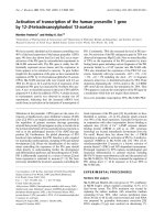
To investigate the role of CD73 in human endothelial
cells, HUVECs were transfected with three different
siRNAs to CD73 (si1, si2 and si3), all of which reduced
surface levels of CD73 by at least 70%, whereas a con-
trol non-targeting siRNA (siControl; siC) did not affect
CD73 levels (Figure 1A). Adenosine is the product of
CD73 enzymatic activity. It was constitutively produced
by HUVECs, and this was markedly reduced in CD73
knockdown cells (Figure 1B), indicating that CD73 is
the major source of extracellular adenosine in these
cells.
CD73 regulates adhesion molecule expression in
endothelial cells
Pro-inflammatory cytokines up-regulate the expression
of the leukocyte adhesion molecules ICAM-1, V-CAM-1
and E-selectin in endothelial cells [19]. To investigate
whether CD73 regulates cell surface levels of these
adhesion molecules, we tested the effects of CD73
depletion. Unstimulated HUVECs expressed low levels
of ICAM-1 on the cell surface, whereas VCAM-1 and E-
selectin levels were not above background (data not
shown). CD73 depletion induced an increase in ICAM-
1, VCAM-1 and E-selectin levels, whereas siControl had
no effect (Figure 1C-E). Taken together, these results
are consist ent with a role of constitutive adenosine pro-
duction by C D73 in suppressing ex pression of leukocyte
adhesion molecules in endothelial cells.
TNF-a induces ICAM- 1, VCAM-1 and E-selectin
expression in part through activation o f the tr anscrip-
tion factor NF-B [19]. NF-B activity was reported to
be increased in endothelial cells derived from CD73
-/-
mice, and thus could contribute to upregulation of
VCAM-1 levels [6]. To test if NF-B activity was
increased in HUVECs depleted of CD73, cells were
stained with antibodies to NF-B. NF-B translocates to
the nucleus when it is act ivated [23], and TNF-a ,which
is well known to stimulate NF-B activity, stimulated
NF-B nuclear translocation in over 60% of HUVECs
(Figure 2). CD73 depletion also increased the proportion
of cells with nuclear NF-Bstaining(Figure2).These
results suggest that CD73 knockdown induces a pro-
inflammatory phenotype in HUVECs, which could be
mediated in part by NF-B activation.
CD73 depletion induces morphological changes in
HUVECs
Since CD73 knockdown induced upregulation of adhesion
molecules similar t o TNF-a, we tested whether CD73
affected endothelial morphology. We have previously
shown that TNF-a induces cell elongation and actin stress
fibre formation in HUVECs [24]. CD73 knockdown
induced an elongated morphology s imilar to m orphological
changes occurring after TNF-a tre atment (Figure 3). CD73
depletion also increased stress fibres, although to a lesser
extent than 10 ng/ml TNF-a (Figure 3). These r esults
Grünewald and Ridley Journal of Inflammation 2010, 7:10
/>Page 3 of 7
further strengthen the hypothesis that CD73 depletion
induces a pro-inflammatory phenotype.
CD73 regulates leukocyte adhesion
The increase in adhesion molecule expression in CD73-
depleted endothelial cells suggests that leukocyte adhe-
sion could be affected. To study this we incubated THP-
1 monocytic leukaemia cells with HUVECs. Adh esion of
THP-1 cells to HUVECs was significantly increased by
CD73 knockdown (Figure 4A). In contrast, CD73 deple-
tion did not affect THP-1 adhesion to TNF-a-treated
HUVECs, reflecting the 4 to 6 fold increase in the levels
of ICAM-1, VCAM-1 and E-selectin expression induced
by TNF-a alone (data not shown).
Endothelial permeability is increased in CD73-depleted
cells
TNF-a is known to increase endothelial permeability in
HUVECs [24,25], whereas adenosine, the product of
CD73 enzymatic activity, has been shown to reduce per-
meability [11,12,26]. The decrease in extracellular ade-
nosine production due to CD73 knockdown (Figure 1C)
would therefore be predicted to lead to an increase in
permeability. In agreement with this, the permeability of
HUVEC monolayers was higher following CD73 deple-
tion than in control cells (Figure 4B ). The 1.5 to 2-fol d-
increase in permeability following CD73 knockdown was
in the same range to that induced by 10 ng/ml TNF-a
(2 to 2.5 fold; data not shown and [24])
Discussion
The endothelium of CD73
-/-
mice has been shown to
have increased VCAM-1 levels, but the effect of CD73
depletion on human endothelial cells has not been
described. We show here that CD73 normally functions
to suppress multiple different aspects of a pro-inflam-
mato ry phenotype of endothelial cells, including expres-
sion of ICAM-1, VCAM-1 and E-selectin, translocation
of the transcription factor NF-B to the nucleus,
endothelial cell morphology, actin cytoskeletal organisa-
tion and permeability. CD73-depleted cells exhibited a
similar phenotype to treatment with TNF-a.
Consistent with the lower levels of leukocyte adhesion
molecules and leukocyte adhesion we observe in CD73-
depleted endothelial cells, leukocyte infiltration in inflam-
matory situations is reduced in CD73
-/-
mice [7,27,28].
Figure 1 CD73 regulates ICAM-1, VCAM-1 and E-selectin expression. HUVECs were transfected with CD73 siRNAs or control oligonucleotide
(siC). A, Cell surface expression levels of CD73. B, CD73 activity. C-E, ICAM-1, VCAM-1 and E-selectin, shown as mean fluorescence of the
population. Results were normalised to siC. ***p < 0.001, **p < 0.01, *p < 0.05 determined by Student’s t-test and Bonferroni post-test, compared
to siC.
Grünewald and Ridley Journal of Inflammation 2010, 7:10
/>Page 4 of 7
Endothelial CD73 is important for these responses [28],
although lymphocyte CD73 als o contributes to reducing
cardiac graft rejection [8]. In lymphocytes it has been sug-
gested that CD73 has non-enzymatic functions in modu-
lating the clustering of the integrin LFA-1 or in inhibiting
apoptosis, but so far no such role of CD73 has b een
described in endothelial cells [1,29]. However, an A
2B
ade-
nosine receptor agonist rescues the defect in lymphocyte
recruitment to lymph nodes in CD73
-/-
mice [28], indicat-
ing that in this case the phenotype i s probably due to
decreased levels of adenosine.
It is likely that the signalling pathway whereby CD73
and adenosine suppress leukocyte adhesion molecule
expression differs from that regulating morphology and
endothelial permeability. The regulation of en dothelial
permeability and stress fibre levels by adenosine is
attributed to an increase in cAMP, which in turn
induces both inhibition of RhoA, and hence decrease s
actomyosin contractility and stress fibre formation, and
activation of Rap1, thereby strengthening adherens junc-
tion integrity [30]. Although the mechanistic b asis for
adenosine-mediated inhibition of leukocyte adhesion
molecule expression is less clear, it is possible that it
also involves cAMP production, since increased cAMP
inhibits TNF-a-and thrombin-induced transcription of
NFB-regulated genes, including ICAM-1 and VCAM-1
[31,32], an effect that could be mediated through
cAMP-induced repression of p38 MAPK activity [31].
It is not clear whether the pro-inflammato ry phenoty-
picchangesweobserveinresponsetoCD73depletion
represent t he constitutive activity of an intrinsic signal-
ling pathway in endothelial cells that is suppressed by
Figure 2 CD73 depletion increases nuclear localisation of NF-
B. HUVECs were transfected with CD73 siRNAs or control siC. A,
Immunolocalization of NF-B (p65) and b-catenin. Bar = 50 μm. B,
Quantification of NF-B localization; at least 100 cells were counted
in each of three independent experiments. * p < 0.05 determined
by Student’s t-test and Bonferroni post-test, compared to siC.
Figure 3 CD73 regulates endothelial morphology. HUVECs were
transfected with CD73 siRNAs or control oligonucleotide (siC), and
stimulated with or without TNF-a. Representative phase-contrast
images (A) and confocal images of actin filaments (B) of at least five
independent experiments are shown. Bars = 50 μm.
Grünewald and Ridley Journal of Inflammation 2010, 7:10
/>Page 5 of 7
CD73 and adenosine or are mediated by an external sti-
mulus. It is possible that HUVECs themselves produce
some TNF-a or other pro-inflammatory cytokines,
although TNF-a production by endothelial cells is nor-
mally only induced by inflammatory stimuli such as LPS
or interleukin 1b [33,34]. In the future it would be inter-
esting to determine whether the anti-inflammatory
effects of CD73 are mediated by alterations in the con-
stitutive activity of GTPases such as RhoA or Rap1. It
will also be important to investigate whet her the effects
of reduced CD73 e xpression we report with human
endothelial cells in vitro correlate with in vivo observa-
tions on human endothelium.
Conclusions
CD73 depletion in HUVECs induces a pro-inflammatory
phenotype similar to low levels of TNF-a,including
increased expression of leukocyte adhesion molecules
and changes in endothelial morphology. Since we found
that HUVECs normally produce extracellular adenosine
and that this is predominantly due to CD73, it is likely
that reduced levels of adenosine are responsible for the
phenotypes we observe upon CD73 knockdown.
Acknowledgements
We are grateful to Sirpa Jalkanen (University of Turku, Finland) for providing
antibody to human CD73. This research was supported by European
Commission contract no. LHSG-CT-2003-502935 (MAIN), by the Ludwig
Institute for Cancer Research and Cancer Research UK.
Authors’ contributions
JKGG and AJR designed the study. JG carried out all experimental work and
prepared the figures. JKGG and AJR wrote the manuscript. Both authors
have read and approved the final manuscript.
Competing interests
The authors declare that they have no competing interests.
Received: 7 September 2009
Accepted: 5 February 2010 Published: 5 February 2010
References
1. Jalkanen S, Salmi M: VAP-1 and CD73, endothelial cell surface enzymes in
leukocyte extravasation. Arterioscler Thromb Vasc Biol 2008, 28:18-26.
2. Yegutkin GG: Nucleotide- and nucleoside-converting ectoenzymes:
Important modulators of purinergic signalling cascade. Biochim Biophys
Acta 2008, 1783:673-694.
3. Koszalka P, Ozuyaman B, Huo Y, Zernecke A, Flogel U, Braun N,
Buchheiser A, Decking UK, Smith ML, Sevigny J, Gear A, Weber AA,
Molojavyi A, Ding Z, Weber C, Ley K, Zimmermann H, Godecke A,
Schrader J: Targeted disruption of cd73/ecto-5’-nucleotidase alters
thromboregulation and augments vascular inflammatory response. Circ
Res 2004, 95:814-821.
4. Eckle T, Krahn T, Grenz A, Kohler D, Mittelbronn M, Ledent C, Jacobson MA,
Osswald H, Thompson LF, Unertl K, Eltzschig HK: Cardioprotection by ecto-
5’-nucleotidase (CD73) and A2B adenosine receptors. Circulation 2007,
115:1581-1590.
5. Thompson LF, Eltzschig HK, Ibla JC, Wiele Van De CJ, Resta R, Morote-
Garcia JC, Colgan SP: Crucial role for ecto-5’-nucleotidase (CD73) in
vascular leakage during hypoxia. J Exp Med 2004, 200:1395-1405.
6. Zernecke A, Bidzhekov K, Ozuyaman B, Fraemohs L, Liehn EA, Luscher-
Firzlaff JM, Luscher B, Schrader J, Weber C: CD73/ecto-5’-nucleotidase
protects against vascular inflammation and neointima formation.
Circulation 2006, 113:2120-2127.
7. Reutershan J, Vollmer I, Stark S, Wagner R, Ngamsri KC, Eltzschig HK:
Adenosine and inflammation: CD39 and CD73 are critical mediators in
LPS-induced PMN trafficking into the lungs. FASEB J 2009, 23:473-82.
8. Hasegawa T, Bouis D, Liao H, Visovatti SH, Pinsky DJ: Ecto-5’ nucleotidase
(CD73)-mediated adenosine generation and signaling in murine cardiac
allograft vasculopathy. Circ Res 2008, 103:1410-1421.
9. Ham J, Rees DA: The adenosine a2b receptor: its role in inflammation.
Endocr Metab Immune Disord Drug Targets 2008, 8:244-254.
10. Shryock JC, Belardinelli L: Adenosine and adenosine receptors in the
cardiovascular system: biochemistry, physiology, and pharmacology. Am
J Cardiol 1997, 79:2-10.
11. Comerford KM, Lawrence DW, Synnestvedt K, Levi BP, Colgan SP: Role of
vasodilator-stimulated phosphoprotein in PKA-induced changes in
endothelial junctional permeability. FASEB J 2002, 16:583-585.
12. Srinivas SP, Satpathy M, Gallagher P, Lariviere E, Van Driessche W:
Adenosine induces dephosphorylation of myosin II regulatory light
chain in cultured bovine corneal endothelial cells. Exp Eye Res 2004,
79:543-551.
13. Eckle T, Faigle M, Grenz A, Laucher S, Thompson LF, Eltzschig HK: A2B
adenosine receptor dampens hypoxia-induced vascular leak.
Blood 2008,
111:2024-2035.
14. Eckle T, Grenz A, Laucher S, Eltzschig HK: A2B adenosine receptor
signaling attenuates acute lung injury by enhancing alveolar fluid
clearance in mice. J Clin Invest 2008, 118:3301-3315.
15. Cronstein BN: Adenosine, an endogenous anti-inflammatory agent. J Appl
Physiol 1994, 76:5-13.
16. Wakai A, Wang JH, Winter DC, Street JT, O’Sullivan RG, Redmond HP:
Adenosine inhibits neutrophil vascular endothelial growth factor release
and transendothelial migration via A2B receptor activation. Shock 2001,
15:297-301.
Figure 4 CD73 depletion increases monocyte adhesion to
endothelial cells and endothelial permeability. HUVECs were
transfected with CD73 siRNAs or control siC. A, Adhesion of THP-1
cells to HUVECs was measured after 15 min. B, Monolayer
permeability was determined on Transwell filters. Results were
normalised to the respective control (siC). **p < 0.01, *p < 0.05,
determined by Student’s t-test and Bonferroni post-test, as
compared to siC.
Grünewald and Ridley Journal of Inflammation 2010, 7:10
/>Page 6 of 7
17. Niemela J, Ifergan I, Yegutkin GG, Jalkanen S, Prat A, Airas L: IFN-beta
regulates CD73 and adenosine expression at the blood-brain barrier. Eur
J Immunol 2008, 38:2718-2726.
18. McPherson JA, Barringhaus KG, Bishop GG, Sanders JM, Rieger JM,
Hesselbacher SE, Gimple LW, Powers ER, Macdonald T, Sullivan G, Linden J,
Sarembock IJ: Adenosine A(2A) receptor stimulation reduces
inflammation and neointimal growth in a murine carotid ligation model.
Arterioscler Thromb Vasc Biol 2001, 21:791-796.
19. De Martin R, Hoeth M, Hofer-Warbinek R, Schmid JA: The transcription
factor NF-B and the regulation of vascular cell function. Arterioscler
Thromb Vasc Biol 2000, 20:E83-88.
20. Minguet S, Huber M, Rosenkranz L, Schamel WW, Reth M, Brummer T:
Adenosine and cAMP are potent inhibitors of the NF-B pathway
downstream of immunoreceptors. Eur J Immunol 2005, 35:31-41.
21. Walker G, Langheinrich AC, Dennhauser E, Bohle RM, Dreyer T, Kreuzer J,
Tillmanns H, Braun-Dullaeus RC, Haberbosch W: 3-deazaadenosine
prevents adhesion molecule expression and atherosclerotic lesion
formation in the aortas of C57BL/6J mice. Arterioscler Thromb Vasc Biol
1999, 19:2673-2679.
22. Bouma MG, Wildenberg van den FA, Buurman WA: Adenosine inhibits
cytokine release and expression of adhesion molecules by activated
human endothelial cells. Am J Physiol 1996, 270:C522-529.
23. Karin M, Greten FR: NF-B: linking inflammation and immunity to cancer
development and progression. Nat Rev Immunol 2005, 5:749-759.
24. McKenzie JA, Ridley AJ: Roles of Rho/ROCK and MLCK in TNF-a-induced
changes in endothelial morphology and permeability. J Cell Physiol 2007,
213:221-228.
25. Wojciak-Stothard B, Entwistle A, Garg R, Ridley AJ: Regulation of TNF-a-
induced reorganization of the actin cytoskeleton and cell-cell junctions
by Rho, Rac, and Cdc42 in human endothelial cells. J Cell Physiol 1998,
176:150-165.
26. Lennon PF, Taylor CT, Stahl GL, Colgan SP: Neutrophil-derived 5’-
adenosine monophosphate promotes endothelial barrier function via
CD73-mediated conversion to adenosine and endothelial A2B receptor
activation. J Exp Med 1998, 188:1433-1443.
27. Mills JH, Thompson LF, Mueller C, Waickman AT, Jalkanen S, Niemela J,
Airas L, Bynoe MS: CD73 is required for efficient entry of lymphocytes
into the central nervous system during experimental autoimmune
encephalomyelitis. Proc Natl Acad Sci USA 2008, 105:9325-9330.
28. Takedachi M, Qu D, Ebisuno Y, Oohara H, Joachims ML, McGee ST,
Maeda E, McEver RP, Tanaka T, Miyasaka M, Murakami S, Krahn T,
Blackburn MR, Thompson LF: CD73-generated adenosine restricts
lymphocyte migration into draining lymph nodes. J Immunol
2008,
180:6288-6296.
29. Mikhailov A, Sokolovskaya A, Yegutkin GG, Amdahl H, West A, Yagita H,
Lahesmaa R, Thompson LF, Jalkanen S, Blokhin D, Eriksson JE: CD73
participates in cellular multiresistance program and protects against
TRAIL-induced apoptosis. J Immunol 2008, 181:464-475.
30. Vandenbroucke E, Mehta D, Minshall R, Malik AB: Regulation of endothelial
junctional permeability. Ann N Y Acad Sci 2008, 1123:134-145.
31. Rahman A, Anwar KN, Minhajuddin M, Bijli KM, Javaid K, True AL, Malik AB:
cAMP targeting of p38 MAP kinase inhibits thrombin-induced NF-B
activation and ICAM-1 expression in endothelial cells. Am J Physiol Lung
Cell Mol Physiol 2004, 287:L1017-1024.
32. Ollivier V, Parry GC, Cobb RR, de Prost D, Mackman N: Elevated cyclic AMP
inhibits NF-B-mediated transcription in human monocytic cells and
endothelial cells. J Biol Chem 1996, 271:20828-20835.
33. Nilsen EM, Johansen FE, Jahnsen FL, Lundin KE, Scholz T, Brandtzaeg P,
Haraldsen G: Cytokine profiles of cultured microvascular endothelial cells
from the human intestine. Gut 1998, 42:635-642.
34. Imaizumi T, Itaya H, Fujita K, Kudoh D, Kudoh S, Mori K, Fujimoto K,
Matsumiya T, Yoshida H, Satoh K: Expression of tumor necrosis factor-a in
cultured human endothelial cells stimulated with lipopolysaccharide or
interleukin-1a. Arterioscler Thromb Vasc Biol 2000, 20:410-415.
doi:10.1186/1476-9255-7-10
Cite this article as: Grünewald and Ridley: CD73 represses pro-
inflammatory responses in human endothelial cells. Journal of
Inflammation 2010 7:10.
Submit your next manuscript to BioMed Central
and take full advantage of:
• Convenient online submission
• Thorough peer review
• No space constraints or color figure charges
• Immediate publication on acceptance
• Inclusion in PubMed, CAS, Scopus and Google Scholar
• Research which is freely available for redistribution
Submit your manuscript at
www.biomedcentral.com/submit
Grünewald and Ridley Journal of Inflammation 2010, 7:10
/>Page 7 of 7