Gãy xương, chấn thương potx
Bạn đang xem bản rút gọn của tài liệu. Xem và tải ngay bản đầy đủ của tài liệu tại đây (205.1 KB, 10 trang )
192 Journal of the American Academy of Orthopaedic Surgeons
The pain and decreased mobility
resulting from the loss or degener-
ation of the articular cartilage in
synovial joints compromise the pro-
ductivity and quality of life of mil-
lions of persons.
1-3
Unfortunately, all
of the current treatments of synovial
joint problems due to damaged or
degenerating articular cartilage
have significant limitations. Anal-
gesic and anti-inflammatory med-
ications, activity modification, and
physical therapy may provide par-
tial symptomatic relief, but they do
not restore damaged articular carti-
lage to its normal state; thus, they
rarely allow patients to return to full
function for prolonged periods of
time.
2
Surgical procedures, includ-
ing arthrodesis and joint replace-
ment, relieve joint pain, but they also
have important limitations, espe-
cially for the younger patient who
wants to pursue vigorous physical
activities. Arthrodesis restricts
mobility, causes muscular atrophy,
and over time may have undesired
effects on adjacent joints. Most
artificial joints cannot withstand
prolonged and frequent heavy load-
ing, and wear and loosening of the
prosthesis may lead to implant fail-
ure even in patients who have
significantly restricted their activi-
ties. For these reasons, substantial
efforts have been devoted to the
search for biologic methods of
restoring degenerating articular car-
tilage to normalcy before it reaches
end-stage osteoarthritis.
The inability of cartilage to repair
itself after traumatic injuries and the
incapacity of treatment to arrest the
osteoarthritic process have been
described repeatedly for at least 250
years.
4
However, the clinical experi-
ence of treating damaged articular
surfaces by osteotomy, abrasion
arthroplasty, and fascial, periosteal,
and perichondral interposition
arthroplasties has shown that there
is the potential of restoring some
form of cartilaginous articulating
surface even in severely injured or
degenerated joints.
2,5
In addition,
recent advances in cartilage bio-
chemistry and morphology, cellular
and molecular biology, and joint and
tissue biomechanics
6
suggest that
better methods of restoring articular
Restoration of Injured or Degenerated Articular Cartilage
Joseph A. Buckwalter, MD, Van C. Mow, PhD, and Anthony Ratcliffe, PhD
Dr. Buckwalter is Professor of Orthopaedic
Surgery, Department of Orthopaedic Surgery,
University of Iowa Hospitals and Clinics, Iowa
City. Dr. Mow is Professor of Mechanical Engi-
neering and Orthopaedic Bioengineering,
Columbia University, New York; and Director,
Orthopedic Research Laboratory, Department of
Orthopedic Surgery, Columbia-Presbyterian
Medical Center, New York. Dr. Ratcliffe is Asso-
ciate Professor of Orthopaedic Biochemistry,
Columbia University; and Head, Biochemistry
Section, Orthopedic Research Laboratory,
Department of Orthopedic Surgery, Columbia-
Presbyterian Medical Center.
Reprint requests: Dr. Mow, Orthopedic Research
Laboratory, Department of Orthopedic Surgery,
Columbia-Presbyterian Medical Center,
BB1412, 630 West 168th Street, New York, NY
10032.
Copyright 1994 by the American Academy of
Orthopaedic Surgeons.
Abstract
Intra-articular fractures, ligamentous and meniscal injuries, and articular carti-
lage breakdown are major causes of degenerative joint disease. Lesions on the artic-
ular surface seem to have a limited capacity for repair and often progress
inexorably toward osteoarthritis. Recent studies on joint immobilization and car-
tilage atrophy, however, have shown that repair and remodeling of articular car-
tilage may be possible. Currently used clinical methods of stimulating cartilage
repair and remodeling include alteration of the loading on degenerated joints (pri-
marily by using osteotomies), introduction of new cartilage-forming cells by per-
foration of subchondral bone, and soft-tissue arthroplasty. These procedures
provide temporary relief in selected patients, but they often do not predictably
restore long-term joint function. Experimentally, cartilage repair has been stimu-
lated successfully with the use of allografts of periosteum and perichondrium,
which serve as sources of cells with chondrogenic potential; introduction of cells
grown in culture (stem cells or chondrocytes); stimulation by fibrin clot forma-
tion; artificial collagen matrices combined with cell transplants; and chondrogenic
growth factors. The long-term success of all these methods has not been explored
thoroughly, even in animal studies. Nevertheless, some research results are
sufficiently encouraging to suggest that repair of the degenerating articular car-
tilage may be possible in the future.
J Am Acad Orthop Surg 1994;2:192-201
Vol 2, No 4, July/Aug 1994 193
Joseph A. Buckwalter, MD, et al
surfaces can eventually be devel-
oped. This has long been one of the
goals of orthopaedic surgery.
This review summarizes some
potentially useful approaches to
restoring injured or degenerating
articular cartilage. It must be borne in
mind that although both injury and
degeneration involve disruption or
loss of the cartilaginous articulating
surfaces, the clinical problems, nat-
ural history, and potential for restora-
tion of joint function are different.
Form and Function of
Articular Cartilage
Articular cartilage consists of a large
extracellular matrix with a sparse
population of specialized cells, the
chondrocytes (Fig. 1, A and B). The
extracellular matrix is composed pri-
marily of a mix of collagen (mainly
type II) and proteoglycan aggrecan,
with smaller amounts of other colla-
gens, proteoglycans, proteins, and
glycoproteins.
6
In normal articular
cartilage, the collagen network has a
well-defined ultrastructure (Fig. 1, C
and D), which dictates the tensile
stiffness and strength of each carti-
lage layer.
2,6
Because collagen and
proteoglycan form a fiber-reinforced
composite material, the collagen net-
work also provides shear stiffness
and strength to the tissue. The tex-
ture of the normal articular cartilage
surface is relatively smooth and is
composed of tightly woven sheets of
collagen.
In normal articular cartilage, many
aggrecan molecules (Fig. 2) bind to a
chain of hyaluronan, and this interac-
tion is stabilized by a separate link
protein (Fig. 3). Thus, the aggrecans
are effectively immobilized within
the fine collagen network, which pro-
duces a strong, cohesive collagen-pro-
teoglycan solid matrix.
7
An aggrecan
molecule comprises many gly-
cosaminoglycan chains (keratan sul-
fate and chondroitin sulfate), which
contain numerous charged carboxyl
and sulfate groups. Together with
their counterions, they create the
swelling pressure of the tissue, which
has a major influence on cartilage
hydration and hence its deformational
properties.
6,8
The chondrocytes are responsi-
ble for the synthesis of the articular
cartilage during development, for
the maintenance of normal adult
cartilage, and for the degradation
of cartilage during osteoarthritis.
6
The chondrocytes orchestrate the
balance between the synthesis of
matrix components, the incorpora-
tion of these components into the
established extracellular matrix,
and the breakdown of matrix as
part of the normal maintenance
process (Fig. 4). The chondrocytes
respond to a variety of factors,
including the composition of the
surrounding matrix, the mechani-
cal load, and soluble mediators,
such as growth factors and
cytokines (small proteins that
influence multiple cell functions,
including migration, proliferation,
differentiation, and matrix synthe-
sis).
8-10
In a variety of ways, the
chondrocytes are able to receive
signals from their environment and
transduce these signals into bio-
chemical products, which then
maintain a biomechanically normal
articular cartilage.
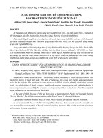
Fig. 1 Structure of articular cartilage. A, Histologic section of cartilage from a young, healthy adult shows even
safranin O staining and distribution of chondrocytes. B, Schematic diagram of chondrocyte organization in the three
main zones of the uncalcified cartilage (STZ = superficial tangential zone), the tidemark, and the subchondral bone.
C, Sagittal cross-sectional diagram of collagen fiber architecture shows the three salient zones of articular cartilage. D,
Scanning electron micrographs depict arrangement of collagen in the three zones (top = STZ; center = middle zone;
bottom = deep zone). (Photographs in A and D reproduced with permission from Mow VC, Proctor CS, Kelly MA:
Biomechanics of articular cartilage, in Nordin M, Frankel VH [eds]: Basic Biomechanics of the Musculoskeletal System,
2nd ed. Philadelphia: Lea & Febiger, 1989, pp 32 and 34, respectively.)
A
B
C
D
194 Journal of the American Academy of Orthopaedic Surgeons
Restoration of Articular Cartilage
visible disruption of the articular
surface, (2) macrodisruption of the
articular cartilage alone (chondral
fractures), and (3) fracture of the
articular cartilage and the subchon-
dral bone (osteochondral fractures).
Cartilage Injury Without Tissue
Disruption
A single moderately severe impact
or less severe repetitive trauma can
damage cartilage. This type of damage
is measurable in terms of decreased
proteoglycan concentration in the
matrix, increased tissue hydration,
and possibly altered fibrillar organiza-
tion of collagen. More important, the
trauma can also injure chondrocytes
or alter their synthetic and degrada-
tive activities.
6,11-13
The exact nature of
this type of damage has not been well
studied, although the decrease in pro-
teoglycan concentration, the increase
in hydration, and the disorganization
of the collagen ultrastructure may rep-
resent some of the earliest detectable
cartilage damage.
B
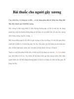
Fig. 3 A, A proteoglycan aggregate is composed of a long hyaluronan chain to which many aggrecans are attached, forming macromolec-
ular complexes that are effectively immobilized within the collagen network. The length of the hyaluronan chain determines the size of the
aggregate. The total molecular weight may be as high as 200 million daltons in immature cartilage; in adult and aging articular cartilage, the
aggregate gradually decreases in size. (Reproduced with permission from Simon SR [ed]: Orthopaedic Basic Science. Rosemont, Ill: American
Academy of Orthopaedic Surgeons, 1994, p 10.) B, Electron micrographs of proteoglycan aggregates in bovine articular cartilage from a skele-
tally immature calf (1) and a skeletally mature steer (2). The aggregates consist of a central hyaluronan filament and multiple attached
monomers. Bar represents 500 µm. (Reproduced with permission from Buckwalter JA, Kuettner KE, Thonar EJ: Age-related changes in artic-
ular cartilage proteoglycans: Electron microscopic studies. J Orthop Res 1985;3:251-257.)
A
Articular Cartilage Injury
Mechanical injuries to articular car-
tilage occur when repetitive and
prolonged joint overloading or sud-
den impact produces high compres-
sive stress throughout the tissue and
high shear stress at the subchondral
bone junction.
3
These stresses cause
injuries that can be separated into
three distinct types: (1) microdam-
age to the cells and matrix without
Fig. 2 Diagram of an aggrecan molecule. The protein core has several globular domains
(G1, G2, and G3). Other regions contain the keratan sulfate (KS) and chondroitin sulfate (CS)
glycosaminoglycan chains. The N-terminal G1 domain is able to bind specifically to hyaluro-
nan; this binding is stabilized by link protein. The total molecular weight of an aggrecan
ranges from 0.5 million to 1.0 million daltons. (Reproduced with permission from Simon SR
[ed]: Orthopaedic Basic Science. Rosemont, Ill: American Academy of Orthopaedic Surgeons,
1994, p 9.)
A slightly more severe impact may
produce severe chondrocyte abnor-
malities and deaths and a weakened
collagenous network (Fig. 5). These
injuries may also be accompanied by
shear damage to the junction between
the articular cartilage and the sub-
chondral bone and may cause reac-
tive bone remodeling with increasing
replication of the tidemark.
12,14
Loss of
proteoglycans and an increase in
water content are strongly correlated
with a decrease in cartilage stiffness
and an increase in its hydraulic per-
meability. These changes in material
properties act to decrease fluid pres-
surization within the interstitium (i.e.,
the interstitial fluid can be more easily
squeezed from the more permeable
cartilage) and thus impair the normal
load-carrying capacity of the intersti-
tial fluid.
3
Both of these effects cause
greater loading to be exerted on the
collagen-proteoglycan solid matrix,
thereby increasing the vulnerability
of the extracellular matrix to further
damage.
In addition to the injuries sus-
tained by cartilage due to mechan-
ical trauma, disruption of the
synovial membrane often occurs.
This leaves the articular surface ex-
posed to synovial inflammation
factors, which can enzymatically
cause articular cartilage injury.
Conversely, when synovial joints
are immobilized or are otherwise in a
state of disuse, an active remodeling
process develops within the articular
cartilage.
9,15
The attendant functional
changes can result in a dramatic loss
of proteoglycans from the cartilage.
On remobilization, however, the
apparently dormant chondrocytes
are reawakened to repair the matrix,
and the resulting cartilage appears to
be able to return to its original form
and function. This indicates that the
chondrocytes have the potential to
repair cartilage, at least for some
types of injury. However, this type of
repair may require many weeks
(possibly months) to restore the
affected tissue to normal. There is an
overwhelming body of scientific evi-
dence to support the notion that
chondrocytes have the ability to
detect changes in matrix composi-
tion and to sense altered mechanical
stresses within the surrounding
extracellular matrix, and that they
have the capacity to respond to these
changes by synthesizing new mole-
cules to repair the damaged extracel-
lular matrix.
9,15
However, the
signal-transduction mechanism by
which the cells detect these changes
and the manner with which the
chondrocytes translate these signals
into altered metabolic events are
unknown.
Following intra-articular injuries,
such as a torn meniscus or rupture of
the anterior cruciate ligament, the
capacity of the chondrocytes for
repair is often insufficient to main-
Vol 2, No 4, July/Aug 1994 195
Joseph A. Buckwalter, MD, et al
Fig. 4 Schematic represen-
tation of the metabolic
events controlling the pro-
teoglycans in cartilage. The
chondrocytes synthesize
and secrete the aggrecans,
link protein, and hyal-
uronan and become incor-
porated into functional
aggregates in the extra-
cellular matrix. Enzymes
released by the cells break
down the proteoglycan
aggregates. The fragments
are released from the matrix
into the synovial fluid; from
there, the fragments are
taken up by the lymphatic
vessels and moved into the
circulating blood.
Fig. 5 Scanning electron micrograph of a
human cartilage specimen demonstrates
fissure in the articular surface. This type of
damage not only weakens the surface in ten-
sion but also allows large pores to be created
in the surface, thus decreasing its effective-
ness as a filter and its ability to provide a
membrane to limit the rate of fluid exuda-
tion (original magnification X3,000).
(Reproduced with permission from Mow
VC, Mak AF: Lubrication of diarthrodial
joints, in Skalak R, Chien S [eds]: Handbook
of Bioengineering. New York: McGraw-Hill,
1986, p 5.)
tain a normal, functioning cartilage.
This occurs if the cells fail to repair
the microdamages in the extracellu-
lar matrix at a sufficiently rapid rate,
or if repetitive stress continues to
cause microdamage at a more rapid
rate. It is not currently known, how-
ever, at what point the accumulated
microdamage becomes irreversible.
Presumably, chondrocytes can
restore lost proteoglycans if the rate
of loss does not exceed the rate of
production. If there is concomitant
damage to the collagen network or if
a sufficient number of chondrocytes
have been destroyed, an irreversible
degenerative process ensues.
Reliable, clinically applicable
methods of detecting damage to artic-
ular cartilage in the absence of surface
disruption have yet to be developed,
but identification of decreased carti-
lage stiffness and resiliency by prob-
ing the surface during surgery
represents a crude method of detect-
ing the severe form of this type of
injury. Bone scintigraphy and mag-
netic resonance imaging can detect
alterations in subchondral bone fol-
lowing joint injury, but the relation-
ship between these alterations and
cartilage damage has not been
defined. The use of biochemical mark-
ers in analyzing synovial fluid, serum,
and urine offers a potential means of
assessing cartilage metabolism and
degeneration,
10,11
but such tests are not
currently available for clinical use. If
biochemical markers could be used to
detect the earliest stages of cartilage
damage, clinical treatment could be
devised for joints that have been sub-
jected to trauma, and the effectiveness
of that treatment could be measured.
Chondral Fractures
Compressive forces acting on an
articular surface will produce a vari-
ety of stresses (tension, compression,
shear, and hydrostatic pressure)
within the cartilage. These stresses,
if sufficiently high, can cause chon-
dral fissures, flaps, and fractures, as
well as chondrocyte damage. Loss of
significant segments of the articular
surface will result in joint effusions,
pain, and mechanical symptoms,
such as locking and crepitus, and
may lead to progressive degenera-
tion of the synovial joint.
2
Because
articular cartilage lacks blood ves-
sels, these injuries do not cause hem-
orrhage or fibrin-clot formation or
provoke an inflammatory response.
The chondrocytes respond by prolif-
erating and increasing the synthesis
of matrix macromolecules near the
injury site.
13
Unfortunately, the
newly synthesized matrix and pro-
liferating cells do not fill the tissue
defect and therefore fail to restore
the articular surface. When large
defects are present, increased load-
ing of adjacent articular cartilage
and underlying subchondral bone
can lead to degeneration of the unin-
jured cartilage; over time, the entire
joint is affected.
Current treatments of chondral
injuries include sharp debridement
of the fractured edges and removal
of loose cartilage fragments from the
joint. When there is significant loss
of articular surface, some surgeons
advise abrasion or drilling of the
underlying subchondral bone.
Experimental work suggests that
replacement of cartilage fragments
with tissue adhesives or with chon-
dral or osteochondral allografts may
be beneficial. At present, there are
insufficient long-term studies of the
outcome and no guidelines to direct
the use of these treatments in acute
chondral injuries.
Osteochondral Injuries
Acute joint injuries from more
severe impact may also result in
fractures that extend through the
cartilage into subchondral bone.
Unlike injuries that are limited to
cartilage, fractures that extend into
subchondral bone cause hemor-
rhage and fibrin-clot formation,
thereby activating the inflammatory
response. These events fundamen-
tally alter the synovial fluid and the
joint environment surrounding the
articular cartilage. Soon after injury,
blood escaping from blood vessels
in the damaged bone forms a
hematoma, which temporarily fills
the injury site. The fibrin clot extends
from the bone for a variable distance
into the cartilage defects. Platelets
within the clot release vasoactive
mediators and growth factors or
cytokines. These factors include
transforming growth factor beta
(TGF-
β
) and platelet-derived growth
factor.
Bone matrix also contains mul-
tiple growth factors, including
TGF-
β
, bone morphogenic proteins,
platelet-derived growth factor, and
insulin-like growth factors. Release
of these growth factors may play an
important role in stimulating repair
of osteochondral defects. In particu-
lar, these factors stimulate vascular
invasion and migration of undiffer-
entiated cells, proliferation of these
cells, and differentiation into chon-
drocyte-like cells in the chondral
portion of the defect. Some of the
undifferentiated mesenchymal cells
that migrate into the defect assume
the rounded form of chondrocytes
and begin to synthesize a matrix that
contains type II collagen and rela-
tively high concentrations of proteo-
glycans. These cells produce regions
of hyaline-like cartilage in the chon-
dral and osseous portions of the
osteochondral defect. The cells
within the chondral region produce
a repair cartilage that usually con-
tains a high concentration of type II
collagen and proteoglycans, but
often also contains some type I colla-
gen. The cells in the osseous portion
of the defect eventually produce
immature bone, which is gradually
replaced by mature bone.
The composition of this cartilage
repair tissue rarely replicates the
structure of normal articular carti-
lage.
6
This tissue may occasionally
196 Journal of the American Academy of Orthopaedic Surgeons
Restoration of Articular Cartilage
persist unchanged or may progres-
sively remodel to form a more func-
tional joint surface over time.
2
However, in most instances the
chondral repair tissue and large
osteochondral defects begin to show
evidence of depletion of matrix pro-
teoglycans, fragmentation, fibrilla-
tion, and loss of chondrocyte-like
cells. The remaining cells typically
assume the appearance of fibroblasts
as the surrounding matrix comes to
consist primarily of densely packed
type I collagen fibrils. This fibrous
tissue usually fragments and often
disintegrates within a year, and may
leave areas of exposed bone.
Large osteochondral fractures in
which the cartilage remains intact
with the bone often can be treated by
early open reduction and internal
fixation of the fracture. If the fracture
is not treated soon after injury, the
fragments remodel, which makes
accurate reduction difficult. The avail-
able evidence indicates that the articu-
lar surface heals and remodels at the
site of anatomically or near-anatomi-
cally reduced osteochondral fractures,
especially in skeletally immature per-
sons. Smaller osteochondral fractures
and those in which the cartilage is not
suitable for replacement are currently
treated by debridement. Osteochon-
dral allografts have been used suc-
cessfully as the late treatment of
selected osteochondral fractures in
which the injured region forms the
important frequent-load-bearing
region of the joint.
Articular Cartilage
Degeneration
The clinical appearance of early
degeneration of articular cartilage is
characterized by superficial rough-
ening, fibrillation, or fissuring
16
and
is apparent at arthroscopy. With
time, the fissures extend progres-
sively deeper into the tissue and
eventually reach the region of
calcified cartilage and subchondral
bone. As the disorder progresses, the
articular surface becomes weakened
(Fig. 6), fragments of the cartilage
break free from the surface, and the
remaining tissue becomes increas-
ingly fragmented and fibrous.
6,10,11
Eventually, the cartilage may be lost
entirely, leaving exposed subchon-
dral bone. The more advanced
degenerative changes in the articular
surface typically occur simultane-
ously with increasing density of the
subchondral bone, eburnation, and
osteophyte formation. Joint-space
narrowing, detected radiographi-
cally, is evidence of degeneration of
the joint at a relatively late stage of
the disease.
Many important changes occur
in the articular cartilage before
the development of clinical osteo-
arthritis. These preclinical changes in
cartilage include not only the disor-
ganization of the collagen ultrastruc-
ture (i.e., roughening and fibrillation
of the surface zone) but also cell
cloning and cell necrosis. In
osteoarthritic joints, as opposed to
joints in a state of disuse or immobi-
lization, the tensile stiffness and
strength of the surface zone are
always decreased in association with
the disorganization of its collagen
network (Fig. 6).
6,8,12
The water con-
tent increases, and the proteoglycan
content increases initially, followed
by a dramatic decrease. The increase
in hydration is due to the weakening
of the collagen network of the surface
zone in tension and the concomitant
increase in the swelling pressure
resulting from the temporal increase
in the proteoglycan content.
These preclinical events may be
due strictly to adverse mechanical
loading, such as joint instability
resulting from disruption of the
anterior cruciate ligament, with
physical changes in the surface col-
lagen network. Alternatively, these
cartilage changes may be mediated
by the release by the chondrocytes of
matrix-degrading enzymes that
actively degrade the collagen and
proteoglycan components.
16
This cel-
lular response is certainly ongoing
early in the disease process,
although it is not clear whether the
initiating factor is the mechanical
Vol 2, No 4, July/Aug 1994 197
Joseph A. Buckwalter, MD, et al
Fig. 6 Knee-cartilage ten-
sile stiffness and colla-
gen-proteoglycan ratio in
normal, mildly fibrillated,
and osteoarthritic (OA) tis-
sues (the latter obtained
from a site adjacent to frank
lesions). Note linear correla-
tion in normal and mildly
fibrillated tissues. This rela-
tionship is lost in OA tissue,
likely due to the total disor-
ganization of the cartilage
microstructure during ad-
vanced stages of OA. (Re-
produced with permission
from Akizuki A, Mow VC,
Muller F, et al: The tensile
properties of human knee
joint cartilage: I. Influence of
ionic conditions, weight
bearing, and fibrillation on
the tensile modulus. J
Orthop Res 1986;4:379-392.)
event, the cellular event, or a combi-
nation of the two.
These degradative changes are also
accompanied by an increased rate of
chondrocyte mitosis. The resultant
cloning of the chondrocyte represents
an attempt at repair. However, the
newly synthesized proteoglycan and,
to a lesser degree, the collagen are
often lost into the joint fluid.
10,11
Thus,
the reparative responses ultimately
fail, the joint progresses to overt
degeneration, and clinically apparent
osteoarthritis develops.
Synovial joint degeneration typi-
cally causes pain and decreased
mobility. These symptoms, com-
bined with articular cartilage degen-
eration and osteophyte formation,
are recognized as representing clini-
cal osteoarthritis. However, osteo-
arthritis is generally not considered
to be a single condition and may best
be thought of as the clinical result of
joint degeneration due to a variety of
underlying causes.
16
The degenera-
tion of a synovial joint may be pri-
mary, in the sense that there is no
known cause, or it may be secondary
to conditions such as severe joint
trauma, joint instability, lack or loss
of joint or limb innervation, joint dys-
plasia, Paget’s disease, and metabolic
diseases, including hemochromatosis
and ochronosis The natural history of
degeneration varies considerably
among joints and among individuals.
Occasionally, the disorder may spon-
taneously arrest or appear to
improve, but in most instances over a
prolonged period of time it pro-
gresses. Medical and physical ther-
apy treatments have not been shown
to favorably alter this natural history.
Methods of Stimulating
Restoration of an Articular
Surface
In considering methods of restoring
an injured or degenerated articular
surface, it is important to distin-
guish articular cartilage repair from
articular cartilage regeneration.
Repair refers to the healing of
injured tissues or replacement of
lost tissues by cell proliferation and
synthesis of new extracellular
matrix. Unfortunately, the repaired
articular cartilage generally fails to
replicate the structure, composition,
and function of normal articular car-
tilage.
2
Regeneration in this context
refers to the formation of an entirely
new articulating surface that essen-
tially duplicates the original articu-
lar cartilage.
The success of a given method of
restoring an injured or degenerated
articular surface has frequently
been assessed by determining
whether repair tissue fills the chon-
dral defect and by comparing the
composition and mechanical prop-
erties of the repair tissue with those
of normal articular cartilage. How-
ever, filling a chondral defect with
repair tissue does not necessarily
relieve or even decrease pain or
improve joint function, nor has it
been shown that repair tissue that
more closely resembles normal
articular cartilage necessarily pro-
duces better clinical results. The
ultimate measure of the success of
any method of restoring the articu-
lar surface must be long-term joint
function and pain relief.
The available clinical and animal
studies show that a number of meth-
ods can stimulate the formation of
repair tissues, and some recent
experimental studies suggest that
regeneration of an articular surface
may be possible. Methods of stimu-
lating cartilage repair that are cur-
rently used in clinical practice
include altering the loading of
degenerated joints, the introduction
of new cartilage-forming cells by
penetration of subchondral bone,
and soft-tissue arthroplasty. Given
the limited ability of mature chon-
drocytes to repair cartilage defects,
one of the potentially most produc-
tive approaches to restoring an artic-
ular surface is introducing a new cell
population into a chondral or osteo-
chondral defect. These cells may be
obtained from populations grown in
culture and may be combined with
artificial matrices and chondrogene-
sis factors to enhance the formation
of new cartilage. Unfortunately, it is
difficult to compare methods of
restoring articular surfaces because
controlled, randomized clinical
studies of the outcomes of these
treatments have not been per-
formed, and few clinical or experi-
mental studies have investigated the
long-term durability of restored
articular surfaces and the long-term
biomechanical function of the joints.
Altering Loads Applied to
Damaged Articular Cartilage
Joint loading and motion can
have a significant impact on the pro-
gression of joint degeneration and
on cartilage repair. Excessive load-
ing of an injured joint can accelerate
degeneration and destroy the repair
tissue. Prolonged immobilization
and unloading of a joint will also
contribute to cartilage deterioration
and make it susceptible to acceler-
ated degeneration when the joint is
suddenly remobilized. However,
reducing the level of loading on
degenerated cartilage can stimulate
repair, and controlled motion and
loading, including passive motion,
may facilitate repair and maintain or
improve joint motion.
Two methods have been used clini-
cally to promote repair of degenerated
cartilage surfaces by decreasing the
loading of the cartilage: osteotomies
and muscle releases. Experimental and
clinical evidence shows that these
approaches allow and even stimulate
some repair of severely damaged artic-
ular surfaces. Unfortunately, the clini-
cal results are not predictable, and the
relationship between altered loads on
degenerative joints and the formation
of cartilage repair tissue has not been
well defined.
198 Journal of the American Academy of Orthopaedic Surgeons
Restoration of Articular Cartilage
Drilling, Abrasion, or Fracture
of Subchondral Bone
Surgical penetration of subchon-
dral bone to disrupt intraosseous
blood vessels leads to fibrin-clot
formation, releases bone-matrix
growth factors, and introduces new
cells into the cartilage defect. These
cells proliferate and will synthesize a
cartilage-repair matrix. The quality
and volume of repair tissue follow-
ing penetration of subchondral bone
vary considerably; they are depen-
dent on the size and location of the
cartilage defect and probably on the
method used for penetrating the
subchondral bone, and can be
influenced by the loading applied to
the joint during the rehabilitation
process following the procedure.
Partially because of these variables,
the clinical results of this approach
are difficult to predict. Some patients
report symptomatic improvement,
and in some instances the repair tis-
sue functions reasonably well as a
normal load-bearing articular
surface for years; however, other
patients experience no improve-
ment.
Periosteal and Perichondral
Grafts
Soft-tissue arthroplasties replace
degenerated or lost cartilage with
grafts of tissues, including fascia,
tendon, muscle, periosteum, and
perichondrium. The ability of peri-
chondral and periosteal cells (most
probably the cells of the cambium
layer adjacent to the bone) to form
hyaline cartilage makes them an
attractive source of new cells to
restore an articular surface.
5,17
The
availability of periosteum in relative
abundance makes this the most
likely of the tissues to be used with
frequency.
Recent experimental surgical
studies with animals have used
osteoperiosteal and osteoperichon-
dral grafts as a source of repair tis-
sue in large osteochondral defects
that have been created in the patel-
lar groove and the high-weight-
bearing region of the femoral
condyles. The results indicate that it
is possible to form a tissue that fills
the defect site, has the gross appear-
ance of hyaline cartilage, and is his-
tologically characteristic of articular
cartilage. Biomechanical and bio-
chemical studies indicate that the
repair tissue closely resembles artic-
ular cartilage.
2,5
Some motion and
normal loading of the joint appear to
be important in this repair process.
The clinical results of periosteal and
perichondral grafts vary consider-
ably among individuals and among
joints, but there is evidence that this
approach produces better results in
younger individuals. Success with
this approach may be achieved by
using highly selected groups of
patients in whom repair is reason-
ably likely (i.e., young patients with
focal osteochondral injury).
Implantation of Chondrocytes
or Mesenchymal Stem Cells
Cartilage-forming cells can also be
introduced into chondral defects by
implantation of cells grown and main-
tained in culture. Experimental inves-
tigations of this approach have shown
that the transplanted cells can survive
and synthesize a cartilaginous matrix
that appears to more closely resemble
normal cartilage than the fibrous tis-
sue that forms in similar defects not
treated with cell transplants. One pos-
sible method of applying this
approach in humans would be to har-
vest mesenchymal stem cells or chon-
drocytes, expand them in culture,
implant them in an artificial matrix,
and then implant the matrix and the
cells in a cartilage defect.
Stimulation of Fibrin-Clot
Formation
In vascularized tissues, formation
of fibrin clots (including release of
growth factors by platelets) proba-
bly has an important role in initiat-
ing repair. The cytokines released
from the clot provide chemotactic
and mitogenic stimuli for mesenchy-
mal cells that will migrate into the
clot, which may provide a tempo-
rary matrix for these cells. Poten-
tially, clot formation could have a
similar effect in nonvascularized tis-
sue, such as articular cartilage.
Because the proteoglycans in the
cartilage matrix may inhibit clot for-
mation in cartilage defects, investiga-
tors have proposed irrigating the
defects with saline or enzyme solu-
tions to degrade the proteoglycans and
to allow fibrin to clot and adhere to the
defects. Experimental studies have
provided some evidence that this
approach does promote clot formation
and adherence and that cell migration
into chondral defects does occur.
Chondrogenesis-Stimulating
Factors
A variety of polypeptide growth
factors (e.g., TGF-
β
, bone mor-
phogenic proteins, insulin-like
growth factor, fibroblast growth fac-
tor, and platelet-derived growth fac-
tor) influence chondrocyte and other
mesenchymal cell functions, such as
cell migration, proliferation, matrix
synthesis, and differentiation. The
effects of these factors on chondro-
cytes are mediated by cell-surface
receptors (integrins). These factors
may also directly modify the extra-
cellular matrix and thus modulate
the signals (e.g., stresses, strains, and
fluid pressure and flow) transmitted
to the cells from the surrounding
extracellular matrix.
Experimental work has shown that
selected growth factors can stimulate
formation of cartilaginous tissue in
vitro and in vivo. All of the growth
factors have shown mitogenic activity
on chondrocytes in vitro, and basic
fibroblast growth factor, insulin-like
growth factor I, and TGF-
β
have been
shown to stimulate matrix synthesis
in vivo. In addition, some growth fac-
tors potentiate the metabolic effects of
Vol 2, No 4, July/Aug 1994 199
Joseph A. Buckwalter, MD, et al
other growth factors. For example,
TGF-
β
can potentiate the mitogenic
effects of basic fibroblast growth fac-
tor or insulin-like growth factor I, and
insulin-like growth factor I and basic
fibroblast growth factor act synergis-
tically to increase matrix synthesis.
Further work is needed to identify the
most effective factors or combination
of factors, the optimal doses and
methods of delivery, and the best
methods of maintaining and releasing
them at the site of cartilage injury.
Implantation of Synthetic
Matrices
Filling cartilage defects with syn-
thetic matrices can provide a frame-
work that promotes cell migration
and gives cells that migrate into the
defect a scaffolding they can use to
create a new matrix. These matrices
can be fabricated from collagen fibers
and possibly other substances (for
example, carbon fibers or gly-
cosaminoglycan gels) to fill specific
defects in articular surfaces, thereby
facilitating regeneration of a normal
joint contour. A more likely approach
is to use such a matrix in vitro as a
three-dimensional scaffold in which
chondrocytes or cells with chondro-
genic potential can be seeded and
allowed to establish a three-dimen-
sional cartilage-like matrix. This
would then be used as the graft tissue
to repair cartilage lesions.
Electromagnetic Fields
Mesenchymal cells respond to
electromagnetic fields by altering
their synthetic and proliferative
activities. In vitro studies have
shown that electromagnetic fields
can stimulate chondrocytes to prolif-
erate and increase synthesis of pro-
teoglycans. Limited in vivo studies
suggest that treatment of osteochon-
dral defects with pulsed electromag-
netic fields enhances the volume and
quality of repair tissue.
2
Summary
Treatment of injured or degener-
ated articular surfaces remains one
of the most challenging clinical
problems in orthopaedics. Despite
the limited capacity of articular
cartilage for repair and regenera-
tion, injured and degenerated syno-
vial joints do have some capacity
for repairing chondral defects. For-
mation of cartilage repair tissue can
be stimulated with several cur-
rently available methods, including
decreasing loading on degenerated
articular cartilage (primarily
through the use of osteotomies),
soft-tissue arthroplasty, and intro-
duction of new cell populations to
repair chondral defects by pene-
trating subchondral bone.
Ultimately, the value of a method
of restoring an articular surface must
be assessed on the basis of outcomes
defined by long-term joint function
and symptomatic improvement, not
just restoration of an articular surface.
When this standard is used, none of
the clinical methods of stimulating
cartilage repair has been shown to
predictably restore long-term syno-
vial joint function, although in
selected patients they may provide
temporary improvement.
Advances in the understand-
ing of the relationships between
joint use or loading and articular
cartilage degeneration and repair
could improve the predictability of
these treatments. However, despite
significant advances in our knowl-
edge of the biologic, biochemical,
and biomechanical processes
involved in articular cartilage degen-
eration, little new information is
available on the ability of cartilage to
effect the necessary repair and regen-
eration.
These same recent advances have
shown the potential methods neces-
sary to pursue future research to
understand the reparative process.
2,6
To some extent, the success of any of
these methods of restoring cartilage
may vary with the cause of the carti-
lage loss, that is, whether it is due to
an acute mechanical injury or to the
osteoarthritic process. The greatest
potential for rapid progress in the
clinical treatment of articular carti-
lage damage or loss is most likely in
the effort to develop effective meth-
ods of restoring articular surfaces
following acute mechanical injuries
to normal articular cartilage.
Treatment of degenerated articu-
lar cartilage presents a more
difficult problem. Once the
osteoarthritic process has caused
substantial cartilage loss and
significant alterations in the sub-
chondral bone to the point of ebur-
nation and osteophyte formation,
clinical attempts to restore an artic-
ular surface and normal joint func-
tion are unlikely to be of any real
benefit. Prosthetic replacement of
the affected joint would then be the
orthopaedic treatment of choice. If
the osteoarthritic process could be
clinically screened sufficiently early
by means of markers or some form
of radiologic imaging in which the
articular cartilage could be clearly
delineated, some of the biologic
treatment modalities identified in
this article could be applied clini-
cally, thus possibly delaying the
development of end-stage osteo-
arthritis. At present, a vast amount
of research is being conducted on
cartilage biology, biochemistry, and
biomechanics. Results from these
basic research efforts may provide
answers to important questions
related to the clinical treatment of
this difficult orthopaedic problem.
Acknowledgments: This work was spon-
sored in part by Bristol-Myers Squibb/Zim-
mer Grants for Centers of Excellence in
Orthopaedic Research at Columbia Univer-
sity and the University of Iowa.
200 Journal of the American Academy of Orthopaedic Surgeons
Restoration of Articular Cartilage
Vol 2, No 4, July/Aug 1994 201
Joseph A. Buckwalter, MD, et al
References
1. Ewing JW (ed): Articular Cartilage and
Knee Joint Function: Basic Science and
Arthroscopy. New York: Raven Press,
1990, p 369.
2. Buckwalter JA, Mow VC: Cartilage
repair in osteoarthritis, in Moskowitz
RW, Howell DS, Goldberg VM, et al
(eds): Osteoarthritis: Diagnosis and
Medical/Surgical Management, 2nd ed.
Philadelphia: WB Saunders, 1992, pp
71-107.
3. Finerman GAM, Noyes FR (eds): Biology
and Biomechanics of the Traumatized
Synovial Joint: The Knee as a Model. Rose-
mont, Ill: American Academy of
Orthopaedic Surgeons, 1992, p 597.
4. Sokoloff L: The Biology of Degenerative
Joint Disease. Chicago: University of
Chicago Press, 1969, p 162.
5. O’Driscoll SW, Keeley FW, Salter RB:
Durability of regenerated articular carti-
lage produced by free autogenous
periosteal grafts in major full-thickness
defects in joint surfaces under the
influence of continuous passive motion:
A follow-up report at one year. J Bone
Joint Surg Am 1988;70:595-606.
6. Mankin HJ, Mow VC, Buckwalter JA, et al:
Form and function of articular cartilage, in
Simon SR (ed): Orthopaedic Basic Science.
Rosemont, Ill: American Academy of
Orthopaedic Surgeons, 1994, pp 1-44.
7. Muir H: Proteoglycans as organizers of
the intercellular matrix. Biochem Soc
Trans 1983;11:613-622.
8. Mow VC, Ratcliffe A, Poole AR:
Cartilage and diarthrodial joints as
paradigms for hierarchical mate-
rials and structures. Biomaterials
1992;13:67-97.
9. Helminen HJ, Jurvelin J, Kiviranta I, et
al: Joint loading effects on articular car-
tilage: A historical review, in Helminen
HJ, Kiviranta I, Tammi M, et al (eds):
Joint Loading: Biology and Health of Artic-
ular Structures. Bristol, England: Wright
Publications, 1987, pp 1-46.
10. Poole AR: Immunochemical markers of
joint inflammation, skeletal damage and
repair: Where are we now? Ann Rheum
Dis 1994;53:3-5.
11. Lohmander LS, Dahlberg L, Ryd L, et al:
Increased levels of proteoglycan frag-
ments in knee joint fluid after injury.
Arthritis Rheum 1989;32:1434-1442.
12. Mow VC, Setton LA, Ratcliffe A, et al:
Structure-function relationships of
articular cartilage and the effects of
joint instability and trauma on carti-
lage function, in Brandt KD (ed): Car-
tilage Changes in Osteoarthritis. Indi-
anapolis: University of Indiana Press,
1990, pp 22-42.
13. Mankin HJ: The response of articular
cartilage to mechanical injury. J Bone
Joint Surg Am 1982;64:460-466.
14. Bullough PG, Jagannath A: The mor-
phology of the calcification front in
articular cartilage: Its significance in
joint function. J Bone Joint Surg Br
1983;65:72-78.
15. Palmoski M, Perricone E, Brandt KD:
Development and reversal of a pro-
teoglycan aggregation defect in nor-
mal canine knee cartilage after
immobilization. Arthritis Rheum
1979;22:508-517.
16. Howell DS, Treadwell BV, Trippel SB:
Etiopathogenesis of osteoarthritis, in
Moskowitz RW, Howell DS, Goldberg
VM, et al (eds): Osteoarthritis: Diagnosis
and Medical/Surgical Management, 2nd
ed. Philadelphia: WB Saunders, 1992, pp
233-252.
17. Mow VC, Ratcliffe A, Rosenwasser MP,
et al: Experimental studies on repair of
large osteochondral defects at a high
weight bearing area of the knee joint: A
tissue engineering study. J Biomech Eng
1991;113:198-207.