Virus suy giảm miễn dịch ở người docx
Bạn đang xem bản rút gọn của tài liệu. Xem và tải ngay bản đầy đủ của tài liệu tại đây (600.91 KB, 9 trang )
Journal of the American Academy of Orthopaedic Surgeons
312
Patients infected with the human
immunodeficiency virus (HIV) dis-
play a wide array of disease pro-
cesses secondary to this systemic
infection.
1
Multiple organ systems,
including the musculoskeletal sys-
tem, may be involved; Berman et al
2
studied 101 consecutive patients
with HIV infection and found that
71% had bone, joint, or muscle in-
volvement. These patients can be
significantly disabled and often pre-
sent a diagnostic dilemma. Myopa-
thies, neuropathies, and arthropa-
thies can be difficult to distinguish
from each other. Certain infections
and inflammatory conditions rarely
seen in the general population are
more prevalent in this patient popu-
lation. Musculoskeletal involve-
ment may occur at any phase of HIV
infection but is more commonly
seen in later stages.
3
Knowledge of
these conditions is a prerequisite to
effective management of musculo-
skeletal complaints in HIV-infected
individuals.
Acute Infection Phase
The signs and symptoms of acute
HIV-1 infection usually are noted
within days or weeks after expo-
sure. Retrospective studies have
shown that 85% of acute infections
are not clinically silent and that 95%
of patients seek medical attention
because of symptoms.
1
The most
common signs and symptoms are
fever, fatigue, and a maculopapular
skin rash. Most patients (50% to
70%) also complain of myalgias,
arthralgias, and paresthesias, which
may be the only symptoms of the
acute infection.
1
Although the acute
illness may last from only a few days
to as many as 10 weeks, it usually
subsides within 2 weeks. Acute HIV
infection should be included in the
differential diagnosis of sudden on-
set of arthralgias and myalgias with
a compatible history of exposure.
1
Myopathies
Pyomyositis
In 1885, Scriba
4
first described the
formation of abscesses in striated
muscle, now known as pyomyositis.
Originally called pyomyositis tropi-
cans because it was endemic to trop-
ical regions, it accounted for up to
4% of all surgical admissions in
some African hospitals.
5
This infec-
tious disease was rare in more tem-
perate climates, with only 50 cases
reported before 1981.
6
These cases
tended to involve patients who
Dr. Biviji is Resident, Department of
Orthopaedic Surgery, University of California,
San Francisco, San Francisco, CA. Dr.
Paiement is Professor, Department of Ortho-
paedic Surgery, University of California, San
Francisco. Dr. Steinbach is Professor, Depart-
ment of Radiology, University of California,
San Francisco.
Reprint requests: Dr. Paiement, Room 3A36,
1001 Potrero Avenue, San Francisco, CA
94110-0842.
Copyright 2002 by the American Academy of
Orthopaedic Surgeons.
Abstract
Musculoskeletal manifestations of the human immunodeficiency virus (HIV) are
common and are sometimes the initial presentation of the disease. Knowledge of
the conditions affecting muscle, bone, and joints in HIV-infected patients is essen-
tial for successful management. Myopathies may be caused by pyogenic infection
(eg, pyomyositis), idiopathic inflammation (eg, polymyositis), or drug effect (eg,
AZT myopathy). Characteristic skeletal infections, such as tuberculosis and
bacillary angiomatosis, require a high index of suspicion for accurate diagnosis.
Neoplastic processes, such as non-Hodgkin’s lymphoma and Kaposi’s sarcoma,
occur more frequently as the immune system deteriorates. Inflammatory and
reactive arthropathies are more prevalent in HIV-positive than HIV-negative
individuals and include Reiter’s syndrome, psoriatic arthritis, HIV-associated
arthritis, painful articular syndrome, acute symmetric polyarthritis, and hyper-
trophic osteoarthropathy. Patients with atypical musculoskeletal complaints and
a suspected history of exposure should be tested for HIV.
J Am Acad Orthop Surg 2002;10:312-320
Musculoskeletal Manifestations of Human
Immunodeficiency Virus Infection
Ayaz A. Biviji, MD, Guy D. Paiement, MD, and Lynne S. Steinbach, MD
Ayaz A. Biviji, MD, et al
Vol 10, No 5, September/October 2002
313
were malnourished or immunocom-
promised from diseases such as dia-
betes, Felty’s syndrome, and lym-
phocytic leukemia. In 1987, Watts
et al
6
first reported pyomyositis in a
British patient with HIV. Pyomyo-
sitis has become more prevalent in
Western countries because of a
growing HIV-infected population,
and it is seen more frequently as the
immune system deteriorates.
7
Most cases in temperate climates
are initially misdiagnosed as muscle
strain, contusion, hematoma, celluli-
tis, deep vein thrombosis, osteomy-
elitis, septic arthritis, or neoplasm.
In a series from Hawaii,
8
one patient
underwent an en bloc excision for a
presumed soft-tissue sarcoma that
in fact was pyomyositis. The diag-
nosis of pyomyositis should be con-
sidered in any HIV-infected individ-
ual who presents with systemic
symptoms and insidious muscle
pain and swelling.
Clinical manifestations of pyo-
myositis are classified in three
stages
9
(Table 1). The first stage is
characterized by cramplike pain
localized to one muscle group, along
with induration and low-grade
fever. Chiedozi
9
reported that this
entity occurred in the quadriceps
muscle group in 75% of 112 patients.
Multiple areas of involvement were
seen in 30% to 40% of patients. The
soft tissues in the first stage have a
“wooden” stiffness on palpation.
The second stage is marked by in-
creasing pain, high-grade fever, and
formation of an abscess within the
muscles. Finally, in the third stage,
the patient becomes septic and the
muscles, necrotic. If the patient is
not adequately treated, septic shock
and death may result, usually within
3 weeks of the onset of initial symp-
toms. The mortality rate in the series
reported by Chiedozi was 1%.
9
More
recently, other series
10
have reported
a mortality rate as high as 20%.
An elevated erythrocyte sedi-
mentation rate usually is noted, and
serum creatine kinase (CK) levels
are often normal, even in advanced
cases of myonecrosis. Blood cul-
tures were positive in 5% of cases in
the largest case series.
6
Staphylo-
coccus aureus is the most common
pathogen (90% of cases) but other
organisms, including Streptococcus
pyogenes, Mycobacterium tuberculosis,
Nocardia asteroides, and Cryptococcus
neoformans, have been identified.
6
Imaging modalities, including
ultrasound, contrast-enhanced com-
puted tomography (CT), and mag-
netic resonance imaging (MRI), help
identify the fluid collection and
delineate the extent of the infection.
Increased radionuclide uptake of
gallium 67– and indium 111–tagged
white blood cells may allow rough
localization to a particular muscle
group. Contrast-enhanced CT can
demonstrate a well-defined abscess
as a rim-enhancing low-attenuation
mass within a muscle belly (Fig. 1).
MRI is especially useful in earlier
stages when the clinical diagnosis
may be difficult and confusing. A
rim of increased signal intensity
around a central isointense area rela-
tive to normal muscle can be seen on
T1-weighted MRI (Fig. 2, A). This
central area appears hyperintense on
T2-weighted images (Fig. 2, B).
The exact etiology of pyomyositis
is debatable. Miyake
11
showed in a
canine model that simple inocula-
tion of a muscle with sublethal doses
of S aureus did not cause pyomyo-
sitis. However, the same inoculation
of traumatized muscle resulted in
the formation of abscesses, leading
to the hypothesis that pyomyositis
develops in patients with preexist-
ing muscle damage who experience
transient bacteremia. Muscle injury
may result from nutritional deficien-
cies, azidothymidine (AZT)-induced
mitochondrial injury, opportunistic
infections, or direct viral invasion of
muscle tissue in HIV-infected indi-
viduals.
5
Some have theorized that
an antecedent parasitic infection of
Table 1
Clinical Stages of Pyomyositis
9
Clinical Stage Symptoms Clinical Signs Workup and Treatment
I Early invasive Muscle pain Induration, CBC, ESR, MRI,
(often low-grade blood cultures,
quadriceps) fever parenteral antibiotics
II Suppurative Severe pain, Abscess Contrast-enhanced
malaise formation, CT scan, ultrasound,
high fever surgical drainage
III Late Shock, death Extensive Resuscitation,
necrosis aggressive
débridement
CBC = complete blood count, ESR = erythrocyte sedimentation rate
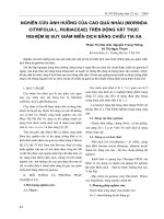
Figure 1 An HIV-infected patient with
pyomyositis (Salmonella) of his upper arm.
Axial CT image demonstrates an infiltrative
process (arrow) of the triceps muscle group.
Musculoskeletal Manifestations of HIV Infection
Journal of the American Academy of Orthopaedic Surgeons
314
muscle may set up the nidus for
infection in tropical regions, where
people often walk barefoot.
5
Neu-
trophil dysfunction in HIV-infected
patients may contribute to the pre-
disposition to bacterial infection.
Decreased chemotaxis, phagocyto-
sis, and leukocyte bactericidal activi-
ty also have been demonstrated in in
vitro studies.
6
An increased fre-
quency of a Staphylococcus carrier
state relative to the general popula-
tion also has been suggested in these
patients.
12
Early recognition and
aggressive management with par-
enteral antibiotics and surgical
drainage are the keys to treatment of
pyomyositis.
Polymyositis
Polymyositis (idiopathic inflam-
matory myositis) may be the first
manifestation of HIV.
13
This condi-
tion is characterized by bilateral,
symmetrical proximal muscle weak-
ness associated with elevated serum
CK levels. Its unique characteristics
make polymyositis distinguishable
from pyomyositis, which usually
occurs in advanced stages of HIV.
Polymyositis may be related to a
direct invasion of muscle tissue by
the virus, causing a cytopathic effect
and subsequent muscle fiber necro-
sis. Dalakas et al
14
showed that
polymyositis developed in 50% of
primates infected with the simian
acquired immunodeficiency syn-
drome virus. The simian virus was
isolated from muscle tissue homoge-
nates in these primates. In humans,
the presence of HIV has been detect-
ed by polymerase chain reaction in
skeletal muscle.
15
Other researchers
have postulated that HIV infection
causes an immunogenic response
that leads to the invasion of muscle
by inflammatory elements.
13
The
demonstration of a mononuclear
cell infiltrate that includes CD4 and
CD8 T lymphocytes in involved
muscle supports this hypothesis. A
third theory is that an underlying op-
portunistic infection causes chronic
antigenic stimulation and a reactive
response in muscle tissue.
16
The diagnosis of polymyositis is
made both on the clinical presenta-
tion of progressive hip and shoulder
girdle weakness and on the data
from objective studies, such as MRI,
electromyogram (EMG), and muscle
biopsy. Because the inflammatory
process is isointense relative to mus-
cle, it may be difficult to discern on
T1-weighted MRI. However, in-
creased signal intensity can be de-
tected on T2-weighted images. Un-
like pyomyositis, rim enhancement
is not present. Histologic examina-
tion shows extensive perivascular
and interstitial lymphocytic infiltra-
tion, necrosis, and phagocytosis of
degenerated muscle tissue. EMG
studies demonstrate myopathic pat-
terns characterized by short dura-
tion, low amplitude, and polyphasic
potentials. Fibrillation potentials at
rest are seen occasionally.
13
Results
of nerve conduction studies are nor-
mal.
Treatment with anti-inflammatory
agents, including oral prednisone,
has been extremely effective. Al-
though the use of systemic steroids
in an immunocompromised patient
must be carefully weighed, the use
of prednisone at doses of up to 60
mg/day does not seem to cause
worsening of the HIV infection or
faster progression to AIDS.
13
AZT Myopathy
Long-term treatment with AZT
can induce a reversible toxic mito-
chondrial myopathy that mimics
polymyositis clinically. AZT myop-
athy is usually dose-related and has
been reported in as many as 17% of
patients on chronic AZT therapy.
16
Patients usually present with myal-
gia, fatigue, proximal muscle weak-
ness, and elevated serum CK levels.
Myopathic changes are evident on
EMG studies. Patients improve with
discontinuation of the medication,
and both the serum CK and EMG
measures subsequently normalize.
AZT myopathy is considered multi-
factorial and may involve nutritional
deficiencies.
16
Discontinuation of
AZT and alternative antiviral ther-
apy is recommended for patients
with AZT myopathy. The ability of
AZT to induce myopathy in nonin-
fected individuals or to cause toxicity
in muscle cell cultures has not been
demonstrated.
A B
Figure 2 An HIV-infected patient with pyomyositis (Staphylococcus aureus) of his thigh.
A, Axial T1-weighted MRI scan demonstrates low-intensity signal (arrow) in adductor
muscles and adjacent ischium. B, T2-weighted MRI scan in the same patient demonstrates
high signal intensity (arrow) in the adductor muscles and ischium.
Ayaz A. Biviji, MD, et al
Vol 10, No 5, September/October 2002
315
Skeletal Infections
Tuberculosis Osteomyelitis
The HIV epidemic has led to a
resurgence of tuberculosis and is a
serious setback in the war against
this Mycobacterium infection.
17
Until
1985, the incidence of tuberculosis
steadily declined in industrialized
countries. Recent increases in tuber-
culosis cases in the United States are
directly attributable to the rising
number of patients immunocom-
promised by HIV infection. The
prevalence of tuberculosis is 500
times greater in HIV-infected per-
sons than in the non–HIV-infected
population.
17
This increased sus-
ceptibility is caused by a depletion
and functional impairment of CD4
lymphocytes, macrophages, and
monocytes. These patients are also
at increased risk of latent tuberculo-
sis reactivation and extrapulmonary
disease, including tuberculosis
osteomyelitis (Fig. 3). M tuberculosis
is a common pathogen in HIV-
infected individuals who have mus-
culoskeletal infections. In some
regions of Africa, one third of adults
are HIV positive, and tuberculosis
infections have radically changed
orthopaedic practice. Orthopaedic
surgeons in North America and
Europe now must reacquaint them-
selves with treating these infections.
Tuberculosis osteomyelitis devel-
ops from a hematogenous seeding
of M tuberculosis from a newly
acquired or reactivated pulmonary
infection. Of 188 consecutive tuber-
culosis patients treated at the Uni-
versity of Zambia, the spine was in-
volved in 66%, the hip in 18%, the
knee in 10%, other joints in 5%, and
other bones in 1%.
18
The thoracic
and lumbar (especially L1) regions
are most commonly affected; in-
volvement of the cervical spine or
sacrum is atypical. In contrast to
bacterial infections, tuberculosis
starts in the vertebral body and
spreads to adjacent disk spaces.
19
The infection usually begins in the
anterior portion of the vertebral
body and may spread underneath
the anterior longitudinal ligament
and extend into the soft tissues.
Soft-tissue extension in the lumbar
spine may lead to a psoas abscess
with the characteristic calcifications
that are a distinguishing feature of
tuberculosis. In other bones, the
metaphyseal region is most com-
monly involved, sometimes leading
to joint sepsis.
Tuberculosis can be differentiated
from bacterial infections on plain
radiographs by the presence of cal-
cified (cold) soft-tissue abscesses
and late disk involvement relative
to the vertebral body in the spine.
MRI findings are similar to those of
bacterial osteomyelitis, with low
signal on T1-weighted images and
high signal on T2-weighted images.
In the thoracic spine, the infection
may extend posteriorly into the
epidural space and cause cord com-
pression. Treatment consists pri-
marily of chemotherapy, with surgi-
cal débridement and stabilization
procedures reserved for refractory
cases, progressive neurologic deficit,
or structural instability. The dura-
tion of antibiotic tuberculosis thera-
py usually is longer in HIV-infected
patients than in immunocompetent
patients, with standard treatments
often lasting longer than 1 year.
Bacillary Angiomatosis
Bacillary angiomatosis is a unique
multisystem infection caused by the
gram-negative rod Bartonella henselae
(formerly Rochalimaea henselae) that
is seen exclusively in immunocom-
promised patients. Epidemiologic
data suggest that both a cat bite and
cat scratch are strong risk factors.
Distinct vascular proliferations of
skin, viscera, and lymph nodes usu-
ally are seen in this infectious
process. Multiorgan involvement
may include adenitis, intracerebral
mass lesions, aseptic meningitis,
peliosis hepatis, and osteomyelitis.
Cutaneous lesions are characterized
by friable angiomatous papules,
which may be difficult to distinguish
from Kaposi’s sarcoma lesions. The
presence of osseous lesions, which
are not typically seen with Kaposi’s
sarcoma, may help differentiate this
disease. One third of patients with
bacillary angiomatosis have osseous
lesions, which are lytic and can be
associated with periostitis and a
soft-tissue mass
20
(Fig. 4). Extensive
cortical damage and medullary per-
meation are seen on radiographs,
often preceding the cutaneous le-
sions by many months. The overly-
ing skin changes may resemble cel-
lulitis. There is usually an increased
uptake on technetium 99m bone
scan, and MRI shows the nonspecific
changes of osteomyelitis. Warthin-
Starry silver staining is used to iden-
tify the bacillary organism. Because
bacillary angiomatous osteomyelitis
can be successfully treated with eryth-
romycin, biopsy and early institution
of therapy are recommended in any
HIV-infected patient presenting with
an osteolytic lesion. Such lesions
Figure 3 An HIV-infected patient with
tuberculosis osteomyelitis of the right hand
and wrist. Anteroposterior radiograph of
the hand shows extensive destructive
changes of the carpus with periosteal reac-
tion of the adjacent metacarpals.
Musculoskeletal Manifestations of HIV Infection
Journal of the American Academy of Orthopaedic Surgeons
316
have been shown to regress with anti-
biotics. If left undiagnosed and un-
treated, bacillary angiomatosis may
be fatal, with patients succumbing to
overwhelming infection with the
involvement of many internal organs.
Neoplastic Conditions
Non-Hodgkin’s Lymphoma
The first reports of non-Hodg-
kin’s lymphoma in patients with
AIDS appeared in 1982. It is the sec-
ond most common type of tumor in
HIV-infected persons after Kaposi’s
sarcoma and is 60 times more preva-
lent relative to the general popula-
tion.
21
Non-Hodgkin’s lymphoma
tends to be more aggressive in HIV-
infected individuals, with presenta-
tion at more advanced stages, and it
is one of the diagnostic criteria for
AIDS. Casado et al
7
found that HIV-
positive patients with bone lym-
phoma had a mean CD4 count of
130 cells/mm
3
. Extranodal involve-
ment, including the central nervous
system, bone marrow, abdominal
organs, and mucocutaneous sites, is
frequently seen. Lymphomatous in-
filtration of muscle may be difficult
to differentiate from pyomyositis.
Primary and secondary bone
involvement is reported in 20% to
30% of cases and predominantly
affects the lower extremities.
21
Pa-
tients may present with pain, fever,
weight loss, and a pathologic frac-
ture. On plain radiographs, the
lesion usually appears osteolytic,
with cortical destruction that can
have a permeative pattern. Other
features include purely sclerotic
lesions or mixed sclerotic and osteo-
lytic lesions with an indistinct zone
of transition. Periosteal reaction
with an associated soft-tissue mass
also may be seen. MRI can show
the extent of bone marrow involve-
ment, which typically has a low sig-
nal appearance on T1-weighted
images and a high signal appear-
ance on T2-weighted images. Bac-
terial osteomyelitis also may have
the same appearance on imaging
studies and must be considered in
the differential diagnosis. Biopsy
definitively confirms diagnosis, and
treatment includes chemotherapy
and radiation, with surgical debulk-
ing in selected cases.
Kaposi’s Sarcoma
An unusual clustering of cases of
Kaposi’s sarcoma reported in 1982
led to the discovery of HIV. This
multifocal neoplasm arises from
lymphatic endothelial cells and may
progress to tumorous masses.
Approximately 20% of AIDS pa-
tients eventually develop Kaposi’s
sarcoma.
1
Osseous involvement, al-
though rare, is seen more frequently
with the endemic African type of
Kaposi’s sarcoma. In a Ugandan
study,
22
10 of 16 patients with florid
Kaposi’s sarcoma had bone lesions
ranging from discrete erosions to
diffuse osteopenia on plain radio-
graphs. Cortical destruction directly
beneath the cutaneous tumors was
observed in 13 of these patients.
Treatment consists of chemotherapy
and radiation.
Inflammatory
Arthropathies
Knowledge of rheumatic manifesta-
tions of HIV continues to evolve. In
1985, the National Institute of
Arthritis and Musculoskeletal and
Skin Disease began to investigate the
rheumatic complications of HIV.
Several authors have suggested a
possible co-occurrence of Reiter’s
syndrome and other reactive arthrit-
ides with HIV because of the higher
prevalence of the conditions in HIV-
infected persons.
23
A similar ob-
servation has been made in Africa,
where reactive arthritides, which are
usually related to outbreaks of dys-
entery during the rainy season, also
have become more frequent since
the outbreak of the HIV epidemic.
18
These arthropathies are more severe
and respond poorly to standard
therapies in HIV-positive individu-
als. Interestingly, immune-mediated
rheumatic conditions, such as sys-
temic lupus erythematosus and
rheumatoid arthritis, improve as the
immune system deteriorates.
1,24
One third of HIV-infected patients
experience arthralgias during the
course of their disease, and it may be
the first manifestation of HIV for
some patients.
2
Several arthropa-
thies, such as Reiter’s syndrome,
psoriatic arthritis, HIV-associated
arthritis, painful articular syndrome,
acute symmetric polyarthritis, and
hypertrophic osteoarthropathy, are
either more prevalent in or distinctly
unique to the HIV-infected popula-
tion. The spectrum of presentations
ranges from mild arthralgia to
severe joint disability. The etiology
and pathogenesis of these arthropa-
thies are controversial and poorly
understood. These conditions may
even simulate septic arthritis, mak-
ing their diagnosis and management
even more challenging.
Reiter’s Syndrome
Reiter’s syndrome is 100 to 200
times more frequent in the HIV-
infected population than in the non-
infected, with a prevalence rate of
5% to 10%.
25
In 1987, Winchester et
al
23
described a group of 13 patients
with HIV and Reiter’s syndrome. In
Figure 4 An HIV-infected patient with
bacillary angiomatosis. Lateral radiograph
of the radius shows a lytic lesion of the cor-
tex (asterisk) with an associated periostitis.
*
Ayaz A. Biviji, MD, et al
Vol 10, No 5, September/October 2002
317
this series, the pattern of joint in-
volvement tended to be oligoarticu-
lar, with a predisposition for the
lower extremities. Nine patients
were HLA-B27 antigen positive.
Enthesopathic manifestations, pre-
dominantly Achilles tendinitis, were
noted in 11 of the patients and were
described as painful and incapaci-
tating.
Reiter’s syndrome is remarkable
for its severity in HIV-positive
patients. The debilitating clinical
course is usually refractory to non-
steroidal anti-inflammatory drugs
(NSAIDs). The classic triad of ure-
thritis, conjunctivitis, and seronega-
tive arthritis may be observed, as
well as asymmetric oligoarthritis
that mainly affects the large joints in
the lower extremity. Presentation in
the foot includes an enthesopathy,
which frequently involves the Achil-
les tendon, plantar fascia, and exten-
sor tendons, as well as anterior and
posterior tibial tendons. Clinically
this may be termed AIDS foot,
which presents as a broad-based gait
with weight bearing through the lat-
eral margins of the feet to protect the
painful heel. It can be extremely dis-
abling, forcing some patients to be
wheelchair bound, and may mimic a
peripheral neuropathy. Upper ex-
tremity enthesopathy may include
medial or lateral epicondylitis, rota-
tor cuff tendinitis, de Quervain’s
tenosynovitis, or flexor tendinitis.
Axial skeleton involvement is rare.
Osteopenia and erosion at tendi-
nous insertion sites (eg, Achilles
tendon and plantar fascia) are often
seen. The erythrocyte sedimenta-
tion rate and C-reactive protein
level are usually elevated, and syn-
ovial fluid analysis reveals a white
blood cell count in the range of
27,000 to 50,000/mm
3
.
The etiology of Reiter’s syndrome
is unclear. HLA-B27 antigen is pres-
ent in approximately 70% to 80% of
affected patients, compared with 6%
to 10% in the general population.
23
Many investigators have concluded
that either an enteric or venereal in-
fection is the environmental link in
the pathogenesis of Reiter’s syn-
drome. Winchester et al
25
reported
that 4 of the 13 patients in their
series had an antecedent enteric
infection with either Shigella flexneri
or Campylobacter jejuni. Eleven of the
13 patients had a diarrheal illness
that preceded the arthritis. Enteric
Yersinia infection also has been re-
ported in the development of Reiter’s
syndrome. A growing body of liter-
ature supports the theory that a mo-
lecular mimicry between MHC class
I antigens and the infecting bacteria
in an HLA-B27–positive host trig-
gers these arthropathies. The im-
mune system essentially is tricked
into attacking tissue containing HLA-
B27 antigen as well as the offending
organism. Amino acid sequences
shared with HLA-B27 have been
demonstrated for Klebsiella pneumo-
niae and S flexneri. HIV-infected
patients are more susceptible to these
enteric infections, which also include
Giardia lamblia and Chlamydia tra-
chomatis. This heightened suscepti-
bility may account for the higher
prevalence of these arthropathies in
the HIV-infected population.
The management of Reiter’s syn-
drome is often difficult. Although
NSAIDs are often ineffective, sec-
ond-line agents such as phenylbuta-
zone and sulfasalazine have been
shown to be more effective.
3
Immu-
nosuppressive agents, including
cyclosporine and prednisone, have
been used successfully in patients
with symptoms refractory to initial
treatments. However, the use of
methotrexate is contraindicated.
Full-blown AIDS and Kaposi’s sar-
coma may develop after methotrex-
ate treatment.
23
Psoriatic Arthritis
Psoriatic arthritis is 10 to 40 times
more frequent in the HIV-infected
population than in the general pop-
ulation, with a reported prevalence
between 2% and 3%.
26
These pa-
tients have severe cutaneous disease,
with the development of arthritis
preceding an accelerated progres-
sion to full-blown AIDS. Psoriatic
arthritis may be clinically difficult to
differentiate from Reiter’s syn-
drome. In one report,
26
the arthritis
preceded the psoriasis in 16% of
patients, making the diagnosis even
more challenging. Typical cuta-
neous manifestations include cir-
cumscribed, discrete, and confluent
red, silvery scaled maculopapules
that occur predominantly on the
elbow, knee, scalp, and trunk. Nail
changes can range from pitting to
severe destruction. Five patterns
have been described: asymmetric
oligoarthritis, symmetric polyarthri-
tis, dominant desquamative intersti-
tial pneumonia, arthritis mutilans,
and sacroiliitis or spondylitis with-
out peripheral involvement.
26
Syno-
vial white blood cells typically range
between 7,000 and 15,000/mm
3
.
Radiologic findings include marginal
erosion, soft-tissue swelling, osteo-
penia, osteolysis, and so-called pen-
cil-in-cup deformities in the digits.
Like Reiter’s syndrome, the patho-
genesis of psoriatic arthritis may be
related to the combination of a
genetic predisposition and an arthri-
togenic infection. Management of
psoriatic arthritis is similar to that
for Reiter’s syndrome.
HIV-Associated Arthritis
HIV-associated arthritis, a sub-
acute oligoarthritis, was first de-
scribed by Rynes et al
24
in 1988 and
is unique to HIV-infected patients.
Symptoms typically develop over 1
to 6 weeks and may last up to 6
months. Characteristic features in-
clude exquisite, incapacitating joint
pain, predominantly in the knee and
ankle. Synovial fluid analysis
reveals a noninflammatory reaction
with a white blood cell count in the
range of 50 to 2,600/mm
3
. Radio-
graphic findings are typically unre-
markable, although osteopenia may
be evident from disuse or chronic
Musculoskeletal Manifestations of HIV Infection
Journal of the American Academy of Orthopaedic Surgeons
318
synovitis. Synovial biopsy shows a
chronic process with a predominantly
mononuclear cell infiltrate.
Proposed etiologies include a
reactive mechanism to deposited
immune complexes or direct HIV
infection of the synovium. The latter
theory has been supported by a pos-
itive culture assay for HIV in the
synovial fluid of a patient with this
condition.
27
Neither rheumatoid
factor nor HLA-B27 antigen are
associated with this particular arthri-
tis, which may help distinguish it
from other arthropathies. Intra-
articular steroid injections have
proved to be an extremely effective,
safe, and rapid treatment, and no
cases of secondary infections have
been reported. HIV infection
should be considered with any sus-
picion of bacterial or mycobacterial
arthritis.
Painful Articular Syndrome
Painful articular syndrome is
seen in as many as 10% of patients
with AIDS.
24
The hallmark of this
arthritis is a sharp, severe arthralgia
of acute onset that often simulates a
septic joint. Emergency room care
or hospitalization is required in
more than half of cases.
2
The knee is
most commonly affected, but the
elbow and shoulders also can be
involved. This condition can be dis-
tinguished from a septic joint by its
intermittent pain pattern and lack of
effusion or synovitis on physical
examination. Joint aspiration re-
veals no inflammatory fluid and a
normal percentage of polymor-
phonucleocytes. This self-limited
condition lasts from 2 to 24 hours
and responds well to narcotics and
anti-inflammatory medications. Ra-
diographic features are nonspecific,
with occasional periarticular osteo-
penia seen. The mechanism for
this unique arthritis is speculative;
current theories include cytokine
elaboration and a transient bony
ischemia. Management is expectant
with analgesic medications.
Acute Symmetric Polyarthritis
Acute symmetric polyarthritis is
unique to HIV-infected patients and
resembles rheumatoid arthritis both
clinically and radiographically.
Small joint involvement in the hand
is common, and characteristic fea-
tures include ulnar deviation and
swan neck deformities. This rare
condition differs from rheumatoid
arthritis by its acute onset and the fre-
quent presence of negative rheuma-
toid factor. Periarticular osteopenia,
joint space narrowing, and marginal
erosion are seen on radiographs.
Unlike rheumatoid arthritis, prolifer-
ative changes also may be observed.
Gold has been used successfully as
treatment.
24
Hypertrophic Osteoarthropathy
Hypertrophic osteoarthropathy is
a systemic disorder seen with pul-
monary neoplasms and pyogenic
infections (eg, empyema and lung
abscesses) and can affect bones,
joints, and soft tissues. In HIV-
infected patients with Pneumocystis
carinii pneumonia, this condition re-
solves with treatment of the pneu-
monia.
28
Severe pain in the lower
extremity is typical, and clinical
manifestations include arthralgias,
nonpitting edema, digital clubbing,
and periarticular soft-tissue involve-
ment of the ankle, knee, and elbow.
Neurovascular changes of the hand
and foot, such as chronic erythema,
paresthesias, and increased sweat-
ing, can be observed. The skin
over affected areas often has a glis-
tening appearance and may feel
warm and edematous. Extensive
periosteal reaction and subpe-
riosteal proliferative changes of the
long bones in the lower extremity
are seen on plain radiographs.
Bone scan reveals increased uptake
along cortical surfaces. Treatment
is directed at the underlying pneu-
monia. Surgical or chemical vago-
tomy or radiation therapy has been
used to relieve bone pain in refrac-
tory cases.
Figure 5 A 34-year-old HIV-infected person developed bilateral hip pain 21 months after
initiation of protease inhibitor treatment. Anteroposterior pelvic radiograph demonstrates
subchondral lucency and collapse in the superior portion of the right femoral head and
patchy sclerosis of the left femoral head. These radiographic changes are consistent with
osteonecrosis of both femoral heads.
Ayaz A. Biviji, MD, et al
Vol 10, No 5, September/October 2002
319
Osteonecrosis
An increasing number of cases of
osteonecrosis of the femoral head in
the HIV-positive population have
been reported recently,
29
and a direct
link between HIV and osteonecrosis
has been proposed.
30
Embolic phe-
nomena secondary to the formation
of antiphospholipid antibodies and
immune complexes, protein S defi-
ciency, and hypergammaglobulin-
emia also have been proposed as eti-
ologies. Our series at the University
of California, San Francisco demon-
strated a strong association between
chronic protease inhibitor use and
the development of osteonecrosis in
HIV-infected patients
30
(Figs. 5 and
6). Lipid metabolism disturbances
secondary to protease inhibitors that
result in hyperlipidemic states and
an associated lipodystrophy syn-
drome have been described.
31
These
systemic effects may explain the rela-
tionship between protease inhibitor
use and osteonecrosis of the femoral
head. Osteonecrosis should be con-
sidered in the differential diagnosis
in HIV-infected patients with hip
pain. A higher vigilance for osteo-
necrosis is especially crucial for pa-
tients taking protease inhibitors.
Surgical Outcomes
In urban centers, 3% to 7% of ortho-
paedic patients who receive emer-
gent care and 1% to 3% who undergo
elective procedures are HIV posi-
tive.
32
Complications of greatest
concern include impaired bone and
soft-tissue healing as well as postop-
erative infections. Of HIV-infected
hemophiliac patients undergoing
elective arthroplasty and non-
arthroplasty procedures, asympto-
matic individuals with CD4 counts
>200 cells/mm
3
are not at higher
risk of postoperative infection than
the general population.
33
In the
trauma setting, however, asympto-
matic HIV-infected patients may be
at increased risk. Paiement et al
34
reported a fivefold increase in risk of
infection in HIV-infected patients
after open fractures. A longer dura-
tion of prophylactic parenteral anti-
biotics may be required in this subset
of patients. Careful preoperative
evaluation, including assessment of
the nutritional and immune status of
these patients, should be done before
elective orthopaedic surgery.
Routine HIV testing is a contro-
versial issue. It is not clear whether
there is any benefit to routine HIV
testing of trauma patients or elective
patients, either for the individual
patient or because of concerns for
public health. Guidelines from
national professional organizations,
where they do exist, are not very
specific. Principal consideration for
HIV testing is whether it is of bene-
fit to the patient, which is a basic
tenet of ethical practice. Trauma and
elective patients are not screened
routinely at our institution and we
do not recommend it.
Summary
Musculoskeletal manifestations of
HIV infection are frequent and
involve muscle, bone, and joints.
The disease processes can be inflam-
matory, infectious, or neoplastic and
often are unique to the HIV infec-
tion itself or to the immunocompro-
mising nature of this progressive
condition. Disease processes can be
either a direct consequence of the
virus and circulating immune com-
plexes or secondary to the oppor-
tunistic infections that occur in HIV-
infected persons. Patients often pre-
sent as a diagnostic and treatment
challenge. The treating orthopaedic
surgeon must take into considera-
tion the myriad of debilitating man-
ifestations of the disease and its
treatment to effectively manage this
unique group of patients. Espe-
cially for the orthopaedic surgeon
who does not routinely treat HIV-
positive patients, a multidiscipli-
nary approach is strongly recom-
mended, including a rheumatolo-
gist, physical therapist, neurologist,
infectious diseases specialist, pain
consultant, musculoskeletal radiolo-
gist, and the patient’s primary
physician. Patients presenting with
atypical musculoskeletal complaints
and significant risk factor for HIV
infection should be tested for HIV
as part of a rational diagnostic and
treatment plan.
A B
Figure 6 A 45-year-old HIV-infected person developed right hip pain 31 months after pro-
tease inhibitor treatment. A, T1-weighted coronal MRI scan of both hips shows ischemic
changes (arrow) involving the superior portion of the right femoral head, with loss of the
structural architecture. These findings are consistent with the diagnosis of osteonecrosis.
B, Gross specimen of the right femoral head at the time of total hip replacement demon-
strates collapse of the articular surface. Involvement is predominantly limited to the superior
portion of the femoral head. Pathology results confirmed the diagnosis of osteonecrosis.
Musculoskeletal Manifestations of HIV Infection
Journal of the American Academy of Orthopaedic Surgeons
320
References
1. Kahn JO, Walker BD: Acute human
immunodeficiency virus type I infec-
tion. N Engl J Med 1998;339:33-39.
2. Berman A, Espinoza LR, Diaz JD, et al:
Rheumatic manifestations of human
immunodeficiency virus infection. Am
J Med 1988;85:59-64.
3. Cuellar ML: HIV infection-associated
inflammatory musculoskeletal disor-
ders. Rheum Dis Clin North Am 1998;
24:403-421.
4. Scriba J: Bietrag zur aetiologie der
Myositis acuta. Dtsch Ztschr Chir 1885;
22:497-502.
5. Rodgers WB, Yodlowski ML, Mintzer
CM: Pyomyositis in patients who
have the human immunodeficiency
virus: Case report and review of the
literature. J Bone Joint Surg Am
1993;75:588-592.
6. Watts RA, Hoffbrand BI, Paton DF,
Davis JC: Pyomyositis associated with
human immunodeficiency virus infec-
tion. Br Med J (Clin Res Ed) 1987;294:
1524-1525.
7. Casado E, Olivé A, Holgado S, et al:
Musculoskeletal manifestations in
patients positive for human immuno-
deficiency virus: Correlation with CD4
count. J Rheumatol 2001;28:802-804.
8. Brown JD, Wheeler B: Pyomyositis:
Report of 18 cases in Hawaii. Arch
Intern Med 1984;144:1749-1751.
9. Chiedozi LC: Pyomyositis: Review of
205 cases in 112 patients. Am J Surg
1979;137:255-259.
10. Vassilopoulos D, Chalasani P, Jurado RL,
Workowski K, Agudelo CA: Musculo-
skeletal infections in patients with human
immunodeficiency virus infection.
Medicine (Baltimore) 1997;76:284-294.
11. Miyake H: Beiträge zur Kenntnis der
sogenannten Myositis infectiosa. Mitteil-
Grenzgeb Med Chir 1904;13:155-198.
12. Ganesh R, Castle D, McGibbon D,
Phillips I, Bradbeer C: Letter: Staphy-
lococcal carriage and HIV infection.
Lancet 1989;2:558.
13. Espinoza LR, Aguilar JL, Espinoza CG,
et al: Characteristics and pathogenesis
of myositis in human immunodefi-
ciency virus infection: Distinction from
azidothymidine-induced myopathy.
Rheum Dis Clin North Am 1991;17:
117-129.
14. Dalakas MC, Pezeshkpour GH, Gravell
M, Sever JL: Polymyositis associated
with AIDS retrovirus. JAMA 1986;256:
2381-2383.
15. Seidman R, Peress NS, Nuovo GJ: In
situ detection of polymerase chain
reaction-amplified HIV-1 nucleic acids
in skeletal muscle in patients with
myopathy. Mod Pathol 1994;7:369-375.
16. Chariot P, Gherardi R: Myopathy and
HIV infection. Curr Opin Rheumatol
1995;7:497-502.
17. Barnes PF, Bloch AB, Davidson PT,
Snider DE Jr: Tuberculosis in patients
with human immunodeficiency virus
infection. N Engl J Med 1991;324:1644-
1650.
18. Jellis JE: Orthopaedic surgery and
HIV disease in Africa. Int Orthop 1996;
20:253-256.
19. Bureau NJ, Cardinal E: Imaging of
musculoskeletal and spinal infections
in AIDS. Radiol Clin North Am 2001;39:
343-355.
20. Baron AL, Steinbach LS, LeBoit PE, Mills
CM, Gee JH, Berger TG: Osteolytic
lesions and bacillary angiomatosis in
HIV infection: Radiologic differentiation
from AIDS-related Kaposi sarcoma.
Radiology 1990;177:77-81.
21. Ziegler JL, Drew WL, Miner RC, et al:
Outbreak of Burkitt’s like lymphoma in
homosexual men. Lancet 1982;2:631-633.
22. Taylor JF, Templeton AC, Vogel CL,
Ziegler JL, Kyalwazi SK: Kaposi’s sar-
coma in Uganda: A clinico-pathologi-
cal study. Int J Cancer 1971;8:122-135.
23. Winchester R, Bernstein DH, Fischer
HD, Enlow R, Solomon G: The co-
occurrence of Reiter’s syndrome and
acquired immunodeficiency. Ann
Intern Med 1987;106:19-26.
24. Rynes RI, Goldenberg DL, DiGiacomo
R, Olson R, Hussain M, Veazey J: Ac-
quired immunodeficiency syndrome-
associated arthritis. Am J Med 1988;84:
810-816.
25. Winchester R, Brancato L, Itescu S,
Skovron ML, Solomon G: Implications
from the occurrence of Reiter’s syn-
drome and related disorders in associ-
ation with advanced HIV infection.
Scand J Rheumatol Suppl 1988;74:89-93.
26. Moll JMH, Wright V: Psoriatic arthri-
tis. Semin Arthritis Rheum 1973;3:55-78.
27. Withrington RH, Cornes P, Harris JR,
et al: Isolation of human immunodefi-
ciency virus from synovial fluid of a
patient with reactive arthritis. Br Med
J (Clin Res Ed) 1987;294:484.
28. Bhat S, Heurich AE, Vaquer RA, Dunn
EK, Strashun AM, Kamholz SL: Hyper-
trophic osteoarthropathy associated
with Pneumocystis carinii pneumonia in
AIDS. Chest 1989;96:1208-1209.
29. Calza L, Manfredi R, Mastroianni A,
Chiodo F: Osteonecrosis and highly
active antiretroviral therapy during
HIV infection: Report of a series and
literature review. AIDS Patient Care
STDs 2001;15:385-389.
30. Ries MD, Barcohana B, Davidson A,
Jergesen HE, Paiement GD: Association
between human immunodeficiency
virus and osteonecrosis of the femoral
head. J Arthroplasty 2002;17:135-139.
31. Mallon PW, Cooper DA, Carr A: HIV-
associated lipodystrophy. HIV Medicine
2001;2:166-173.
32. Luck JV Jr, Logan LR, Benson DR,
Glasser DB: Human immunodeficien-
cy virus infection: Complications and
outcome of orthopaedic surgery. J Am
Acad Orthop Surg 1996;4:297-304.
33. Ragni MV, Crossett LS, Herndon JH:
Postoperative infection following or-
thopaedic surgery in human immuno-
deficiency virus-infected hemophiliacs
with CD4 counts < or = 200/mm
3
.
J Arthroplasty 1995;10:716-721.
34. Paiement GD, Hymes RA, LaDouceur
MS, Gosselin RA, Green HD: Postop-
erative infections in asymptomatic
HIV-seropositive orthopedic trauma
patients. J Trauma 1994;37:545-551.