Vai trò của các thuật chỉnh hình ppt
Bạn đang xem bản rút gọn của tài liệu. Xem và tải ngay bản đầy đủ của tài liệu tại đây (448.81 KB, 14 trang )
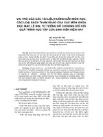
Vol 7, No 4, July/August 1999
217
Type 1 neurofibromatosis (NF-1), or
von Recklinghausen disease, is a
multisystem disease that primarily
affects cell growth of neural tissue.
It is an autosomal dominant disor-
der, with approximately 50% of
cases due to new mutation. The
entity is quite common, affecting 1 in
4,000 individuals, and is one of the
most common dominantly inherited
gene disorders in humans. In 1990,
the gene locus of NF-1 in humans
was cloned, and its protein product,
neurofibromin, was identified.
1
In
1993, the gene for central neurofibro-
matosis (NF-2) was cloned, and its
protein product, merlin or schwanno-
min, was identified.
2,3
Patients with NF-1 may develop
Schwann cell tumors called neuro-
fibromas and pigmentation abnor-
malities. In children with NF-1,
complications are associated with
both the orthopaedic manifesta-
tions of the disease and the treat-
ment thereof. Strategies for their
evaluation and management are
critical.
Neurofibromatosis was first
graphically described by Tilesius
von Tilenau in 1793. Von Reckling-
hausen was the first (in 1882) to
associate the origin of this disorder
with tumors arising from cells of
the nerve sheaths.
Historically, NF-1 is well known
because of the Òelephant man,Ó
Joseph Carey Merrick, who was a
medical curiosity in London in the
1880s.
4
His disfiguring deformity
of the head, extremity involvement,
and vertebral deformities made him
a celebrity. More recently, Cohen
5
has speculated that Merrick had
Proteus syndrome rather than NF-1.
Nevertheless, the interest in his case
brought needed attention to NF-1.
The fact that Weber, one of the
more renowned dermatologists of
his time, examined Merrick and
made no mention of cafŽ-au-lait
spots gives credibility to CohenÕs
treatise. Samples of MerrickÕs skin
were lost during World War II and
are not available for analysis.
Type 1 neurofibromatosis can be
clearly distinguished from NF-2,
which is also an autosomal domi-
nant disorder. The latter occurs
much more rarely and is estimated
to affect 1 in 40,000 individuals.
Characteristically, in NF-2 there are
bilateral schwannomas of the
vestibular portion of the eighth cra-
nial nerve. Schwannomas of other
Dr. Crawford is Professor of Orthopaedics and
Pediatrics, Division of Human Genetics,
ChildrenÕs Hospital Medical Center, Cincinnati.
Dr. Schorry is Assistant Professor of Pediatrics,
Division of Human Genetics, ChildrenÕs
Hospital Medical Center, Cincinnati.
Reprint requests: Dr. Crawford, Division of
Human Genetics, ChildrenÕs Hospital Medical
Center, 3333 Burnet Avenue, Cincinnati, OH
45229-3039.
Copyright 1999 by the American Academy of
Orthopaedic Surgeons.
Abstract
Type 1 neurofibromatosis (NF-1), also known as von Recklinghausen disease, is
one of the most common human single-gene disorders, affecting at least 1 mil-
lion persons throughout the world. It encompasses a spectrum of multifaceted
disorders and may present with a wide range of clinical manifestations, includ-
ing abnormalities of the skin, nervous tissue, bones, and soft tissues. The condi-
tion can be conclusively diagnosed when two of seven criteria established by the
National Institutes of Health Consensus Development Conference are met.
Most children with NF-1 have no major orthopaedic problems. For those with
musculoskeletal involvement, the most important issue is early recognition.
Spinal deformity, congenital tibial dysplasia (congenital bowing and
pseudarthrosis), and disorders of excessive bone and soft-tissue growth are the
three types of musculoskeletal manifestations that require evaluation. Statistics
gathered from the Cincinnati ChildrenÕs Hospital Neurofibromatosis Center
database show the incidence of spinal deformity in children with NF-1 to be
23.6%; pectus deformity, 4.3%; limb-length inequality, 7.1%; congenital tibial
dysplasia, 5.7%; hemihypertrophy, 1.4%; and plexiform neurofibromas, 25%.
The orthopaedic complications can be managed, but only rarely are they cured.
J Am Acad Orthop Surg 1999;7:217-230
Neurofibromatosis in Children:
The Role of the Orthopaedist
Alvin H. Crawford, MD, FACS, and Elizabeth K. Schorry, MD
peripheral nerves, meningiomas,
and ependymomas are also com-
mon. Tumors of the eighth cranial
nerve are not found in NF-1. The
gene for NF-2 has been localized on
the long arm of chromosome 22
and cloned. Because NF-2 does not
seem to have any musculoskeletal
manifestations, it will not be dis-
cussed in this article.
Approximately 50% of all NF-1
cases are new mutations, which is
100-fold higher than the usual
mutation rate for a single locus and
may reflect the huge size of the NF-1
locus (estimated at 350,000 base
pairs). (Most genes are composed
of several tens of thousands of base
pairs; the largest known, the gene
for Duchenne muscular dystrophy,
extends over 2.5 million base pairs.)
Prenatal testing is now possible in
families with multiple affected gen-
erations and in patients with an
identifiable mutation.
Mutations for NF-1 may be iden-
tified in the laboratory with the
protein truncation test. This test
can be used to detect an abnormal-
ly shortened protein product due
to gene mutations. Unfortunately,
the protein truncation assay can
detect only 70% of the NF-1 muta-
tions, making it a less than ideal
diagnostic test for NF-1.
6
The manifestations of NF-1 vary
from one person to another, but
each individual who carries the
gene will eventually show some
clinical features of the disease, the
penetrance for NF-1 being close to
100%. Cloning of the gene has
allowed creation of animal models,
which may ultimately be used to
develop more effective therapy
against the disease.
The Consensus Development
Conference on Neurofibromatosis
at the National Institutes of Health
in 1987 concluded that the diagno-
sis of NF-1 could be assigned on the
basis of the presence of two or more
of the criteria shown in Table 1.
7
These criteria have been shown to
be very useful even in young chil-
dren. Since the consensus panel
meeting, various types of learning
disabilities and magnetic resonance
(MR) imaging abnormalities (espe-
cially in children) have also been
specifically associated with NF-1.
Other disorders of pigmentation,
such as McCune-Albright and
Leopard syndrome, can be con-
fused with von Recklinghausen
neurofibromatosis. Genetic studies
have shown that Watson syndrome,
but not Noonan syndrome, may be
linked to the NF-1 locus.
8,9
Clinical Features
Café-au-Lait Spots
CafŽ-au-lait spots are hyperpig-
mented macules that are usually
ovoid and have smooth, well-
defined borders (Fig. 1). CafŽ-au-
lait spots are present in more than
90% of patients with NF-1. The pig-
mentation is melanotic in origin
and is located both in the basal
layer of the epidermis and in the
melanocytes of the upper layers.
The lesions are usually found in
skin areas not exposed to the sun.
Crowe et al
10
concluded that an
adult who has more than six cafŽ-
au-lait spots measuring 1.5 cm or
more in diameter should be as-
sumed to have NF-1. Whitehouse
11
evaluated 365 children under the
age of 5 and concluded that fewer
than two cafŽ-au-lait spots is a com-
mon and normal phenomenon in
children but more than five spots
with a diameter of at least 0.5 cm
should be considered diagnostic of
NF-1. Commonly, an infant or
young child with no family history
of NF-1 presents with multiple cafŽ-
au-lait spots. Unfortunately, the
diagnosis of NF-1 cannot be made
in the absence of other features. The
family should be told that NF-1 is
likely to be the diagnosis, as familial
cafŽ-au-lait spots are exceedingly
rare. Additional criteria are almost
always met by the age of 10 years.
12
Cutaneous Neurofibromas
Neurofibromas are mixed cell
tumors that are rich in Schwann cells
but also include fibroblasts, endothe-
lial cells, and glandular elements.
The primary cell responsible for
tumor formation is unknown. The
tumor (formerly called fibroma mol-
luscum) is usually raised over the
skin surface and is slightly bluish.
The tumors are generally seen in
small numbers in preadolescence
but tend to appear more extensively
after puberty and pregnancy.
13
Neurofibromatosis in Children
Journal of the American Academy of Orthopaedic Surgeons
218
Table 1
Criteria for Diagnosis of Neurofibromatosis, as Established by the NIH
Consensus Development Conference
7
More than six cafŽ-au-lait spots, at least 15 mm in greatest diameter in adults
and 5 mm in children
Two or more neurofibromas of any type or one plexiform neurofibroma
Freckling in the axillae or inguinal regions (Crowe sign)
Optic glioma
Two or more Lisch nodules (iris hamartomas)
A distinctive bone lesion, such as sphenoid dysplasia or thinning of the cortex
of a long bone, with or without pseudarthrosis
A first-degree relative (parent, sibling, or offspring) with NF-1 by the above
criteria
Plexiform Neurofibroma
Plexiform neurofibroma is a very
sensitive subcutaneous neurofibroma
with a ropy Òbag of wormsÓ feeling.
Plexiform neurofibromas are often
found underlying an area of cuta-
neous hyperpigmentation. The pig-
mentation is purplish with indistinct
edges, and the skin lesion is slightly
raised. When the pigmentation
approaches or crosses the midline of
the body, it is likely that the tumor
originates from the spinal canal and
will be aggressive. A plexiform neuro-
fibroma has the potential to become
malignant.
Elephantiasis
Elephantiasis is another derma-
tologic manifestation of the disease.
This condition is characterized by
large soft-tissue masses with rough,
raised, villous skin. Attempts to
resect sizable portions of the soft
tissue have met with limited suc-
cess. There is also dysplasia of the
underlying bone when the lesions
occur in an extremity.
14
Verrucous Hyperplasia
Verrucous hyperplasia is an in-
frequent and unsightly cutaneous
lesion of neurofibromatosis. There
is tremendous overgrowth of the
skin, with thickening but also a vel-
vety soft, papillary quality. Many
crevices form in this disorder; these
crevices tend to break down easily,
and some weeping occurs in the
skin folds. The sites often become
superficially infected, giving rise to
a foul odor. This condition most
often develops unilaterally and is
one of the most grotesque cuta-
neous lesions of NF-1.
Axillary and Inguinal Freckles
The presence of diffuse, small
(up to 2 to 3 mm in diameter), hy-
perpigmented spots in the axillae
and groin regions is helpful in the
diagnosis of NF-1. Axillary or
inguinal freckling is the second
most common feature after cafŽ-au-
lait spots to appear in children and
is confirmatory of a diagnosis of
NF-1.
12
A frequency of 81% by age
6 years has been reported.
15
The
recognition of this skin-fold freck-
ling in a young child with multiple
cafŽ-au-lait spots will permit an
early diagnosis of NF-1.
Lisch Nodules
Lisch nodules are slightly raised,
well-circumscribed hamartomas in
the iris. They are present in over 90%
of patients with NF-1 who are 6 years
of age or older. Some neurofibro-
matosis centers have reported inci-
dence rates of less than 50% by 5
years old but 90% by adulthood.
The lesions are thought to be spe-
cific for NF-1.
Optic Gliomas
Although optic gliomas account
for only 2% to 5% of all brain
tumors in childhood, as many as
70% of the cases are found in per-
sons with NF-1. In many NF-1 pa-
tients, these tumors change little in
size over a period of years, but a
small percentage may enlarge
rapidly, leading to exophthalmos
and visual impairment.
Severity
Type 1 neurofibromatosis pre-
sents with various degrees of severi-
ty. There seem to be two peaks of
severe clinical problems for NF-1
patients: one at 5 to 10 years and the
other at 36 to 50 years. At the sec-
ond peak, 75% of the clinical prob-
lems are related to malignancy.
16
For children, the most important ele-
ment is early recognition.
Spinal Deformities
Spinal deformities noted to
occur in NF-1 include both dys-
trophic and nondystrophic changes
in the vertebral bodies (Fig. 2). The
radiographic appearance of non-
dystrophic deformity consists of
wedging, angulation, and rotation
similar to that seen in idiopathic
deformities. The radiologic ap-
pearance of dystrophic changes
includes scalloping of the posterior
vertebral margins, severe rotation
of the apical vertebrae, vertebral
wedging, widening of the spinal
canal, enlargement of the neural
foramina, widened interpediculate
distance, defective pedicles, pres-
ence of a paraspinal mass, spin-
dling of the transverse process, and
rotation of the ribs resembling a
twisted ribbon (ÒpencilingÓ).
17
Rib
penciling is diagnosed when a rib
is smaller in diameter than the
midportion of the second rib.
These changes may be due to
intraspinal lesions, such as tumors,
meningoceles, and dural ectasia.
However, the changes may occur
even if the intraspinal contents are
entirely normal. In these cases, the
dystrophic changes have been
explained as a primary bone dys-
plasia.
Dural ectasia, meningoceles,
pseudomeningoceles, and Òdumb-
bellÓ lesions are all related to the
presence of neurofibroma or abnor-
mal pressure phenomena in and
about the spinal canal neuraxis.
Dural ectasia is a circumferential
dilatation of the dural sac. The
mechanism by which this occurs
has not been defined. The neural
elements are not abnormal or
enlarged, and the expanded area
contains increased cerebrospinal
Alvin H. Crawford, MD, FACS, and Elizabeth K. Schorry, MD
Vol 7, No 4, July/August 1999
219
Fig. 1 Multiple cafŽ-au-lait spots and
cutaneous neurofibromas on the trunk of a
patient with NF-1.
fluid and a brownish proteinaceous
material. The expanding dura
erodes the surrounding osseous
structures, widening the spinal
canal, thinning the laminae, and
destabilizing the vertebral ele-
ments. The process occasionally
results in dislocation of the verte-
bral column. Its expansion out-
ward through the neural foramina
causes meningoceles and will give
the radiographic dumbbell appear-
ance. Because of spinal canal
widening and expansion, there may
be tremendous angular deformity
and distortion without spinal cord
compromise or neurologic deficit.
Further destabilization at the costo-
vertebral junction has been associ-
ated with rib penetration into the
spinal canal with neurologic com-
promise.
18,19
Most often, however,
the single dumbbell lesion (visual-
ized as enlargement of a single
neural foramen on an oblique x-ray
film) is caused by a neurofibroma
exiting from the spinal canal rather
than dural ectasia (Fig. 3).
Unrecognized extrapleural tho-
racic tumors have presented as
focal scoliosis. These lesions are
usually plexiform neurofibromas
and are occasionally visible on
plain radiographs.
20
High-volume computed tomo-
graphic (CT) myelography or MR
imaging should be used in the
investigation of all dystrophic
curves before initiating treatment.
21
Intraspinal elements may occasion-
ally compromise the cord directly
when instrumentation and stabi-
lization are attempted or may cause
erosive changes in the bone, pre-
venting primary fusion or weaken-
ing existing fusion. Care must be
taken during surgical exposure to
avoid directly entering the spinal
canal through the very thin laminae
and injuring the spinal cord.
The cervical spine in NF-1 pa-
tients has not received enough
attention and should be evaluated
at the initial scoliosis investigation.
There may be early evidence of
dystrophic changes on lateral radio-
graphs. Cervical abnormalities
occur much more frequently when
scoliosis or kyphoscoliosis is pres-
ent in the thoracolumbar region,
and the examinerÕs attention may
be distracted by the more obvious
deformity. The most common cer-
vical abnormality is kyphosis,
which in itself is highly suggestive
of the disorder.
In one study of 56 patients with
NF-1, Yong-Hing et al
22
found that
17 had cervical abnormalities. Of
these, 7 were asymptomatic, and
the rest had either limited motion
or pain in the neck. Four had neu-
rologic deficits, which could proba-
bly be attributed to cervical insta-
bility. Four patients required
fusion of the cervical spine.
Curtis et al
23
described eight
patients with paraplegia and NF-1.
In four cases, the paraplegia was
due to cervical spine instability or
intraspinal disorders in the cervical
spine. Anterior dislocation of both
the upper and the lower cervical
spine has been reported.
24,25
Attention should also be paid to
C1 and C2. Isu described three
patients with NF-1 who had C1-2
dislocation with neurologic deficit.
All improved after decompression
or fusion.
17
In none of the patients
were there any obvious osseous
changes at the C1-C2 level on plain
radiographs. Cervical instability
sometimes develops after excision
of tumors and resection of the lami-
nae and posterior elements. Pro-
gressive kyphosis is more common
than nonprogressive kyphosis.
It is important to obtain cervical
spine radiographs of all NF-1 pa-
tients who undergo surgery, require
endotracheal anesthesia, are placed
in halo traction, or present with neck
tumors, neck pain, torticollis, or dys-
Neurofibromatosis in Children
Journal of the American Academy of Orthopaedic Surgeons
220
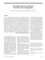
Fig. 2 Spinal deformities associated with NF-1. A, Nondystrophic-appearing changes in
the vertebral body associated with spinal deformities. The appearance is very similar to
that of idiopathic scoliosis. B, Myelogram shows widening of the spinal canal and the
characteristic short, segmented, sharply angulated deformity associated with neurofibro-
matosis.
A B
phagia. Widening of the neural
foramina on oblique views may be
represented by the dumbbell lesions
characteristic of neurofibromas exit-
ing the spinal canal. If there is any
suspicion of subluxation, CT and/or
MR imaging are appropriate.
Scoliosis
Scoliosis is the most common os-
seous defect associated with NF-1.
It may vary in severity from mild,
nonprogressive forms to severe
curvatures. The cause of spinal
deformity is unknown, but it may
be secondary to osteomalacia, a
localized neurofibroma that erodes
bone, an endocrine disturbance, or
mesodermal dysplasia.
The exact prevalence of spinal
deformity in NF-1 is unknown. We
have reservations about estimating
the incidence on the basis of the
occurrence in populations in tertiary
referral institutions with a primary
interest in the disease or in spinal
deformity. In a general orthopaedic
clinic, 2% of the scoliosis population
will have NF-1, whereas in an NF-1
population, perhaps 10% to 30% of
patients will have some disorder of
the spine.
26
Although both dystrophic and
nondystrophic abnormalities are
possible, most patients with spinal
deformities who are seen in neu-
rofibromatosis clinics have nondys-
trophic deformities. Functional
scoliosis resulting from limb hyper-
trophy or long-bone dysplasia
must be ruled out in patients with
NF-1.
27
Preadolescent children
with NF-1 should be evaluated for
scoliosis, which occurs earlier in
children with NF-1.
The dystrophic curvature is
characterized by short segments,
sharp angulation with severe apical
rotation and usually less than six
spinal segments, vertebral scallop-
ing, spindled ribs or a transverse
process, a paravertebral soft-tissue
mass, foraminal enlargement, and
occasionally thinned defective
pedicles. (Not all conditions need
be present in any one individual.)
These anomalies may predispose to
subluxation or dislocation of a ver-
tebral body. The dystrophic type
has a tendency to progress to a
severe deformity.
28,29
Some centers have noted a high-
er incidence of thoracic lordosis in
young patients with dystrophic
curves
30
(Figs. 2, B; 4). Others have
noted a higher incidence of prima-
ry kyphosis in patients with dys-
trophic curvatures and have found
it useful to divide this group on the
basis of whether the deformity is
angular or secondary to severe ver-
tebral rotation.
29,31
A trend to a
higher incidence of left convex dor-
sal curvatures has also been noted.
Left convex spinal deformities are
known for their association with
spinal axis tumors.
32
Nondystrophic curvature is
quite similar to the idiopathic cur-
vature seen in adolescents and
appears to usually involve eight to
ten spinal segments (Fig 2, A). It is
most often convex to the right;
however, this is not consistent. The
majority of patients with spinal
deformities in the NF-1 population
have the nondystrophic type.
Dystrophic curvatures of less
than 20 degrees should be observed
for progression at 6-month inter-
vals. For curvatures greater than 20
to 40 degrees, a posterior spinal
fusion with segmental spinal instru-
mentation is recommended.
33
Be-
cause of the potential for unabated
progression, modulation, and pseud-
Alvin H. Crawford, MD, FACS, and Elizabeth K. Schorry, MD
Vol 7, No 4, July/August 1999
221
Fig. 3 A, Axial computed tomographic (CT) image shows erosive defects and foraminal and spinal canal widening due to a dumbbell
lesion. B, Axial CT image obtained at another level in the same patient shows that soft-tissue instability has allowed subluxation of one
rib head into the spinal canal. C, Dumbbell tumor was removed from the neural foramen at the time of surgery. The dumbbell appear-
ance refers to the conscription of the neurofibroma that occurs at the neural foramen, where the lesion exits the spinal canal.
A B C
arthrosis in patients with dystrophic
curvatures, surgery is recommended
for progressive deformities of lesser
magnitude.
34
This is in contradis-
tinction to the situation in patients
with idiopathic scoliosis, in whom
surgical stabilization of curvatures
measuring less than 40 degrees is
usually inappropriate.
Dystrophic curves greater than
50 degrees should be treated with
anterior and posterior fusion.
Although thoracotomy is generally
necessary, we have recently used
video-assisted thoracoscopy to per-
form anterior release, costoplasty,
and intervertebral fusion.
35
Oblique
x-ray views should be taken every 6
months to rule out pseudarthrosis.
Brace treatment alone has not been
effective in the management of dys-
trophic deformities. For the very
young child, early fusion will result
in minimal stunting of growth.
Nondystrophic curvatures of less
than 20 degrees should be observed,
those measuring 20 to 35 degrees
should be braced, and those over 35
degrees should be treated with fu-
sion. Close observation and follow-
up are essential because of the ten-
dency of these curves to modulate
to dystrophic curves.
33
Funasaki et
al
29
have documented the develop-
ment of certain dystrophic features
over time. It is possible that some
patients are simply too young at
presentation to show the typical
manifestations of vertebral dyspla-
sia. Patients with nondystrophic
deformity have a higher incidence
of pseudarthrosis after attempts at
fusion and surgical stabilization
than patients with idiopathic scolio-
sis.
33,36
If indicated, the fusion mass can
be evaluated with technetium-99m
bone scanning or tomography.
Future refinements of MR imaging
may allow identification of pseud-
arthrosis.
37
These tests may not be
conclusive, and exploration and
graft reinforcement may be neces-
sary. Complications of spinal sur-
gery include bleeding from plexi-
form venous channels, dural leaks,
and paraplegia.
Kyphoscoliosis
The kyphoscoliosis seen in NF-1
is distinguished by acute sagittal-
plane angulation. The term is used
in those cases in which the scoliosis
is accompanied by kyphosis greater
than 50 degrees (Fig. 5). The verte-
bral bodies may be so severely
deformed as to cause them to be
confused with congenital anom-
alies. Occasionally, weakening of
the spinal stabilizers (e.g., facets,
pedicles, and ligaments) by dural
ectasia with meningocele formation
gives rise to kyphosis with subluxa-
tion and dislocation of the spine.
24
Even with severe ÒhairpinÓ angula-
tion, the neurologic status may
remain normal, and the spinal cord
is protected because of the widened
spinal canal.
Bracing is recommended for pa-
tients with kyphosis measuring less
than 50 degrees. Traction may be
dangerous when performed on rigid
deformities because it increases ten-
sion on the midapical spinal cord,
which may cause neurologic dam-
age. Dystrophic rigid kyphotic cur-
vatures greater than 50 degrees can
rarely be stabilized with posterior
fusion alone and are better treated
by combined anterior-posterior seg-
mental instrumentation.
26,32
Once a
curvature exceeds 70 degrees, indefi-
nite bracing may be required even
after anterior-posterior spinal sur-
gery.
Because of the association of
paraplegia with kyphosis, there has
been a tendency to perform lami-
nectomy. When neurologic changes
of cord compression secondary to
kyphosis are present, a trial of halo
traction is warranted. The myelopa-
thy will often improve, at which
time anterior and posterior fusion
with halo immobilization may be
performed. If the myelopathy does
not improve, anterior decompres-
sion followed by anterior-posterior
fusion is indicated.
Laminectomy alone for kyphotic
cord compression is absolutely
contraindicated for two reasons.
First, the cord compression is usu-
ally anterior, and resection of the
posterior element predisposes the
spine to instability. Second, resec-
tion removes valuable bone stock
required for fusion. Spinal fusion
should always be performed after
laminectomy.
Lordoscoliosis
The condition in which the ky-
photic curve is physiologically nor-
mal but the sagittal-plane contour
measures less than normal magni-
tude has been recognized as a sepa-
rate entity in a small percentage of
patients.
26,30,38,39
The prognostic
implications of this deformity have
not been clarified. It is well known
that lordoscoliosis is associated
with mitral valve prolapse and
decreased pulmonary function.
40,41
Lordoscoliosis appears more com-
monly in patients with dystrophic
Neurofibromatosis in Children
Journal of the American Academy of Orthopaedic Surgeons
222
Fig. 4 Severe thoracic lordosis.
deformities with dural ectasia caus-
ing considerable thinning of the
posterior elements. Surgical plan-
ning should include instrumenta-
tion and fusion well above the lor-
dotic area because of the tendency
to junctional kyphosis at the cervico-
thoracic junction.
Spondylolisthesis
Spondylolisthesis is a very rare
finding, most often associated with
pathologic luxation of the lumbar
vertebrae because of elongation
and erosion of the pedicles or pars
by lumbosacral foraminal neurofi-
broma or dural ectasia. It is impor-
tant to evaluate the entire spine,
including the sacral segments, in
every patient. Anterior and poste-
rior stabilization is recommended
for progressive deformity.
39
Failure to recognize intraspinal
lesions in patients with neurofibro-
matosis who undergo manipula-
tion and instrumentation of the
spine may result in neurologic
compromise. Preoperative radiog-
raphy, CT, or MR imaging is essen-
tial for patients with dystrophic
vertebral elements and curvatures
requiring instrumentation and
fusion. Most patients with a signif-
icant deformity have no preopera-
tive neurologic deficit. It is the sur-
geonÕs responsibility to stabilize
the spine in the most expedient,
safe, and permanent method with-
out causing neurologic injury.
Congenital Tibial Dysplasia
Congenital tibial dysplasia
(CTD) was first described by
Hatzoecher in 1708. ÒCongenital
pseudarthrosisÓ is a misnomer;
CTD is the preferred term. It may
present initially as either anterolat-
eral bowing or a frank fracture.
Congenital tibial dysplasia is
rare, occurring in 1 per 140,000
live-born children. In contrast, its
incidence is 1% to 2% in patients
with NF-1. The deformity may
present before the other common
manifestations, such as cafŽ-au-lait
spots. It is usually evident within
the first year of life, with a fracture
not uncommonly occurring by the
age of 2 to 2
1
Ú2 years. Posteromedial
congenital bowing, or Òkyphosco-
liosis tibia,Ó is a benign condition
associated with occasional limb-
length inequality.
Tibial bowing associated with
skin dimples, bilateral presentation,
ring constrictions, and foot deformi-
ties is rarely associated with NF-1.
The early appearance of callus and
subperiosteal new bone on the pos-
teromedial concavity of an antero-
laterally bowed tibia and the lack of
involvement of the fibula are diag-
nostic of a spontaneously resolving
benign condition.
42
The management of CTD associ-
ated with NF-1 is frustrating, and
complications are frequent. Frac-
ture and refracture are common
after treatment and are often more
frequent when the residual angular
deformity is excessive. Stiffness of
the ankle joint invariably occurs
because of the need for rigid immo-
bilization during treatment. Be-
cause of the possibility of nonunion
or pseudarthrosis after osteotomy,
an increase in angular deformity
should not be addressed surgically
if the limb has not fractured and
can be braced. Limb-length in-
equality may occur because of dis-
use atrophy as well as deficiency of
growth potential of the distal tibial
physis.
Most patients with CTD and
angular deformity have a valgus
deformity of the ankle. The valgus
is caused by a deficiency in the
fibular lateral buttress due to either
fracture and/or pseudarthrosis in
the lower part of the fibula or a
sloping distal tibial epiphysis.
43
Surgical treatment of CTD does not
appear to be particularly success-
ful; the results of prefracture brace
treatment are more encouraging.
We have recently performed epiph-
yseal stapling and percutaneous-
screw epiphysiodesis of the distal
medial tibia to correct ankle valgus.
Efforts have been made to classify
the diverse forms of CTD by either
Alvin H. Crawford, MD, FACS, and Elizabeth K. Schorry, MD
Vol 7, No 4, July/August 1999
223
Fig. 5 This child presented with severe kyphoscoliosis. He had undergone three
attempts at posterior spinal fusion, all of which were unsuccessful. The kyphosis was in
excess of 100 degrees.
radiographic or pathologic criteria to
aid in determining prognosis and
selecting appropriate treatment
alternatives. Unfortunately, incon-
sistency of treatment and results is
the rule.
44-46
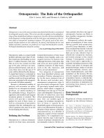
There are two basic
types of CTD, nondysplastic and
dysplastic (Table 2, Fig. 6).
The fibula may be primarily
involved by pseudarthrosis inde-
pendently or may be angulated
and/or pseudarthrotic with the
tibia. However, its management
will not be discussed here.
Treatment
Bracing
Bracing can be both preventive
and therapeutic (Fig. 7). Once the
diagnosis of NF-1 is suspected and
anterolateral bowing has been con-
firmed, the prewalker child should
be placed in an ankle-foot orthosis.
When the child starts to stand, a
change should be made to a poly-
propylene knee-ankle-foot orthosis.
The Òdrop-lockÓ type of knee-joint
hinge is added as the child gets
older, which allows sitting with the
knee flexed.
Orthotic support should be main-
tained until maturity whether or
not surgical osteosynthesis has been
achieved, because of the disastrous
consequences of fracture and refrac-
ture. An articulated above-knee
ankle brace is recommended arbi-
trarily after 10 years of age if the
stabilizing instrumentation has not
been extended across the ankle.
Pulsating Electromagnetic Fields
Treatment with a pulsating elec-
tromagnetic field is advocated for
some patients with progressive
CTD. The source may be externalÑ
the ÒclamshellÓ device over an
ankle-foot orthosis or an internal
unit implanted in the soft tissue
around the area of pseudarthrosis,
usually in conjunction with an autog-
enous bone graft. The effectiveness
of these forms of treatment remains
highly controversial.
47,48
Surgical Bone Grafting
Autogenous bone is placed into
the excised pseudarthrosis site. An
intramedullary rod is placed from
the proximal tibia across the pseud-
arthrosis site, incorporating the
graft and extending down through
the ankle across the talus and into
the calcaneus. The Williams tech-
nique of transankle stabilization
with a Rush rod has provided the
best surgical results in our center.
Residual ankle stiffness, growth
disturbance to the distal tibia, ankle
valgus, and lateral plantar nerve
entrapment have been documented
as complications of the proce-
dure.
49
The possibility of fractur-
ing at the tip of the nail during its
distal migration or during removal
or reinsertion is a further concern
with this procedure. Rod elonga-
tion and/or replacement is re-
quired as the limb grows, and con-
tinuous bracing is necessary.
50-52
Bypass prophylactic bone grafting
as a form of management as well
Neurofibromatosis in Children
Journal of the American Academy of Orthopaedic Surgeons
224
Table 2
Basic Types of Congenital Tibial
Dysplasia
Nondysplastic (type I)
Anterolateral bowing with
increased bone density
Sclerosis of the medullary canal
Possibility of conversion to dys-
plastic type after osteotomy to
correct the angulation
Dysplastic (type II)
Subtype A: Anterolateral bowing
with failure of tubularization
Subtype B: Anterolateral bowing
with cystic prefracture or canal
enlargement from previous
fracture
Subtype C: Frank pseudarthrosis
and atrophy with Òsucked candyÓ
narrowing of the ends of the two
fragments
Fig. 6 Classification of congenital tibial dysplasia. Type I is characterized by anterior lat-
eral bowing with increased cortical density and a narrow but normal medullary canal;
type IIA, by anterior lateral bowing with failure of tubularization and a widened
medullary canal; type IIB, by anterior lateral bowing with a cystic lesion before fracture or
canal enlargement from a previous fracture; type IIC, by frank pseudarthrosis and bone
atrophy with Òsucked candyÓ narrowing of the ends of the two fragments.
Type I Type IIA
Type IIB
Type IIC
as after fracture is used less fre-
quently.
53
Vascularized Autogenous Graft
The most commonly used vas-
cularized graft is the contralateral
fibula (Fig. 8), followed by the iliac
crest and rib. The graft is removed
extraperiosteally and placed into
the pseudarthrosis site. The blood
vessels are then anastomosed to
those normally supplying the tibia.
It is necessary to stabilize the grafted
segment. It is most important to
fuse or transfix the distal tibia and
fibula of the donor leg to prevent
proximal migration of the fibula
and ankle valgus.
52,54
Problems
associated with vascularized grafts
include failure to unite, further
pseudarthrosis, progressive angu-
lar deformity, failure to achieve
normal length, valgus ankle insta-
bility, and disability of the donor
limb.
18
Coleman and Coleman
50
have
recommended using the ipsilateral
fibula transferred with a vascular
pedicle. Their rationale is that two
abnormal legs are created with a
contralateral graft, whereas the
long-term effects of loss of the fibu-
la on the ankle joint of the normal
leg are unknown. This procedure is
recommended only after conven-
tional treatment has failed. It should
be noted that only five cases with
relatively short follow-up (average,
2.0 years) were reported.
Compression and Distraction
Histiogenesis
Compression and distraction
histiogenesis of bone and soft tis-
sue by the Ilizarov method pro-
vides many theoretical advantages
in the treatment of problems asso-
ciated with CTD. This method
allows the surgeon to address
limb-length inequality, angular
deformity, fibular nonunion, prox-
imal fibular migration, ankle val-
gus, and foot contractures. The
technique encompasses various
methods for treating the pseud-
arthrosis, including open reduc-
tion, resection and shortening,
compression/distraction, resection
and bone transport, and invagina-
tion of one end of the affected bone
in the other.
The Ilizarov method results in a
high rate of initial union; however,
the rate of refracture is extremely
high.
43
Another disadvantage of
the Ilizarov method has to do with
its external location, which is less
well tolerated by pediatric patients.
The myriad complications with the
procedure include joint stiffness,
cartilage necrosis, cystic bone le-
sions, dorsiflexion ankle contrac-
ture with calcaneovalgus deformity,
pin-track infection, loosening, pin
breakage, nerve injury, and com-
partment syndrome.
43
The pin-
holes also may create stress risers
after union.
Considerable preoperative plan-
ning is essential to identify solu-
tions for every possible problem.
Boero et al
46
recently concluded
that patients who were aged 5
years or older at operation had bet-
ter results with this technique.
Resection of the pseudarthrotic
Alvin H. Crawford, MD, FACS, and Elizabeth K. Schorry, MD
Vol 7, No 4, July/August 1999
225
A B
Fig. 7 Congenital tibial dysplasia treated with chronic bracing after a fracture. A,
Radiograph obtained after early union of a distal tibial fracture. An ankle-foot orthosis
was used at that time. When the patient started walking, a change was made to a knee-
ankle-foot orthosis. B, Radiograph taken 9 years later shows complete union of previous
fracture.
stumps, followed by short-term
compression with corticotomy or
epiphyseal distraction to correct
limb-length discrepancy, gave bet-
ter results than other combinations.
This technique avoids risking in-
jury to the contralateral leg, and
other treatment methods are not
excluded. Most authors who use
the Ilizarov technique advocate
continuing orthotic management
after successful treatment or until
skeletal maturity is achieved.
Amputation
The logic of performing multi-
ple surgical procedures after fail-
ure of three attempts at bone union
is questionable. If the ultimate
outcome will be a short, scarred
limb with a fibrotic ankle, amputa-
tion is a viable alternative. The
weight-bearing surface of the foot
should be maintained by means of
a Boyd-Syme procedure rather
than a midleg transbone amputa-
tion, which predisposes the child
to subsequent surgeries for bony
stump overgrowth. The resultant
length after the Boyd-Syme proce-
dure adds biomechanical stability
for prosthetic wear. Contrary to
previous reports, removing the
foot and then placing the limb in a
prosthesis that allows continuous
compression forces across the
pseudarthrosis has not resulted in
osseous union of the pseudarthro-
sis.
55
The new ÒSeattle footÓ and
ÒrunnerÕs footÓ have made chil-
drenÕs prosthetics much more
functional, and team sports like
soccer are not out of the question
after amputation.
Long-term Bracing
Regardless of the procedure
used, some form of bracing is re-
quired for all patients until skeletal
maturity, because of the marked
propensity for refracture and re-
currence of pseudarthrosis. The
diminished quality of life that is
the outcome of numerous unsuc-
cessful operative procedures makes
chronic bracing a reasonable alter-
native.
Treatment of Pseudarthrosis
Pseudarthrosis of other bones,
including the fibula (Fig. 9), ulna,
os pubis, and clavicle, is not as fre-
quent as pseudarthrosis of the
tibia. Nevertheless, the problems
of achieving synostosis are similar.
Fortunately, except for the fibula,
most of these bones are not weight
bearing and as a result pseudarthro-
sis is easier to manage; however,
there is a consistent tendency to de-
lay of synostosis after pseudarthro-
sis. Pseudarthrosis may develop
spontaneously, after fracture, or
after osteotomy of the involved
bone. The treatment and complica-
tions associated with treatment are
similar to those for pseudarthrosis
of the tibia.
Treatment of Bone-Growth
Disorders
Three disorders of bone growth
are segmental hypertrophy, cystic
lesions, and subperiosteal bone
growth and proliferation (Fig. 10).
Overgrowth of an extremity is not a
rare complication of NF-1 and may
be related to changes in the soft tis-
sues, such as hemangiomatosis,
lymphangiomatosis, elephantiasis,
and beaded plexiform neurofibro-
mas.
56
The zones of overgrowth in
the bone and soft tissues are usually
unilateral, involving the extremities
or the head and neck. The osseous
changes characteristically cause the
bone to elongate with wavy irregu-
larity or thickening of the cortex.
Macrodactyly is commonly seen in
NF-1, with disproportionate en-
largement of either the toes or the
fingers. Treatment is extremely em-
pirical and individualized. Some
combination of epiphysiodesis,
debulking, and neurofibroma resec-
tion is recommended.
57,58
The incidence of neoplasia asso-
ciated with segmental hypertrophy
is higher than that of other lesions.
Neurofibromatosis in Children
Journal of the American Academy of Orthopaedic Surgeons
226
A B
Fig. 8 Images of a child who underwent
vascularized fibular bone grafting. Previous
attempts at bone grafting and implanted
electromagnetic bone stimulation were
unsuccessful. The stimulator from the pre-
vious surgical procedure was not removed.
A, Radiograph shows construct of vascular-
ized fibular bone graft. B, Successful union
of vascularized graft.
Fig. 9 Anteroposterior (A) and lateral (B)
views of the leg of a child with congenital
fibular dysplasia and neurofibromatosis.
Note valgus deformity of the ankle joint
shown on anteroposterior view.
A B
Attempts to debulk the soft tissue
and resect the bone have not neces-
sarily resulted in a significant cos-
metic improvement. Early epiphy-
seal arrest of the involved bone and
leg lengthening on the normal
short side have achieved less than a
fair amount of success.
Neurosegmental overgrowth is a
condition in which hypertrophy of
the limb is due to nerve dysplasia
rather than primary bone involve-
ment. Autonomous hypertrophy of
the roots or trunks of the brachial or
lumbosacral plexus causes an in-
crease in the girth and length of the
limb. Unfortunately, the nerve is
the tumor, and the tumor is the
nerve. Therefore, resection of the
hypertrophied nerve causes neuro-
logic loss, and amputation may be
indicated.
Cystic lesions of bone may result
from extrinsic tumors as well as
intraosseous tumors similar to
those seen in the spine (Fig. 11). In-
traosseous lesions are often multi-
cystic and may represent intraos-
seous fibroma, neurofibroma, or a
manifestation of a bone dyspla-
sia.
38,59
The characteristic lesions of
NF-1 are radiologically similar to
nonossifying fibroma and may be
seen in the distal femur and proxi-
mal tibia. Biopsy specimens of
these lesions rarely show evidence
of neurofibroma and usually reveal
fibrous tissue variants.
Subperiosteal bone proliferation
is one of the protean manifestations
of NF-1.
60
Most cases are consid-
ered to be initiated by minor frac-
tures with subperiosteal bleeding,
followed by osseous dysplasia of
the subperiosteal hematoma. The
early onset of subperiosteal hema-
toma may be identified on techne-
tium bone scanning by the presence
of the Òdoughnut signÓ (a peripher-
al rim of increased activity sur-
rounding a relatively photopenic
center appearing on blood-pool and
delayed imaging).
61
This sign may
also arise because of central necro-
sis and surrounding hyperemia due
to tumor or abscess. In NF-1, the
doughnut sign is produced by
radionuclide accumulation in the
areas of microcalcification occur-
ring at the periphery of the subpe-
riosteal hematoma and enveloping
the nonossified organizing center.
The periosteum in NF-1 has been
described as less adherent to bone,
predisposing it to periosteal hemor-
rhage. As a result, extensive subpe-
riosteal hemorrhage can occur in
the bone underlying elephantoid
soft-tissue hypertrophy.
Other Conditions
Children with NF-1 tend to be
somewhat shorter than their unaf-
fected siblings, with relative macro-
cephaly. As many as 50% have some
form of learning disorder. It has
been suggested that there is a corre-
lation between cognitive dysfunction
and high-signal-intensity lesions,
(Òunidentified bright objectsÓ) on T2-
weighted MR images of the brain
(Fig. 12).
62,63
Development problems
may include speech delay, late
attainment of gross motor skills, and
mental retardation. Other complica-
tions of NF-1 include optic gliomas
and malignant tumors of the brain,
nerve, and/or spinal cord; congeni-
tal and acquired defects of the sphe-
noid bone of the skull; high blood
pressure, which may be related to
Alvin H. Crawford, MD, FACS, and Elizabeth K. Schorry, MD
Vol 7, No 4, July/August 1999
227
Fig. 10 The segmental hypertrophy and
overgrowth in this child are related to sub-
periosteal bone overgrowth. The patient
presented at 2
1
Ú2 years of age with trauma
and subperiosteal bone proliferation. The
resulting overgrowth and dysplasia pro-
gressed over a 3-year period. Attempts at
distal femoral and proximal tibial epiphy-
siodesis were unsuccessful.
Fig. 11 Cystic bone lesions in a child with
NF-1. The patient, a 12-year-old boy, pre-
sented with complaints of pain in his left
knee. Radiographs revealed a cystic lesion
that was considered to represent a patho-
logic fracture through the distal femoral
metaphyseal cyst. Subsequent radiographs
of both knees revealed the multiple cystic
areas associated with NF-1.
renal artery stenosis in children and
pheochromocytoma in adults; cere-
brovascular occlusion (stroke); and
pruritus.
Summary
In a 280-patient series from our
pediatric neurofibromatosis center,
most patients had cutaneous mani-
festations as their only presenting
symptoms. CafŽ-au-lait spots were
noted in 99% of patients, skin-fold
freckling in 82%, and cutaneous
neurofibromas in 37%. Neurologic
problems included frequent head-
aches in 28%, mental retardation in
6%, seizures in 9%, and learning
disabilities or unspecified school
performance problems in 36%. The
most common tumor type was
plexiform neurofibroma, in 25% of
patients. Thirteen percent of pa-
tients had optic nerve glioma (al-
though only 2% had progression to
visual impairment), 2.5% had
brainstem glioma, and 2% had
astrocytoma. Malignant neoplasms
were diagnosed in 2% of patients
and included neurofibrosarcoma,
rhabdomyosarcoma, and leukemia.
In all, 106 patients (38%) had one or
more orthopaedic findings, with
scoliosis being the most common.
Optimal management of the or-
thopaedic complications of NF-1
depends on recognition of the pro-
tean manifestations of the deformi-
ty and a complete evaluation of
both the patient and the family by a
multidisciplinary team according
to the nature of the lesions present.
Most important is the specific treat-
ment of individual lesions with a
highly organized approach, espe-
cially in reference to radical opera-
tive procedures. Observation must
be continued for the remainder of
the patientÕs life.
There is little doubt that NF-1
predisposes affected persons to an
increased risk of certain cancers.
Underrecognition of this relation-
ship may occur when the cuta-
neous signs of NF-1 are over-
looked or are not yet apparent in
young children. Neurofibrosar-
coma, childhood leukemia, rhab-
domyosarcoma of the urogenital
tract, and Wilms tumor have all
been reported in patients with NF-1.
Large plexiform neurofibromas
that present with rapid enlarge-
ment or progressive pain should
be critically evaluated for malig-
nant degeneration.
Although MR imaging is recom-
mended as a controlled investiga-
tory tool, consistently significant
enhancement of aggressive lesions
has not been documented. Several
multicenter trials for treatment of
NF-1Ðrelated tumors are currently
ongoing. These include a thera-
peutic trial of interferon, cis-
retinoic acid, and etoposide for
treatment of optic pathway gliomas
and plexiform neurofibromas,
which is being coordinated through
the ChildrenÕs Hospital of Phila-
delphia, and a National Cancer
Institute trial of phenylacetate for
plexiform neurofibromas. It is ex-
pected that additional medications,
including drugs that act by block-
ing the RAS pathway, will be avail-
able for clinical trials in the near
future.
It is important to remember that
approximately 65% of all patients
with NF-1 have only mild or mod-
erate involvement throughout their
lifetime. Fewer than 10% of pa-
tients will ever require orthopaedic
treatment. Furthermore, NF-1 is a
frustrating disease to treat effective-
ly because many orthopaedic com-
plications can only be managed, not
cured.
Neurofibromatosis in Children
Journal of the American Academy of Orthopaedic Surgeons
228
Fig. 12 MR image of the brain of a patient
with NF-1 shows the typical location of
increased signal intensity on T2-weighted
images (unidentified bright objects) in the
globus pallidus (arrow) and posterior thal-
amus (arrowhead).
References
1.Goldberg NS, Collins FS: The hunt for
the neurofibromatosis gene. Arch
Dermatol1991;127:1705-1707.
2.Rouleau GA, Merel PE, Lutchman M,
et al: Alteration in a new gene encod-
ing a putative membrane-organizing
protein causes neuro-fibromatosis
type 2. Nature1993;363:515-521.
3.Trofatter JA, MacCollin MM, Rutter
JL, et al: A novel moesin-, ezrin-,
radixin-like gene is a candidate for the
neurofibromatosis 2 tumor suppres-
sor. Cell1993;72:791-800.
4.Seward GR: The elephant man: Part I.
Br Dent J1990;169:173-175.
5.Cohen MM Jr: Further thoughts about
the elephant man. Am J Med Genet
1988;29:777-782.
6.Heim RA, Silverman LM, Farber RA,
Kam-Morgan LNW, Luce MC: Screening
for truncated NF-1 proteins. Nat Genet
1994;8:218-219.
7.NIH Consensus Development Con-
ference Statement: Neurofibromatosis.
Neurofibromatosis1988;1:172-178.
8.Ahlbom BE, Dahl N, Zetterqvist P,
AnnerŽn G: Noonan syndrome with
cafŽ-au-lait spots and multiple lentig-
ines syndrome are not linked to the
neurofibromatosis type 1 locus. Clin
Genet1995;48:85-89.
9.Allanson JE, Upadhyaya M, Watson
GH, et al: Watson syndrome: Is it a
subtype of type 1 neurofibromatosis?
J Med Genet1991;28:752-756.
10.Crowe FW, Schull WJ, Neel JW: A
Clinical, Pathological, and Genetic Study
of Multiple Neurofibromatosis. Spring-
field, Ill: Charles C Thomas, 1956.
11.Whitehouse D: Diagnostic value of
the cafŽ-au-lait spot in children. Arch
Dis Child1966;41:316-319.
12.Korf BR: Diagnostic outcome in chil-
dren with multiple cafŽ au lait spots.
Pediatrics1992;90:924-927.
13.Listernick R, Charrow J: Neurofibro-
matosis type 1 in childhood. J Pediatr
1990;116:845-853.
14.Pollnitz R: Neurofibromatosis in
childhood: A review of 25 cases. Med J
Aust1976;2:49-52.
15.Obringer AC, Meadows AT, Zackai
EH: The diagnosis of neurofibromato-
sis-1 in the child under the age of 6
years. Am J Dis Child1989;143:717-719.
16.Riccardi VM, Kleiner B: Neurofibro-
matosis: A neoplastic birth defect with
two age peaks of severe problems. Birth
Defects Orig Artic Ser1977;13:131-138.
17.Loop JW, Akeson WH, Clawson DK:
Acquired thoracic abnormalities in
neurofibromatosis. AJR Am J Roent-
genol1965;93:416-424.
18.Jacobsen FS, Crawford AH: Complica-
tions in neurofibromatosis, in Epps CH
Jr, Bowen JR (eds): Complications in
Pediatric Orthopaedic Surgery. Philadel-
phia: JB Lippincott, 1995, pp 649-683.
19.Major MR, Huizenga BA: Spinal cord
compression by displaced ribs in neu-
rofibromatosis: A report of three cases.
J Bone Joint Surg Am1988;70:1100-1102.
20.Schorry EK, Crawford AH, Egelhoff
JC, Lovell AM, Saal HM: Thoracic
tumors in children with neurofibro-
matosis-1. Am J Med Genet1997;74:
533-537.
21.Angtuaco EJC, Binet EF, Flanigan S:
Value of computed tomographic mye-
lography in neurofibromatosis. Neuro-
surgery1983;13:666-671.
22.Yong-Hing K, Kalamchi A, MacEwen
GD: Cervical spine abnormalities in
neurofibromatosis. J Bone Joint Surg
Am1979;61:695-699.
23.Curtis BH, Fisher RL, Butterfield WL,
Saunders FP: Neurofibromatosis with
paraplegia: Report of eight cases. J
Bone Joint Surg Am1969;51:843-847.
24.Stone JW, Bridwell KH, Shackelford
GD, Abramson CL: Dural ectasia as-
sociated with spontaneous dislocation
of the upper part of the thoracic spine
in neurofibromatosis: A case report
and review of the literature. J Bone
Joint Surg Am1987;69:1079-1083.
25.Samoto T, Watanabe Y, Suda A: At-
lantoaxial dislocation with neurofibro-
matosis: A case report. Orthop Trauma-
tol Surg (Japan)1981;24:289.
26.Akbarnia BA, Gabriel KR, Beckman E,
Chalk D: Prevalence of scoliosis in neu-
rofibromatosis. Spine1992;17(suppl 8):
S244-S248.
27.Crawford AH, Gabriel KR: Dysplastic
scoliosis in neurofibromatosis, in
Bridwell KH, Dewald RL (eds): The
Textbook of Spinal Surgery, 2nd ed.
Philadelphia: Lippincott-Raven, 1997,
vol 1, pp 277-298.
28.Hsu LCS, Lee PC, Leong JCY: Dys-
trophic spinal deformities in neurofi-
bromatosis: Treatment by anterior and
posterior fusion. J Bone Joint Surg Br
1984;66:495-499.
29.Funasaki H, Winter RB, Lonstein JB,
Denis F: Pathophysiology of spinal
deformities in neurofibromatosis: An
analysis of seventy-one patients who
had curves associated with dystrophic
changes. J Bone Joint Surg Am1994;76:
692-700.
30.Winter RB: Thoracic lordoscoliosis in
neurofibromatosis: Treatment by a Har-
rington rod with sublaminar wiringÑ
Report of two cases. J Bone Joint Surg
Am1984;66:1102-1106.
31.Winter RB, Lonstein JE, Anderson M:
Neurofibromatosis hyperkyphosis: A
review of 33 patients with kyphosis of
80¡or greater. J Spinal Disord1988;
1:39-49.
32.Coonrad RW, Richardson WJ, Oakes
WJ: Left thoracic curves can be differ-
ent. Orthop Trans1985;9:126-127.
33.Crawford AH: Pitfalls of spinal defor-
mities associated with neurofibro-
matosis in children. Clin Orthop1989;
245:29-42.
34.Savini R, Parisini P, Cervellati S,
Gualdrini G: Surgical treatment of ver-
tebral deformities in neurofibromato-
sis. Ital J Orthop Trauma1983;9:13-24.
35.Crawford AH: Anterior release of
spinal deformities, in Dickman CA,
Rosenthal DJ, Perrin NE (eds): Thoraco-
scopic Spine Surgery. New York:
Thieme Medical Publishers, 1999, pp
161-183.
36.Crawford AH: Neurofibromatosis, in
Weinstein SL (ed): The Pediatric Spine:
Principles and Practice. New York:
Raven Press, 1994, vol 1, pp 619-649.
37.Sirois JL III, Drennan JC: Dystrophic
spinal deformity in neurofibromatosis.
J Pediatr Orthop1990;10:522-526.
38.Joseph KN, Bowen JR, MacEwen GD:
Unusual orthopedic manifestations of
neurofibromatosis. Clin Orthop1992;
278:17-28.
39.Winter RB, Moe JH, Bradford DS,
Lonstein JE, Pedras CV, Weber AH:
Spine deformity in neurofibromatosis:
A review of one hundred and two pa-
tients. J Bone Joint Surg Am1979;61:
677-694.
40.Hirschfeld SS, Rudner C, Nash CL Jr,
Nussbaum E, Brower EM: Incidence
of mitral valve prolapse in adolescent
scoliosis and thoracic hypokyphosis.
Pediatrics1982;70:451-454.
41.Winter RB, Lovell WW, Moe JH: Ex-
cessive thoracic lordosis and loss of
pulmonary function in patients with
idiopathic scoliosis. J Bone Joint Surg
Am1975;57:972-977.
42.Tuncay IC, Johnston CE II, Birch JG:
Spontaneous resolution of congenital
anterolateral bowing of the tibia. J
Pediatr Orthop1994;14:599-602.
43.Paley D, Catagni M, Argnani F, Prevot
J, Bell D, Armstrong P: Treatment of
congenital pseudoarthrosis of the tibia
Alvin H. Crawford, MD, FACS, and Elizabeth K. Schorry, MD
Vol 7, No 4, July/August 1999
229
using the Ilizarov technique. Clin
Orthop1992;280:81-93.
44.Boyd HB: Pathology and natural his-
tory of congenital pseudarthrosis of
the tibia. Clin Orthop1982;166:5-13.
45.Crawford AH: Neurofibromatosis in
children. Acta Orthop Scand Suppl
1986;218:1-60.
46.Boero S, Catagni M, Donzelli O,
Facchini R, Frediani PV: Congenital
pseudarthrosis of the tibia associated
with neurofibromatosis-1: Treatment
with IlizarovÕs device. J Pediatr Orthop
1997;17:675-684.
47.Paterson D: Clinical pseudarthrosis of
the tibia. Clin Orthop1989;247:44-54.
48.Kort JS, Schink MM, Mitchell SN,
Bassett CAL: Congenital pseudarthro-
sis of the tibia: Treatment with puls-
ing electromagnetic fieldsÑThe inter-
national experience. Clin Orthop1982;
165:124-127.
49.Rao SB, Crawford AH: Lateral plantar
nerve entrapment following Rush rod
fixation for congenital pseudarthrosis of
the tibia. Orthopedics1966;19:1043-1046.
50.Coleman SS, Coleman DA: Congenital
pseudarthrosis of the tibia: Treatment
by transfer of the ipsilateral fibula
with vascular pedicle. J Pediatr Orthop
1994;14:156-160.
51.Anderson DJ, Schoenecker PL, Sher-
idan JJ, Rich MM: Use of an intramed-
ullary rod for the treatment of congen-
ital pseudarthrosis of the tibia. J Bone
Joint Surg Am1992;74:161-168.
52.Weiland AJ, Weiss APC, Moore JR,
Tolo VT: Vascularized fibular grafts in
the treatment of congenital pseudar-
throsis of the tibia. J Bone Joint Surg
Am1990;72:654-662.
53.McFarland B: Pseudarthrosis of the
tibia in childhood. J Bone Joint Surg Br
1951;33:36-46.
54.Gordon L, Weulker N, Jergesen H:
Vascularized fibular grafting for the
treatment of congenital pseudarthrosis
of the tibia. Orthopedics1986;9:825-831.
55.Jacobsen ST, Crawford AH, Millar EA,
Steel HH: The Syme amputation in
patients with congenital pseudarthro-
sis of the tibia. J Bone Joint Surg Am
1983;65:533-537.
56.Praharaj KC, Mohanta KD, Nanda BK,
Nanda CN: Congenital hemihypertro-
phy with neurofibromatosis. Indian J
Med Sci1969;23:146-150.
57.Crawford AH: Neurofibromatosis, in
Drennan JC (ed): The ChildÕs Foot and
Ankle. New York: Raven Press, 1992,
pp 439-459.
58.Turra S, Frizziero P, Cagnoni G,
Jacopetti T: Macrodactyly of the foot
associated with plexiform neurofibro-
ma of the medial plantar nerve. J
Pediatr Orthop1986;6:489-492.
59.Mandell GA, Dalinka MK, Coleman
BG: Fibrous lesions in the lower ex-
tremities in neurofibromatosis. AJR
Am J Roentgenol1979;133:1135-1138.
60.Yaghmai I, Tafazoli M: Massive sub-
periosteal hemorrhage in neurofibro-
matosis. Radiology1977;122:439-441.
61.Mandell GA, Harcke HT: Subperiosteal
hematoma: Another scintigraphic
Òdoughnut.Ó Clin Nucl Med1986;11:35-37.
62.North K, Joy P, Yuille D, et al: Specific
learning disability in children with
neurofibromatosis type 1: Significance
of MRI abnormalities. Neurology1994;
44:878-883.
63.Moore BD III, Slopis JM, Schomer D,
Jackson EF, Levy BM: Neuropsycho-
logical significance of areas of high sig-
nal intensity on brain MRIs of children
with neurofibromatosis. Neurology
1996;46:1660-1668.
Neurofibromatosis in Children
Journal of the American Academy of Orthopaedic Surgeons
230