Báo cáo y học: " Inflammatory cytokines, goblet cell hyperplasia and altered lung mechanics in Lgl1+/- mice" pptx
Bạn đang xem bản rút gọn của tài liệu. Xem và tải ngay bản đầy đủ của tài liệu tại đây (1.98 MB, 15 trang )
BioMed Central
Page 1 of 15
(page number not for citation purposes)
Respiratory Research
Open Access
Research
Inflammatory cytokines, goblet cell hyperplasia and altered lung
mechanics in Lgl1
+/-
mice
Jie Lan
1
, Leslie Ribeiro
†1,2
, Isabel Mandeville
†1
, Katia Nadeau
1,2
, Tim Bao
1,3
,
Salomon Cornejo
1,2
, Neil B Sweezey
4,5
and Feige Kaplan*
1,2,3,6
Address:
1
McGill University - Montreal Children's Hospital Research Institute Montreal, Quebec, H3Z2Z3, Canada,
2
Department of Human
Genetics, McGill University, Montreal, Quebec H3A1B1, Canada,
3
Department of Biology, McGill University Montreal, Quebec H3A1B1, Canada,
4
Hospital for Sick Children Research Institute, Toronto, Ontario M5G 1X8, Canada,
5
Departments of Pediatrics and Physiology, University of
Toronto, Toronto, Ontario, Canada and
6
Department of Pediatrics, McGill University, Montreal, Quebec, Canada
Email: Jie Lan - ; Leslie Ribeiro - ; Isabel Mandeville - ;
Katia Nadeau - ; Tim Bao - ; Salomon Cornejo - ;
Neil B Sweezey - ; Feige Kaplan* -
* Corresponding author †Equal contributors
Abstract
Background: Neonatal lung injury, a leading cause of morbidity in prematurely born infants, has
been associated with arrested alveolar development and is often accompanied by goblet cell
hyperplasia. Genes that regulate alveolarization and inflammation are likely to contribute to
susceptibility to neonatal lung injury. We previously cloned Lgl1, a developmentally regulated
secreted glycoprotein in the lung. In rat, O
2
toxicity caused reduced levels of Lgl1, which normalized
during recovery. We report here on the generation of an Lgl1 knockout mouse in order to
determine whether deficiency of Lgl1 is associated with arrested alveolarization and contributes to
neonatal lung injury.
Methods: An Lgl1 knockout mouse was generated by introduction of a neomycin cassette in exon
2 of the Lgl1 gene. To evaluate the pulmonary phenotype of Lgl1
+/-
mice, we assessed lung
morphology, Lgl1 RNA and protein, elastin fibers and lung function. We also analyzed tracheal
goblet cells, and expression of mucin, interleukin (IL)-4 and IL-13 as markers of inflammation.
Results: Absence of Lgl1 was lethal prior to lung formation. Postnatal Lgl1
+/-
lungs displayed
delayed histological maturation, goblet cell hyperplasia, fragmented elastin fibers, and elevated
expression of T
H
2 cytokines (IL-4 and IL-13). At one month of age, reduced expression of Lgl1 was
associated with elevated tropoelastin expression and altered pulmonary mechanics.
Conclusion: Our findings confirm that Lgl1 is essential for viability and is required for
developmental processes that precede lung formation. Lgl1
+/-
mice display a complex phenotype
characterized by delayed histological maturation, features of inflammation in the post-natal period
and altered lung mechanics at maturity. Lgl1 haploinsufficiency may contribute to lung disease in
prematurity and to increased risk for late-onset respiratory disease.
Published: 21 September 2009
Respiratory Research 2009, 10:83 doi:10.1186/1465-9921-10-83
Received: 2 April 2009
Accepted: 21 September 2009
This article is available from: />© 2009 Lan et al; licensee BioMed Central Ltd.
This is an Open Access article distributed under the terms of the Creative Commons Attribution License ( />),
which permits unrestricted use, distribution, and reproduction in any medium, provided the original work is properly cited.
Respiratory Research 2009, 10:83 />Page 2 of 15
(page number not for citation purposes)
Background
Impaired alveolar development is a leading cause of neo-
natal morbidity in premature infants weighing less than
one kilogram. Deficient alveolar maturation in these chil-
dren is often characterized by distal airspace enlargement,
disruption of elastin fibers and mucus cell hyperplasia.
Antenatal exposures of the premature lung may increase
susceptibility to inflammation and subsequent postnatal
(PN) lung injury. Genes that regulate alveolarization,
innate immunity and inflammation are likely to contrib-
ute to susceptibility to and outcome in neonatal lung dis-
ease.
In a search for downstream targets of glucocorticoid (GC)
that regulate lung maturation, we cloned Lgl1 (late gesta-
tion lung 1), a developmentally regulated gene in the lung
[1-3]. Lgl1 is a CRISP family (cystine rich secretory pro-
tein) protein characterized by a secretory signal and two
LCCL (also known as FCH) domains [4-6]. The LCCL
domain, an as yet poorly understood module found in
over 100 extracellular proteins, has been implicated in
directional cell migration and differentiation [5,7], extra-
cellular matrix deposition [5,8], cell adhesion [9] and
innate host-defense mechanisms [4,10,11]. While Lgl1
synthesis is almost exclusively restricted to the mesen-
chyme, Lgl1 protein is associated with lung epithelial cells
from the late canalicular period onward [2,3]. We showed
previously that Lgl1 protein stimulates airway branching
in lung explant culture [12]. Maximal fetal expression of
Lgl1 was, however, concordant with the onset of aug-
mented surfactant production in late gestation [1,3]. In
postnatal rat lung, Lgl1 concentrated at the tips of bud-
ding alveolar septa [3]. Levels of Lgl1 were drastically
reduced in rat O
2
toxicity models of bronchopulmonary
dysplasia (BPD), a chronic lung disease of impaired alve-
olarization in premature infants, and were restored during
recovery in air [3]. Taken together, these observations sug-
gested that Lgl1 may regulate both early and late events in
lung organogenesis.
We now report on the development of an Lgl1 knockout
mouse to investigate the in vivo function of Lgl1 in regulat-
ing multiple aspects of lung development. Absence of Lgl1
in homozygous null (Lgl1
-/-
) mice was not compatible
with life. We describe here the lung phenotype of Lgl1
+/-
heterozygous mice. Lungs of developing Lgl1
+/-
mice were
characterized by disorganized elastin fibers, early expres-
sion of inflammatory cytokines and goblet cell hyperpla-
sia. In mature Lgl1
+/-
mice, reduced Lgl1 expression was
associated with altered lung mechanics.
Methods
Generation of Lgl1 knockout mice
All procedures involving animals were conducted accord-
ing to criteria established by the Canadian Council for
Animal Care and approved by the Animal Care Commit-
tee of the McGill University Health Centre. A 13.7 kb
EcoR1 fragment of BAC clone 34304 containing the Lgl1
gene was subcloned into pQZ1BamH1 and a Neo cassette
was used to replace exon 2. Not1-Sal1 digestion of this
construct produced a 9.6 kb targeting fragment that was
electroporated into ES cells. Southern analysis and PCR
were used to verify accuracy of targeting. Mouse genotypes
were verified by PCR. The primers used were (5'-3'):
reverse (wild type) CACTGCTCCGTGTATCAAGCATA-
CAC; reverse (NeoI) GACAATCG GCTGCTCTGATG; or
reverse (5' to3') TCGTCGTGACCCATGGCGAT (NeoII)
and forward (for all 3 reactions) CAGGTCTGGCTCTGAG-
GTTCTTGCA. The expected amplification products were:
0.8 kb (wild type), 1 kb (Neo1) and 0.46 kb (Neo2). For
details on mouse preparation see Additional file 1.
Isolation of Total Lung RNA
Total RNA was prepared from lungs, brain, heart, kidney,
thymus and spleen using Trizol reagent (Invitrogen, Burl-
ington, ON, Canada) according to the manufacturer and
was resuspended in 1× RNASecure (Ambion, Austin, TX,
USA). RNA was pooled for each litter according to geno-
type (N ≥ 4).
Quantitative Real-Time RT-PCR
Quantitative real-time RT-PCR was performed on the
Mx4000 QPCR system (Stratagene, La Jolla, CA, USA)
using the Quantitect One-Step Probe RT-PCR Kit (Qiagen,
Mississauga, ON, Canada) as directed by supplier. Gene-
specific primers and FAM labeled probes for mouse LGL1,
IL-4, IL-13, Mucin5AC and Tropoelastin were designed
using Qiagen's online QuantiProbe Design Software.
Quantitect Gene Expression Assay for mouse 18S (Qia-
gen, Mississauga, ON, Canada) was used to normalize for
the input of RNA. The results were analyzed according to
the standard curve method. One-step real-time RT-PCR
reactions were performed in 25 μL volumes for 40 cycles,
using 20 ng of total RNA for Lgl1, IL-4, IL-13, Mucin5AC
and Tropoelastin and 50 pg for 18S. For a list of primers
and probes used see Additional file 2.
Bronchoalveolar Lavage (BAL)
BAL was performed by instilling the lungs four times with
1 ml cold phosphate-buffer saline through a tracheal can-
nula. Lavage fluid was centrifuged and pellets were resus-
pended in 0.5 ml cold saline. Total cell numbers were
counted with a haemocytometer. For differential cell
counts, cytospin slides were prepared (Cytospin 4; Shan-
don, Pittsburgh, PA) and stained with Diff-Quick; at least
200 cells/slide were counted and percentage of each cell
type was calculated.
Lung fixation
Lungs were inflated with 4% paraformaldehyde at a pres-
sure of 20 cm of water. Lungs were gently extracted and
fixed in 4% paraformaldehyde overnight. Samples were
Respiratory Research 2009, 10:83 />Page 3 of 15
(page number not for citation purposes)
dehydrated through a series of increasing ethanol washes
and embedded in paraffin. 5 μm thick tissue slices were
cut through the entire lung.
Antibodies
Lgl1, 1:100 (Covance, Quebec, Canada), β-actin, 1:5000
(Sigma-Aldrich, Oakville, ON, Canada), Anti-Rabbit IgG
HRP conjugated, 1:5000 (Amersham, Little Chalfont,
Buckinhamshire, UK), Anti-Rabbit IgG AlexaFluor 594
and Anti-Rat IgG AlexaFluor 488 conjugated, 1:200 (Inv-
itrogen, Burlington, ON, Canada), CD34, 1:100 (Abcam,
Cambridge, MA, USA).
Immunohistochemistry
Sections of paraffin-embedded lung tissue were stained
with hematoxylin and eosin or used for histochemical
staining. Ten sections from each of at least 4 and up to 7
animals were assessed for all histochemical experiments
and representative images shown in all figures. For immu-
nohistochemistry, slides were rehydrated through a series
of decreasing ethanol washes, rinsed with PBS-0.03% Tri-
ton and incubated in warm 10 mM sodium citrate for
antigen retrieval. Slides were then incubated in H
2
O
2
and
methanol for 20 minutes to block endogenous peroxidase
activity. To block non-specific binding, slides were incu-
bated in PBS-0.03% Triton containing 5% normal goat
serum and 1% BSA. Primary antibodies were incubated
overnight at 4°C and the following day in corresponding
fluorescent conjugated secondary antibody for 30 min-
utes at room temperature. For CD34 and Lgl1 co-immu-
nohistochemistry, the slides were then washed with PBS-
0.03% Triton and blocked again with 5% normal goat
serum and 1% BSA. The second primary antibody was
incubated overnight at 4°C and the following day in cor-
responding fluorescent conjugated secondary antibody
for 30 minutes at room temperature. Slides were washed
with PBS-0.03% Triton and mounted with pro-long anti-
fade media containing DAPI (Invitrogen, Burlington, ON,
Canada). For the co-staining, the DAPI is not shown to
improve visualization. For Lgl1 immunohistochemistry,
protein levels were quantified using the Northern Eclipse
program (Northern Eclipse software, Empix Imaging, Inc.
Mississauga, ON, Canada) as per Nadeau et al. 2006 [3].
Identification of Goblet cells
Slides were rehydrated through a series of decreasing eth-
anol washes and stained with Periodic Acid-Schiff kit
(Sigma-Aldrich, Oakville, ON, Canada) to visualize gob-
let cells. For goblet cell staining, sections from at least 6
and up to 10 animals were assessed and representative
images shown in all figures.
Elastin Staining
Slides were rehydrated through a series of decreasing eth-
anol washes and elastin fibers were stained with Fuschin
Weigert stain and counterstained with methyl green for
better visualization. For elastin staining sections from at
least 4 and up to 7 animals were assessed and representa-
tive images shown in all figures.
Morphometry
Mouse lungs were fixed under constant distending pres-
sure of 20 cm of fixative. Morphometric measurements
were made on hematoxylin and eosin stained lung sec-
tions (n > 5 mice). A minimum of 10 representative fields
were studied in each lung. A computer generated grid
(384 intersections) was superimposed on digital images
and grid intersections were examined to determine
whether they localized to airspace or tissue. Percent frac-
tional airspace or fractional area of lung parenchyma was
quantified using Northern Eclipse software.
Lung Mechanics
At 4 weeks of age, mice were deeply anaesthetized by an
i.p. injection of xylazine (8 mg/kg) and pentobarbital (70
mg/kg), tracheotomized and placed on a small animal
ventilator (flexiVent, SCIREQ, Canada). Animals were
ventilated quasi-sinusoidally (150 breaths/min and tidal
volume of 10 ml/kg) and subsequently paralyzed by an
i.p. injection of pancuronium bromide (0.8 mg/kg). Max-
imal resistance and elastance were recorded before and
after increasing doses of aerosolized methacholine.
Statistical Analysis
All results are expressed as mean ± standard error of the
mean. P ≤ 0.05 was considered to be statistically signifi-
cant as measured by student t-test or ANOVA as appropri-
ate.
The numbers of goblet cells in postnatal day 14 lungs dis-
played a single modal distribution in wild type, but a
marked bimodal distribution in heterozygous (Lgl1
+/-
)
mice. Subgroup analyses of the heterozygous mice
revealed the data were not normally distributed; a Mann-
Whitney U test showed a significant difference between
the subgroups (p < 0.02).
Results
Absence of Lgl1 is associated with embryonic lethality
An Lgl1 knockout mouse was generated by introduction
of a neomycin cassette in exon 2 of the Lgl1 gene. Analysis
of progeny (79 litters, 133 wild type and 290 heterozy-
gotes) revealed no homozygous Lgl1
-/-
progeny or resorp-
tion sites (from embryonic day (E) 9.5 until birth).
We explored the lung phenotype of Lgl1
+/-
heterozygotes
from E14.5 until maturity at postnatal day (PN) 28.
Lgl1
+/-
mice have altered lung morphology
Heterozygous (Lgl1
+/-
) mice appeared normal at birth and
exhibited no obvious changes in gross morphology. Lung
and body weight and lung to body weight ratios were nor-
Respiratory Research 2009, 10:83 />Page 4 of 15
(page number not for citation purposes)
mal. On morphometric analysis, the ratio of respiratory
tissue to airspace throughout the entire lung was signifi-
cantly increased in Lgl1
+/-
mice at PN1 (tissue fraction
[39.0 ± 3.51] % vs [23.48 ± 2.46] % in wild type, p < 0.04).
Visual inspection of the histological sections revealed that
this difference was distributed unevenly, with patchy areas
of distinctly thickened respiratory interstitium alternating
with areas of relatively normal appearance (Figure 1). No
such patches were seen in wild type lungs. With advancing
PN age, distinct areas with thickened interstitium could
still be identified in the lungs of heterozygote (but not
wild type) mice to a diminishing degree; however, the
observed trends towards an increased tissue to airspace
ratio for the entire lung no longer reached statistical sig-
nificance. At PN14, when lungs of wild type animals
showed advanced alveolarization, lungs of Lgl1
+/-
mice
appeared to be at an earlier, more active phase of second-
ary septation with fewer alveolar secondary septa and rel-
atively enlarged distal airspaces. By PN28, the lung
morphology of Lgl1
+/-
mice was indistinguishable from
that of wildtype littermates.
Lgl1 mRNA and protein expression in Lgl1
+/-
mice
Given the observed morphological changes, we expected
that PN Lgl1
+/-
mice would display aberrant expression of
Lgl1 mRNA and Lgl1 protein. We used real-time quantita-
tive RT-PCR to compare lung Lgl1 mRNA levels in Lgl1
+/-
and Lgl1
+/+
mice (Figure 2). No significant differences
were observed in Lgl1 mRNA in the lungs of Lgl1
+/-
mice
compared to controls during the course of lung develop-
ment from E9.5 until PN14 (Figure 2A, B). By contrast, at
4 weeks of postnatal age, when lung development in the
mouse is essentially complete (alveolarization occurs
mainly between PN1-PN14), a significant reduction in
Lgl1 mRNA (~ 50%) was observed (Figure 2C). Given the
absence of significantly altered Lgl1 expression in the
lungs of developing Lgl1
+/-
mice, we considered the possi-
bility that effects on lung morphology were indirect and
resulted from aberrant Lgl1 expression in other organs. No
significant differences in Lgl1 mRNA expression were
observed in developing heart, brain, kidney, spleen and
thymus (not shown). Mature Lgl1
+/-
mice, however,
showed a limited but significant reduction in Lgl1 mRNA
expression in the heart-concordant with the observed
changes in lung Lgl1 mRNA expression (Figure 2D). These
findings suggest that aberrant expression of Lgl1 in Lgl1
+/-
mice is tissue- and temporal-specific and may depend on
availability of local and circulating regulatory factors.
We next assessed whether variance in Lgl1 mRNA would
be reflected in coordinate changes in levels and/or distri-
bution of Lgl1 protein. Several Lgl1 antibodies were pre-
pared but none consistently detected Lgl1 on Western
blots. Lgl1 protein was therefore analyzed by immunohis-
tochemistry (IHC). Representative images of Lgl1 IHC (n
≥ 4) are illustrated in Figure 3. Northern Eclipse software
was used to quantify Lgl1 immunostaining. From PN7
onward, lungs of Lgl1
+/-
mice appeared to have reduced
levels of Lgl1 protein, most markedly at PN28. There was
considerable variability among pups. In previous studies,
we showed that Lgl1 protein concentrated at the tips of
septating alveoli in PN7 rat lung[3]. In PN7 mice, Lgl1
appeared to be more widely distributed in the lung and
accumulation at septal tips was more clearly noted at
PN14. In Lgl1
+/-
mice, a reduction in Lgl1 protein at the
tips of alveolar septa was observed at PN14 (Figure 3, see
arrows).
Lgl1 protein does not localize to PN pulmonary
endothelial cells
Lung alveoli are lined by specialized Type 1 and Type 2
epithelial cells and are vascularised by an extensive capil-
lary bed[13]. The developing lung mesenchyme under-
goes vasculogenesis and angiogenesis. The eventual
juxtaposition of Type 1 cells with pulmonary endothelial
cells is required to facilitate gas exchange. Consistent with
previous findings in rat lung,Lgl1 localized to both mesen-
chyme and epithelium in PN murine lung with high con-
centrations noted in epithelium surrounding the larger
airways (Figure 3). To assess whether Lgl1 is present in
endothelial cells, we evaluated colocalization of Lgl1
immunoreactivity (Figure 4, red color) with the endothe-
lial marker CD34 (green color) in PN1-PN14 lung sec-
tions prepared from wild type and Lgl1
+/-
mice.
Representative images are shown in Figure 4 (n ≥ 4). No
evidence of colocalization, which would appear as yellow
color in merged images, was observed.
Abnormal pulmonary mechanics in methacholine
challenged Lgl1
+/-
mice
To clarify the significance of the observed reduction of
Lgl1 expression in mature mouse lung, we assessed lung
function in Lgl1
+/+
and Lgl1
+/-
mice at PN28. Initially, base-
line lung mechanics were analyzed using the flexiVent
small animal ventilator (Figure 5). No significant changes
in lung resistance (R), compliance (C) or elastance (E)
were observed when Lgl1
+/-
mice were compared with
Lgl1
+/+
littermates. We next administered methacholine
(MCh), a smooth muscle agonist, to assess the effects on
R and E of transient bronchoconstriction. Interestingly,
increasing doses of MCh provoked a significantly greater
increase in airway resistance (Figure 5A) and elastance
(Figure 5B) in wild type mice than that observed in Lgl1
+/
-
mice. The observed effects on lung elastance suggested
the possibility of altered elastin expression and/or deposi-
tion in developing lungs of Lgl1
+/-
mice.
Lungs of Lgl1
+/-
mice display altered tropoelastin
expression and disorganized elastin fibers
Chronic lung injury in the newborn has been associated
with disordered elastin deposition[14]. Reduced lung
elastance is also a characteristic feature of emphysematous
Respiratory Research 2009, 10:83 />Page 5 of 15
(page number not for citation purposes)
Lgl1
+/-
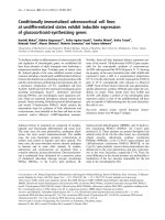
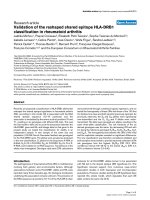
mice display altered lung morphologyFigure 1
Lgl1
+/-
mice display altered lung morphology. Representative micrographs of hematoxylin and eosin stained lung sections
showing areas of increased interstitial tissue in Lgl1
+/-
mice at PN1 and PN7, and enlarged airspace with fewer alveolar septa at
PN14. For Lgl1
+/-
mice, representative images of regions of impaired lung morphology are shown together with images of
regions indistinguishable from those of wild type lungs. Magnification: 200×.
PN1
PN7
PN28
PN14
Lgl1
+/-
Lgl1
+/+
Normal histology Increased interstitium
Normal histology Increased airspace
Lgl1
+/-
Lgl1
+/+
Respiratory Research 2009, 10:83 />Page 6 of 15
(page number not for citation purposes)
lung[15]. Organization of the complex pulmonary elastin
network is initiated in the pseudoglandular lung and
peaks during alveolarization [16,17] Elastin deposition by
myofibroblasts in late gestation is believed to have a spa-
tially instructive role in alveolarization as specific sites of
elastic fiber formation correspond to the location of
future buds[18]. Elastin synthesis is initiated by expres-
sion of tropoelastin. To assess whether observed effects on
lung elastance in Lgl1
+/-
mice were associated with altered
elastin synthesis, we measured tropoelastin mRNA (Fig-
ure 5C). A significant reduction in lung tropoelastin
expression was observed at E14.5. From E16.5 until PN1
no significant differences in tropoelastin mRNA were
detected until maturity. Interestingly, at PN28, concomi-
tant with the observed reduction in Lgl1 expression, there
was a significant increase in expression of tropoelastin
mRNA (Figure 5C).
We next used Weigert's elastin stain to visualize elastin fib-
ers in the lungs of Lgl1
+/-
and wild type Lgl1
+/+
mice. Rep-
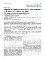
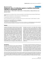
Lgl1 mRNA is reduced in the lungs of Lgl1
+/-
miceFigure 2
Lgl1 mRNA is reduced in the lungs of Lgl1
+/-
mice. Lgl1 mRNA isolated from total lungs (A and C) or whole embryos (B)
of Lgl1
+/+
and Lgl1
+/-
mice was quantified by quantitative real-time RT PCR. A. and B. No significant differences in lung Lgl1
mRNA was observed from E9.5 until PN14 (p > 0.05). C. At PN28, there was significantly less Lgl1 mRNA in the lungs of Lgl1
+/
-
mice compared to their wild type littermates (p ≤ 0.05). D. Mature Lgl1
+/-
mice display significantly reduced levels of Lgl1
mRNA in the heart when compared to their wild type littermates (p ≤ 0.05). (A. N ≥ 3 pooled litters, B. N ≥ 4, C. N ≥ 5, D. N
≥ 4)
Respiratory Research 2009, 10:83 />Page 7 of 15
(page number not for citation purposes)
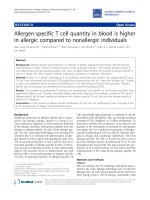
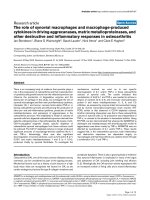
Lgl1 protein is reduced in Lgl1
+/-
miceFigure 3
Lgl1 protein is reduced in Lgl1
+/-
mice. Lgl1 immunohistochemistry (N ≥ 4, all groups) was performed as described in
Methods and quantified with Northern Eclipse as per Nadeau et al. 2006 [3]. Representative images are shown. Modest to
moderate differences in Lgl1 protein levels were observed between Lgl1
+/+
and Lgl
+/-
mice at ages PN1, 7 and 14 (Arrows indi-
cate the reduction of Lgl1 at tips of alveolar septa in Lgl1
+/-
mice at PN 14). At PN28, there was considerable variability between
Lgl1
+/-
mouse lung samples. In some cases, Lgl1 protein appeared to be reduced, consistent with the observed reduction in Lgl1
mRNA (shown) while in others, effects on protein expression were not detectible. Magnification: 400×
Respiratory Research 2009, 10:83 />Page 8 of 15
(page number not for citation purposes)
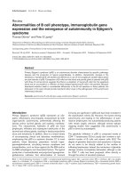
Lgl1 does not localize to lung endothelial cellsFigure 4
Lgl1 does not localize to lung endothelial cells. Immunohistochemistry for Lgl1 (red color) and the endothelial marker
CD34 (green color; N ≥ 4, all groups) was performed as described in Methods. Representative images are shown. No colocal-
ization (would have shown yellow colour) of Lgl1 and CD34 was detected in the lungs of Lgl1
+/+
or Lgl
+/-
mice at ages PN1, 7
and 14. Magnification: 630×
Respiratory Research 2009, 10:83 />Page 9 of 15
(page number not for citation purposes)
Lgl1
+/-
mice display reduced resistance and elastance in response to MCh challengeFigure 5
Lgl1
+/-
mice display reduced resistance and elastance in response to MCh challenge. Airway hyperresponsiveness
(AHR) in response to increasing doses of aerosolized MCh was assessed in 4 week old Lgl1
+/+
and Lgl1
+/-
mice (N = 13 and 9
respectively). Response was measured as maximal resistance (A) and elastance (B). Means are presented ± SEMs. A. Lgl1
+/-
mice
displayed similar resistance to their wild type littermates at baseline. At doses of MCh ranging from 25 to 50 mg/ml Lgl1
+/-
mice
showed reduced resistance compared to wild type littermates. (p ≤ 0.05). B. Lgl1
+/-
mice displayed similar elastance to their
wild type littermates at baseline. In the presence of 50 mg/ml MCh, Lgl1
+/-
mice displayed significantly less elastance than their
wild type littermates (p ≤ 0.05). C. Tropoelastin mRNA isolated from total lungs of Lgl1
+/+
and Lgl1
+/-
mice was quantified by
quantitative real-time RT PCR (N ≥ 4). A significant reduction in tropoelastin mRNA was observed at E14.5 in the Lgl1
+/-
mice.
From E16.5 until PN1 tropoelastin mRNA levels in lungs of Lgl1
+/-
mice were similar to those observed in wild type littermates
(p < 0.05). At PN28, tropoelastin mRNA levels were significantly elevated relative to controls (p < 0.01).
Respiratory Research 2009, 10:83 />Page 10 of 15
(page number not for citation purposes)
resentative images are shown in Figure 6 (n > 4). At E18.5,
elastin fibres in Lgl1
+/+
mice run longitudinally along the
alveolar walls and protrude into the airspaces at the sites
of secondary septal crests (Figure 6, see arrows). By con-
trast, elastin fibres in the lungs of Lgl1
+/-
mice appeared
disorganized and fragmented with trapping in the intersti-
tium (See insets, Figure 6). At PN1 trapping of elastin frag-
ments in the interstitium was still apparent. Moreover,
less elastin was observed at sites of septal crests. At PN7
and PN14 effects on elastin distribution were much less
pronounced. By PN28, these effects on elastin appeared to
be resolved and elastin structure looked normal despite
the presence of elevated levels of tropoelastin mRNA.
Post-natal Lgl1
+/-
mice display goblet cell hyperplasia
Increased numbers of mucin-positive goblet cells are a
characteristic feature of inflammation in multiple respira-
tory disease states including BPD. In order to determine
whether observed changes in Lgl1 expression caused
abnormalities in airway epithelial cells in the trachea and
bronchi, lungs of Lgl1
+/-
and Lgl1
+/+
mice were stained for
mucin-positive goblet cells (PAS stain). Representative
images are shown in Figure 7B (n ≥ 6). Whereas PAS pos-
itive cells were rarely seen in wild type mice from PN4 -
PN14, a considerable number of mucin-positive goblet
cells were observed in both the bronchi and trachea of a
subset Lgl1
+/-
mice during this period (Figure 7B). At
PN14, Lgl1
+/-
mice fell into two very distinct subgroups
based on goblet cell number, those with an increased
number of goblet cells and those that were not distin-
guishable from wild type littermates (significant differ-
ence between subgroups, p = 0.017, Mann Whitney U test;
See scatter plot, Figure 7C). In no case were goblet cells
elevated in wild type pups.
In the lungs, goblet cell hyperplasia of surface epithelial
cells in inflammatory disease generally correlates with
increased expression of mucin (MUC5AC) mRNA[19].
We therefore assessed MUC5AC mRNA expression in
lungs of Lgl1
+/-
and Lgl1
+/+
mice. Increased staining of gob-
let cells in PN lung of Lgl1
+/-
mice was accompanied by an
increase in expression of mucin (MUC5AC) mRNA (Fig-
ure 7A).
Inflammatory cytokines, IL-4 and IL-13 are elevated at
PN7 in Lgl1
+/-
mice
Inflammatory cytokines stimulate MUC5AC expression.
The findings of altered mucin expression and goblet cell
hyperplasia in PN lungs of Lgl1
+/-
mice led us to ask
whether Lgl1 may have a role in the development of
immune modulation. We assessed levels of two inflam-
matory cytokines, IL-4 and IL-13. Dramatically elevated
levels of both IL-4 and IL-13 were observed at PN7 (Figure
8). At PN14 IL-4 remained significantly elevated. The
increase in IL-13 expression was no longer significant at
this time. To determine whether elevated cytokine levels
were associated with induction of recruitment of inflam-
matory cells, BAL cell differentials were determined at
PN10 and at maturity. No significant differences in BAL
cell counts were detected when Lgl1+/- mice were com-
pared to wild type littermates.
Discussion
The finding that null mutation of the Lgl1 gene in mouse
embryos is lethal prior to the onset of lung morphogene-
sis classifies Lgl1 as an essential early gene in organismal
development. The determinants of embryonic lethality in
Lgl1
+/-
mice remain to be determined and pre-date lung
organogenesis. Mutation in Lgl1 has pleiotropic effects.
Moreover, it is possible that effects on Lgl1 expression in
other organ systems contribute to the postnatal pheno-
type observed in the lung.
The present study demonstrates that the heterozygous
Lgl1 genotype is sufficient to induce a complex pheno-
type. In postnatal lung, histologically immature areas
with distinctly thickened respiratory interstitium and the
appearance of delayed secondary septation alternate with
areas of relatively normal appearance. Disorganized elas-
tin fibers, goblet cell hyperplasia and high levels of
inflammatory cytokines are present in the absence of
detectible differences in Lgl1 mRNA levels. Our inability
to detect changes in Lgl1 mRNA expression in total RNA
isolated from developing lung is likely to reflect a specific
requirement for Lgl1 in a subpopulation of cells in which
suppressed expression of Lgl1 is sufficient to produce the
observed phenotype but escapes detection by RT-PCR of
total lung RNA. For example, we showed previously that
Lgl1 is maximally expressed in fibroblasts adjacent to the
epithelium [1,2]. We also demonstrated that Lgl1 is
secreted and taken up by epithelial cells beginning in late
gestation and continuing in PN life [2]. Deficient Lgl1
expression in a subset of fibroblasts important in mesen-
chymal-epithelial interactions that regulate alveolariza-
tion may contribute to the observed phenotype.
We have backcrossed our Lgl1
+/-
mice onto the C57BL/6
background (eight generations). Neonatal Lgl1
+/-
mice on
this background faithfully reproduce the phenotype of
disorganized elastin, goblet cell hyperplasia with elevated
levels of MUC5AC and increased IL-4 and IL-13 expres-
sion, demonstrating that the phenotype we report is not
due to a mixed genetic background.
The association of a distinct respiratory phenotype in the
absence of significant reduction in mRNA has been
reported previously [13,20]. Foxf1 heterozygotes can be
divided on the basis of pulmonary levels of Foxf1
mRNA[13]. Low Foxf1 producers fail to undergo differen-
tiation of terminal airspaces. An albeit less-severe defect in
Respiratory Research 2009, 10:83 />Page 11 of 15
(page number not for citation purposes)
Lgl1
+/-
mice display disorganization of elastin fibers at E18Figure 6
Lgl1
+/-
mice display disorganization of elastin fibers at E18.5 and PN1. Lung sections of Lgl1
+/+
and Lgl1
+/-
mice were
treated with Weigert's elastin stain to visualize elastin fibres. Representative images are shown. In wild type mice, elastin fibres
run longitudinally along the alveolar walls of lungs and protrude into the airspaces at the sites of secondary septal crests (see
arrows). By contrast, at E18.5 and PN1 lungs of Lgl1
+/-
mice show disorganization and fragmentation of elastin fibres (See also
insets). Effects on elastin structure were less pronounced from PN7-PN14 and resolved at maturity. Magnification: 400×,
insets: 1000×.
Respiratory Research 2009, 10:83 />Page 12 of 15
(page number not for citation purposes)
Postnatal Lgl1
+/-
mice display goblet cell hyperplasia and increased expression of MUC5ACFigure 7
Postnatal Lgl1
+/-
mice display goblet cell hyperplasia and increased expression of MUC5AC. A. MUC5AC was
quantified in mRNA isolated from total lungs of Lgl1
+/+
and Lgl1
+/-
mice by quantitative real-time RT PCR (N ≥ 5). A significant
increase in MUC5AC mRNA was observed at PN7 and PN14 in the Lgl1
+/-
mice (p < 0.05). B. Lung sections of Lgl1
+/+
and Lgl1
+/
-
mice were stained with Period Acid Schiff (PAS) stain to visualize the goblet cells. Representative images are shown.PAS posi-
tive cells were rarely seen in wild type mice during the early post natal period, however a considerable number of PAS positive
cells were observed in both the trachea and upper bronchi of the Lgl1
+/-
mice during this same time period. Magnification: 400×
C. Scatter plot illustrating bimodal distribution of goblet cells in heterozygous Lgl1
+/-
mice (significant difference between sub-
groups, p = 0.017, Mann Whitney U test).
Respiratory Research 2009, 10:83 />Page 13 of 15
(page number not for citation purposes)
septation of the lung periphery is observed in High Foxf1 1
producing heterozygotes mice that express normal or
nearly normal (90%) levels of Foxf1 mRNA[13]. Moreo-
ver, these animals display aberrant expression of a
number of developmentally important genes in the lung.
There was considerable variability in Lgl1 protein levels in
Lgl1
+/-
mice. No such variability was observed in wild type
animals. We demonstrated regional differences in tissue
fraction in postnatal Lgl1
+/-
mice. Given that Lgl1 is of
mesenchymal origin and that mesenchymal thinning is a
prominent feature of lung maturation, variability in Lgl1
protein levels may reflect varying degrees of developmen-
tal delay. The observed reduction in Lgl1 protein in the
lungs of Lgl1
+/-
mice may also reflect effects on RNA stabil-
ity or protein turnover Absence of Lgl1 in endothelial cells
suggests that Lgl1 does not have a direct role in PN angio-
genesis.
Genetic modifiers of tissue- and temporal-specific expres-
sion of Lgl1 may also contribute to the observed variation
in penetrance of the Lgl1
+/-
phenotype. In this context it is
important to recall that low levels of Lgl1 in pseudoglan-
dular lung are essential to airway branching[12]. Haplo-
sufficiency for Lgl1 is clearly sufficient to rescue the
branching program. At the same time, effects on tropoe-
lastin expression were observed at E14.5 suggesting that
the heterozygous phenotype does impact lung develop-
ment in the pseudoglandular period.
Reduced Lgl1 expression in mature Lgl1
+/-
mice was asso-
ciated with normal baseline lung function. As expected,
MCh challenge provoked an increase in airway resistance
and elastance in both Lgl1
+/-
and wild type littermates.
However, the effects on both these parameters were signif-
icantly greater in wild type mice. Moreover, tropoelastin
expression in mature Lgl1
+/-
mice was elevated. Elastic
interdependence of the lung accounts for orderly elastic
recoil of lungs during passive expiration [21]. Organiza-
tion of the elastin network is initiated in the pseudoglan-
dular lung and peaks during alveolarization [16,17]. At
the alveolar level, elastic interdependence is mediated by
the correct expression, cross-linking, and orientation of
elastin and collagen fibers. Absence of a correctly cross-
linked and oriented elastin matrix predisposes to aberrant
alveolarization. Deficient alveolar maturation in BPD
includes disruption of elastin fibers, distal airspace
enlargement, and mucus cell hyperplasia [14]. All of these
properties were present in newborn Lgl1
+/-
mice. Dysregu-
lation of elastin synthesis is also a prominent feature of
BPD in murine [22] and preterm lamb models [23].
Excessive degradation of the elastin matrix underlies the
loss of elastic interdependence and alveolar degeneration
associated with chronic lung disease in adults. Indeed, it
is believed that individuals with even limited elastin
insufficiency may suffer damage sufficient to preclude
alveolar repair from a less severe injury than would be
required to have this effect in normal subjects. Survivors
of preterm birth are at particular risk to develop premature
COPD [21,24]. The disrupted elastin architecture in Lgl1
+/
-
mice appears to resolve at maturity. Yet these animals
have elevated tropoelastin levels and impaired lung func-
tion at maturity. It is tempting to speculate that latent
effects on elastin integrity may increase vulnerability of
Lgl1
+/-
mice to respiratory insult at maturity and that Lgl1
haploinsufficiency may modify risk to respiratory insult in
later life.
Postnatal Lgl1
+/-
mice display increased expression of IL-4 and IL-13 mRNAFigure 8
Postnatal Lgl1
+/-
mice display increased expression of IL-4 and IL-13 mRNA. IL-4 and IL-13 mRNA was quantified in
mRNA isolated from total lungs of Lgl1
+/+
and Lgl1
+/-
mice by quantitative real-time RT PCR (N ≥ 4). A. Lgl1
+/-
display signifi-
cantly elevated IL-4 levels at PN7 and PN14 compared to wild type controls (p ≤ 0.05). B. Lgl1
+/-
mice display significantly ele-
vated levels of IL-13 at PN7 compared to wild type controls (p ≤ 0.05).
Respiratory Research 2009, 10:83 />Page 14 of 15
(page number not for citation purposes)
Given that the disruption of elastin expression and/or
organization has been associated with lung injury in the
newborn period, it was of interest to establish whether
Lgl1
+/-
mice displayed any other characteristic features
associated with neonatal lung injury. Inflammation is
known to interfere with lung development in model sys-
tems and is present chronically in the lungs of preterm
infants who develop BPD [25]. Goblet cell hyperplasia is
associated with multiple respiratory disorders including
asthma, BPD and emphysema. It has been suggested that
exposure of the developing lung to inflammation may be
central to the development of BPD[25]. Analysis of goblet
cell number in PN14 lungs of Lgl1
+/-
mice identified a
bimodal distribution, with one subgroup of animals dis-
playing significantly elevated numbers of goblet cells in
trachea and bronchi associated with elevated mucin
(MUC5AC) production. A second group of animals
showed no increase in goblet cell number. The presence of
these 2 distinct groups is likely to reflect the incomplete
penetrance of the Lgl1
+/-
phenotype and may be attributed
to the genetic contribution from C57BL/6 and 129/J
strains. Indeed, we have found that on the C57BL6 back-
ground, goblet cell hyperplasia in Lgl1
+/-
mice is much less
variable.
The differentiation of epithelial cells to goblet cells is
induced by inflammatory cytokines [19]. We found dra-
matically elevated levels of the T
H
2 cytokines IL-4 and IL-
13 in the postnatal Lgl1
+/-
mouse lung, concordant with
evidence of goblet cell hyperplasia. Both IL-4 and IL-13
have been shown to induce goblet cell hyperplasia and
mucus hypersecretion in mouse airways [26-28]. It is of
interest that despite the pronounced rise in IL-4 and IL-13
observed at PN7, we did not detect an inflammatory infil-
trate in BAL. Both Il-4 and IL-13 are cytokines associated
with the inflammatory process in diseases such as asthma,
but neither of these cytokines has chemokine properties.
Indeed, Wills-Karp et al. [28] have shown that these
cytokines can induce pathophysiological features of
asthma via mechanisms independent of eosinophil
recruitment. Thus the inflammation observed in postnatal
Lgl1
+/-
mice that are free of allergen and infection is un
likely to involve recruitment of cellular infiltrates. The
combined findings of goblet cell hyperplasia and induc-
tion of inflammatory cytokines are nevertheless consist-
ent with a role for Lgl1 in innate immunity suggesting that
Lgl1 may function to protect the lung from external
insults.
To date, the domain structure of Lgl1 has offered little to
our understanding of its molecular function. The LCCL
domain family now includes more than 100 proteins.
Orthologues of Lgl1 are typified by having two LCCL
modules. Multiple functions have been attributed to the
LCCL module (also known as FCH[5]). These include
roles in cell differentiation, motility and migration [5,7];
cell adhesion and matrix deposition [8,9]; and host-
defense and innate immunity [4,10,11]. The results of our
studies provide the first evidence for a potential role of
Lgl1 in innate immunity. Mutagenesis of the LCCL mod-
ules in Lgl1 will be necessary to explore such effects in
vitro.
There are a number of limitations to this study. While
haploinsufficiency for Lgl1 is the only reasonable explana-
tion for the pulmonary phenotype in Lgl1
+/-
heterozy-
gotes, we were not able to identify the precise time and
localization of deficient Lgl1 that triggers the postnatal
phenotype. There was considerable variability in several
outcome measures among Lgl1
+/-
heterozygotes. The
development of a mouse model with conditionally regu-
latable expression of Lgl1 will allow a more definitive
analysis of the role of Lgl1 in early lung development.
Conclusion
Absence of Lgl1 in null mice is embryonic lethal, making
Lgl1 an essential gene. Neonatal Lgl1
+/-
mice exhibit
increased interstitial tissue, goblet cell hyperplasia and
elevated cytokine levels. The disorganized elastin architec-
ture seen in neonatal Lgl1
+/-
mice would be expected to
interfere with normal elastic recoil and may increase vul-
nerability to alveolar degeneration. Indeed, in adult Lgl1
+/
-
mice, which express 50% Lgl1 mRNA, reduced lung
elastance is associated with elevated tropoelastin levels.
Mice with Lgl1 haploinsufficiency display changes in lung
phenotype that resemble those seen in chronic neonatal
lung disease.
Competing interests
The authors declare that they have no competing interests.
Authors' contributions
JL generated the knockout mouse. LR and IM character-
ized Lgl1 mRNA and protein, elastin and tropoelastin
mRNA, and goblet cells and mucin mRNA in Lgl1
+/-
heter-
ozygotes and wild type controls. TB participated in H and
E histochemistry and protein quantitation. KN and SC
carried out lung function studies. NBS contributed to the
design of the study and the preparation of the manuscript.
FK conceived and participated in the design of the study
and had a primary role in preparation of the manuscript.
Additional material
Additional file 1
Generation of Lgl1 KO Mouse. Detailed description of the generation of
the Lgl1 KO mouse
Click here for file
[ />9921-10-83-S1.DOC]
Publish with BioMed Central and every
scientist can read your work free of charge
"BioMed Central will be the most significant development for
disseminating the results of biomedical research in our lifetime."
Sir Paul Nurse, Cancer Research UK
Your research papers will be:
available free of charge to the entire biomedical community
peer reviewed and published immediately upon acceptance
cited in PubMed and archived on PubMed Central
yours — you keep the copyright
Submit your manuscript here:
/>BioMedcentral
Respiratory Research 2009, 10:83 />Page 15 of 15
(page number not for citation purposes)
Acknowledgements
The authors thank Laura Montermini for assistance in preparation of this
manuscript. This work was supported by an operating grant (MOP68954)
from the Canadian Institutes of Health Research (FK and NBS) and gradu-
ate scholarships from the Montreal Children's Hospital Research Institute
(LR, TB and SC) and from The Respiratory Health Network of the FRSQ
and the Canadian Institutes of Health Research (LR).
References
1. Kaplan F, Ledoux P, Kassamali FQ, Gagnon S, Post M, Koehler D,
Deimling J, Sweezey NB: A novel developmentally regulated
gene in lung mesenchyme: homology to a tumor-derived
trypsin inhibitor. Am J Physiol Lung Cell Mol Physiol 1999,
276:L1027-L1036.
2. Oyewumi L, Kaplan F, Sweezey NB: Biochemical characteriza-
tion of lgl1, a mesenchymal protein in fetal lung that regu-
lates epithelial airway branching. Biochem J 2003, 376:61-69.
3. Nadeau K, Jankov RP, Tanswell AK, Sweezey NB, Kaplan F: Lgl1 is
suppressed in oxygen toxicity animal models of bronchopul-
monary dysplasia and normalizes during recovery in air. Pedi-
atr Res 2006, 59:389-395.
4. Trexler M, Banyai L, Patthy P: The LCCL module. Eur J Biochem
2000, 267:5751-5757.
5. Robertson NG, Hamaker S, Aster J, Morton CC: Subcellular local-
isation, secretion, and post-translational modification of nor-
mal cochlin, and of mutants causing the sensineural deafness
and vestibular disordar, DFNA9. J Med Gen 2003, 40:479-486.
6. Gibbs GM, Roelants K, O'Bryan MK: The CAP Superfamily:
Cysteine-Rich Secretory Proteins, Antigen 5, and Pathogen-
esis-Related 1 Proteins Roles in Reproduction, Cancer, and
Immune Defense. Endocrine Reviews 2008, 29:865-897.
7. Vogt DL, Gray CD, Young WS III, Orellana SA, Malouf AT:
ARHGAP4 is a novel RhoGAP that mediates inhibition of
cell motility and axon outgrowth. Molecular & Cellular Neuro-
sciences 2007, 36:332-342.
8. Grabski R, Szul T, Sasaki T, Timpl R, Mayne R, Hicks B, Sztul E: Muta-
tions in COCH that result in non-syndromic autosomal dom-
inant deafness (DFNA9) affect matrix deposition of cochlin.
Hum Genet 2003, 113:406-416.
9. Ahsan M, Ohta K, Kuriyama S, Tanaka H: Novel soluble molecule,
Akhirin, is expressed in the embryonic chick eyes and exhib-
its heterophilic cell-adhesion activity. Developmental Dynamics
2005, 233:95-104.
10. Greer P: Closing in on the biological functions of FPS/FES and
FER. Nat Rev Mol Cell Biol 2002,
3:278-289.
11. Dessens JT, Sinden RE, Claudianos C, Dessens JT, Sinden RE, Claudi-
anos C: LCCL proteins of apicomplexan parasites. Trends in
Parasitology 2004, 20:102-108.
12. Oyewumi L, Kaplan F, Gagnon S, Sweezey NB: Antisense Oligode-
oxynucleotides Decrease LGL1 mRNA and Protein Levels
and Inhibit Branching Morphogenesis in Fetal Rat Lung. Am
J Respir Cell Mol Biol 2003, 28:232-240.
13. Kalinichenko V, Lim L, Stolz DB, Shin B, Rausa F, Clark J, Whitsett JA,
Watkins SC, Costa RH: Defects in pulmonary vasculature and
perinatal lung hemorrhage in mice heterozagous null for
Forkhead box f1 transcription factor. Dev Biol 2001,
235:489-507.
14. Pierce RA, Albertine KH, Starcher BC, Bohnsack JF, Carlton DP,
Bland RD: Chronic lung injury in preterm lambs: disordered
pulmonary elastin deposition. Am J Physiol 1997, 272:L452-L460.
15. Bates JH, Irvin CG: Measuring lung function in mice: the pheno-
typing uncertainty principle. J Appl Physiol 2003, 94:1297-306.
16. Mariani TJ, Reed JJ, Shapiro SD: Expression profiling of the devel-
oping mouse lung: insights into the establishment of the
extracellular matrix. American Journal of Respiratory Cell & Molecu-
lar Biology 2002, 26:541-548.
17. Shifren A, Mecham RP: The Stumbling Block in Lung Repair of
Emphysema: Elastic Fiber Assembly. Proc Amer Thoracic Soc
2006, 3:428-433.
18. Bourbon J, Boucherat O, Chailley-Heu B, Delacourt C: Control
mechanisms of lung alveolar development and their disor-
ders in bronchopulmonary dysplasia. Pediatr Res 2005,
57:38R-46R.
19. Thai P, Loukoianov A, Wachi S, Wu R: Regulation of mucin gene
expression. Annu R Physiol 2008, 70:405-429.
20. Kalinichenko V, Zhou Y, Shin B, Stolz DB, Watkins S, Whitsett J,
Costa RH: Wild-type levels of the mouse Forkhead Box f1
gene are essential for lung repair. Am J Physiol Lung Cell Mol Phys-
iol 2002, 282:L1253-L1265.
21. Warburton D, Gauldie J, Bellusci S, Shi W: Lung Development and
Susceptibility to Chronic Obstructive Pulmonary Disease.
Proc Am Thorac Soc 2006, 3:668-672.
22. Bland RD, Ertsey R, Mokres L, Xu L, Jacobson B, Jiang S, Alvira CM,
Rabinovitch M, Shinwell ES, Dixit A: Mechanical ventilation
uncouples synthesis and assembly of elastin and increases
apoptosis in lungs of newborn mice.: Prelude to defective
alveolar septation during lung development? Am J Physiol Lung
Cell Mol Physiol 2008, 294:L3-14.
23. Bland R, Xu L, Ertsey R, Rabinovitch M, Albertine KH, Wynn KA, et
al.: Dysregulation of pulmonary elastin synthesis and assem-
bly in preterm lambs with chronic lung disease. Am J Physiol
Lung Cell Mol Physiol 2007, 292:L1370-L1384.
24. Baraldi E, Filippone M: Chronic lung disease after premature
birth. N Engl J Med 2007, 357:1946-1955.
25. Kallapur SG, Jobe AH: Contribution of inflammation to lung
injury and development. Archives of Disease in Childhood Fetal &
Neonatal Edition 2006, 91:F132-F135.
26. Perkins C, Wills-Karp M, Finkelman FD: IL-4 induces IL-13-inde-
pendent allergic airway inflammation. J Allergy Clin Immunol
2006, 118:410-419.
27. Tanabe T, Fujimoto K, Yasuo M, Tsushima K, Yoshida K, Ise H,
Yamaya M: Modulation of mucus production by interleukin-13
receptor alpha-2 in human airway epithelium. Clin Exp Allergy
2007, 38:122-134.
28. Wills-Karp M, Luyimbazi J, Xu X, Schofield B, Neben TY, Karp CL,
Donaldson DD: Interleukin-13: central mediator of allergic
asthma. Science 1998, 282:2258-2261.
Additional file 2
Additional methods. Detailed explanation of methods used for Lgl1
immunohistochemistry, quantitative real-time PCR and pulmonary func-
tion studies.
Click here for file
[ />9921-10-83-S2.DOC]