Core Topics in Operating Department Practice Anaesthesia and Critical Care – Part 3 pdf
Bạn đang xem bản rút gọn của tài liệu. Xem và tải ngay bản đầy đủ của tài liệu tại đây (571.46 KB, 23 trang )
the risk of aspiration even further include, for
example:
• patients who have a hiatus hernia (where part
of the stomach pushes up into the lower chest
through a defect in the diaphragm leading to an
increased potential for gastric reflux into the
oesophagus)
• patients in the late stages of pregnancy (where
the position of the foetus causes gastric reflux)
• patients who have suffered traumatic injury
(traumatic injury slows digestion and stomach
emptying)
• cases of severe head injury (unconscious patients
have no natural ability to protect their airway
from regurgitated stomach contents)
• patients who are intoxicated with drug or alcohol
use (deeply unconscious patients through misuse
of alcohol and drugs are unable to protect their
own airway naturally from regurgitated stomach
contents)
• any other clinical situation where gastric empty-
ing is delayed.
There are also emergency situations where the use
of cricoid pressure is not advised, including for
example, active vomiting, unstable cervical spine
injury and cricotracheal injury. Cricoid pressure
is a part of an anaesthetic technique known as
Rapid Sequence Induction (RSI). RSI is often
carried out where gastric emptying is delayed.
Conditions such as these present difficulties for
anaesthetists and healthcare providers and wher-
ever possible alternatives to general anaesthesia
may be sought.
Applying cricoid pressure
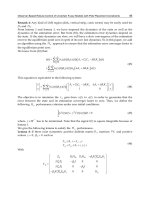
When applying cricoid pressure, the cricoid
cartilage (the only complete ring of cartilage in
the trachea) is manually pushed back against the
cervical spine at the level of the C5/C6 vertebrae
to occlude the oesophagus, which lies directly
beneath the trachea. All other cartilage rings
contained in the trachea are made up of semi-
circles and are therefore not suitable for use in this
technique. The manoeuvre is achieved by using
the thumb and index finger usually of the right
hand to compress the cricoid cartilage (Figure 4.2).
The right hand is normally used because of the
design of many anaesthetic rooms in the UK.
The anaesthetic equipment is usually located on
the patient’s right or at the head end of the patient
trolley/bed and the anaesthetic assistant is mainly
positioned to the right side of the patient, making
use of the right hand naturally more effective than
the left. Nevertheless, dependent on the situation,
the use of either hand is equally effective.
A formally qualified and experienced anaes-
thetic practitioner is required to apply ‘effective’
cricoid pressure. According to Anaesthesia UK
(2004), the following components are essential
for undertaking RSI:
• Tilting table/trolley
• Full monitoring of blood pressure, ECG, pulse
oximetery and End Tidal CO
2
monitor
• Suction ready (switched to the ON position
and placed under the patient’s pillow)
• Fully trained assistant
• IV access
• Pre-oxygenation for 3 minutes
Figure 4.2 Applying cricoid pressure.
The use of cricoid pressure during anaesthesia 31
• Suitable sleep dose of induction agent
• Cricoid pressure
• Suxamethonium
Õ
• Laryngoscopy and intubation
• Check position
• Secure tube.
If conscious, the practitioner should tell the patient
about the procedure before induction. The practi-
tioner must firstly find and identify the anatomy of
the cricoid cartilage and position fingers lightly
over the correct area, telling the patient the reasons
for these actions.
The patient is pre-oxygenated for a full 3 minutes,
to create a reservoir of oxygen in the lungs.
This provides the anaesthetist with the maximum
time available to intubate the patient without
compromising the patient’s oxygen saturation.
Forced ventilation using a Bag Valve Mask (BVM)
technique is contraindicated in patients who are
at high risk of gastric aspiration as there is a risk
of forcing air into the stomach (causing possible
gastric distension) thus increasing the likelihood
of regurgitation. Cricoid pressure is also recom-
mended for mask ventilation during cardio pulmo-
nary resuscitation (CPR), if there are two or more
rescuers, to reduce gastric distension and conse-
quent regurgitation (MERCK, 2004).
The anaesthetist then gives Thiopentone
Õ
or
Etomidate
Õ
À depending on the patient’s cardio-
vascular stability. Etomidate
Õ
may be an alterna-
tive if the patient is losing large volumes of blood,
or has underlying cardiovascular problems, as it
does not drop the blood pressure as rapidly as
Thiopentone
Õ
. Both these drugs act to induce
narcosis (sleep). Cricoid pressure is applied grad-
ually as the patient closes their eyes and the
patient’s ‘lash reflex’ has decreased. The lash
reflex is used by anaesthetists to decide if the
patient is unconscious, by gently touching the
eyelash and establishing if the patient’s eyes
blink. When blinking is absent, the anaesthetist
will then give the depolarising muscle relaxant
Suxamethonium
Õ
to achieve total muscle paralysis
in readiness for intubation. Suxamethonium
Õ
is
a short-acting muscle relaxant which has a rapid
onset of about 45 seconds, the effects of which
last about 2À5 minutes (Yentis et al., 2004).
Cricoid pressure should not be removed until
the anaesthetic practitioner is directed to do so
by the anaesthetist. Removal of pressure usually
occurs once the patient has been intubated,
the cuff of the endotracheal tube is inflated and
the anaesthetist is satisfied that the tube is in the
correct position. If pressure is removed too early
the patient could be at risk of regurgitation and
aspiration.
The practitioner should be ready to release
the pressure if the patient shows signs of vomiting.
During vomiting the patient may be prone to
oesophageal rupture if cricoid pressure is not
removed immediately. The stomach is normally
relaxed, but when squeezed forcefully by the
abdominal wall, it ejects any food or fluid up
through the oesophagus and vomiting occurs.
A pressure over 60 cm of H
2
O can develop which
may tear the oesophagus at the oesophagogastric
junction if the oesophagus is occluded because of
cricoid pressure. Oesophageal rupture is normally
fatal to the patient. If vomiting occurs during
induction of anaesthesia and the use of cricoid
pressure, the pressure should be removed and the
patient should be tilted head down or turned to the
left lateral position. Suction is then applied to
remove the vomit from the oropharynx.
Training the technique of cricoid pressure
The technique used to apply cricoid pressure varies
from practitioner to practitioner. The force of
pressure required to be exerted on the larynx is
estimated at between 20 and 40 Newtons À where
10 Newtons equal about 1 kilogram of pressure.
As with many clinical skills, there are good
and poor techniques, several contraindications for
its use and few signs, apart from the absence of
regurgitation, of whether the manoeuvre has been
carried out successfully. Most healthcare provid-
ers were, in the past, often taught the technique
‘in-house’ and told to ‘just put your hand there and
32 C. Wayne-Conroy
press’. Nevertheless, this is not enough training
for what is a difficult technique to perfect, which is
frightening for both the patient and the practitioner
when first encountered.
Patients are at risk of harm from practitioners
who fail to apply cricoid pressure consistently or
correctly. Death from Mendelson’s syndrome can
result from applying cricoid pressure inefficiently,
not applying cricoid pressure at all, or relaxing the
pressure before intubation has been successfully
established (Murray et al., 2000).
Cricoid pressure also has the potential to cause
anatomical distortion to the upper airway. Failed
intubations using conventional laryngoscopy can
sometimes be increased during the use of cricoid
pressure. Nevertheless, pressure can be adjusted
slightly, to aid the view of the vocal cords if
requested by the anaesthetist. Other useful items
of equipment such as the Gum-Elastic Bougie
(see Figure 4.3) can be employed to aid airway
management during intubation if the view of the
larynx is in anyway distorted either due to the
cricoid pressure or pre-existing anatomical diffi-
culties (The Ambulance Service Association, 2001).
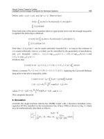
There are now simulation manikins or task
trainers available in most clinical skills training
environments (see Figure 4.4) which students and
health professionals can use to practise and learn
this technique successfully, without compromising
patient safety.
The manikin contains an electronic monitor
which displays the correct and incorrect hand
placement and continuously shows the force
being applied to the cricoid cartilage. When using
the task trainer, many healthcare providers are
surprised by the force required and the difficulty
in maintaining that force correctly.
Various research studies into the use of cricoid
pressure during RSI raise questions about the
effectiveness of the technique in preventing
regurgitation and the practical application of
this manoeuvre. A recent magnetic resonance
Figure 4.3 Sample picture on right
Left À the Gum-Elastic Bougie. Right À the Bougie in use.
The use of cricoid pressure during anaesthesia 33
imaging (MRI) study carried out in Texas in the
United States on healthy volunteers suggests that
the cricoid cartilage and oesophagus are not always
anatomically aligned in the same axis and that
application of cricoid pressure further displaced
both the oesophagus and larynx laterally.
The researchers suggested that gastric content
aspiration may occur during the induction of
anaesthesia despite the application of cricoid
pressure (Hernandez et al., 2004).
Much debate will undoubtedly remain among
the medical profession about the use of cricoid
pressure. All patients who present for emergency
surgery, especially patients who require intestinal
surgery, where there is suspicion of delayed gastric
emptying, should be induced using RSI technique.
For example, even a patient requiring an appendi-
cectomy, who has been a hospital inpatient for a
week or more and fasted of oral solids and fluids
and was showing no signs of recent vomiting,
should not undergo general anaesthetic induction
without the use of cricoid pressure.
No definite alternative has currently been devised
or developed to replace the use of cricoid pressure
during rapid sequence induction. Therefore, the
priority for health professionals is to standardise
the use and technique of cricoid pressure and start
training programmes for those who teach and
practise this technique. This would help to reduce
errors and poor techniques and ensure future
patient safety throughout the procedure.
REFERENCES
Amersham Health Medical Dictionary. (2005). Avail-
able at: www.amershamhealth.com (Accessed 6 April
2005).
Anaesthesia UK. (2004). The Components for Rapid
Sequence Induction. Available at: www.frca.co.uk
(Accessed 4 April 2005).
Hernandez, A., Wolf, S. W., Vijayakumar, V., Solanki., D. R.
& Mathru, M. (2004). Sellick’s Manoeuvre for the
Prevention of Aspiration Is It Effective? Available
at: www.asaabstracts.com/strands (Accessed 9 April
2005).
MERCK Manual. (2004). Cardiopulmonary Resuscitation.
Available at: www.merck.com/mrkshared/mmanual/
section16 (Accessed 30 March 2005).
Mijumbi, C. (1994). Anaesthesia for the Patient with a Full
Stomach. Available at: www.nda.ox.ac.uk (Accessed
5 April 2005).
Murray, E., Keirse, M., Neilson, J. et al. (2000). A Guide to
Effective Care in Childbirth and Pregnancy. Available at:
www.maternitywise.org (Accessed 6 April 2005).
Owen, H., Follows, K., Reynolds, J., Burgess, G. &
Plummer, J. (2002). Learning to apply effective cricoid
pressure using a part task trainer. Continuing Education
in Anaesthesia, Critical Care & Pain, 5(2), 45À8.
Sinclair, R. C. F. & Luxton, M. C. (2005). Rapid sequence
induction. Continuing Education in Anaesthesia,
Critical Care and Pain, 5(2), 45À8.
Smith, B. & Williams, T. (eds.) (2004). Operating Depart-
ment Practice AÀZ. London: Greenwich Medical Ltd.
The Ambulance Service Association. (2001). Difficult
Intubation Protocol: Use of the Endotracheal
Tube Introducer (Gum-Elastic Bougie). Available at:
www.asancep.org.uk/Endotrachealtubeintroducer.htm
(Accessed 9 April 2005).
Yentis, S., Nicholas, P. H. & Smith, G. B. (2004).
Anaesthesia and Intensive Care AÀZ À An Encyclopaedia
of Principles and Practice, 2nd edn. Edinburgh:
Elsevier Ltd.
Figure 4.4 Life/form
Õ
Cricoid Pressure Trainer 2005.
34 C. Wayne-Conroy
5
Anaesthetic breathing circuits
Norman Wright
Key Learning Points
• Discuss the basic design of breathing circuits
• Describe the evolution of breathing circuits
• Identify the benefits and disadvantages of each
circuit
An anaesthetic breathing circuit is an assembly of
parts, which connects the patient’s airway to the
anaesthetic machine creating an artificial atmo-
sphere, from and into which a patient breathes
(Ravi Shankar, 2004).
Shankar also states that a breathing circuit
mostly consists of:
• a tube through which fresh anaesthetic gases are
delivered from the anaesthetic machine to the
patient
• a method of connecting the circuit to the
patient’s airway
• a rebreathing bag or corrugated rubber tubing
(used in the early circuits) which acts as a gas
reservoir, which would meet the peak inspiratory
flow requirements
• an expiratory valve which allows the expired
gases to pass into the scavenging circuit
• a carbon dioxide absorber for total rebreathing,
and tubing to connect all the parts; as stated
earlier in the early stages the tubing was com-
posed of corrugated rubber. (Ravi Shankar, 2004).
Even though the design and materials used for
breathing circuits have developed over the years,
the individual component’s roles have remained
almost unchanged.
Since introducing ether as an anaesthetic in
1846, many improvements in the design of breath-
ing circuits have occurred. Initially, inventors
developed apparatus to deliver a single anaesthetic
agent, such as nitrous oxide. Nitrous oxide fell from
favour as a single-agent anaesthetic but was
reintroduced in 1868, stored in cylinders, as part
of a combination of anaesthetic agents. Barth, in
1907, developed a method of delivering nitrous
oxide to patients using a valve, a reservoir bag and
a Clover’s inhaler. A Clover’s inhaler consists of a
black triangular mask attached to one side of a
central silver drum with a flattened black rubber
elliptical bag attached. By changing the lever’s
position in the valve, Barth could allow patients to
either completely rebreathe the anaesthetic gases,
or alternatively breathe completely from the
atmosphere.
The Boyle’s machine was developed in 1917.
This development coincided with Magill and
Rowbotham mastering endotracheal intubation
using a single-lumen red rubber tube. Following
from this, a simple anaesthetic delivery circuit
called the ‘Magill’s Circuit’ was developed.
The next 20 years saw many of the core advances
in anaesthetic technology:
• 1929 À Cycloprane (C
3
H
6
): a flammable colour-
less gas which was used as an anaesthetic.
• 1931 À Cuffed endotracheal tubes: the cuff sits
beyond the vocal chords to form a seal within the
trachea to prevent anaesthetic gases escaping
Core Topics in Operating Department Practice: Anaesthesia and Critical Care, eds. Brian Smith, Paul Rawling, Paul Wicker and
Chris Jones. Published by Cambridge University Press. ß Cambridge University Press 2007.
35
and to prevent gastric contents from entering the
lungs.
• Water’s ‘to and fro’ circuit for closed circuit
anaesthesia: this is a complete circuit consisting
of tubing, a soda lime canister and a swivel
connector.
• 1936 À Sword’s circle circuit: this circuit was
similar to earlier circle circuits but required
smaller amounts of fresh gas each minute.
• 1937 À Ayre’s T piece: used for paediatric
anaesthesia, later modified by Jackson Rees.
• 1941 À The EMO inhaler: an early version of a
vaporiser using the ‘drawover’ method.
(Online Medical Dictionary, 1997).
The fifties and sixties saw breathing circuits
develop at an increased rate, which was due in
part to the new methods of providing anaesthesia.
The ether ‘open drop’ method was no longer used
and modern anaesthetic machines had vaporisers.
The classification of anaesthetic machines
depended on the whim of the developer, however,
most developers agreed that breathing circuits
should essentially deliver gases from the machine
to the alveoli in the concentration that was set by
the user and in the shortest possible time. The
circuit also has to effectively eliminate dead-space
(areas in the circuit where no movement of gases
occurs), provide minimal apparatus dead-space
and have a low resistance to the inspiration and
expiration of air, to and from the patient’s lungs.
There are also several other requirements when
developing breathing circuits, which include econ-
omy of fresh gas, conservation of heat and the
ability to humidify fresh gas adequately. The cir-
cuits should also be lightweight, which was not
possible in the days of corrugated rubber tubing,
but is now possible because of modern plastics.
They should be efficient during both spontaneous
and controlled ventilation, to ensure good CO
2
elimination and fresh gas use. They also need to be
adaptable for adult, paediatric and mechanical
ventilation. One of the most important develop-
ments of breathing circuits is the provision for
scavenging (collecting, reusing and expelling from
the operating department) waste anaesthetic gases,
thus reducing theatre pollution. This followed the
introduction of CO
2
absorbers which used soda
lime to absorb the exhaled CO
2
.
The purpose of breathing is to maintain a supply
of oxygen to the lungs for the blood to transport to
the tissues and to remove CO
2
and other waste
products from the body. A breathing circuit must
enable a patient to breathe satisfactorily without
significantly increasing the work of breathing or
increasing the physiological dead-space, caused by
the resistance to airflow in the air passages of the
respiratory system. It must also conduct inhala-
tional anaesthetic agents. The volume of gas expired
with each breath is called the tidal volume (normally
6À10 ml/kg). The total volume breathed in a minute
is the minute volume and the volume of gas in the
lungs at the end of normal expiration is the
functional residual capacity (FRC).
There are several breathing circuits commonly
in use in anaesthesia today. W. W. Mapelson
classified the circuits in 1954 as A, B, C, D and E,
later adding the Mapelson F system to the list
(Figure 5.1).
The Mapleson A system
Sir Ivan Magill designed the Mapleson A system
(Figure 5.2) in the 1930s. This is an ideal circuit
for spontaneous respiration. The expiratory
(Heidbrink) valve reduces dead-space by position-
ing it close to the patient. During spontaneous
respiration this circuit has a three-phase cycle;
inspiration, expiration and respiratory pause. The
patient inhales the gas from the reservoir bag
during inspiration. The reservoir bag is also a visual
indicator that breathing is taking place, as it
partially collapses during inspiration.
During the early part of expiration the pressure
does not increase, because the bag is not full.
The exhaled gas of which the first portion is dead-
space gas passes along the tubing to the bag, which
is also filled with gas from the anaesthetic machine.
As shown in Figure 5.2 the bag fills during expira-
tion, which increases the pressure within the circuit,
36 N. Wright
the Heidbrink valve opens thus allowing alveolar
gas, which contains CO
2
, to leave the circuit.
The expiratory pause allows more fresh gas
to enter the circuit, thus forcing any remaining
alveolar gas back along the tubing and out through
the valve.
If used effectively this circuit can provide a
respiratory cycle in which no rebreathing takes
place. This requires a high fresh gas flow rate,
which drives all the alveolar gas from the circuit
before the next inspiratory phase takes place. With
careful adjustment, the anaesthetist can reduce the
fresh gas flow, which would allow only fresh gas
and dead-space gas to be in the breathing circuit at
the start of inspiration.
In practice the fresh gas flow would be near to the
patient’s total minute volume. A patient weighing
75 kg would therefore need a fresh gas flow of
around 6 l per minute to prevent rebreathing. This
figure is obtained from the formula for an average
person’s minute volume being 80 ml/kg/min.
This circuit is efficient for spontaneous res-
piration where no CO
2
absorption is available.
Nevertheless, it is inefficient for controlled ven-
tilation because a fresh gas flow rate of 2.5 times
a patient’s minute volume is required to minimise
Figure 5.2 The Mapleson A circuit (Milner, 2004).
Figure 5.1 The Mapleson classification of anaesthetic breathing circuits (Milner, 2004).
Anaesthetic breathing circuits 37
rebreathing resulting in a fresh gas flow rate of
12À15 l/min. This high flow rate would be exhaust-
ing for the patient and would result in the use of
high quantities of anaesthetic agent.
Therefore, the Mapleson A (Magill) circuit should
not be used for positive pressure ventilation.
The Lack system
The Lack circuit (Figure 5.3) is a variation of
Mapleson A. A four-way block is attached to the
fresh gas outlet (F). This block is connected to an
outer reservoir tube (R) attached to the patient (P),
an inner exhaust tube (E), a breathing bag (B) and a
spring-loaded expiratory valve (V).
The Lack circuit is essentially similar in function
to the Magill circuit, except that the expiratory
valve is placed at the machine-end of the circuit,
being connected to the patient adaptor by the inner
coaxial tube.
The valve’s location is more convenient, helping
intermittent positive pressure ventilation and
scavenging of expired gas.
In common with other coaxial circuits, if the
inner tube becomes disconnected or breaks, the
entire reservoir tube becomes dead-space. This
situation can be avoided by use of the ‘parallel
Lack’ circuit, which replaces the inner and outer
tubes by conventional breathing tubing and a
Y-piece (Figure 5.4).
The Mapleson B system
The Mapleson B circuit (Figure 5.5) features
the fresh gas inlet near the patient, distal to the
expiratory valve. The expiratory valve opens when
Figure 5.3 The Lack system (Anaesthesia UK, 2005).
Figure 5.4 The parallel Lack circuit (Anaesthesia UK, 2005).
38 N. Wright
pressure in the circuit increases, and discharges
a mixture of alveolar gas and fresh gas. During
the next inspiration the patient inhales a mixture of
retained fresh gas and alveolar gas. Using fresh gas
flow rates of greater than twice the minute
ventilation for both spontaneous and controlled
ventilation avoids the problems of rebreathing
waste anaesthetic gases.
The Mapleson C system
The Mapleson C circuit (Figure 5.6) is also known
as the Water’s circuit, but without an absorber. It is
similar in construction to the Mapleson B circuit,
but the main tubing is shorter. The prevention of
rebreathing requires a low fresh gas flow, equal to
twice the patient’s minute ventilation.
Carbon dioxide builds up slowly with this circuit
when compared with the Mapleson A and B sys-
tems. This is because both Mapleson A and B
systems mix alveolar and fresh gas during sponta-
neous or controlled ventilation, leading to a fairly
high chance of rebreathing expired gases and
therefore increasing CO
2
intake. The shorter main
tubing of the Mapleson C circuit makes rebreathing
less of a risk and easier to control using lower
gas flow rates.
The Mapleson C system is an ideal circuit to use
during resuscitation and when transferring patients
because the valve and the rebreathing bag are
close to the patient (Gwinnutt, 1996).
The Mapleson D system
The Mapleson D system (Figure 5.7) may be
described as a coaxial modification (an inner tube
to deliver the fresh gas and an outer tube for the
waste gases) of the basic T-piece circuit, developed
to help scavenging of waste anaesthetic gases.
The Bain circuit is a modification of the
Mapleson D system. It is a coaxial circuit in
which the fresh gas flows through a narrow
inner tube within the outer corrugated tubing.
The Bain circuit therefore works in the same way
as the T-piece, except that the tube supplying
fresh gas to the patient is placed inside the
reservoir tube.
During spontaneous ventilation, normocarbia
requires a fresh gas flow of 200À300 ml/kg.
During controlled ventilation, a fresh gas flow of
only 70 ml/kg is required to produce normocarbia.
J. A. Bain and W. E. Spoerel have recommended
the following:
• 2 l/min fresh gas flow in patients weighing less
than 10 kg
•6À9 l/min fresh gas flow in patients weighing
between 10 and 50 kg
Figure 5.6 The Mapleson C system (Anaesthesia UK,
2005).
Gas flow during inspiration and expiration in the
Lack circuit
Inspiration: the valve closes and the patient inspires
fresh gas from the outer reservoir tube.
Expiration: the patient expires into the reservoir tube.
Towards the end of expiration, the bag fills and positive
pressure opens the valve, allowing expired gas to escape
through the inner exhaust tube.
Expiratory pause: fresh gas washes the expired gas out of
the reservoir tube, filling it with fresh gas for the next
inspiration.
Figure 5.5 The Mapleson B System (Anaesthesia UK,
2005).
Anaesthetic breathing circuits 39
• 70 ml/kg fresh gas flow in patients weighing more
than 60 kg.
The recommended tidal volume is 10 ml/kg and
respiratory rate is 12À16 breaths per minute.
The advantage of this circuit is the reduced
volume of dead-space, low resistance to breathing
and efficient scavenging of waste gases.
The disadvantages of the circuit are that it needs
a high fresh gas flow rate which may cause
problems when using the oxygen emergency flush
valve and that it may also cause barotraumas (i.e.
trauma to the airways or sinuses).
Another major problem with coaxial circuits is
that if the inner gas supply tube becomes discon-
nected or breaks, the entire breathing tube
becomes dead-space, which leads to severe alveo-
lar hypoventilation. The practitioner can check for
broken or disconnected tubes in circuits fitted with
a bag, by closing the valve and pressing the oxygen
emergency flush button. If the inner tube is intact,
the force of the rapid stream of gas leaving the
inner tube will empty the bag of gas. Conversely, if
there is inner tube damage the gas flows into the
bag, which will fill.
As with the Lack circuit, the so-called ‘parallel
Bain circuit’ removes these disadvantages. This
circuit replaces the inner and outer tubes with
conventional circle absorber tubing and a Y-piece.
This circuit can also be used in the Humphrey ADE
circuit.
The Mapleson E system
The Mapleson E system (Figure 5.8) is a modifica-
tion of Ayre’s T-piece which Phillip Ayre (a
Newcastle anaesthetist) developed in 1937 for use
in paediatric patients undergoing cleft palate repair
or intracranial surgery.
The circuit comprises a three-way T-tube whose
limbs are connected to (F) the fresh gas supply
from the anaesthesia machine, (R) a length of
corrugated reservoir tube and (P) the patient
connector. It has minimal dead-space, no valves
and minimal resistance. Jackson Rees further
varied the circuit (described later in this chapter)
(Gwinnutt, 1996).
During spontaneous ventilation the fresh gas
and exhaled gas flow down the expiratory limb.
Peak expiratory flow occurs early in exhalation.
Figure 5.7 The Mapleson D system (Anaesthesia UK, 2005).
Gas flow during inspiration and expiration in the
Mapleson D system
Inspiration: the patient inspires fresh gas from the outer
reservoir tube.
Expiration: the patient expires into the reservoir tube.
Even though fresh gas is still flowing into the circuit at this
time, it is wasted as it is contaminated by expired gas.
Expiratory pause: fresh gas from the inner tube washes
the expired gas out of the reservoir tube, filling it with fresh
gas for the next inspiration.
40 N. Wright
Thus, the proportion of fresh gas added to the
exhaled gases increases. During the next breath,
the patient draws fresh gas from the fresh gas inlet
and the expiratory limb.
The original analysis of the Mapleson E circuit
suggested that a gas flow rate of 2.5À3 times the
minute volume was required to prevent rebreathing
of expired gas. However, this assumed a square-
wave respiratory pattern, and investigations using a
more realistic breathing pattern have suggested
that 1.5À2 times the minute volume is acceptable in
spontaneously breathing patients (Table 5.1).
Again, these values are guidelines only À if there
is evidence of rebreathing (i.e. build-up of CO
2
),
the flow rate should be increased.
Controlled ventilation
In contrast with Mapleson A circuits, Mapleson D
and E circuits are more efficient during controlled
ventilation. This is because the tidal volume must
be supplied during the expiratory pause. With
the almost sinusoidal respiratory pattern of spon-
taneous respiration, there is relatively little time
for this volume to be supplied, so the fresh gas
flow rate must be high. The pattern of controlled
ventilation, however, is usually one of a rapid
inspiration, expiration and a relatively prolonged
expiratory pause. This long expiratory pause gives
enough time for the tidal volume requirement to
be supplied, even with a low fresh gas flow rate.
Thus, during controlled ventilation, the recom-
mended fresh gas flow rate is similar to that of the
Mapleson A circuits during spontaneous ventila-
tion (see above). Intermittent positive pressure
ventilation may be performed by intermittently
occluding the end of the reservoir tube.
The use of the T-piece
Figure 5.9 shows the most commonly used T-piece
circuit known as the Jackson-Rees’ modification of
the Ayre’s T-piece (sometimes also known as the
Mapleson F circuit). This circuit connects an open-
ended bag to the expiratory limb of the circuit; gas
escapes through the ‘tail’ of the bag.
The bag allows respiratory movements to be
more easily seen and allows intermittent positive
ventilation if necessary. The bag is, however,
not essential to the circuit functioning as it
would operate in the same way as the original
Ayre’s T-piece. Nevertheless, anaesthetists had to
tape a feather or a piece of tissue paper to the end of
Figure 5.8 The Mapleson E system (Anaesthesia UK,
2005).
Gas flow during inspiration and expiration in the
Mapleson E system
Inspiration: the patient inspires fresh gas from the
reservoir tube.
Expiration: the patient expires into the reservoir tube.
Even though fresh gas is still flowing into the circuit, it is
wasted, as it is contaminated by expired gas. An expiratory
limb volume greater than the patient’s tidal volume
prevents entrainment of room air (which would dilute
anaesthetic gases and oxygen).
Expiratory pause: fresh gas washes the expired gas out of
the reservoir tube, filling it with fresh gas for the next
inspiration.
A fresh gas flow greater than three times the minute
ventilation prevents rebreathing.
Table 5.1 Fresh gas flow requirements appropriate to
patient body weights
Body weight (kg) Fresh gas flow (l/min)
5 1.4À1.8
10 2.4À3.2
20 4.1À5.4
40 7.2À9.6
Source: Anaesthesia UK, 2005.
Anaesthetic breathing circuits 41
the tubing to discover whether the patient was
breathing. This practice is considered unacceptable
today.
Intermittent positive pressure ventilation (IPPV)
may be performed by occluding the tail of
the bag between the ring finger and the
little finger squeezing the bag. Alternatively, a
‘bag-tail valve’, which employs an adjustable
resistance to gas flow, may be attached to the
bag tail. This causes the bag to remain partially
inflated and so helps one-handed performance
of IPPV.
Several different designs of T-piece are available,
which work in essentially the same way. Modern T-
pieces incorporate 15-mm fittings for the reservoir
tube and endotracheal adaptor.
The advantages of the modern T-piece circuit
are that they are compact, inexpensive and have no
valves. This circuit produces minimal dead-space,
minimal resistance to breathing and is economical
for controlled breathing.
A major disadvantage with this circuit is that the
bag may become twisted and impede breathing.
The circuit also needs a high flow rate and it is
therefore only suitable for children who weigh less
than 20 kg.
Humphrey ADE
David Humphrey designed a single circuit that can
be changed from a Mapleson A to a Mapleson D by
moving a lever on the block which connects the
circuit to the fresh gas supply on the anaesthetic
machine (see Figure 5.10).
Humphrey Block
The Humphrey Block circuit (Figure 5.11) can
be used for spontaneous or controlled ventilation.
It consists of two lengths of tubing with a Y
connector at the patient end: one for the fresh
Figure 5.9 The Jackson-Rees’ modification of the Ayre’s T-piece (Mapleson F circuit).
Figure 5.10 The Humphrey ADE circuit.
42 N. Wright
gas and one for the exhaled gas. In addition it
consists of an APL valve, a lever to select controlled
or spontaneous respiration, a reservoir bag, a port
to connect to the ventilator and a safety pressure
relief valve.
Conclusion
Breathing circuits have undergone major
changes from the days of the heavy corrugated
rubber tubing, which practitioners had to sterilise
regularly, to the modern circuits which are plastic,
single use and lightweight.
The modern-day emphasis on safety and
efficiency of use has resulted in several different
types of breathing circuits developing. It is
essential for the anaesthetic practitioner to be
familiar with the most common of these circuits
to provide the best patient care. The misuse of
circuits can severely affect the patient’s respiration
and breathing pattern and could eventually lead to
harm or even death. The practitioner should check
the anaesthetic circuit before each patient and the
circuits should be changed in accordance with
the manufacturer’s guidelines, in line with trust
policy. These days with the new lightweight circuits
the chances of disconnection or dislodging the
endotracheal tube are much reduced, but practi-
tioners must always take great care to ensure the
highest level of patient safety.
REFERENCES
Anaesthesia UK (2005) is available at www.frca.co.uk.
Gwinnutt, C. (1996). Clinical Anaesthesia. Oxford:
Blackwell Science Ltd.
Milner, Q. (2004). Anaesthetic Breathing Systems. Available
at: www.nda.ox.ac.uk/wfsa/html/u07/u07À012.htm
(Accessed February 2005).
Online Medical Dictionary. (1997). Available at: http://
cancerweb.ncl.ac.uk/omd/index.html (Accessed March
2006).
Ravi Shankar, M. (2004). Anaesthetic Breathing
Systems. Available at: www.capnography.com/Circuits/
Breathingsys/ravi.htm (Accessed January 2005).
Further Reading
Aitkenhead, A. R., Rowbotham, D. J. & Smith, G. (2001).
Textbook of Anaesthesia, 4th edn. London: Elsevier
Science Ltd.
Al-Shaikh, B. & Stacey, S. (2002). Essentials of Anaes-
thetic Equipment, 2nd edn. London: Churchill
Livingstone.
Figure 5.11 The Humphrey Block.
Anaesthetic breathing circuits 43
Clarke, P. & Jones, J. (1998). Brigden’s Operating Depart-
ment Practice. Edinburgh: Churchill Livingstone.
Davey, A. & Ince, C. (2000). Fundamentals of Operating
Department Practice. London: Greenwich Medical
Media Ltd.
Kumar, B. (1998). Working in the Operating Department.
New York: Churchill Livingstone.
Robson, N. (2004). Anaesthesia Breathing Systems.
Available at: www.usyd.edu.au/su/anaes/lectures/
breathing-sys-nr.html (Accessed February 2005).
44 N. Wright
6
Deflating the endotracheal tube pilot cuff
Martin Maguire
Key Learning Points
• Understanding the literature behind safe deflation
of the ET tube cuff
• Implications of non-deflated pilot tubes
• Review of manufacturers’ ET guidelines
Introduction
Tracheal extubation of patients following anaesthe-
sia is a complex and skilled procedure that carries
potential risks of various complications. These risks
range from minor, such as a sore throat, to major
life-threatening complications, such as airway
obstruction. Minimisation of these risks is essential
if recovery from anaesthesia is to be smooth and
trouble free. There are many different methods
employed by anaesthetists and perioperative staff
for the extubation of post-operative patients within
theatre or in the recovery room. The deflation of the
endotracheal tube cuff with a syringe is generally
advocated, but there are times when the cuff is
deflated by snapping or cutting off the pilot tube
apparatus. This practice infringes all guidelines and
advice given in textbooks, journals and by endo-
tracheal tube manufacturers. There is evidence that
this practice could lead to, or aggravate, some
potentially harmful post-anaesthetic complications.
Defining the problem
Asai et al.(1998) studied respiratory problems
associated with both intubation and extubation
and found the incidence of complications asso-
ciated with extubation were significantly higher
than during the induction of anaesthesia
(p < 0.001). They therefore implied that ‘the inci-
dence of respiratory complications associated with
tracheal extubation may be higher than that during
tracheal intubation’ (Asai et al., 1998). Even though
their list of factors that could contribute to post-
extubation complications does not include the
snapping of pilot tubes, other studies (notably
Grap et al., 1995 and Hartley & Vaughan, 1993)do
suggest that unplanned tracheal extubation, where
there is no deflation of the endotracheal tube cuff,
can lead to respiratory problems such as airway
spasm, oedema and trauma.
Few would recommend the removal of an
endotracheal tube without first deflating the cuff.
Unplanned extubation has been associated with
many complications including: trauma, laryngeal
spasm, bronchospasm, coughing and pain.
Maguire and Crooke (2001) showed that snapping
of the pilot tube causes the tracheal tube cuff to
deflate more slowly and less predictably than
deflation using a syringe. Sometimes the cuff has
failed to deflate at all (see Figure 6.1) therefore
snapping of the pilot tube is often tantamount to
extubating a patient without deflating the cuff. The
resultant complications seen in the recovery room
are comparable to those seen following unplanned
extubation. Patients may experience stridor
because of laryngeal trauma or laryngeal spasm.
They may cough or suffer varying degrees of
Core Topics in Operating Department Practice: Anaesthesia and Critical Care, eds. Brian Smith, Paul Rawling, Paul Wicker and
Chris Jones. Published by Cambridge University Press. ß Cambridge University Press 2007.
45
respiratory distress. They may also complain of
sore throat or suffer hoarseness of voice. The
increased stimulation of the laryngeal and
pharyngeal mucosa may precipitate excess secre-
tions, which would further aggravate coughing or
laryngeal spasm. Rare but significant complica-
tions include arytenoid dislocation and recurrent
laryngeal nerve paralysis.
The two arytenoid cartilages are pyramidal in
shape and attach to the vocal cords. Their move-
ment (rocking and sliding) enables the adduction
and abduction of the vocal cords leading to the
activation of the main functions of the larynx À
airway protection, respiration and voice produc-
tion (see Figure 6.2).
The recurrent laryngeal nerves branch from the
vagus nerve and innervate the intrinsic muscles of
the larynx. They are vulnerable to damage during
surgery in the neck, particularly thyroid surgery.
Minor damage to the recurrent laryngeal nerves
results in changes in vocal tone, usually causing
hoarseness. Major damage (e.g. severing) can lead
to total obstruction of the airway because of the
vocal cords becoming totally adducted.
Confounding issues
There is however some controversy about the
causes of these post-extubation complications.
There are several confounding issues that may
contribute to the problems associated with diffi-
culty in extubation. It sometimes seems there are
as many different techniques for extubation as
there are anaesthetists! They will all favour their
own particular method as the best. Within what
might be described as the correct method there can
be several variations. For example, some will stress
the importance of timing of extubation. ‘Deep’
versus ‘light’ has long been debated. Some prefer to
extubate patients while they are still deeply
anaesthetised, especially if the patient is under-
going intracranial or intra-ocular surgery, because
this is claimed to lessen the incidence of coughing,
straining or cardiovascular effects. Dyson et al.
(1990) showed increases of over 20% in the heart
rate and arterial pressure of 70% of patients during
or immediately following extubation. Lowrie et al.
(1992) identified a significant increase in plasma
concentrations of adrenaline after tracheal extuba-
tion in a small group of patients who had under-
gone major elective surgery. These cardiovascular
effects can undoubtedly be minimised by deep-
plane extubation, but there are problems asso-
ciated with early extubation. Asai et al.(1998)
found the incidence of other respiratory compli-
cations following extubation will be greater
when the trachea is extubated when the patient is
still deeply anaesthetised. Deep-plane anaesthesia
Figure 6.1 Endotracheal tube showing snapped pilot tube and undeflated cuff.
46 M. Maguire
extubation can avoid cardiovascular stimulation,
but may lead to subsequent difficulty in manage-
ment of the airway. The current thinking is
that extubation should be carried out when
the patient’s defensive airway reflexes have
returned. The possible adverse effects of late
extubation are a small price to pay for ensuring
that protective mechanisms for the patient’s
airway are fully functional before removing an
endotracheal tube.
Some would advise suctioning of the upper
airway above the cuff before extubation. Others
would suggest passing a suction catheter down the
lumen of the endotracheal tube and applying
suction while removing both the tube and the
suction catheter at the same time. Some say that if
you extubate the patient when the cough reflex has
already returned then there is no need to suction
before extubation, as the patient will cough or
swallow to protect their own airway from possible
aspiration. Some anaesthetists recommend the
use of positive pressure by a Mapleson C breathing
circuit or similar to force out foreign material from
the larynx at the time of extubation. The removal of
secretions during extubation should reduce the risk
of laryngeal spasm developing. Many more varia-
tions of correct technique exist, and the possibility
of deflating the tracheal cuff with a syringe or
Figure 6.2 Action of the cricoarytenoid joint.
Deflating the endotracheal tube pilot cuff 47
by snapping the pilot tube further confuses the
issue regarding the causes which contribute to
post-extubation complications.
Manufacturers’ recommendations
Endotracheal tube manufacturers provide training
manuals and video or DVD recordings which give
very precise and detailed guidance on intubation
techniques, but little instruction on how best to
extubate. Each box of endotracheal tubes con-
tains an advice insert, which provides users with
suggested directions for use. One manufacturer,
Malinckrodt, includes an advice insert that
contains the following:
9. Prior to extubation, deflate the cuff by
inserting a syringe into the valve housing and
removing the air until a definite vacuum is noted in
the syringe and the pilot balloon is collapsed.
10. Extubate the patient, following currently
accepted medical techniques.
Under the heading: ‘Warnings/Precautions’ it also
states:
Deflate the cuff prior to repositioning the tube.
Movement of the tube with the cuff inflated could
result in patient injury.
Another manufacturer, Rusch, states in its direc-
tions for use:
10. Extubate the tube only after complete
deflation . . . with a luer tip syringe . . .
When approached, representatives from both of
these companies made it clear that they in no way
condoned the practice of snapping of pilot tubes to
deflate tracheal cuffs. There is no reason to assume
that any other manufacturers of endotracheal
tubes would advise differently.
Medical education
Anaesthetists and anaesthetic practitioners learn
the difficult skill of extubation ‘on-the-job’. No
formalised standardised method for teaching
the skill exists. Junior anaesthetists learn their
technique (good or bad) from whichever consul-
tant they happen to be working with at the time.
This in itself causes a problem, because juniors are
assigned to different consultants daily and what
one consultant tells them one day could be
contradicted the next day by another. The age-old
principle of ‘see one, do one teach one’ may persist
within many hospitals. The added problem of
junior staff having to learn several different
‘correct’ techniques leads to a very unsatisfactory
way of learning a difficult skill that, if done wrongly
could lead to potentially devastating complica-
tions. Since clinical governance, the standardisa-
tion of practices should be aimed to ensure
patients’ safety. That standardisation must include
training skills such as intubation and extubation.
The theory related to intubation and extubation
is accessed from recommended texts and/or
anaesthetic journals. Lee’s Synopsis of Anaesthesia,
which describes itself as ‘a summary of current
teaching and practice’, contains less than one page
on extubation, in which it states that ‘difficulty in
extubation is unusual, but may be caused by the
cuff failing to deflate’.
The Textbook of Anaesthesia, edited by Smith, G.
and Aitkenhead, A.R. does describe a method
for extubation, but again takes barely more than
half a page to do so. Both of these texts are
among those most often recommended to those
entering the anaesthetic speciality, and neither
devotes much space to the practicalities and
problems of extubation. Considering the findings
of Asai et al.(1998) that more complications occur
at or just following extubation than intubation, the
relative importance granted to each in the texts
seems paradoxical.
Examples of ‘snapping of pilot tubes‘
Very little literature exists relating to the snapping
of pilot tubes to deflate tracheal cuffs. Much has
been written on the post-anaesthetic complica-
tions of intubation and/or extubation, and there is
some literature on the problems associated with
48 M. Maguire
unplanned extubation. The lack of literature on the
subject of snapping of pilot tubes could be because
the practice is known to be incorrect, against
manufacturers’ guidelines, and may be looked on
as poor practice. There may be legal concerns to
think about. If it is known that a certain action
carries risks for the patient (that can be avoided by
using a different method), that action could be
considered negligent if a patient suffered harm
because of that action. The following two cases
deal directly with the issue of snapping the pilot
tube. The first is a case report and the second is a
letter. Both describe a failure of the cuff to deflate
following snapping of the pilot tube. In each case a
method is described for the subsequent deflation
of the cuff to simplify safe extubation.
Brock-Utne et al.(1992) describe a patient who
became alert in the post-anaesthetic care unit,
and attempted to extubate herself. In an effort to
deflate the cuff rapidly, the pilot balloon and valve
assembly were pulled off the pilot tube. In the
process, the pilot tube was stretched and the
remaining stump of the pilot tube was occluded.
The endotracheal tube could not then be removed.
Direct laryngoscopy confirmed the tracheal cuff
was still inflated. A 25-gauge needle attached to a
1-ml syringe was inserted into the pilot tube and
air was withdrawn from the tracheal tube cuff.
The patient was awake and reassured throughout.
She was subsequently extubated easily and made
an uneventful recovery from anaesthesia. Brock-
Utne et al. go on to describe various other causes of
difficult extubation. These include: tracheal tubes
inadvertently wired to facial bones; tubes sutured
to the pulmonary artery; tubes transfixed by screws
or drill bits, or entangled with nasogastric tubes;
and one case of a tube being stuck below the cords
by folds in a large deflated cuff. They claim that
their report is the first to present a complication at
extubation directly attributable to pulling off the
pilot balloon to deflate the cuff. Literature searches
would seem to support their claim, but it is hardly
something to boast about! They also concede that
this is an increasingly common practice, and the
justification for it appears to be in the difficulty in
quickly finding a syringe with which to deflate the
tube cuff. They advise that this practice should be
‘strongly discouraged’ for two reasons: the first
is the risk of the tube cuff not deflating and the
second is the importance of having a functional
endotracheal tube of the correct size should
reintubation become necessary in an emergency.
If the pilot tube and valve assembly have been
snapped off, then the choice of reusing the existing
tube is no longer available.
The second case is a letter from Singh et al.
(1995). They describe how the inflating tube was
detatched in an attempt to deflate the tracheal cuff
rapidly. As with the Brock-Utne case, the pilot
tube stretched and the endotracheal tube could not
be removed. Laryngoscopy revealed the failure to
extubate was because of the tracheal cuff remain-
ing inflated. As in the previously described case,
the stretched pilot tube had become occluded.
Nevertheless, Singh et al.’s management was dif-
ferent. They describe how a small V-shaped cut was
made through the wall of the endotracheal tube
across the pilot tube just beyond the attachment of
the pilot tube. The cut segment was then lifted to
allow air to escape. The tube was subsequently
removed easily. They claim that this method is
quick, easy and safer than other methods described
in the literature. Unlike Brock-Utne et al. they do
not warn against the practice of snapping pilot
tubes. Had a syringe been used to deflate the cuff in
both cases, the complication would not have arisen
and the solutions described would not have
become necessary. In both cases the reason given
for snapping the pilot tube was to extubate quickly,
and in both cases extubation was delayed more
than if a syringe had been obtained.
Incidence
Perioperative staff working in recovery rooms will
no doubt identify with finding the evidence of
snapping of pilot tubes. All too often the pilot
balloon and valve assembly is found lying next to a
patient’s head, having been left there following
Deflating the endotracheal tube pilot cuff 49
extubation. It is difficult to establish with any
certainty the incidence of snapping within the
anaesthetic and allied professions, because there is
bound to be reluctance to admit taking part in an
incorrect activity that may lead to harmful conse-
quences. Any audit would undoubtedly lead to
change in practice once it was known what the
audit entailed, but this should not deter those
wishing to carry out such an audit as the resultant
change in behaviour can only be to the benefit
of future patients. From a North West of England
hospital, Aintree Hospitals, study where an anon-
ymous questionnaire was completed by 24 senior
anaesthetists, Figure 6.3 shows 18 out of 24 (75%)
admitted to snapping of the pilot tube at some
time. One third snapped the pilot tube at least
50% of the time.
Some anaesthetists and theatre practitioners
refuse to accept the practice of snapping of pilot
tubes can cause problems. They point out that
they have snapped the pilot tubes for a long time
without facing any difficulties. They might even
refuse to use a syringe that has been offered to
them for cuff deflation. One argument put forward
by those who continue to snap pilot tubes is the
cuff is always deflated when the endotracheal tube
is removed; therefore, there is no difference from
when a syringe is used. This may be because as the
cuff is slowly deflating, the act of pulling it through
the vocal cords has the effect of squeezing out any
residual air from the tracheal cuff. This partially
deflated cuff could still have the potential to cause
laryngeal trauma and post-extubation airway
problems.
The mechanics of the problem
Maguire and Crooke (2001) carried out a bench test
to prove that snapping the pilot tube was less
reliable than use of a syringe for the deflation of the
tracheal cuff. They used a model trachea and
timed the deflation of the cuff when a syringe
was used and when a snapping technique was
used. Fifty cuffs were deflated using a syringe
and 50 pilot tubes were snapped to deflate the
cuffs. They found the deflation of the cuff using
a syringe was significantly quicker and more
predictable than when the pilot tube was snapped
(p < 0.001).
Figure 6.3 Numbers of anaesthetists using syringe deflation of tracheal cuff.
50 M. Maguire
As this is due to the small internal diameter of
the pilot tube, air escaping passively from the cuff
will do so more slowly than when negative pressure
is applied using the suction effect of a syringe.
As the pilot tube is stretched, the internal diameter
of the tube is reduced even further and the pilot
tube can become occluded as discussed earlier.
If the cuff fails to deflate during attempted
extubation of a patient, then the extubation is
either impossible or could lead to trauma of the
vocal cords. If the cuff deflates slowly, then the
risk of trauma is lessened but still remains, because
it is usual that the endotracheal tube is removed
almost immediately following deflation of the
tracheal cuff.
Conclusion
Snapping pilot tubes could be a widespread
practice despite lack of evidence for best practice
within any relevant text, literature or manufac-
turers’ guidance. There is some evidence to suggest
the snapping of pilot tubes leads to slow, unpre-
dictable deflation of the tracheal cuff and may
result in a failure of the cuff to deflate at all. There
is also support for the belief that extubation of a
patient when the tracheal cuff is not deflated can
lead to complications in the early post-operative
period. Some would argue the new high volume,
low-pressure cuffs are unlikely to cause trauma to
the larynx, but the vocal cords and associated
anatomy consist of highly sensitive and fragile
structures. It is accepted that anaesthetists and
other professionals are autonomous practitioners,
and that each develops his or her own strategies
and methods for practice. Nevertheless, in areas
where there is potential for causing harm, there is a
need for at least a standardised training and the
formulation of a definitive ‘correct’ technique.
This way junior anaesthetists can be certain they
are given the right advice. If they choose not to
follow that advice, they do so at their peril. In areas
such as theatres, where patients are vulnerable
to many risks, the first priority must always be
to uphold patients’ safety and wellbeing. This is
impossible if we allow practices to continue which
we know to be unsafe and potentially harmful to
those patients. The rarity of a complication does
not justify actions, which could lead to that
complication arising.
REFERENCES
Asai, T., Koga, K. & Vaughan, R. S. (1998). Respiratory
complications associated with tracheal intubation
and extubation. British Journal of Anaesthesia, 80,
767À75.
Brock-Utne, J. G., Jaffe, R. A., Robins, B. & Ratner, E.
(1992). Difficulty in extubation. A cause for concern.
Anaesthesia, 47, 229À30.
Dyson, A., Isaac, P. A., Pennant, J. H., Griesecke, A. H. &
Lipton, J. M. (1990). Esmolol attenuates cardiovascular
responses to extubation. Anaesthesia and Analgesia,
71, 675À8.
Grap, M. J., Glass, C. & Lindamood, M. O. (1995). Factors
related to unplanned extubation of endotracheal tubes.
Critical Care Nurse, 15(2), 57À65.
Hartley, M. & Vaughan, R. S. (1993). Problems associated
with tracheal extubation. British Journal of Anaesthesia,
71, 561À8.
Lowrie, A., Johnston, P. L., Fell, D. & Robinson, S. L.
(1992). Cardiovascular and plasma catecholamine
responses at tracheal extubation. British Journal of
Anaesthesia, 68, 261À3.
Maguire, M. P. & Crooke, J. (2001). Pilot tubes: to snap
or not to snap. British Journal of Anaesthesia, 86(2),
308À9.
Singh, B., Gupta, M. D. & Sham, L. S. (1995). Difficult
extubation: a new management. Anaesthesia and
Analgesia, 81, 433.
Deflating the endotracheal tube pilot cuff 51
7
How aware are you? Inadvertent awareness
under anaesthesia
Paul Rawling
Key Learning Points
• Potential causes of awareness under general
anaesthesia
• Introduce what the incidence of awareness is
believed to be
• Monitor methods used to detect awareness
• Appreciate the possible patient outcomes
Have you ever tried to imagine what it may feel
like to be awake, yet paralysed? Have you
ever experienced a dream where an event was
happening to you but you were unable to
respond in any way and the screams are only in
your mind?
Awareness during anaesthesia is widely accepted
as being defined as the spontaneous recall of
events by a patient that took place during an
episode of general anaesthesia. Inadvertent aware-
ness during anaesthesia is not a new occurrence
but one which has attracted increased media
attention in recent years. Some 159 years ago
Dr John Snow commented that:
the advent of the third degree of narcotism is marked by
the cessation of all voluntary motion . . . . . . as there are
no signs of ideas in this degree, I believe that there
are none, and the mental faculties are completely
suspended: consequently the patient is perfectly secured
against mental suffering from anything that may be done
(Snow, 1847 cited in Power, 1998).
Snow’s comment clearly shows that in the early
days of anaesthesia the issue of awareness during
surgery was given consideration. In more recent
times patients have become more inclined to
report episodes of awareness following anaesthesia
and surgical interventions, due partly to the
increase in popular media reporting.
The problem is undergoing re-evaluation
because of recent studies suggesting that aware-
ness is a relatively common event of greater pro-
portion than was previously believed. Inadvertent
awareness during anaesthesia has considerable
potential for patient morbidity including severe
emotional distress. More importantly, the extreme
psychological insult and trauma experienced by
patients who have been aware and sometimes in
pain have been likened to post-traumatic stress
disorder (PTSD). Bailey and Jones (1997) asserted
that because of the ‘profound physical and
psychological trauma’ experienced by patients
who experience awareness episodes, successful
litigation is almost certain. Also suggested is
thorough investigation to ensure that fraudulent
claims are not made. It must be remembered
that situations of intra-operative awareness not
only involve the patient but can also be potentially
damaging to the anaesthetist and the entire team
who contribute to the patient’s care. A potentially
more disturbing issue as Gravenstein (1991)
suggests is that awareness under general anaes-
thesia may occur without spontaneous recall of
intra-operative events, which has the potential to
be just as psychologically destructive to the patient.
Core Topics in Operating Department Practice: Anaesthesia and Critical Care, eds. Brian Smith, Paul Rawling, Paul Wicker and
Chris Jones. Published by Cambridge University Press. ß Cambridge University Press 2007.
52
Every patient who receives general anaesthesia is at
risk of experiencing an inadvertent awareness
event. The nature of awareness under general
anaesthesia may be more difficult to identify in
clinical practice than other anaesthetic-related
problems.
It is widely believed possible that as many as
1À2 patients per 1000 (0.1À0.2%) have some
degree of recall of events which occurred during
perioperative procedures involving general anaes-
thesia, which rises to as many as 3 patients per
1000 (0.3%) or greater in cardiac surgery, obstetric
surgery and trauma surgery (Myles et al., 2003).
Placing the problem into a local context of an
average district general hospital of moderate size,
it is likely that 30 patients out of 15 000 undergoing
perioperative procedures involving general anaes-
thesia might experience awareness under anaes-
thesia. Between 2001 and 2002, over 6 million
surgical procedures were carried out in the
United Kingdom, many of which would have
involved general anaesthesia (National Statistics,
2003). The simple mathematical calculation shows
that if 3 million general anaesthetics took place a
very significant number, potentially 3000À6000
cases of inadvertent intra-operative awareness
may have taken place during this period. Many
will have been reported by the patients, others
will not.
Much of the research into this subject has been
carried out in Australia and Scandinavia over a
number of years. Myles et al.(2004) published
findings from a large randomised controlled trial
using Bispectral Index (BIS) monitoring as a poten-
tial monitor for detecting awareness in patients
undergoing general anaesthesia which included the
use of muscle relaxant drugs. Of 1225 patients in
the BIS-monitored group two cases of awareness
were reported, though in the standard anaesthetic
care group of similar but not identical size, 11 cases
were identified. It was found that patients in the
BIS-monitored group recovered faster than the
standard anaesthetic care group to a predeter-
mined point of eye opening. In this study of almost
2500 patients the use of BIS monitoring reduced
the incidence of awareness by 82% in adult patients
who were described as being at risk of potential
intra-operative awareness under general anaesthe-
sia involving muscle relaxant drugs. The definitive
identification and interpretation of potential
awareness cases remains extremely problematic.
This may be due to the difficulty of differentiation
between the possibility of patients dreaming, and
experiencing awareness and recollection in the
early post-operative stages of recovery.
A recent Anaesthetic Incidence Monitoring Study
(AIMS), a voluntary incident reporting system used
widely in Australia that reviewed 8372 incidents,
identified 81 cases of definite or highly probable
incidents of awareness between 1998 and 2001.
Most of the cases included in the study were
believed to be preventable by the researchers
but 13 had no obvious cause (Bergman et al.,
2002). The 81 cases were subsequently divided into
those without any obvious cause, those due to low
inspired volatile agent and those because of drug
error. The largest part of these cases was attributed
to drug error at the beginning of anaesthesia
resulting in inadvertent paralysis and recall of
intubation during the induction phase of the
procedure. Within the study, there was no sugges-
tion of labelling of syringes, which would aid the
elimination of this hazard.
Pederson and Johansen (1989) (cited in Bailey
and Jones, 1997) studied 5926 non-obstetric
patients reporting an incidence of 0.1% episodes
of awareness under general anaesthesia. This study
however was later criticised for relying purely on
patient reporting rather than using a structured
interview technique post-operatively as a more
scientific and true alternative. A controlled trial
conducted by Moerman et al.(1993) involved
experienced anaesthetists who were shown anaes-
thetic records of patients, some of whom had been
previously proven to experience an awareness
episode. Of the patients who had experienced an
awareness episode none were accurately identified
using routine autonomic parameters (increasing
pulse rate and blood pressure, tear formation and
sweating).
Inadvertent awareness under anaesthesia 53