Applied Radiological Anatomy for Medical Students Applied - part 5 doc
Bạn đang xem bản rút gọn của tài liệu. Xem và tải ngay bản đầy đủ của tài liệu tại đây (1.09 MB, 18 trang )
The renal tract, retroperitoneum and pelvis andrea g. rockall and sarah j. vinnicombe
61
• inferior mesenteric artery (at L3), dividing into the superior left colic
artery, inferior left colic arteries, and the superior rectal artery.
Three pairs of lateral visceral arteries:
• adrenal arteries
• renal arteries
• gonadal arteries (testicular or ovarian).
Five pairs of lateral abdominal wall arteries:
• inferior phrenic arteries (supplying the diaphragm)
• four pairs of lumbar arteries (supplying the abdominal wall).
Imaging the aorta
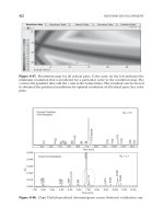
Ultrasound: The abdominal aorta may be imaged from the diaphragm
to the bifurcation, although occasionally the distal aorta is obscured by
overlying bowel gas. It is normally 2–3 cm in diameter (Fig. 6.21).
CT and MRI: The aorta and its main branches are well depicted on CT
and MRI following intravenous contrast enhancement (Figs. 6.1, 6.2
and 6.7). The celiac axis, superior mesenteric artery and renal arteries
are always visible when normal. The inferior mesenteric artery and
several lumbar arteries may also be seen. Multi-detector CT or MR
angiography enable image reformatting, to demonstrate the vessels in
any anatomical plane.
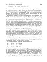
Angiography: A pigtail catheter introduced into the upper abdominal
aorta is used to inject iodinated contrast medium directly into the
aorta, followed by rapid imaging (Fig. 6.22). Selective catheterization
of the aortic branches may also be performed.
Inferior vena cava (IVC) (Figs. 6.2, 6.7)
The IVC is formed by the union of the common iliac veins from the
pelvis, just behind the right common iliac artery, at the level of the 4th
or 5th lumbar vertebra. The IVC runs up along the anterior aspects of
Fig. 6.21. Longitudinal ultrasound scan through the aorta, celiac, and superior
mesenteric arteries.
Liver
Celiac axis
Vertebrae
Stomach
Superior
mesenteric
artery
Aorta
Left hepatic artery
Intercostal artery
Hepatic artery
Common
hepatic artery
Gastroduodenal
artery
Right renal
artery
Ileocolic artery
Distal superior
mesenteric artery
Left gastric artery
Splenic artery
Left renal
arteries (2)
Superior
mesenteric
artery
Jejunal branches
Lumbar arteries
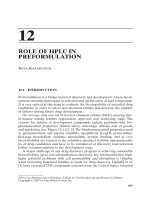
Fig. 6.22. Flush aortogram, frontal projection. Note the left hepatic artery arises
from the left gastric artery (a variant seen in 25% of normal individuals). The
patient has two left renal arteries.
The renal tract, retroperitoneum and pelvis andrea g. rockall and sarah j. vinnicombe
62
the lumbar vertebral bodies, just to the right of the aorta. It runs ante-
rior to the right adrenal gland and right crus of diaphragm. Superiorly,
the IVC runs through the liver (the intrahepatic IVC). It then crosses
through the central tendon of the diaphragm at the level of the 8th tho-
racic vertebra to drain into the right atrium of the heart.
Tributaries that drain into the IVC closely follow the branches of
the aorta (apart from the venous drainage of the small and large
bowel, which is via the mesenteric veins that drain into the portal
circulation):
• abdominal wall veins drain into the IVC via the right and left
phrenic veins and the 3rd and 4th lumbar veins
• the right gonadal, renal and adrenal veins each drain directly into
the IVC
• the left gonadal and adrenal veins drain into the left renal vein,
which then crosses the midline and drains into the IVC
• the right, middle and left hepatic veins drain into the
intrahepatic IVC.
Imaging the inferior vena cava
Ultrasound: The intrahepatic part of the IVC can be seen throughout
its length, up to the junction with the right atrium. The upper abdom-
inal portion of IVC can usually be well seen, but the lower part of the
IVC, common iliac, internal and external iliac veins are often partly
obscured by overlying bowel gas.
CT: The IVC can be seen throughout its length. The major pelvic veins
are also well demonstrated.
MRI: This is the method of choice for the demonstration of flow in
the IVC. The images are best performed as an MR venogram, with
administration of intravenous contrast medium (Fig. 6.2).
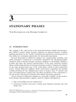
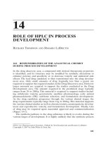
The pelvic vasculature
A pelvic arteriogram is shown in Fig. 6.23.
The aorta bifurcates in front of the fourth lumbar vertebral body at
the level of the iliac crest into the common iliac arteries, which enter
the pelvis on the medial border of the psoas muscles, lying just ante-
rior to the common iliac veins. The common iliac arteries divide at the
pelvic brim anterior to the lower sacroiliac joints into internal and
external iliac arteries.
The external iliac artery runs along the medial border of psoas,
passing under the inguinal ligament to become the femoral artery. It
is larger than the internal iliac artery. Just above the inguinal liga-
ment, it gives off the inferior epigastric artery and the deep
circumflex iliac artery, which supply the anterior abdominal wall
muscles.
The internal iliac artery enters the true pelvis anterior to the
sacroiliac joint, with the ureter anterior to it. From its origin, the
artery runs inferomedially, anterior to the sacrum, its length varying
from 2–5 cm. It has the most variable branching pattern of all the
arteries in the body; the commonest pattern is described here. It
divides into anterior and posterior divisions at the upper border of
the greater sciatic foramen.
The anterior division courses down towards the ischial spine and
gives off the following branches:
(a) the obturator artery
(b) the inferior vesical artery, supplying the lower bladder, ureter,
prostate gland and seminal vesicles
(c) the middle rectal artery, supplying the prostate gland, seminal
vesicles and rectum
(d) the uterine artery, supplying the uterus, upper vagina, Fallopian
tubes and ovary
(e) the vaginal artery, equivalent to the inferior vesical artery in
the male
(f) the internal pudendal artery, supplying the genitalia in the
perineum
(g) the superior vesical artery, supplying the upper bladder
(h) the inferior gluteal artery, which passes through the lower part
of the greater sciatic foramen.
Branches of the posterior division of the internal iliac artery are as
follows:
(a) the iliolumbar artery, supplying psoas and iliacus
(b) the lateral sacral artery, which supplies the sacral canal and the
muscles and skin over the back
(c) the superior gluteal artery, the largest branch of the internal
iliac artery, passing through the greater sciatic foramen to the
gluteal region.
The internal and external iliac veins accompany the arteries. MR and
CT can demonstrate the pelvic vasculature.
Lymphatics of the abdomen and pelvis
Lymph nodes and lymphatic vessels accompany the major vessels
of the abdomen and pelvis and are classified accordingly. In the
pelvis, the internal and external iliac lymph nodes drain to common
iliac lymph nodes and thence to para-aortic lymph nodes (see below).
Internal iliac
artery
Internal iliac
artery
Iliolumbar
artery
External iliac
artery
Lateral sacral
artery
Superior
gluteal
artery
Obturator artery
Uterus
Uterus
Deep
circumflex
iliac artery
Vesicle
Uterine
artery
Inferior
gluteal
artery
Divisions
of internal
iliac artery
Inferior mesenteric artery
Common iliac artery
Median sacral artery
Posterior
Anterior
Catheter
Common
femoral
artery
Fig. 6.23. Normal pelvic arteriogram in a female patient.
The renal tract, retroperitoneum and pelvis andrea g. rockall and sarah j. vinnicombe
63
Pre-aortic nodes are clustered around the origins of the celiac axis,
the superior mesenteric artery and the inferior mesenteric artery.
These drain the gastrointestinal tract from the lower esophagus to
half-way down the anal canal, as well as the spleen, pancreas, gall
bladder, and part of the liver.
The left para-aortic nodes are grouped along the left lateral
aspect of the aorta. The right para-aortic nodes lie anterior
and lateral to the IVC. The para-aortic nodes drain lymph from
the kidneys and adrenal glands, from the testes in the male and
the ovaries, Fallopian tubes and uterine fundus in the female. The
para-aortic nodes drain into two lymph vessels, the right and
left lumbar trunks. The right and left lumbar trunks join the
intestinal trunk to form the cisterna chyli. This lies just to the
right of the aorta, behind the right crus of diaphragm, at the level
of L1/L2 and is approximately 6 cm long. The cisterna chyli then
drains into the thoracic duct (see chapter “Thorax” section titled
“thoracic duct”).
Imaging the abdominal lymphatic system
Ultrasound: Although the para-aortic lymph nodes in the upper
abdomen may be seen in thin patients, lymph node assessment is
usually incomplete because of overlying bowel gas.
CT and MRI: Lymph nodes can be seen when they measure approxi-
mately 3 mm or more in short axis diameter. Normal para-aortic
nodes may measure up to 1 cm in short axis diameter. Pelvic lymph
nodes rarely exceed 8 mm in short axis diameter.
Lumbosacral plexus
The lumbar plexus is formed in the psoas muscle from the anterior
rami of the L1 to L4 nerve roots. The nerves that form include:
• the iliohypogastric and ilioinguinal nerves
• the lateral cutaneous nerve of the thigh
• the femoral nerve (L 2,3,4), which may be visualized as it runs down
and laterally between the psoas and iliacus to enter the thigh
beneath the inguinal ligament
• the genitofemoral nerve
• the obturator nerve (L2, 3, 4), which crosses the pelvic brim anterior
to the sacroiliac joint, runs behind the common iliac vessels, and
down the pelvic side-wall into the obturator canal (Fig. 6.8)
• the L4 root of the lumbosacral trunk, which joins sacral roots in the
sacral plexus.
The sacral plexus, formed from the lumbosacral trunk (L4, 5) and the
ventral rami of the first to fourth sacral nerves, lies on the piriformis
muscle (Fig. 6.10c). The largest branch is the sciatic nerve, which may
be visualized by CT and MR as it passes through the greater sciatic
foramen into the gluteal region (Fig. 6.8b).
Abdominal sympathetic trunk and sympathetic plexus
The abdominal sympathetic trunks enter the abdomen through the
medial arcuate ligaments as continuations of the thoracic sympathetic
trunks and run along the anterior lumbar vertebrae, then continue
as the pelvic sympathetic chains in the pelvis, posterior to the
common iliac vessels. They are not usually seen using current
imaging techniques.
64
Anatomical Overview
The brain is supported by the skull base and enclosed within the skull
vault. Within, the cranial cavity is divided into the anterior, middle
and posterior fossae. The anterior and middle cranial fossae contain
the two cerebral hemispheres. The posterior fossa contains the brain-
stem, consisting of the midbrain, pons and, most inferiorly, the
medulla, and the cerebellum. Twelve paired cranial nerves arise from
the brainstem, exit the skull base through a number of foramina, and
innervate a variety of structures in the head proper. The largest of
these foramina is the foramen magnum, through which the brainstem
and spinal cord are in continuity. The brain is invested by the
meninges and bathed in cerebrospinal fluid (CSF), circulating in the
Section 4
The head, neck, and vertebral column
Chapter 7 The skull and brain
PAUL BUTLER
Maxillary antrum
Medulla
Foramen magnum
Vertebral artery
Medulla
basilar artery
hypoglossal nerve
canal
foramen of Luschka
Temporal lobe
Meckel’s cave
Middle cerebellar
peduncle
Inferior cerebellar
peduncle
Pons
Middle cerebellar
peduncle
Globe
Lateral rectus
Sphenoid sinus
Internal carotid artery
Trigeminal nerve
Fourth ventricle
Pons
Middle cerebellar
peduncle
Globe
Lateral rectus
Sphenoid sinus
Internal carotid artery
Trigeminal nerve
Fourth ventricle
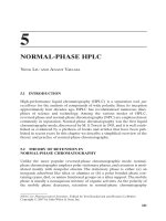
Fig. 7.1.
Routine T2
weighted
axial cranial
MRI: (a) to (o),
base to
vertex.
(a)
(b)
(c)
(d)
(e)
Applied Radiological Anatomy for Medical Students. Paul Butler, Adam Mitchell, and Harold Ellis (eds.) Published by Cambridge University Press. © P. Butler,
A. Mitchell, and H. Ellis 2007.
subarachnoid space. Part of the meninges, the dura, forms an incom-
plete partition between the cerebral hemispheres, known as the falx
and roofs the posterior fossa as the tentorium cerebelli. There is a gap
in the tentorium, called the hiatus, through which the midbrain joins
the hemispheres.
Within the brain are a number of cavities, the lateral, third and
fourth cerebral ventricles, which contain CSF produced by the choroid
plexuses within the ventricles. CSF flows from the ventricles into the
subarachnoid spaces over the cerebral surface and around the spinal
cord.
Blood reaches the brain by the carotid and vertebral arteries and is
drained by cerebral veins into a series of sinuses within the dura into
the internal jugular veins.
Imaging overview
CT and MRI scanning are central to neuroimaging. The role of skull
radiography is very limited and arguably the only situation where it
enjoys a primary role is in the investigation of skull fractures in sus-
pected non-accidental injury in children. The relative merits of MRI
and CT in are summarized below and routine series of axial MRI and
CT are illustrated in Figs. 7.1 and 7.2.
The skull and brain paul butler
65
Optic nerve
Pituitary gland
Pons (upper part)
Superior cerebellar
peduncle
(f)
Crista galli
Gyrus rectus
Sylvian fissure
Posterior cerebral
artery
Midbrain
Occipital lobe
Cerebellar vermis
(g)
Cerebellar vermis
at the tentorial
hiatus
Frontal sinus
Anterior
communicating artery
Middle cerebral
artery
Optic tract
Mamillary body
Quadrigeminal
plate cistern
Superior sagittal
sinus
Cerebellar vermis
at the tentorial
hiatus
Frontal sinus
Anterior
communicating artery
Middle cerebral
artery
Optic tract
Mamillary body
Quadrigeminal
plate cistern
Superior sagittal
sinus
(h)
Anterior cerebral
arteries
Anterior commissure
Insula
Sylvian fissure
Third ventricle
(i)
Corpus callosum
Frontal operculum
Internal capsule
Lentiform nucleus
Fornix
Foramen of Monro
Head of caudate
nucleus
Thalamus
Occipital horn of
lateral ventricle
Visual (calcarine)
cortex
(j)
Body of lateral
ventricle
Insula
Splenium of
corpus callosum
Straight sinus
(k)
Body of lateral
ventricle
(l)
Interhemispheric
fissure
(m)
Centrum
semiovale
(n)
precentral gyrus
central sulcus
postcentral gyrus
(o)
Fig. 7.1.
Continued
The skull and brain paul butler
66
Foramen
magnum
Foramen
magnum
Fig. 7.2.
Cranial CT
after
intravenous
contrast
medium:
(a) to (l),
base to
vertex.
(a)
Anterior clinoid
process
Pituitary gland
Cavernous sinus
Basilar artery
Air cells within
the petrous
temporal bone
(c)
Anterior
communicating
artery
Middle cerebral
artery
Internal carotid
artery
(e)
Lentiform
nucleus
Internal
capsule
Thalamus
Anterior
cerebral
arteries
(g)
Internal cerebral vein
Choroid plexus within
lateral ventricle
(i)
Frontal sinus
Frontal lobe
Sphenoid ridge
Temporal lobe
in middle
cranial fossa
Frontal sinus
Frontal lobe
Sphenoid ridg
e
Temporal lobe
in middle
cranial fossa
(b)
Pons
Cerebellum
(d)
Fourth
ventricle
Midbrain
(f)
Calcification
in pineal
gland
(h)
Superior
sagittal sinus
(j)
The skull and brain paul butler
67
MRI
Advantages
• Superior anatomical detail
• Superior contrast resolution
• Multiplanar capability
• Better for middle and posterior cranial fossae
• No ionizing radiation.
Disadvantages
• Longer investigation
• Claustrophobia
• A number of contraindications relating to various metallic implants
(surgical clips, pacemakers, etc.) and the use of high field-strength
magnets
• Insensitive to subarachnoid haemorrhage and calcification.
CT
Advantages
• Excellent for the emergency situation, both traumatic and non-
traumatic.
• Quick and simple for the patient
• Good for hemorrhage and calcification.
Disadvantages
• Ionizing radiation
• Streak artifacts from bone limit visualization of the adjacent struc-
tures (e.g., the contents of the middle and posterior fossae).
• Usually restricted to axial images with the patient supine, although
high quality, multiplanar views can be reconstructed on the
modern scanners.
MRI is concerned with proton (hydrogen nucleus) imaging and differ-
ent images can be produced depending on the parameters used (the
different pulse sequences). On the T1 weighted (T1W), images, gray
matter is darker (lower signal intensity) than white matter. On T2
weighted (T2W), sequences, the reverse is true. Broadly, T1W images
are good for anatomy, T2W for the detection of pathology. CT is a
digital X-ray investigation. Due to this, and somewhat paradoxically,
white matter is depicted as being darker than gray matter because of
the radiolucency of lipid-containing material.
Iodinated contrast material administered intravenously enhances
blood within the cerebral arteries, veins, and dural venous sinuses.
Enhancement is also seen in the highly vascular choroid plexuses and
in those structures external to the blood–brain barrier such as the
pituitary gland and infundibulum.
With MRI, the mechanism of enhancement with its own intra-
venous contrast agent, gadolinium DTPA, is quite different but, on
T1W images, those structures which enhance become hyperintense
(i.e., whiter) with similar appearances to CT. There are some impor-
tant differences, however. Rapidly flowing blood is displayed as black
“signal voids,” a property shared with both air and cortical bone but
for a different reason (paucity of protons) (Fig. 7.3). The role of angiog-
raphy is primarily for the diagnosis and, in some cases, for the treat-
ment of vascular abnormalities. Increasingly, non- or minimally
invasive forms, magnetic resonance angiography (MRA) or CT angiog-
raphy (CTA), are used for diagnosis. Depending on the technique, MRA
may or may not require gadolinium DTPA. CTA necessitates an intra-
venous injection of iodinated contrast medium.
Catheter angiography, where iodinated contrast medium is injected
directly into an artery (or vein), remains the gold standard. It is nearly
always performed using digital subtraction, showing the vasculature
in near isolation, free of bone detail.
The cervical carotid and vertebral arteries are usually cannulated via
the femoral artery at the groin, although a brachial arterial approach
can be used. The cervical carotid artery can be punctured directly but
this time-honoured method is seldom used now. Angiographic inter-
pretation is the province of the specialist neuroradiologist or clinical
neuroscientist.
Falx
Pituitary stalk
Suprasellar cistern
Posterior cerebral
artery
Midbrain
(k)
Centrum
semiovale
(l)
Fig. 7.3. T1 weighted axial MRI after intravenous gadolinium DTPA. Suprasellar
cistern.
Fig. 7.2. Continued
The skull and brain paul butler
68
CT and MRI interpretation
The way in which a scan is “read” will be determined by the patient’s
suspected clinical diagnosis and the initial observations on the study.
These same considerations will also influence the scan protocol and
whether contrast agents are given. In any case, a sound appreciation
of the normal appearances is essential.
First, the ventricular system should be assessed. Are the ventricles
normal in size or enlarged? Is any enlargement part of generalised
atrophy or is it obstructive? Are all the ventricles enlarged or, say, just
the lateral ventricles, sparing the third and fourth? In this example,
one would search for a lesion in the region of the foramen of Monro.
Next, one should look for abnormal density (CT) or signal intensity
(MRI) within the cerebral substance, comparing the two sides. Is this
associated with mass effect, manifest by sulcal effacement or distortion
of the ventricles (“shift”)? Examination of the basal CSF cisterns, with
CT, will reveal subarachnoid hemorrhage, and their effacement is a
vital clue to cerebral swelling. The appearance of the normal
quadrigeminal plate cistern resembling a smile is reassuring (Fig. 7.2(g)).
Normal scan appearances alter with age. In the normal child, for
instance, the cerebral ventricles and CSF cisterns can be very small. In
the aging population, with some normal “volume loss,” the CSF spaces
may be prominent.
There are also “review areas” on scans, which repay a second look to
identify a subtle change. For instance, on CT the interpeduncular
cistern can harbor a small amount of subarachnoid blood. On MRI, the
region of the posterior part of the third ventricle, cerebral aqueduct
and pineal gland should be studied on the sagittal image. It is also the
case that lesions seen easily on CT may not be clearly shown on MRI
and vice versa. For example, a colloid cyst of the third ventricle can be
difficult to see on MRI in its typical site at the foramen of Monro.
The skull (Fig. 7.4)
The skull vault or calvarium is formed from the frontal, temporal,
parietal, and occipital bones. The skull vault consists of inner and
outer bony “tables” or diploe separated by a diploic space containing
marrow and large, thin-walled diploic veins. In children, marrow is
typically “red,” being active in blood production. It is hypointense on
T1W MRI and, in the adult, is gradually replaced by “yellow” or fatty
marrow, which becomes hyperintense on T1W images.
The bones of the vault are joined at various sutures, which consist
of dense connective tissue. The sagittal suture joins the two parietal
bones in the midline and the coronal suture joins them to the frontal
bone.
In the infant there is a midline defect between the frontal and pari-
etal bones at the junction of the sagittal and coronal sutures. This
anterior fontanelle or bregma closes in the second year.
The occipital bone forms most of the walls and floor of the posterior
cranial fossa, the largest of the three fossae. The single lambdoid suture
separates the parietal and occipital bones. The clivus is formed from the
basal portions of the sphenoid bone anteriorly and of the occipital bone
posteriorly. The articulation is known as the basisphenoid synchondro-
sis and is also the site where the petrous apex joins the clivus.
Sutures are smooth in the newborn but throughout childhood,
interdigitations develop followed by perisutural sclerosis (increased
bone density) and ultimately fusion in the third or fourth decades
or even later. However, for practical purposes sutural fusion occurs
in adolescence because only in children does raised intracranial
pressure, due for instance to a brain tumor, cause head enlargement.
Sutures must be distinguished from fractures of the skull and
important features of the former include interdigitation, sclerosis and
predictable positions.
The skull is invested in periosteum, both externally (pericranium)
and internally (endosteum). The endosteum is firmly adherent to the
connective tissues of the sutures.
The skull base is formed by contributions from the sphenoid, tem-
poral, and occipital bones centrally and from the frontal and
Lambdoid suture
Sagittal suture
Dural calcification
Frontal sinus
Crista galli
Cribriform
plate
Floor of
the anterior
cranial
fossa
Anterior
clinoid
process
Zygomatic
bone
Maxilla
Lesser wing
of sphenoid
Greater wing
of sphenoid
Superior
orbital
fissure
Fig. 7.4. (a) Frontal, (b) lateral skull radiographs.
(a)
The skull and brain paul butler
69
ethmoidal bones anteriorly. The inner surface of the skull base is
divided into the anterior, middle, and posterior fossae. The anterior
fossa is occupied by the frontal lobe; the middle fossa by the temporal
lobe. The posterior fossa contains the brainstem and cerebellum.
The orbital plates of the frontal bones form most of the floor of the
anterior fossa floor with a contribution from the ethmoid bone in the
midline. The inner suface of the frontal bone, forming the floor of
the anterior cranial fossa, has a relatively “rough” surface, which
Maxillary antrum
Zygoma
Foramen ovale
Foramen spinosum
Foramen lacerum
Carotid canal
Jugular foramen
Carotid
canal
Jugular
foramen
(a)
(b)
Fig. 7.5. CT of the skull base: (a) to (c) are contiguous axial images, (a) the most
inferior.
(c)
Fig. 7.4. Continued
Orbital roof
Frontal Parietal
Temporal
Occipital
Pterion
Anterior clinoid
process
Dorsum sellae
Habenular
commissure
(calcifed)
Pineal gland
calcification
Calcified
choroid plexus
Normal temporal
bone ‘thinning’
Clivus
(basiocciput
and basisphenoid)
Mandibular condyle
Zygomatic recesses
of the maxillary antra
Lamina dura
of pituitary
fossa
Sphenoid
sinus
Cribriform
plate
Floor of
the anterior
cranial fossa
Frontal sinus
(b)
accounts for the frequent occurrence of traumatic contusions in the
inferior frontal lobes.
The sphenoid bone consists of a central body and greater and lesser
wings. The greater wing forms the floor of the middle fossa. The lesser
wing forms the posterior part of the anterior fossa and the “ridge,”
bordering the anterior part of the middle fossa. The body is pneuma-
tized by the eponymous air sinus and bears the pituitary fossa on its
superior surface.
A number of foramina occur in the skull base, transmitting a variety
of structures and providing potential routes for the spread of extracra-
nial disease (notably infection or tumor) into the vault (Fig. 7.5).
The foramina ovale, rotundum and spinosum are within the greater
wing of the sphenoid bone. The foramina ovale and spinosum are
often symmetrical, the foramen rotundum rarely so.
The foramen rotundum travels from Meckel’s cave to the ptery-
gopalatine fossa and transmits the maxillary (V2) division of the
trigeminal nerve. On coronal CT it is identified inferior to the anterior
clinoid processes. The foramen ovale transmits the mandibular (V3)
division of the trigeminal nerve. On coronal CT it is identified inferior
to the posterior clinoid processes (Figs. 7.6, 7.16).
The foramen spinosum is situated posterolateral to the larger
foramen ovale and transmits the middle meningeal artery and vein.
The foramen lacerum contains cartilage and separates the apex of
the petrous bone, the body of the sphenoid, and the occipital bone. It
is crossed by the internal carotid artery.
The squamous portion of the temporal bone forms part of the
lateral wall of the middle cranial fossa and its petromastoid consti-
tutes part of the floor of the middle and posterior fossae. The occipital
Carotid
canal
Jugular
foramen
Sphenoid
sinus
The skull and brain paul butler
70
Anterior
clinoid
process
Foramen
rotundum
Posterior clinoid
process
Foramen ovale
Fig. 7.6. Coronal CT of the skull base: (a) is anterior to (b).
(a) (b)
Pyramid
Olive
Fourth ventricle
Cerebellar
hemisphere
Sphenoid sinus
Meckel’s cave
Internal carotid artery
Basilar artery
Internal auditory canal
Middle cerebral peduncle
Inferior cerebellar
peduncle
Fourth ventricle
Fig. 7.7. T2 weighted axial MRI: (a) to (f), inferior to superior. The brainstem.
bone forms most of the floor and walls of the posterior fossa, the
largest of the three.
The skull radiograph
Skull radiograph interpretation
Interpretation of skull radiographs (skull “series”) can be challenging.
It is relatively simple to obtain but is an insensitive indicator of
intracranial pathology with roles limited to trauma and as a prelimi-
nary to cranial surgery. Of course, CT can largely meet these diagnos-
tic requirements and, if necessary, a digital radiograph can be
obtained as part of the CT examination.
Broadly, when confronted with a frontal radiograph, in the attempt
to interpret the many overlapping and irregular lines and lucencies, it
is helpful to compare the two sides. The lateral view gives a relatively
clear view of the vault and pituitary fossa. One will also be influenced
by the external clinical findings as to where an abnormality might be
discovered. For practical purposes, the usual reason for requesting
skull radiographs is to identify a fracture. There are normal vault
“lucencies,” which need to be considered, mainly due to blood vessel
impressions, especially veins. A fracture will usually have a more
distinct margin and, unlike blood vessels, does not often branch.
Normal calcifications may be encountered on skull radiographs
arising in the pineal gland, choroid plexus, dura, and habenular com-
missure (Figs. 7.4, 7.17).
The brainstem (Fig. 7.7)
The brainstem consists of medulla, pons, and midbrain. The medulla,
pons, and cerebellum together constitute the hindbrain.
The medulla commences at the foramen magnum as a continuation
of the spinal cord. Initially it is “closed,” possessing a central canal
like the spinal cord. More superiorly, it becomes “open” as the central
canal leads into the fourth ventricle. In the brainstem, the motor
tracts are generally anterior to the sensory, hence the clinical usage of
“anterior” columns meaning motor and “posterior” column, sensory.
A number of decussations occur within the brainstem where both
motor and sensory fibers cross the midline in accordance with the
general principle that functional control of one-half of the body is
largely exercised by the contralateral cerebral hemisphere. The
sensory decussation is craniad to the motor, but both occur in the
closed portion of the medulla.
The medulla leads superiorly into the pons, which has an anterior
“belly” and a posterior tegmentum.
The midbrain has two cerebral peduncles transmitting the motor
tracts. Its posterior portion is pierced by the cerebral aqueduct
(of Sylvius), to connect the third and fourth cerebral ventricles.
(c)
(b)
Vertebral
artery
Pyramid
Central
canal
(a)
Trigeminal
nerve
Pons
Semicircular
duct
Fourth
ventricle
(d)
The skull and brain paul butler
71
The posterior portion is known as the tectum or tectal plate. It con-
sists of four colliculi or quadrigeminal bodies concerned with auditory
and visual reflexes.
The cerebellum
The cerebellum consists of two hemispheres joined by a central
vermis. The cortical mantle of the cerebellum overlies the white
matter core as in the cerebral hemispheres but the cerebellar cortical
ridges, known as the folia, and the intervening sulci are approxi-
mately parallel to one another and are linked to the brainstem by the
paired cerebellar peduncles (Fig. 7.8). They are named logically. The
inferior cerebellar peduncles join the medulla to cerebellum; the
middle cerebellar peduncles (the largest), pons to cerebellum; the
superior cerebellar peduncles, midbrain to cerebellum.
The cranial nerves
There are 12 paired cranial nerves, most of which are analogous to seg-
mental nerves arising from the spinal cord. They variously provide
sensory and motor nerves to structures in the extracranial head and
neck and their distribution is complex.
The olfactory (first) cranial nerve consists of about 20 bundles of
sensory nerves, which pass through the cribriform plate from the
nose to the olfactory bulb inferior to the frontal lobe. The fibers pass
posteriorly from the bulb along the olfactory tract and thence to the
olfactory cortex.
The optic (second) cranial nerve is not a true nerve but rather an
evagination (outpouching) of the brain. The nerve carries with it a
meningeal sheath and is surrounded by CSF (Fig. 7.9). It passes, along
with the ophthalmic artery, into the orbit through the optic canal.
The two optic nerves converge to form the optic chiasm, which lies in
the suprasellar CSF cistern above the pituitary gland. From the chiasm,
two optic tracts diverge toward the lateral geniculate bodies on each
side of the midbrain. From there, the optic pathway continues through
the temporal lobes towards the visual cortex in the occipital lobes.
The oculomotor (third) cranial nerve supplies the extraocular
muscles with the exceptions of the lateral rectus and superior oblique.
It arises in the midbrain from a nucleus at the level of the superior
colliculi and emerges medial to the cerebral peduncle and is often
seen on FLAIR sequence MR images (Fig. 7.10). It passes anteriorly
between the posterior cerebral and superior cerebellar arteries to
enter the superior part of the cavernous sinus and thence to the orbit
through the superior orbital fissure, accompanied by the trochlear
(fourth) and abducent (sixth) cranial nerves.
The oculomotor nerve is accompanied by parasympathetic fibers,
which constrict the pupil. An intracranial aneurysm arising at the
origins of either the posterior communicating or superior cerebellar
arteries may result in an oculomotor palsy. This will be accompanied
by dilatation of the pupil because the parasympathetic constrictor
fibers travel peripherally in the nerve making them vulnerable
to extrinsic pressure. The nerve also supplies levator palpebrae
superioris – so that a third nerve palsy is associated with ptosis.
fornix
Tectum,
consisting of superior
and inferior colliculi
Inferior cerebellar
peduncle
Superior cerebellar
peduncle
Middle cerebellar
peduncle
Fig. 7.8. T2 weighted coronal MRI. The cerebellar peduncles.
Optic nerve
CSF within sheath
Meningeal sheath
Fig. 7.9. T2 weighted axial MRI. The optic nerve.
Oculomotor
nerve
Fig. 7.10. FLAIR axial MRI.
The oculomotor nerve.
Middle cerebral artery
in Sylvian fissure
Anterior communicating
artery
Optic tract
Substantia nigra
Red nucleus
Ambient cistern
Tectum
Quadrigeminal
plate cistern
(e)
(f)
Fig. 7.7. Continued
Pituitary gland
Cavernous sinus
Pons
Superior cerebellar
peduncle
The skull and brain paul butler
72
Cavernous sinus
Meckel’s cave
Trigeminal nerve
Fig. 7.14. (a) T2 weighted axial MRI, (b) Axial CT on bone algorithm. The
hypoglossal nerve and canal.
The trochlear (fourth) cranial nerve is the only one arising from the
posterior surface of the brainstem, looping around the midbrain and
passing with the oculomotor nerve between the superior cerebellar
and posterior cerebral arteries. It is not seen on routine MRI scans.
The trigeminal (fifth) cranial nerve is the largest and most complex.
The nerve arises from the pons and passes anteriorly to the trigeminal
ganglion located in Meckel’s cave at the posterior end of the cav-
ernous sinus (Fig. 7.11).
The motor root, supplying the muscles of mastication, travels
beneath the ganglion and exits the skull through the foramen ovale.
The ophthalmic (V
I
) division exits through the superior orbital fissure
and the maxillary (V
II
) division through the inferior fissure. The
mandibular (V
III
) division does not enter the cavernous sinus but exits
inferiorly through the foramen ovale. The motor fibers to the muscles
of mastication are confined to the mandibular divisions of the nerve.
The abducent (sixth) cranial nerve supplies the lateral rectus muscle
and has a long intracranial course from pons to cavernous sinus,
which makes it vulnerable in trauma to the skull base. The nerve may
be seen on thin section MR images (Fig. 7.12).
The facial (seventh) cranial nerve, which innervates the muscles
of facial expression, passes with the vestibulocochlear (eighth)
cranial nerve from the pons to the internal auditory canal across the
cerebellopontine angle cistern and these are routinely visualized on
MRI (Fig. 7.12).
There is a sensory root, the intermediate nerve, which transmits
secretomotor fibers to the lacrimal, submandibular, and sublingual
glands and fibers conveying taste from the anterior two-thirds of the
tongu (the chorda tympani).
The glossopharyngeal (ninth), vagus (tenth) and spinal accessory
(eleventh) cranial nerves are not seen on routine cranial MRI but
can be resolved on special sequences. They arise from the medulla
and form a bundle which leaves the cranium through the jugular
foramen (Fig. 7.13).
The hypoglossal (twelfth) cranial nerve can be identified exiting
through the hypoglossal, or anterior condylar, canal after emerging
from the medulla between the olive and pyramid (Fig. 7.14). Again the
nerve is not often seen on routine MRI.
The diencephalon, between the brainstem and cerebral hemi-
spheres includes the thalamus, hypothalamus, and pineal gland,
which all border the third ventricle. The thalami are paired, olive-
shaped nuclear masses extending anteriorly as far as the foramen of
Monro and forming most of the lateral walls of the third ventricle
(Fig. 7.15). Medially, the thalami are apposed (not joined) at the massa
intermedia or interthalamic adhesion. Laterally, the posterior limb of
the internal capsule separates thalamus and lentiform nucleus. The
posterior part of the thalamus is the pulvinar, which overlies the
midbrain.
Fig. 7.11. T2 weighted axial MRI. The trigeminal nerve.
Abducent nerve
Facial nerve
Vestibulo-
cochlear nerve
Bundle containing
glossopharyngeal,
vagus, and spinal
accessory nerves
Fig. 7.13. T2 weighted axial MRI. The glossopharyngeal, vagus and spinal
accessory nerves.
Fig. 7.12. T2 weighted axial MRI. The abducent, facial and vestibulocochlear
nerves.
Hypoglossal
nerve (anterior
condylar) canal
(b)
Hypoglossal
nerve
(a)
The skull and brain paul butler
73
The hypothalamus forms the floor and part of the walls of the
third ventricle. Posterior to the optic chiasm the pituitary stalk or
infundibulum desscends to the pituitary gland. The tuber cinereum
extends posteriorly from the stalk to the mamillary bodies, thence
to the midbrain
The pituitary gland and perisellar region
The pituitary gland and perisellar region are frequently imaged in
cases of endocrine disturbance, or in visual failure.
The pituitary gland lies in the sella turcica (“Turk’s saddle”) on top
of the body of the sphenoid bone (Fig. 7.16). The body of the sphenoid
bone contains the sphenoid air sinus, which provides a route for surgi-
cal access to the pituitary gland via the nose. The sella is roofed by the
dural diaphragma sellae, which is pierced by the pituitary stalk
leading to the hypothalamus (Fig. 7.17).
There is no blood–brain barrier around the pituitary gland, which
therefore takes up intravenous contrast media avidly, either the iodi-
nated agents used for CT or gadolinium DTPA used in MRI.
The pituitary gland should be no more than 9 mm in height,
although it varies in size. In some normal individuals it appears as a
thin rim of tissue at the base of the sella. Its upper margin is usually
concave, although it is often convex in children and in females of
reproductive age.
The cavernous sinuses lie lateral to the sella on either side (Fig. 7.18).
These are extradural venous spaces through which the internal
carotid arteries pass, and damage to the artery here, due to trauma,
can result in a carotico-cavernous fistula.
The third, fourth, branches of the fifth and the sixth cranial nerves
pass through the cavernous sinus to the orbit. The cavernous sinuses
receive blood from a number of facial veins and venous plexuses pro-
viding a potential route for sepsis to spread intracranially (Fig. 7.27).
Above the pituitary gland is the appropriately named suprasellar or
chiasmatic CSF cistern, which contains the circle of Willis and the
optic chiasm (Fig. 7.19). The basal ganglia are part of the extrapyrami-
dal system and consist of the caudate and lentiform nuclei, together
known as the corpus striatum, the amygdala, and claustrum.
The caudate nucleus is C-shaped with a head indenting the frontal
horn of the lateral ventricle, a body running alongside the body of the
lateral ventricle and a tail lying just above the temporal horn of the
lateral ventricle.
The lentiform nucleus is divided in the parasagittal plane into the
medial globus pallidus and larger lateral putamen.
The motor and sensory tracts
The upper motor neurones controlling voluntary movement are found
in the precentral gyrus of the frontal lobe. Axons pass from the cell
Fornix
Internal cerebral
vein
Massa intermedia
Splenium of corpus
callosum
Tectum
Cerebral aqueduct
Fourth ventricle
Mamillary bodySphenoid sinus
Pituitary
gland
Optic chiasm
Hypothalamus
Genu of corpus
callosum
Fig. 7.17. T1 weighted sagittal MRI. Showing the major midline structures.
Internal carotid
artery within the
sphenoid sinus
Pituitary gland
Fig. 7.18. T1 weighted axial MRI after intravenous gadolinium DTPA. The
cavernous sinuses.
Optic foramen
Optic groove
Planum sphenoidale
Tuberculum sellae
Lesser wing
of sphenoid
Anterior clinoid
process
Foramen rotundum
Greater wing
of sphenoid
Foramen
ovale
Foramen
spinosum
Foramen of Vesalius
Dorsum sellae
Posterior clinoid process
Middle clinoid
process
Internal
carotid artery
Fig. 7.16. The bony anatomy of the sellar region.
Fig. 7.15. T1 weighted axial MRI. The basal ganglia.
Head of caudate
nucleus
Fornices
Lentiform nucleus
Internal capsule
Foramen of
Monro
Thalamus
The skull and brain paul butler
74
bodies via the corona radiata to the internal capsule to the motor
nuclei in the brainstem and to the anterior horns of the spinal cord.
The internal capsule is a V-shaped myelinated tract with the genu
(bend) pointing medially separating the anterior and larger posterior
limbs.
From the various cutaneous receptors, sensory neurones with cell
bodies in the dorsal root ganglia synapse in the thalami. Axons of
second-order neurones synapse in the thalami. Third-order neurons
pass from thalami to sensory cortex.
The cerebral hemispheres
The cerebral hemispheres lie above the tentorium and are divided by
fissures and sulci into frontal, parietal, temporal, and occipital lobes.
The limbic system (see below) is also considered to be a lobe.
The hemispheres are linked by the corpus callosum, the largest of
the commissural tracts, which interconnect paired structures across
the midline. Other examples of commissural tracts are the anterior,
posterior, and habenular commissures. The anterior and posterior
commissures are landmarks used in image-guided neurosurgical pro-
cedures.
The corpus callosum is a myelinated tract and appears curved in
sagittal images. The anterior rostrum blends with the anterior com-
missure inferiorly. The curved genu (knee) leads posteriorly to the
body thence the largest and most posterior part, the splenium. Fibers
from the corpus callosum sweep anteriorly into the frontal white
matter as the forceps minor and posteriorly into the occipital white
matter as the forceps major.
There is considerable individual variation in gyral anatomy but the
more constant gyri are shown in Fig. 7.20. It is also important to
appreciate that the relationship of function to structure may be vari-
able and that speech, for example, may be represented over a number
of gyri with intervening white matter. Equally, it may be difficult to
identify the central sulcus and adjacent motor strip accurately. In
specialized centers, functional MRI is carried out with patients per-
forming appropriate intellectual, motor, or sensory tasks. Regional
variations in cerebral oxygen utilization can then be registered and
the eloquent area identified.
The anatomical boundaries of the individual lobes may be indistinct,
depending on the aspect. The frontal lobe is the largest of the anatomi-
cal lobes occupying the anterior cranial fossa and extending posteriorly
to the central sulcus. In common with the temporal lobe, the frontal
lobe has three major gyri, superior, middle, and inferior, which are ori-
ented horizontally. The temporal lobe occupies the middle cranial fossa
The anterior limit of the parietal lobe is the central sulcus, which,
running in the coronal plane, separates the precentral (motor) gyrus of
the frontal lobe from the postcentral (sensory) gyrus. The boundary
between the parietal and temporal lobes laterally is indistinct but the
parieto-occipital incisure medially defines the two lobes. The main cor-
tical supply of the occipital lobe relates to vision. The calcarine (visual)
cortex can be seen to indent the posterior (occipital) horns of the lateral
ventricles. The cortex here is deeply infolded with little intervening
white matter. Inferiorly and laterally the temporo-occipital fissure
marks the division between the two lobes.
The Sylvian or lateral fissure separates the superior surface of the
temporal lobe from the inferior frontal lobe and the anterior parietal
lobe. During development, the cortex overlying the basal ganglia is
invaginated to form the insula (Fig. 7.21). The cortex in front of, above,
and below this depression expands to form covering folds termed the
operculum.
The Sylvian fissure is formed between these folds. On axial imaging
it runs in the coronal plane on the lower cuts and in the sagittal
plane on the higher slices. On coronal MRI, it resembles the shape of
a T lying on its side.
The limbic system
The anatomy of the limbic system is complex, its many components
retaining their descriptive, classical names, unfortunately with some
Fig. 7.19. T2 weighted coronal MRI. The pituitary gland and suprasellar cistern.
Paracentral lobule
M
e
d
ia
l fro
nta
l
g
y
ru
s
Uncus
P
re
cu
n
e
u
s
C
un
e
u
s
Calcarine sulcus
Pa
rahip
po
cam
pal gyrus
M
edial occipitote
mpora
l gyrus
Lateral occipitotemporal gyrus
(a)
Superior frontal gyrus
M
idd
le fro
nta
l
gyr
u
s
Precentral gyrus
C
entral sulcrus
P
o
stcen
tral gyru
s
S
up
e
r
io
r
p
a
rie
ta
l
lo
bu
le
Inferior parieta
l
lobul
e
Temporo-occipital
incisure
In
fe
rio
r
te
m
p
o
r
a
l g
y
r
u
s
In
f
e
rio
r
f
ro
n
t
a
l
gy
r
u
s
P
ars
tria
n
g
u
la
ris
M
id
dle
te
mp
ora
l
g
yru
s
Su
pe
rio
r te
mpo
ral
g
yru
s
Parieto-
occipital
sulcus
(b)
Fig. 7.20. The cortical gyri: (a) medial and (b) lateral aspects.
Cingulate gyrus
Corpus callosum
Septum pellucidum
Frontal horn of lateral ventricle
Head of caudate nucleus
Anterior cerebral artery
Middle cerebral artery
Optic chiasm
Internal carotid artery
Pituitary gland
The skull and brain paul butler
75
synonyms. The limbic system is also classified as one of the cerebral
lobes (the limbic lobe).
The limbic system can be regarded as two C-shaped gyral arches in
each hemisphere, running from near to the midline in the frontal
lobes to the medial part of the temporal lobe (Fig. 7.22). Their course
mirrors the curved relationship of the frontal and temporal lobes and
the various components can be identified in Fig. 7.23.
The arches comprise the following.
The outer limbic gyrus (the larger arc)
subcallosal area, cingulate gyrus, parahippocampal gyrus, subiculum,
uncus,
The inner limbic gyrus (the smaller arc)
supracallosal and paraterminal gyri, hippocampus, The outer and
inner limbic gyri are separated by the hippocampal sulcus and its con-
tinuation, the callosal sulcus.
The hippocampus (sea horse or monster), consists of a head, body,
and tail, and is the first part of the cerebral cortex to form (Figs. 7.24,
7.25). The broadest part is the head anteriorly. More posteriorly, the
body of the hippocampus forms the floor of the temporal horn of the
lateral ventricle. The tail extends around the splenium of the corpus
callosum and is continuous with the supracallosal indusium griseum.
The indusium griseum is closely applied to the surface of the corpus
callosum and anteriorly it merges with the paraterminal gyrus.
Fornices
Hippocampus
Parahippocampal
gyrus
Temporal horn of
lateral ventricle
Fornix
Third ventricle
Temporal horn of
lateral ventricle
Hippocampus
Fornix
Thalamus
Hippocampus
Tectum
Tentorium cerebelli
Cerebellum
Internal
cerebral
veins
Fornix
Hippocampus
Insula
Fig. 7.21. T1 weighted parasagittal MRI. The insula.
Dorsal fornix
Cingulate gyrus and cingulum
Indusium griseum
Septum pellucidum
Column
of fornix
Olfactor
y
bulb
Amygdala nuclear
complex
Uncus
Mamillary body
Parahippocampal gyrus
Dentate gyrus
Hippocampus
Fimbria of fornix
Isthmus
Fig. 7.22. Medial aspect of cerebral hemisphere showing the limbic system.
Fig. 7.23. T2 weighted extended coronal MRI. Obtained perpendicular to the long
axes of the temporal lobes, (a) to (h), anterior to posterior.
(a)
(b)
(c)
(d)
(e)
(f)
(g)
Amygdala
Insula
Globus pallidus
Putamen
Anterior
commissure
Termination of
basilar artery
The skull and brain paul butler
76
The internal structure of the hippocampus is complex and beyond
the resolution of MRI at the time of writing. The dentate gyrus and
cornu Ammonis infold into one another in the form of interlocking
Us (Fig. 7.26).
The parahippocampal gyrus is inferior to the hippocampus and
forms the inferomedial aspect of the temporal lobe. Superior to it is
the hippocampal fissure and laterally, the collateral sulcus. The
parahippocampal gyrus becomes continuous with the cingulate gyrus
which continues anteriorly into the subcallosal area.
The subiculum is a cortical layer of the parahippocampal gyrus and
is separated by the hippocampal fissure from the dentate gyrus.
The most anterior part, the hippocampal head, is separated by the
temporal horn of the lateral ventricle from the amydala (almond),
which lies more anteriorly and a little superiorly.
Splenium of corpus
callosum
Quadrigeminal
plate cistern
Fig. 7.23. Continued
Hippocampus
(h)
7.24. T1 weighted parasagittal MRI. The hippocampus.
Axons from the subiculum and hippocampus form the alveus
(white matter) and converge as the fimbria, which leads into the
fornix (arch) at the posterior hippocampus. The two fornices converge
near to the foramen of Monro. The uncus is formed anteriorly from
the parahippocampal gyrus and posteriorly from the medial part of
the hippocampal head.
The subcortical structures of the limbic system comprise the amyg-
dala, habenula, mamillary body, and septal nuclei.
The septal area is in the medial part of the frontal lobes and
includes the subcallosal area and paraterminal gyri, from the outer
and inner limbic gyri, respectively. There are also limbic connections
with the thalamus and hypothalamus.
The cerebral envelope (Fig. 7.27)
The meninges invest the brain and spinal cord. The three constituent
parts are the outer, fibrous dura mater, the avascular, lattice-like,
arachnoid mater, and the inner vascular layer, the pia mater.
The subarachnoid space contains the cerebrospinal fluid (CSF),
which surrounds the cerebral arteries and veins. It is situated between
the arachnoid, which bridges the sulci, and the pia, which is closely
applied to the cerebral surface.
The dura consists of two layers which separate to enclose the
venous sinuses (Fig. 7.27). The outer layer is the periosteum of the
inner table of the skull. The inner layer covers the brain and gives rise
to the falx and tentorium.
The falx cerebri is a sickle-shaped fold of dura, which forms an
incomplete partition between the cerebral hemispheres. The superior
and inferior sagittal sinuses mark its upper and lower margins.
The “point of the sickle” is anterior, the falx being broader posteri-
orly. When there is swelling of one hemisphere, subfalcine herniation
“midline shift” will be more pronounced anteriorly as a result.
The tentorium cerebelli (“tent”) forms a roof over the contents of
the posterior fossa. Anteriorly and superiorly, the tentorial hiatus con-
stitutes a gap in the tent through which the midbrain passes. The free
medial edge of the tentorium extends anteriorly to form the lateral
wall of the cavernous sinus on each side.
On axial CT, the anterior margin of the tentorium migrates medially
on the higher scans. Structures lateral to the line are supratentorial,
structures medial to the line are infratentorial (or lie within the
posterior fossa).
Temporal horn
Fig. 7.25. T1 weighted
axial image in the plane
of the temporal lobe.
The hippocampus.
Temporal horn of
lateral ventricle
Tail of caudate nucleus
Alveus
Choroid plexus
Ammon’s horn
Fimbria of
fornix
Dentate gyrus
Subiculum
Fig. 7.26. Diagram of
components of limbic
lobe.
The skull and brain paul butler
77
The cerebral ventricular system cerebrospinal fluid
spaces (Fig. 7.28)
The cerebral ventricular system consists of the paired lateral and
single third and fourth ventricles.
Cerebrospinal fluid (CSF), is produced in the choroid plexuses,
and most of it is in the lateral ventricles, entering medially through the
choroidal fissures. It flows from the lateral ventricles to the third ventri-
cle through the foramen of Monro, in the anterior portion of the roof of
the third and from the third to fourth via the cerebral aqueduct of the
midbrain. From the fourth ventricle, the CSF enters the subarachnoid
spaces, leaving through the paired foramina of Luschka, laterally and
the midline, single foramen of Magendie. These foramina provide a
potential route of spread for intraventricular tumors.
At the base of the brain, there are relatively large CSF spaces, the
basal CSF cisterns, which are important both anatomically and in CT
or MRI diagnosis. Although named individually, according to adjacent
structures, they interconnect freely with each other and with the CSF
spaces generally (Figs. 7.29, 7.3, 7.7(f)).
The cerebral blood circulation
Cerebral arteries
The brain is supplied with oxygenated blood by the paired internal
carotid and vertebral arteries. The common carotid artery in the neck
divides at the approximate level of the upper border of the thyroid
cartilage (C4) into its internal and external branches, the latter supply-
ing the various craniofacial structures.
The internal carotid artery
The internal carotid artery is the larger of the two branches, receiving
70% of the common carotid blood flow. It lies posterolateral to the
external carotid near to the bifurcation and neither common nor
internal carotid arteries have cervical branches.
Falx cerebri
Superior sagittal sinus
Inferior sagittal sinus
Inferior petrosal sinus
Internal carotid
artery
Optic nerve
Superior
ophthalmic vein
Anterior
facial vein
Pterygoid venous plexus
Superior petrosal sinus
Cavernous sinus
Great vein
of Galen
Transverse
sinus
Straight sinus
Fig. 7.27. The cranial dura.
Anterior
cerebral
artery
Suprasellar
(chiasmatic)
cistern
Prepontine
cistern
Parieto-
occipital
fissure
Quadrigeminal
plate cistern
Cerebral
aqueduct
Vermis
Fourth
ventricle
Cisterna magna
Fig. 7.29. T2 weighted sagittal MRI. The basal CSF cisterns.
Lateral
Third
Fourth
Cerebral
aqueduct
Foramen of Monro
Fig. 7.28. The cerebral
ventricles.
The skull and brain paul butler
78
Most cerebral arterial aneurysms are borne on the circle of Willis
and so their rupture results in hemorrhage into the subarachnoid
space in the first instance. This includes an aneurysm at the origin of
the ophthalmic artery
The circle of Willis is not circular in shape but rather a five- or six-
pointed star, (Fig. 7.31). It is also complete in only a minority of indi-
viduals. Indeed, the intracranial arterial anatomy is subject to so many
(usually minor) variations that a broad picture will be given here.
The terminal branches of the internal carotid artery are the ante-
rior and middle cerebral arteries. The anterior cerebral artery passes
horizontally towards the midline and links to the other anterior
cerebral by the anterior communicating artery, (Figs. 7.1(h), 7.2(e)).
This first part is known as the A1 segment. From the origin of the
anterior communicating artery both anterior cerebral arteries next
turn superiorly and run in close proximity, above the corpus callo-
sum, following a curving path posteriorly, again near the midline
(Fig. 7.29).
The middle cerebral artery passes horizontally and laterally towards
the Sylvian fissure. There is then a division into two or three branches
(middle cerebral artery bifurcation or trifurcation). These ascend
within the Sylvian fissure and then loop infreolaterally over the oper-
cular cortex over the cerebral surface to supply the parietal and
temporal lobes.
Arising from the proximal anterior and middle cerebral
arteries, a leash of small, perforating arteries (the lenticulostriates)
supplies a variety of structures including the basal ganglia and
internal capsule.
The vertebral arteries are the first branches of the subclavian arter-
ies. They ascend vertically within the foramina transversaria of the
6th to the 2nd cervical vertebrae and posterolaterally through the
foramen transversarium of the atlas, (first cervical vertebra). The
arteries then travel superomedially to pass into the skull through the
foramen magnum, piercing the dura and entering the subarachnoid
space (Figs. 7.7 (a, b)). At the level of the pontomedullary junction,
the two arteries join to form the midline basilar artery (Figs. 7.1
(a)–(f), 7.2 (c)–(e)), which runs anterior to the brainstem. The cerebel-
lum is supplied by the posterior inferior cerebellar arteries, arising
from the vertebral arteries just before the confluence, and the ante-
rior inferior- and superior cerebellar arteries, arising from the basilar
artery.
The internal carotid artery enters the skull through the carotid
canal and courses anteromedially and horizontally (the petrous
segment) before turning superiorly into the cavernous sinus (Fig. 7.5).
In this position the artery forms the shape of a siphon. Emerging from
the cavernous sinus, the artery enters the subarachnoid space and
divides into its terminal branches, the anterior and middle cerebral
arteries (Fig. 7.19).
There are no angiographic markers of the position of the intracav-
ernous portion of the internal carotid artery, but the origin of the oph-
thalmic artery is usually within the subarachnoid space (Fig. 7.30).
The posterior communicating artery passes on each side from the
internal carotid to the posterior cerebral arteries.
The anterior choroidal artery arises from the internal carotid artery
just above the posterior communicating artery
The circle of Willis is situated in the suprasellar cistern and links
the internal carotid arteries with each other and with the verte-
brobasilar system, via the single anterior and paired posterior commu-
nicating arteries. It affords some protection in the event of occlusion
of major arteries by facilitating “cross-flow.”
(b)
(a)
Anterior cerebral
artery
Ophthalmic artery
Middle cerebral
artery branches
Posterior cerebral
artery
Posterior
communicating
artery
Superior cerebellar
artery
Basilar artery
Anterior cerebral artery
Posterior cerebral artery
Middle
cerebral
artery
Basilar artery
Vertebral artery
Posterior
communicating
artery
Anterior communicating
artery
Anterior cerebral artery
Middle cerebral artery
Internal carotid artery
Posterior cerebral artery
Superior cerebellar arter
y
Anterior inferior
cerebellar artery
Posterior inferior cerebellar
artery
Vertebral artery
Fig. 7.30. Internal carotid angiograms: (a) lateral, (b) frontal projections. Although
only the internal carotid artery has been injected with contrast medium, the
vertebrobasilar system and contralateral middle cerebral artery are opacified
due to the Circle of Willis. The triangle in (a) encloses the proximal middle
cerebral artery branches. Note that the anterior ceebral arteries are near to
the midline, whereas the middle cerebral artery branches are laterally situated.
Fig. 7.31. Magnetic resonance angiography. Circle of Willis.