Introduction to Forensic Sciences 2nd Edition phần 4 pot
Bạn đang xem bản rút gọn của tài liệu. Xem và tải ngay bản đầy đủ của tài liệu tại đây (7.51 MB, 38 trang )
8
Forensic Toxicology
ALPHONSE POKLIS
Introduction
To xicology is the study of poisons. More specifically, toxicology is concerned
with the chemical and physical properties of toxic substances and their phys-
iological effects on living organisms, qualitative and quantitative methods
for their analysis in biological and nonbiological materials, and the develop-
ment of procedures for the treatment of poisoning. A poison may be regarded
as any substance which, when taken in sufficient quantity, will cause ill health
or death. The key phrase in this definition is “sufficient quantity”. The inges-
tion of large amounts of water over an extended period of time has been
known to cause fatal electrolyte imbalance. This seemingly bizarre
behavior — ingestion of massive amounts of water — is known as psy-
chogenic polydipsia and occurs in certain forms of schizophrenia. Conversely,
minute quantities of arsenic, cyanide, and other poisons may be ingested,
causing no apparent toxicity. As the 16th century physician Paracelsus
observed, “All substances are poisons; there is none which is not a poison.
The right dose differentiates a poison from a remedy.”
Recently, the science of toxicology has expanded to include a wide range
of interests, including the evaluation of the risks involved in the use of
pharmaceuticals, pesticides, and food additives, as well as studies of occupa-
tional poisoning, exposure to environmental pollution, the effects of radia-
tion, and, regretfully, biological and chemical warfare. However, it is the
forensic toxicologist who has held the title of toxicologist for the longest
period of time. The forensic toxicologist is concerned primarily with the
detection and estimation of poisons in tissues and body fluids obtained at
autopsy or, occasionally, in blood, urine, or gastric material obtained from
a living person. Once the analysis is completed, the forensic toxicologist then
interprets the results as to the physiological and/or behavioral effects of the
poison upon the person from whom the sample was obtained. In the case of
tissues collected at autopsy, the analytical results may reveal that the decedent
©1997 CRC Press LLC
died from poisoning. In living persons, the presence of a drug in a blood or
urine sample may explain coma, convulsions, or erratic behavior.
The complete investigation of the cause or causes of sudden death is an
important civic responsibility. Establishing the cause of death rests with the
medical examiner, coroner, or pathologist, but success or failure in arriving
at the correct conclusion frequently depends upon the combined efforts of
the pathologist and the forensic toxicologist. Poisoning as a cause of death
cannot be proven beyond contention without toxicologic analyses that dem-
onstrate the presence of the poison in the tissues or body fluids of the
deceased. Most drugs and poisons do not produce characteristic or observ-
able lesions in body tissues, and their presence can be demonstrated only by
chemical methods of isolation and identification. If toxicological analyses are
avoided, death may be ascribed to poisoning without definite proof, or a
death due to poisoning may be erroneously attributed to some other cause.
In instances where death is not due to poisoning, the forensic toxicologist
can often provide valuable evidence concerning the circumstances surround-
ing a death. The erratic driving behavior of the victims of automotive acci-
dents is often explained by the presence of alcohol in blood or tissues.
Psychoactive drugs, those which affect behavior, often play a significant role
in circumstances associated with sudden or violent death. The detection of
alcohol, narcotics, hallucinogens, or other drugs may substantiate the testi-
mony of witnesses as to the aggressive, incoherent, or irrational behavior of
the decedent at the time of a fatal incident. Conversely, negative toxicology
findings may dispel stories of the decedent’s drug use. Negative findings are
also significant in persons who should be regularly taking medications to
control pathological conditions. In the case of epileptics, negative or low drug
concentrations may indicate the decedent was not taking his medication in
the prescribed manner and as a result experienced a fatal seizure.
History of Forensic Toxicology
Until the 19th century, physicians, lawyers, and law enforcement officials
harbored extremely faulty notions about the signs and symptoms of poison-
ing. It was traditionally believed that if a body was black, blue, or spotted in
places or “smelled bad” the decedent had died from poison. Other mistaken
ideas were that the heart of a poisoned person could not be destroyed by fire,
or that the body of a person dying from arsenic poisoning would not decay.
Unless a poisoner was literally caught in the act, there was no way to establish
that the victim died from poison. In the early 18th century, a Dutch physician,
Hermann Boerhoave, theorized that various poisons in a hot, vaporous con-
dition yielded typical odors. He placed substances suspected of containing
©1997 CRC Press LLC
poisons on hot coals and tested their smells. While Boerhave was not suc-
cessful in applying his method, he was the first to suggest a chemical method
for proving the presence of poison.
During the middle ages, professional poisoners sold their services to both
royalty and the common populace. The most common poisons were of plant
origin (such as hemlock, aconite, belladonna) and toxic metals (arsenic and
mercury salts). During the French and Italian Renaissance, political assassi-
nation by poisoning was raised to a fine art by Pope Alexander VI and Cesare
Borgia.
The murderous use of white arsenic (arsenic trioxide) became so wide-
spread among the general population that the poison acquired the name
“inheritance powder”. Given this popularity, it is small wonder the first mile-
stones in the chemical isolation and identification of a poison in body tissues
and fluids would center around arsenic. In 1775, Karl Wilhelm Scheele, the
famous Swedish chemist, discovered that white arsenic was converted to
arsenous acid by chlorine water. The addition of metallic zinc reduced the
arsenous acid to poisonous arsine gas. If gently heated, the evolving gas would
deposit metallic arsenic on the surface of a cold vessel. In 1821, Sevillas used
the decomposition of arsine to detect small quantities of arsenic in stomach
contents and urine in poisoning cases. In 1836, James M. Marsh, a chemist
at the Royal British Arsenal in Woolwich, used the generation of arsine gas
to develop the first reliable method to determine an absorbed poison in body
tissues and fluids, such as liver, kidney, and blood.
The 1800s witnessed the development of forensic toxicology as a scientific
discipline. In 1814, Mathieiv J. B. Orfila (1787–1853), the “father of toxicol-
ogy”, published
Traité des Poisons
— the first systemic approach to the study
of the chemical and physiological nature of poisons. Orfila’s role as an expert
witness in many famous murder trials, and particularly his application of the
Marsh Test for arsenic in the trial of the poisoner Marie Lafarge, aroused
both popular and scholarly interest in the new science. As Dean of the Medical
Faculty at the University of Paris, Orfila trained many students in forensic
toxicology.
The first successful isolation of an alkaloid poison was performed in 1850
by Jean Servials Stas, a Belgian chemist, using a solution of acetic acid in
ethyl alcohol to extract nicotine from the tissues of the murdered Gustave
Fougnie. Modified by the German chemist, Friedrich Otto, the Stas-Otto
method was quickly applied to isolation of numerous alkaloid poisons,
including colchicine, conin, morphine, narcotine, and strychnine; the
method is still used today.
In the second half of 19th century, European toxicologists were in the
forefront of the development and application of forensic sciences. Procedures
were developed to isolate and detect alkaloids, heavy metals, and volatile poisons.
©1997 CRC Press LLC
In America, Rudolph A. Witthaus, Professor of Chemistry at Cornell
University Medical School, made many contributions to toxicology and called
attention to the new science by performing analyses for New York City in
several famous poisoning cases: the murders of Helen Potts by Carlyle Harris
and of Annie Sutherland by Dr. Robert W. Buchanan, both of whom used
morphine. In 1911, Tracy C. Becker and Professor Witthaus edited a four-vol-
ume work on medical jurisprudence,
Forensic Medicine and Toxicology,
the
first standard forensic textbook published in the U.S. In 1918, the City of
New York established a medical examiner’s system, and the appointment of
Dr. Alexander O. Gettler as toxicologist marked the beginning of modern
forensic toxicology in America. Although Dr. Gettler made many contribu-
tions to the science, perhaps his greatest was the training and direction he
gave to future leaders in forensic toxicology. Many of his associates went on
to direct laboratories within coroner and medical examiner systems in major
urban centers throughout the country.
In 1949, the American Academy of Forensic Sciences was established to
support and further the practice of all phases of legal medicine in the U.S.
The members of the toxicology section represent the vast majority of forensic
toxicologists working in coroners’ or medical examiners’ offices. Several other
international, national, and local forensic science organizations, such as the
Society of Forensic Toxicologists and the California Association of Toxicol-
ogists, offer forums for the exchange of scientific data pertaining to analytical
techniques and case reports involving new or infrequently used drugs and
poisons. The International Association of Forensic Toxicologists, founded in
1963, with over 750 members in 45 countries, permits worldwide cooperation
in resolving technical problems confronting the toxicologist.
In 1975, the American Board of Forensic Toxicology was organized to
examine and certify forensic toxicologists. One of its stated objectives is “to
make available to the judicial system, and other public, a practical and equi-
table system for readily identifying those persons professing to be specialists
in forensic toxicology who possess the requisite qualifications and compe-
tence”. In general, those certified by the Board must have an earned Doctor
of Philosophy or Doctor of Science degree, have at least 3 years full-time
professional experience, and pass a written examination. At present, only
about 200 toxicologists are certified by the Board.
Deaths Investigated by Toxicologists
Accidental Poisoning
Most accidental poisonings occur in the home. Children, due to their innate
curiosity and adventurous nature, may gain access to and ingest prescription
©1997 CRC Press LLC
drugs, detergents, pesticides, and household cleaners. Fortunately, public
awareness of the safe storage of household chemicals, safety top containers,
the availability of poison control information centers, and better emergency-
room procedures for treating child poisonings have all contributed to a
marked decrease in this type of death. Accidental poisoning in adults is
usually the results of mislabeling, storage of a toxic substance in a container
other than the original one. As often as not, the improper container is an
old whiskey bottle! Arsenic, weed killer, strychnine, cyanide, cleaning solu-
tions, and numerous other deadly poisons have been eagerly and mistakenly
drunk from cider jugs and old whiskey bottles. An open container of cyanide
next to a tin of sugar on a basement work bench has been known to sweeten
a final cup of coffee.
Accidental poisonings may occur in industry due to carelessness or mis-
haps which expose workers to toxic substances. While the potential for acci-
dental poisonings in industry is great, safety standards and regulations and
the availability of emergency medical services today prevent industry from
being a source of many fatal intoxications.
Deaths from Drug Abuse
Drug abuse, the nonmedical use of drugs or other chemicals for the purpose
of changing mood or inducing euphoria, is the source of many poisonings.
Drug abuse may involve the use of illicit drugs such as heroin or phencycli-
dine; the use of restricted or controlled drugs such as cocaine, barbiturates,
and amphetamine; or use of chemicals in a manner contrary to their intended
purpose — such as inhaling solvents and aerosol products. Since the devel-
opment and glorification of the “drug culture” in the mid-1960s, deaths due
to illicit drug use are the most common fatal poisonings investigated by
toxicologists, particularly in large urban areas. Table 8.1 presents the drugs
most commonly encountered in death investigations; note the high incidence
of cocaine, alcohol, and heroin/morphine.
In a broader sense, drug abuse may also include the excessive use of
legal substances, such as alcohol and prescription drugs. The use of alcohol
is the biggest drug problem in the U.S. Alcohol plays a significant role in
violent deaths. Of the 40,000 automobile accident deaths that occur annu-
ally in the U.S., 50% involve drinking drivers, and 60% of pedestrians killed
have significant blood alcohol levels. Of urban adults who were admitted
to a hospital with a fractured bone, 50% fractured the bone during or after
drinking. Significant blood alcohol levels are found at autopsy in 35% of
all persons committing suicide and in 50% of all murder victims. Also,
many people die each year due to many pathologic conditions directly
attributed to alcohol or complications of other pathologic conditions
aggravated by alcohol consumption. Alcohol is a self-limiting poison:
©1997 CRC Press LLC
people usually lose consciousness before a lethal dose is ingested. Therefore,
overdose deaths due to the ingestion of excessive quantities of alcohol are
uncommon. However, numerous accidental deaths occur from the concur-
rent ingestion of potent prescriptions drugs and alcohol.
Suicidal Poisoning
Suicide is a common manner of death in cases of poisoning. In general, about
twice as many men successfully commit suicide as women. However, twice
as many women attempt to commit suicide with poison as men. The most
common suicidal agent is carbon monoxide, a gas generated by the incom-
plete combustion of carbonaceous compounds. Automobile exhaust contains
a substantial concentration of carbon monoxide. Allowing a car motor to
run in a closed garage is the usual method used by those who commit suicide
with carbon monoxide. While cyanide, arsenic, and other well known poisons
may be occasionally used as suicidal agents, most deaths result from pre-
scription drugs. Persons suffering from depression and other emotional dis-
turbances usually have available a supply of potent and, if taken in excess,
deadly drugs to combat the symptoms of their psychological disorders. Today,
most suicidal poisonings involve multiple drug ingestion; usually three to
seven different drugs are ingested at one time. By analyzing the gastric and
bowel contents, blood, urine, and the major organs of the body, the toxicol-
ogist can determine the minimum quantity of the poison ingested. In sui-
cides, the results of such analysis demonstrate that a massive quantity was
taken; this establishes beyond doubt that the decedent could not have acci-
dentally taken such a dose.
Table 8.1 Drugs Most Frequently Encountered
in Medical Examiners Cases, 1991
a
Rank Drug Name Number of Mentions Percent
b
of Total Episodes
1Cocaine 3,020 45.75
2Alcohol — in combination 2,436 36.90
3Heroin/Morphine 2,333 35.3
4Codeine 783 11.86
5Diazepam 587 8.89
6Amitriptyline 437 6.62
7Methadone 430 6.51
8Nortriptyline 379 5.74
9 d-Proxpoxyphene 325 4.92
10 Diphenhydramine 241 3.65
a
Drug Abuse Warning Network, National Institute on Drug Abuse data from 27 metropolitan areas.
b
Percent of total episodes may exceed 100%, as a single case may involve more than one drug.
©1997 CRC Press LLC
Homicidal Poisoning
Accidental and suicidal poisonings are common today; murder by poison is
rare. Determining that a person died as the result of homicidal poisoning is
often the most difficult type of investigation for law enforcement officers and
medical experts. The general evidence of poisoning is obtained from a knowl-
edge of the symptoms displayed by the decedent before death, the postmor-
tem examination of the body by the pathologist, and the isolation and
identification of the poison by the toxicologist. For successful prosecution of
a suspect, law enforcement officers must establish that the perpetrator had
access to a supply of the poison, that the suspect was aware of the lethal
effects of the poison, and that the suspect had opportunity to administer the
poison to the decedent.
When the victim is attended to, before death, by a physician, the doctor
seldom, if ever, considers poisoning as a cause of the patient’s ills. Only if
the patient’s occupation brings him into contact with toxic substances (works
in a refinery, chemical, or smelting plant; works on a farm and uses pesticides
and herbicides) will the physician suspect a chemical intoxication. Murder
by poison most commonly occurs within the home, and the physician will
seldom suspect a bereaved husband, wife, son, or daughter of poisoning
another family member. Also, there is rarely any symptom of poisoning which
cannot equally well be caused by disease. Vomiting, diarrhea, rapid collapse,
and weak pulse, all symptoms of arsenic poisoning, may also be due to a
ruptured gastric ulcer or an inflammation of the pancreas or appendix.
Likewise, both strychnine and tetanus cause convulsions. Contracted pupils
and narcosis may be from narcotic drugs or brain lesions. However, there
are circumstances which render a diagnosis of poisoning moderately certain.
The onset and progression of symptoms to rapid death immediately after
eating or drinking indicate acute poisoning, since bacterial food poisoning
has a delayed onset of symptoms.
The pathologist can recognize the effects of certain poisons at autopsy.
Strong acids and alkalis may cause extensive burns around the mouth or the
surface of the body, with severe destruction of the internal tissues. Metallic
poisons may cause intensive damage to the gastrointestinal tract, liver, and
kidneys. Phosphorus, chlorinated hydrocarbons, and poisonous mushrooms
cause gross fatty degeneration of the liver. However, most poisons do not
produce observable changes in body tissue; hence, in many instances of
poisoning, the value of the pathologist’s examination of the body is estab-
lishing that death was not due to natural causes or traumatic injury and that
there is no evidence for cause of death except from possible poisoning. In
most cases, toxicological analysis produces evidence for murder by poison.
©1997 CRC Press LLC
Toxicological Investigation of a Poison Death
The toxicological investigation of a poison death may be divided into three
steps:
1. Obtaining the case history and suitable specimens
2. The toxicological analyses
3. The interpretation of the results of the analyses
Case History and Specimens
To day, there are readily available to the public thousands of compounds that
are lethal if ingested, injected, or inhaled. The toxicologist has only a limited
amount of material on which to perform his analyses; therefore, it is imper-
ative that, before beginning the analyses, he or she is given as much infor-
mation as possible concerning the facts of the case. The toxicologist must be
aware of the age, sex, weight, medical history, and occupation of the decedent,
as well as any treatments administered before death, the gross autopsy find-
ings, drugs available to the decedent, and the time interval between the onset
of symptoms and death. In a typical year, the toxicology laboratory of a
medical examiner’s office will perform analyses on tissues for such diverse
poisons as prescription drugs (analgesics, antidepressants, hypnotics, tran-
quilizers), drugs of abuse (hallucinogens, narcotics, stimulants), commercial
products (antifreeze, aerosol products, insecticides, rodenticides, rubbing
compounds, weed killers), and gases (carbon monoxide, cyanide). Obviously,
the possible identity of the poison prior to analysis would greatly help.
The collection of specimens for toxicological analysis is usually per-
formed by the pathologist at autopsy. Specimens from numerous body fluids
and organs are necessary as drugs and poisons display varying affinities for
the body tissues (see Table 8.2). Drugs and poisons are not distributed evenly
Table 8.2 Exhibits Collected at Autopsy for Toxicological Analysis
Specimen Quantity Toxicant Sought
Adipose tissue 200 g Insecticides, thiopental
Bile All available Codeine, morphine
Blood 15 ml Alcohols, carbon monoxide
Brain 500 g Volatile poisons
Kidney One whole organ Heavy metals
Liver 500 g Most toxicants
Lung organ One whole Methadone, gases, inhalants
Stomach and intestinal contents All available All toxicants taken orally
Urine All available Most toxicants
Vitreous humor All available Digoxin, electrolytes, glucose
©1997 CRC Press LLC
throughout the body, and the toxicologist usually first analyzes those organs
expected to have the highest drug concentrations, Figure 8.1. A large quantity
of each specimen is needed for thorough toxicological analysis because a
procedure which extracts and identifies one compound or class of com-
pounds may be ineffective in extracting or identifying others.
In collecting the specimens, the pathologist labels each container with
the date and time of autopsy, the name of the decedent, the identity of the
sample, and the signature of the pathologist. The toxicologist, when receiving
the specimens, gives the pathologist a written receipt and stores the specimens
in a locked refrigerator until analysis. This procedure provides an adequate
chain of custody for the specimens which enables the toxicologist to intro-
duce his results into any legal procedures arising from the case.
Specimens should be collected before embalming, as this process may
destroy or dilute the poisons present and render their detection impossible.
For example, cyanide is destroyed by the embalming process. Conversely,
methyl or ethyl alcohol may be a constituent of an embalming fluid, thus
giving a false indication of the decedent’s drinking prior to death.
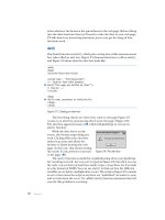
Toxicological Analysis
Before beginning the analysis, the toxicologist must consider several factors:
the amount of specimen available, the nature of the poison sought, and the
possible biotransformation of the poison. Because he is working with a
Figure 8.1
Distribution of cocaine in cases of fatal intravenous injection. (Data from
Poklis, et al.,
J. Anal. Toxicol.,
9, 227, 1985. With permission.)
©1997 CRC Press LLC
limited amount of specimen, the toxicologist must devise an analytical
approach which will allow the detection of the widest number of compounds.
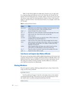
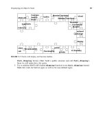
Figure 8.2 outlines a schema for the isolation of poisons when the offending
compound is not known. In cases involving oral administration of the poison,
the gastrointestinal contents are analyzed first, since large amounts of residual
unabsorbed poison may be present. The urine may be analyzed next, as the
kidneys are the major organ of excretion for most poisons and high concen-
trations of toxicants are often present in urine. Following absorption from
the gastrointestinal tract, drugs or poisons are first carried to the liver before
entering the general systemic circulation; therefore, the first analysis of an
internal organ is conducted on the liver. If a specific poison is suspected or
known to be involved in a death, the toxicologist chooses to first analyze
those tissues and fluids in which the poison concentrates.
Figure 8.2
Schema for isolation of poisons.
©1997 CRC Press LLC
Biotransformation is a term used to denote the conversion by the body
of a foreign chemical to a structurally different chemical. The new compound
is called a metabolite. Biotransformation of a drug or poison usually, but not
always, results in formation of a physiologically inactive substance which is
more readily excreted from the body than the parent compound. Figure 8.3
presents the biotransformation of cocaine. Metabolites may be physiologi-
cally active or inactive and nontoxic, less toxic, or more toxic than the parent
compound. Cocaine exemplifies this process as norcocaine is physiologically
active, while benzoylecgonine and methylecgonine have no physiologic action
(Figure 8.3). Thus, the toxicologist must have an understanding of biotrans-
formation reactions. In some instances, the metabolites are the only evidence
that a drug or poison has been administered. Evidence of heroin or cocaine
use is indicated by the presence of their respective metabolites, morphine
and benzoylecgonine.
The toxicologist must be aware of the normal chemical changes which
occur during a body’s decomposition. The autopsy or toxicological analysis
should be started as soon after death as possible, as natural decomposition
processes may destroy a poison initially present at death or may produce
substances or compounds with chemical or physical properties similar to
those of commonly encountered poisons. For example, during decomposi-
tion, phenylalanine, an amino acid normally present in the body, is converted
Figure 8.3
Biotransformation of cocaine.
©1997 CRC Press LLC
to phenylethylamine, which has chemical and physical properties very similar
to amphetamine. The ethyl alcohol and cyanide content of blood may be
decreased or increased depending on the degree of putrefaction and microbial
activity. However, many poisons, such as arsenic, barbiturates, mercury, and
strychnine, may still be detectable many years after death.
In the investigation of a poisoning, it is first necessary for the toxicologist
to isolate and identify the poison. Therefore, forensic toxicologists group
poisons according to the method used to isolate the substances from body
tissues or fluids.
Group I: Gases
Most gases of toxicological significance are not detectable in autopsy speci-
mens. However, some may be isolated from blood or lung tissue by aeration
processes. Usually, air samples are collected at the scene of exposure.
Group II: Steam Volatile Poisons
Compounds in this group are isolated by steam distillation. The sample
(blood, urine, or a tissue homogenate) is made acidic with hydrochloric acid
or basic with solid magnesium oxide. A stream of steam is passed through
the sample and the volatile poisons are distilled off in an aqueous distillate.
Poisons distillable from an acid medium include carbon tetrachloride, chlo-
roform, cyanide, ethanol, methanol, phenols, nitrobenzenes, and yellow
phosphorus. Poisons distillable from a basic medium include amphetamine,
aniline, meperidine, methadone, and nicotine.
Group III: Metallic Poisons
Metals are isolated from tissue by destroying all the organic matter compris-
ing the tissue. The tissue may be destroyed by excessive heat (dry ashing) or
by boiling with concentrated acids or strong oxidizing agents (wet ashing).
Various methods may be used to identify specific metallic poisons remaining
in the ash.
Group IV: Nonvolatile Organic Poisons
This group contains most of the drugs of interest to toxicologists in the U.S.
today. Compounds in this group are usually present in tissues only in minute
quantities. Some drugs (e.g., barbiturates) may be directly extracted from
tissue homogenates by organic solvents. However, many compounds are
often separated from the bulk of the tissue matrix by preparing a protein-free
filtrate of tissue. This filtrate is then subjected to selective extraction with
organic solvents under varying conditions of acidity. Using such techniques,
drugs are isolated into five subgroups.
©1997 CRC Press LLC
1. Strong acids (e.g., chlorothiazide, salicylates)
2. Weak acids (e.g., acetaminophen, barbiturates)
3. Neutrals (e.g., meprobamate, methaprylon)
4. Bases (e.g., codeine, phenothiazines, quinine, strychnine)
5. Amphoterics (e.g., hydromorphone, morphine)
Group V: Miscellaneous Poisons
This group includes all poisons not classified in the previous four groups.
The substances included in this group are inorganic anions (e.g., bromine),
highly water soluble organic ions (e.g., curare, fluoroacetate, paraquat), and
organic compounds insoluble in water or alcohol. Generally, specific techniques
must be used to isolate and identify these compounds from biological samples.
In performing an analysis, the toxicologist has available all the techniques
of modern analytical chemistry. If the poison which caused the death is
known, a specific analysis may be performed; however, if the agent is not
known, or more than one toxicant is suspected, the toxicologist must first
perform a series of analyses to determine which toxicants are present and
then determine by quantitative analysis the amount of each toxic substance
present in the various specimens. While numerous chemical methods are
available to the toxicologist, only a few of the more common procedures are
discussed. All of these methods can be applied to qualitative (identification)
and quantitative (concentration) analysis.
Color Test
A color test is a chemical procedure in which the substance tested for is acted
on by a reagent which causes a change in the reagent, thereby producing an
observable color or color change. Color tests may be used to determine the
presence of specific compounds or a general class of compounds. The pro-
cedures are usually rapid and easily performed. The greatest utility of color
tests in toxicology is the rapid screening of urine specimens, as the urine may
be analyzed directly without time-consuming extraction procedures. An
example of color test is the “Trinder’s test” for the detection of salicylates in
blood or urine. A reagent of ferric nitrate and mercuric chloride is mixed
with 1 ml of blood or urine; if salicylates are present, a violet color is observed.
As in all other toxicology testing, the presence of salicylates must be con-
firmed by another method of analysis. A positive Trinder’s test is observed
for salicylic acid (a metabolite of aspirin), salicylamide, and methyl salicylate.
A false-positive, that is the development of a color when no salicylate is
present, may be observed in urine of diabetic patients excreting acetoacetic
acid and in patients receiving high therapeutic doses of phenothiazine drugs.
The toxicologist must be aware of the limitations of the tests he performs
and particularly the sources of false-positive reactions.
©1997 CRC Press LLC
Microdiffusion Test
Microdiffusion analysis is used for the rapid isolation and detection of volatile
poisons. A simple microdiffusion apparatus consists of a small porcelain dish
with two separate compartments, an inner well surrounded by an outer well
formed between the periphery of the wall of the inner compartment and the
higher outside wall of the dish. The outer well is the sample cell, to which a
small quantity, 1 to 5 ml, of blood, urine, or tissue homogenate is added. To
the inner well an “absorbent” is added. The absorbent is a reagent or solvent
in which particular volatile substances will readily dissolve. After the sample
and absorbent are added to the proper cell, the dish is sealed with a viscous
sealant material and a ground-glass cover plate. If allowed to sit at room
temperature or gently heated, the volatile poison will diffuse from the sample
into the atmosphere of the dish and be entrapped by the absorbent solution,
which often is a color reagent. As the poison is liberated from the sample,
the toxicologist may observe a color formation or color change in the absor-
bent in the inner well. Numerous volatile poisons and gases may be detected
by microdiffusion techniques; they include acetaldehyde, carbon monoxide,
cyanide, ethanol, fluoride, halogenated hydrocarbons, and methanol.
Chromatography
Chromatography is a separation technique. The components of a sample
mixture are distributed between two phases, one of which is stationary while
the second one, the mobile phase, percolates through a matrix or over the
surface of a fixed phase. The components of a sample mixture exhibit varying
degrees of affinity for each phase, and as they are carried along by the mobile
phase, a differential migration occurs. Some components are retained on the
stationary phase longer than others, producing a separation of the com-
pound. The retention of a component by the stationary phase depends on
several factors, including the chemical and physical nature of the stationary
and mobile phases, as well as the experimental conditions, such as temper-
ature or pressure. It is essential, therefore, that pure reference standards be
chromatographed under the same conditions as the unknown materials.
Compounds are tentatively identified by comparing their retention on the
stationary phase with that of the reference standards. Following chromatog-
raphy, the identity of the compounds must be substantiated by other methods
of analysis. There are many varieties of chromatographic analysis; however,
only the three most commonly applied by toxicologists will be briefly dis-
cussed. These are thin-layer chromatography (TLC), gas liquid chromatog-
raphy (GLC), and high-performance liquid chromatography (HPLC).
Thin-Layer Chromatography.
In TLC, the stationary phase is a “thin layer”
of an absorbent, usually silica gel, which is spread on a solid support, such
©1997 CRC Press LLC
as a glass plate. Concentrated sample extracts and drug standards are applied
as a series of spots along the bottom of the plate and allowed to dry. The
plate is then placed in a closed tank, in which the absorbent layer makes
contact with a “developing solvent” (mobile phase) below the applied spots.
The solvent moves up the plate by capillary action, dissolving and separating
the components of the extracts. When the solvent has reached the top of the
plate or ascended a predesignated distance, the plate is removed from the
tank and the solvent evaporated from the plate. Each individual drug in the
standard mixture and in the extracts will separate during migration, produc-
ing a series of spots or narrow bands extending from the bottom to the top
or solvent front on the plate. The migration of compounds is expressed by
the retention factor (Rf) which is defined as the ratio of the distance moved
by the compound to the distance the mobile phase ascends the plate from
the point of application of the compound. The presence of a drug is visualized
by spraying or dipping into the plate various reagents which produce colored
reactions with particular components. Several sprays may be used in sequence
to aid in identification of compounds. Some drugs will react with certain
reagents but not with others. For example, in screening urine extracts for the
presence of drugs of abuse, the toxicologist may first spray the chromatogram
with ninhydrin, which produces a red or pink color with primary amines
such as amphetamine or ephedrine. Next, he may apply ethanol in sulfuric
acid, which produces a series of brightly colored pink, orange, blue, or green
spots with phenothiazine tranquilizers and their metabolites. The plate may
then be sprayed with iodoplatinate, which reacts with all nitrogenous bases.
There are numerous TLC spray reagents to choose from, but the toxicologist
must be guided by the chemical nature of the compounds it is desired to
identify. If a compound from the extract migrates the same distance and
reacts to the applied sprays in the same manner as the reference drug, the
toxicologist then has a tentative identification of the compound, which must
be confirmed by another chemical test; however, he has ruled out all com-
pounds which do not migrate the observed distance in this TLC solvent
system and do not react in the same manner to the spray reagents. Table 8.3
presents the Rfs and reactions with visualization reagents of several drugs
commonly sought in toxicology screening.
Gas Liquid Chromatography.
In GLC, the mobile phase is an inert carrier
gas (e.g., helium, nitrogen) which flows through a column packed with a
solid support coated liquid stationary phase (packed column) or over a
stationary phase coating the walls of narrow column (capillary column).
Numerous types of liquid materials are available, and the toxicologist varies
the stationary phase depending upon the nature of the compounds or groups
of compounds he wishes to separate and identify. Extracted samples are
©1997 CRC Press LLC
Table 8.3 Thin Layer Chromatographic Data of Some Drugs of Toxicological Interest
Spray Reagent
Drug Rf Ninhydrin
Diphenyl-Carbazone in
Mercuric Sulfate Heat U.V. Light Iodoplatinate
Morphine 0.15 Blue
Phenylpropanolamine 0.27 Red Light brown
Codeine 0.30 Brown
Quinine 0.38 Blue Brown
Amphetamine 0.39 Pink
Phenobarbital 0.53 Purple
Amobarbital 0.75 Purple
Chlorpromazine 0.78 Red Brown Brown
Thioridazine 0.78 Blue Dark brown
Diazepam 0.88 Yellow-green Red-brown
Amitriptyline 0.98 Blue Light brown
1. Developing solvent: ethyl acetate, 170 ml; methanol, 20 ml; ammonium hydroxide, 10 ml. (B. Davidow et al.,
Am. J. Clin. Pathol.,
38, 714,
1968. )
©1997 CRC Press LLC
©1997 CRC Press LLC
vaporized and carried through the column by the gas. As the components
are eluted from the column, they are carried by the gas stream to a detector,
which produces an electronic signal that is amplified and displayed on a
recorder. The migration of a compound through the column is usually
expressed by the retention time (Rt), which is defined as the time elapsed
between injection of the sample and the detection of the compound. The
retention time provides a tentative identification of the compound, and the
strength of electronic signal to the recorder may be used to determine the
quantity of the compound present in the sample. An extract of a specimen
chromatographed under the same conditions as reference drugs and produc-
ing a peak at the same time would be tentatively positive for the reference
drug in the specimen. The height of the peak and the area under the peak
are directly related to the concentration of the drug present. Gas chromatog-
raphy is particularly suitable for the analysis of volatile substances such as
alcohols (Figure 8.4).
Figure 8.4
Gas chromatographic separation of common volatiles:
(A) methanol, (B) acetone, (C) ethanol, (D) isopropanol, (E) butanol.
©1997 CRC Press LLC
High-Performance Liquid Chromatography.
In HPLC the mobile phase
is a liquid which flows through a column packed with solid stationary phase
under continuous pressure. Numerous types of stationary materials are avail-
able, and the toxicologist may use almost any solvent or numerous aqueous
mixtures as the liquid phase. Therefore, specific procedures can be developed
for separating compounds which are not easily resolved by other chromato-
graphic methods. The method is particularly suited for heat liable com-
pounds, which may decompose when volatilized for GLC separations. As
with GLC, eluted drugs are identified by their Rt, and detector responses are
proportional to the concentration of drug present in the sample.
Spectroscopy
Spectroscopy concerns the absorption or production of radiant energy. The
absorption of radiation is a characteristic of all molecules; however, the
wavelength of the absorbed radiation may vary from X-rays through ultra-
violet, visible, and infra-red and on to microwave and radio frequencies.
Therefore, the interaction between a chemical compound and radiation is
dependent on its molecular structure and the wavelength of the radiation.
When the absorption of radiation by a compound is determined relative to
the wavelength of the radiation, an absorption spectrum is observed which
is characteristic of that compound. The specificity of the spectrum is related
to the region of absorption. For example, numerous compounds have iden-
tical ultraviolet (200 to 350 nm) spectra while infra-red (2.8 to 25 M) spectra
are extremely specific “fingerprints” of a given compound. Also, there is a
direct relationship between the magnitude of the absorption of radiant
energy and the quantity of absorbing material present. This applies to the
absorption of any radiant energy, from X-rays to radio waves. By experimen-
tally choosing the wavelength of maximum absorption, the concentration of
a compound present in a sample can be determined.
The spectrophotometer used to measure the absorption of radiant energy
consists of a radiation source, a sample cell through which the radiation
passes, and a detector for measuring the absorption of the radiation. The
wavelengths most applicable to toxicological analysis are the ultraviolet, vis-
ible, and infra-red. The commercial instruments used for measuring the
absorption of these forms of light may vary from simple colorimeters, used
to measure absorption in the visible range, to highly sophisticated spectro-
photometers employing monochromatic light and sensitive electronics to
detect, amplify, and record low levels of radiation. While various forms of
spectroscopic analysis may be applied to forensic toxicology analysis, only
ultraviolet spectrophotometry will be discussed here.
Absorption of ultraviolet (UV) light may result in electronic transitions
in organic molecules, causing the promotion of electrons from low-energy
©1997 CRC Press LLC
to high-energy orbitals. The actual wavelength of maximum absorption will
depend on the chemical groups present in the molecule, the solvent in which
the compound is dissolved, the pH, and the temperature of the solution.
Aqueous and alcoholic solutions are the most common solvents used by
toxicologists. Plotting or electronically graphing the absorbance of a com-
pound vs. wavelength (210 to 350 nm) results in an ultraviolet absorption
spectrum. The majority of drugs of toxicological interest absorb light in the
ultraviolet region. The UV spectrum is characteristic of a compound under
the experimental conditions and may be used for tentative identification of
the presence of a given drug. However, identification is not unequivocal as
numerous compounds display the same UV spectrum. For example, amphet-
amine, ephedrine, methamphetamine, phenylethylamine, propoxyphene,
and many other drugs possess UV absorption maxima in acidic solution at
263, 257, and 252 nm. Also, if other UV absorbing compounds are present
in a sample, a mixed spectrum (that is, the composite spectrum of all com-
pounds) will be observed. Today, these limitations may be overcome by
separating compounds by HPLC and then recording the UV spectrum as the
isolated drugs elute from the column. The concentration of the drug may be
determined by comparing the magnitude of absorption at the maximum
wavelength of absorption to that of a series of concentrations of pure drug
standards analyzed under the same experimental conditions.
Mass Spectrometry
In mass spectrometry, a sample is bombarded with a beam of electrons which
produces a charged molecule or shatters the sample into ionic fragments of
the original sample. The assortment of charged particles is then separated
and detected according to their atomic masses. A “mass spectrum” is a display
of the different mass-to-charge fragments produced and their relative abun-
dance. Under experimental conditions, the fragmentation patterns of com-
plex molecules yield a characteristic spectrum that is highly specific and often
establishes an unequivocal identification. A typical fragmentation pattern of
triazolam, a hypnotic drug used to treat insomnia, is presented in Figure 8.5.
Identification of triazolam is based upon the molecular ion at 343, the char-
acteristic mass-to-charge (m/e) fragmentation pattern, and the relative abun-
dance of each ion. For example: 313 m/e, abundance 100; 238 m/e, abundance
87; 75 m/e, abundance 60; 342m/e, abundance 50; and so on. Generally, seven
matches of an unknown sample compared to a reference standard are suffi-
cient for identification. While simple in principle, the instrumentation used
to produce mass spectra is highly complex.
In toxicological analysis, drugs or poisons are usually first separated by
gas chromatographic analysis. As the compounds elute from the column,
they are carried into the bombardment chamber of the mass spectrometer.
©1997 CRC Press LLC
A computerized system displays the resultant mass spectrum and automati-
cally searches stored spectra of known compounds to identify the unknown
samples.
Immunoassay
Immunoassay is a technique which requires antibodies that bind tightly to
the drug of interest and only weakly or not at all to other substances. At
present, there are three commercially available systems, widely used in foren-
sic toxicology: enzyme multiplied immunoassay technique (EMIT), fluores-
cent polarization immunoassay (FPIA), and radioimmunoassay (RIA). An
immunoassay consists of a mixture of the drug-specific antibody and a
“labeled drug” for which the antibody was prepared. The “label” may be a
radioactive atom (RIA), or chemically attached fluorescent compound
(FPIA), or an enzyme (EMIT). When a sample containing the drug of interest
is added to the mixture, it competes with the “labeled drug” for binding to
the antibody. The presence of the drug sought is indicated by a change in
radioactivity (RIA), fluorescence polarization (FPIA), or enzyme reaction
rate (EMIT). These techniques may be used for both qualitative and quan-
titative analysis. The techniques are rapid and often simple to apply, and
samples may be analyzed directly without prior extraction. Therefore, immu-
noassay techniques are extremely useful in the rapid screening of specific
toxicants in biological specimens such as urine. These techniques are highly
specific for a given drug or class of drugs, as the drug is the only antigen
Figure 8.5
Electron impact mass spectrum of triazolam.
©1997 CRC Press LLC
which will react or bind with the prepared antibody. Any drug for which a
specific antibody can be produced can theoretically be analyzed by immu-
noassay techniques.
Interpretation of Findings
Once the analysis of the specimens is completed, the toxicologist must inter-
pret the findings as to the physiological effects of the toxicants on the dece-
dent at the concentrations found. Specific questions as to route of
administration, whether or not the concentration of the toxicant present was
sufficient to cause death or to alter the decedent’s actions so as to contribute
to his death, must be answered. Assessing the physiological meanings of
analytical results is often the most difficult problem faced by the forensic
toxicologist.
In determining the route of administration, the toxicologist notes the
results of the analysis of the various specimens. As a general rule, the highest
concentrations of a toxicant will be found at the site of administration.
Therefore, the presence of large amounts of drugs and/or poisons in the
gastrointestinal tract and the liver indicate oral ingestion, while higher con-
centrations in the lungs compared to other visceral organs indicate inhala-
tion, and high toxicant concentrations in tissues surrounding an injection
site will indicate a fresh intramuscular injection. Intravenous injection intro-
duces a drug directly into the systemic circulation, thus bypassing the initial
effect of concentration in the liver. An examination of the relative drug
concentrations in multiple tissues may indicate intravenous rather than oral
injection (Figure 8.6).
Figure 8.6
Comparison of the tissue distribution of pentazocine following oral and intra-
venous administration.
©1997 CRC Press LLC
The presence of a toxic material in the gastrointestinal tract, no matter
the quantity, is not sufficient evidence to establish that agent as the cause of
death. The toxicologist must demonstrate that absorption of the toxicant
occurred and that it has been transported by the general circulation to the
organ where it has exerted a fatal effect. This is established by blood and
tissue analysis. An exception to the rule is strong corrosive chemicals which
exert their deleterious effects by directly digesting the tissues, thus causing
hemorrhage and shock. Examples are concentrated hydrochloric and sulfuric
acid, lye, and phenol.
The results of urine analysis are often of little benefit in determining the
physiological effects of a toxic agent. In general, urine results establish only
that sometime prior to collection of specimen a toxicant was present in the
body. Correlation of urine values with physiological effects is poor, due to
various factors influencing the rate of excretion of specific compounds and
urine volume.
The physiological effects of most drugs and poisons correlate with the
concentration in the blood and establish that absorption has taken place.
Therefore, blood concentrations are often the best indicators of toxicity;
consequently, blood is a most valuable specimen to the toxicologist.
To interpret blood or tissue levels properly, the toxicologist must consider
all factors which influence obtaining a given toxicant concentration in a
specimen. Interpretation of blood or tissue values may be divided into three
categories: (1) normal or therapeutic, (2) toxic, and (3) lethal. A normal
value is that concentration of a substance found in the general population
and which has no toxic effect on the body. For example, cyanide is usually
readily identified as a highly poisonous chemical; however, minute quantities
of cyanide are generated following the ingestion of certain foods. Also, small
amounts of cyanide are generated and absorbed during tobacco smoking.
Therefore, small amounts of cyanide are a normal constituent in the body
and low concentrations are tolerated without toxicity. Many heavy metals,
such as arsenic, lead and mercury, which are not essential to normal body
functions, are present in the general population due to environmental con-
tamination. A therapeutic value is that concentration of a drug present fol-
lowing a therapeutically effective dose — the sufficient amount of drug
necessary to treat a medical disorder, but not enough to cause toxicity. A
toxic value is a concentration of a compound which is associated with harm-
ful effects which may or may not be life threatening. A lethal value is that
concentration of a toxicant whose effect as the cause of death is consistently
established in well documented and investigated cases. A comparison of
normal and lethal tissue values for arsenic is presented in Figure 8.7.
In certain instances, the toxicologist may differentiate acute from chronic
poisoning. For example, hair is the preferred specimen for the diagnosis of
©1997 CRC Press LLC
chronic arsenic exposure. The analysis of sequential sections of hair provides
reliable correlation to the pattern of arsenic exposure. Arsenic circulating in
the blood is deposited in the hair follicle, where it is trapped by keratin and
carried up the follicle in the growing hair. The germinal cells of the hair are
in relatively close equilibrium with the circulating arsenic, and as arsenic
concentrations in blood rise or fall, so does the amount of arsenic deposited
in the growing hair. Hair grows approximately 0.4 to 0.5 mm/day or about
half an inch (12.5 mm) per month. Therefore, analysis of 1.0-cm segments
or less provides a monthly pattern of exposure. Normal arsenic content of
hair varies with nutritional, environmental, and physiological factors; how-
ever, the maximum upper limit in persons not exposed to arsenic is about 5
ppm. Once an individual is removed from the source of arsenic exposure,
hair values return to normal within several weeks. The profile of arsenic in
the hair of a murder victim presented in Figure 8.8 is consistent with chronic
arsenic poisoning. The murderer prepared the victim’s meals the last
2 months of his life. The victim was in the hospital the third through the
fifth months prior to death, and with the murderer before that time.
Factors which may influence the response of an individual to a given
toxicant concentration include age, sex (normal status), body weight, matu-
rity, and nutritional, genetic, and immunological status. Also, the presence
of disease or specific organ pathology and central nervous system activity
(depression, stress, etc.) must be considered. An additional factor which often
complicates interpretation is the pharmacological phenomenon of “toler-
ance”. Tolerance is a state of decreased responsiveness to a toxicant as a result
of prior exposure to it or its chemical congener, usually over a long period
Figure 8.7
Arsenic tissue concentrations found in the normal population compared to
those in a case of fatal poisioning.
©1997 CRC Press LLC
of time. There are several physiologic mechanisms for the development of
tolerance; however, cellular adaptation is the most troublesome to the toxi-
cologist. Cellular adaptation is a form of tolerance in which ever-increasing
blood or tissue levels of a drug are necessary to elicit the desired pharmaco-
logical response. For example, narcotic addicts may regularly take doses of
methadone which for them do not produce central nervous system depression,
while the same dose may cause death in someone not regularly receiving opiates.
Factors that influence a given blood or tissue concentration following
the administration of a toxicant are related to both the nature of the com-
pound and the biological makeup of the individual. The chemical composi-
tion and physical characteristic of a material often affects its toxicity. For
example, the hydrochloride or oxide salts of certain metals are much more
soluble in the gastrointestinal tract and, hence, more rapidly absorbed than
their sulfide salts. In general, the faster the absorption of an agent, the higher
the blood concentration. Pharmaceutical preparations may be formulated in
such a way that, following oral ingestion, the drug is absorbed either rapidly
or extremely slowly. The biological factors primarily affecting blood concen-
tration are its binding to tissue proteins and the rate of biotransformation
of the toxicant. The rate of biotransformation of a substance is genetically
controlled and is often subject to significant individual variations. If several
individuals are given the same dose of drug per body weight, the blood
concentration of each may vary greatly due to a difference in their rates of
biotransformation of the drug.
Figure 8.8
Distribution of arsenic in hair from a case of homicidal poisoning.
©1997 CRC Press LLC
Toxicants are eliminated from the body by various routes. Gases such
as carbon monoxide are removed by the lungs in expired air. Others, such
as toxic metals, DDT, and morphine, are primarily eliminated through the
bile and, hence, feces. While these are not major routes of excretion, most
poisons are present to some degree in all body secretions: milk, sweat, and
tears. The major route of elimination of most toxicants is through the urine.
The rate of elimination of toxicant by urine greatly affects the amount
present in blood or tissues at any time. However, urinary excretion is often
highly variable, depending upon the volume and acidity of the urine.
Theoretically, it is possible to produce a tenfold change in rate of elimina-
tion of weakly acidic or basic drugs by changing the acidity of urine one
pH unit.
Only after reviewing the case history, taking into account all the above
factors of toxicity, distribution, and biotransformation and comparing the
analytic results with similar cases reported in the professional literature or
similar cases from his own experiences, does the toxicologist write his final
interpretation of a case.
The Toxicologist as Expert Witness
The forensic toxicologist is often called on to testify in court as to his or
her analytical findings and their interpretation. Although few toxicologists
have medical degrees, they are frequently permitted to state in court the
effects of drugs or poisons on the human body. When questioned as to his
analytical findings, the toxicologist must first establish that he has main-
tained a proper chain of custody of all specimens analyzed. The written
chain of custody establishes that all specimens received were from the stated
decedent and were stored before, during, and after analysis in a manner
which prevented unauthorized persons from tampering with the speci-
mens. The toxicologist must be completely familiar with the principles,
procedures, and limitations of all tests which he performed. His interpre-
tation must reflect a knowledge of the professional literature, as well as his
own experience with similar cases. Although he may disagree with other
experts in the field, all his conclusions must be based on sound scientific
or medical knowledge. As with all expert witnesses, the forensic toxicologist
must present all testimony with honesty and integrity. If he does not know
the correct answer to a question, he should state that he does not know.
No one knows all things about any given field of medical or scientific
endeavor. At best, a successful testimony in court may further the ends of
justice; at worst, it may serve as an educational experience.
©1997 CRC Press LLC