Introduction to Forensic Sciences 2nd Edition phần 6 ppt
Bạn đang xem bản rút gọn của tài liệu. Xem và tải ngay bản đầy đủ của tài liệu tại đây (7.47 MB, 38 trang )
Medium-Velocity Bloodstain Patterns
Medium-velocity impact spatter is defined as bloodstains produced on a
surface when the exposed blood source has been subjected to a force other
than gravity of at least 5 to 25 feet per second up to 100 feet per second.
Impact force greater than 100 feet per second would be in the range of high-
velocity impact. The energy of the impact causes the blood to be broken up
into small droplets. The resultant bloodstains produced on surfaces are usu-
ally within the range of 1 to 4 mm in diameter with smaller and larger stains
not uncommon (Figure 10.17). Blows administered to a victim with a blunt
Figure 10.15
Bloods soaking of left knee and leg of trousers resulting from kneeling in
blood.
Figure 10.16
Diagrammatic representation of dense zone at lowest edge of bloodstain
produced by the effect of gravity.
©1997 CRC Press LLC
instrument, as well as a sharp object, will produce medium-velocity blood
spatter once the blood has been exposed to receive impact. The distribution
of medium-velocity blood spatter and determination of directionality and
angle of impact on nearby surfaces assist with the positioning of the victim
and assailant during bloodshed. Blood droplets are often radially distributed
away from the impact site, and spatters may be seen on the assailant’s person
and clothing. The quantity and location of blood spatter observed on an
assailant depends upon the relative position of the assailant and the victim,
as well as the angle and number of blows struck. For example, an assailant
delivering blows with overhead swings to a prone victim would likely receive
blood spatter on the lower legs as well as the hand and arm wielding the
weapon. On the other hand, when the direction of force is away from the
assailant, such as with side swings of a blunt weapon, little if any spatter may
impact upon the assailant.
Events other than beatings can produce bloodstains in the size range of
medium-velocity impact spatter. Examples include coughing and expiration
of blood through the nose and mouth, minor events such as the slapping of
a hand or object in blood, cast-off blood on some occasions, minor arterial
spurting, as well as fly activity. The occurrence of these events can often be
recognized and distinguished through careful examination of the entire
scene, the victim’s injuries, and condition of the body.
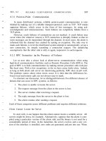
Figure 10.17
Medium-velocity impact
blood spatter produced by beating.
©1997 CRC Press LLC
An understanding of fly activity at scenes of exposed blood, as well as
body decomposition, is essential for proper interpretation of blood spatters.
The mouth parts of the common housefly are specialized for lapping and
sucking while the horsefly is characterized as a biter. The mosquito is spe-
cialized for piercing and sucking. Many flies ingest blood and regurgitate it
onto a surface. These surfaces may also show evidence of excretion or defe-
cation of digested or partly digested blood. The blood spatters produced as
the result of these activities are usually a millimeter or less in size with no
definite point of origin (Figures 10.18 and 10.19). They may be observed on
many surfaces at a scene, including the decomposing body and the clothing.
Often these surfaces would appear to be protected from receiving impact
spatter which occurred during injury to the victim. Conclusions should be
conservative and carefully considered when evaluating blood spatter, espe-
cially when there is a limited number of stains available for examination.
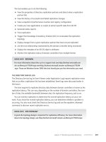
Figure 10.18
Diagrammatic representation of types of bloodstain produced as the result
of fly activity.
Figure 10.19
Bloodstains produced by fly activity on blue jeans of victim.
©1997 CRC Press LLC
High-Velocity Bloodstain Patterns
High-velocity impact blood spatter is produced by a high-velocity force
striking a source of blood. A high-velocity impact is considered to be approx-
imately 100 feet per second or more and is usually associated with gunshot
and high-speed machinery. A mist-like dispersion of minute blood droplets
is characteristic of high-velocity impact blood spatter patterns (Figures 10.20
and 10.21). Due to the low mass of these droplets, their distance travelled in
space is limited (approximately 3–4 feet). The resultant bloodstains have
diameters of 0.1 mm or less. However, bloodstains associated with high-
velocity impact are produced in the medium-velocity spatter size range and
larger. Due to their greater mass, the larger droplets can travel greater distances.
At crime scenes, evidence of high-velocity impact blood spatter is most
frequently associated with gunshot injury. Blood spatter may originate from
either an entrance or exit wound, but the blood droplet dynamics differ
between the two locations. Spatter from an entrance wound is referred to as
back spatter; the blood droplets travel opposite to the direction of the pro-
jectile toward the weapon and the shooter. Back spatter is more commonly
observed with close-range discharge of a firearm. The amount of back spatter
is also affected by the type of weapon and ammunition and the anatomic
features of the wound site. It may be absent due to the blocking effect of hair
and clothing.
Figure 10.20
High-velocity impact blood spatter on wall from gun shot exit wound. Note
projectile hole in wall.
©1997 CRC Press LLC
Blood may be drawn back into the barrel of the firearm with close-range
discharge in addition to back spatter impacting on the exterior of the weapon
and the hand, arm, and chest of the shooter.
Forward spatter is associated with an exit wound, the blood droplets
travelling in the same direction as the projectile. The quantity and distribu-
tion of forward spatter is generally greater than observed with back spatter.
The determination of the point of origin of high-velocity blood spatter assists
with positioning of the victim and assailant at the time of discharge of the
weapon and initial bloodshed. As with medium-velocity blood spatter, care
should be exercised with interpretation of small bloodstains and consider-
ation given to other activities that may have been responsible for the blood-
stains.
Photographic Documentation of Bloodstain Patterns
Photographic documentation of physical evidence at the crime scene, includ-
ing bloodstains, is an essential part of the overall investigative effort and
reconstruction. Crime scene investigators responding to death cases and
nonfatal violent crime frequently do not appreciate the valuable information
available from careful examination and interpretation of bloodstain patterns.
As a result, the photographic documentation of the victim, scene, physical
evidence, and assailant with respect to bloodstains may be incomplete and
lacking in detail for subsequent evaluation and courtroom presentation.
Figure 10.21
Areas of back spatter resulting from gun shot circled on shirt of shooter.
©1997 CRC Press LLC
Persons trained in bloodstain pattern interpretation may be consulted on a
case for prosecution or defense after the event has occurred and the crime
scene is no longer available. Reconstruction of the scene and ultimate con-
clusions regarding bloodstain patterns in a given case may then be limited
in scope, and important details may be impossible to resolve due to poor
photographic technique at the time of the original scene investigation. Fur-
thermore, investigators trained in bloodstain pattern interpretation, when
testifying in court, depend on good photographic documentation of blood-
stains.
The examination and serological studies of bloodstains in the crime
laboratory, such as the precipitin test for human origin, ABO grouping,
genetic marker profiling, and DNA studies must also include photographic
documentation of bloodstains on clothing and other items of physical evi-
dence prior to the removal of bloodstains from the material submitted for
examination. Samples of suspected blood that are cut or otherwise removed
from articles of clothing or other physical evidence may represent portions
of an important bloodstain pattern. Sometimes the bloodstained area may
be minute in size and quantity such as with high-velocity impact blood
spatter. Complete removal of these small bloodstains for serological testing
may be required in many cases. When that occurs, it is extremely important
that the bloodstains be photographed properly; otherwise, the interpretative
value of those bloodstains is irretrievably lost.
Good photographic documentation of the bloodstains, of both overall
bloodstain patterns and individual bloodstains, is crucial. Crime scene pho-
tography including documentation of bloodstain patterns is easily and effec-
tively done with the use of a 35-mm camera with a 35- to 50-mm lens for
overall photographs, close up or macro lens capability, flash attachment, and
high quality color film. Color enlargements of 8
×
10 inches are a good size
for analysis and courtroom presentation. Color slides are also very useful for
courtroom presentation. Color slides can be made from original scene pho-
tographs with a copy stand and photo lamps; good results have been obtained
with reflecting the light from white cardboards. This reduces the amount of
glare on the subject photograph. Color slide film 100 ASA is used, with the
camera set at 125 ASA with the lens setting on automatic. The exposure time
is adjusted to 1/15 or the closest setting that will allow a reading of f8 to f11
on the internal light meter of the camera. These parameters have been effec-
tive in reducing overexposure of the slides which can be a problem.
Personal experience has shown that Polaroid reproductions have limited
value for crime scene work and bloodstain pattern interpretation. Black-and-
white photographs are of use with Luminol but generally do not suffice for
bloodstain pattern interpretation, since stains other than blood will appear
similar to bloodstains and tend to confuse the issues.
©1997 CRC Press LLC
A most important tool for the forensic photographer is a measuring
device scaled in millimeters and inches to be included in all photographs or
slides of bloodstain patterns or individual stains in order to document the
size of the bloodstains. Experience has shown that blue or gray 6-inch rulers
work well to eliminate glare and provide a good guide for color reproduction.
The Crime Scene
The indoor crime scene is for the most part protected from the elements and
easily preserved for extended periods of time, unless the incident occurred
in a public place and there is pressure to clean up the scene as quickly as
possible. On the other hand, bloodstains present at outdoor crime scenes
may be altered in appearance by the terrain and weather. Photography of the
outdoor crime scene should be done as soon as possible, to minimize changes
or obliteration of bloodstains and other physical evidence due to prevailing
conditions. It may be necessary to photograph an outdoor scene at night
with a strong light source. A ladder or truck with a boom is useful for overall
photographs of an outdoor scene. If weather is not a problem, significant
bloodstains should be rephotographed in the daylight hours. Whether
indoors or outdoors, it is important to limit access to any crime scene,
especially bloody scenes, to avoid unnecessary tracking of wet blood or alter-
ation of existing bloodstain patterns that might compromise proper inter-
pretation.
Bloodstain evidence at the crime scene should be documented with high
quality color photographs and/or slides before the body is moved or the scene
otherwise altered. A reference scale should be used. It is important to coor-
dinate the photography of the victim and visible injuries with photographs
of bloodstains and patterns on the body and clothing. Overall views from
above should be taken, as well as close-up photographs of small bloodstains
on the body with a ruler in place. Bloodstains on the body should be pho-
tographed in conjunction with the bloodstains in the immediate area of the
body before the victim is turned or moved. When the body position has been
altered, the area should be rephotographed to document any changes of
previously formed bloodstains or the creation of new or artifactual blood-
stains.
Much of the critical bloodstain pattern photography of the scene relating
to walls, ceilings, floors, and other objects is best accomplished after the
overall scene photography and photographic documentation of the body
have been completed and the body removed. Bloodstain patterns should be
photographed with the camera held at 90° to the bloodstains if possible.
When individual bloodstains are photographed closeup, the general area of
©1997 CRC Press LLC
these bloodstains should be recognizable from prior scene photographs so
that a point of reference is established. If bloodstain convergences and points
of origin are established through measurements and string reconstruction,
these procedures should also be photographed.
The bloodstained clothing of a victim should be carefully removed after
initial photography at the scene and the postmortem examination. The gar-
ments should not be folded or packaged in a damp condition. The best
procedure is to hang and air-dry over clean paper before packaging in paper
bags; this will minimize the alteration of bloodstains and the production of
additional bloodstains or artifacts.
The examination of clothing and bodies of suspects for bloodstains and
trace physical evidence often yields valuable evidence to associate that person
with a victim. Assailants frequently receive bloodstains and spatters on
exposed parts of their bodies, such as on the face and hands; these should
be photographed promptly.
The photographic documentation of bloodstains on clothing should be
done before any suspect bloodstains are removed for serological testing. The
use of a mannikin is helpful in duplicating the proper orientation and loca-
tion of bloodstains as they were while the victim or assailant wore the gar-
ments.
The Use and Photographic Documentation of Luminol
Luminol is a well known chemiluminescent compound and is used as a
presumptive, catalytic test for the presence of blood, taking advantage of the
peroxidase-like activity of heme for the production of light as an end product
rather than a true color reaction. Luminol reagent is applied on objects or
areas containing traces of suspected bloodstains. A bluish-white lumines-
cence or light production on the suspected area observed in the dark is a
positive test. Luminol is best used for the detection of traces of blood which
are not readily observable at crime scenes. This includes light tracking of
blood on dark floors and carpeted areas, cracks and crevices in floors and
walls, and areas where previous attempts at cleaning bloodstained areas are
suspected. The patterns of blood resolved with Luminol may be as important
as the detection of blood itself. The sensitivity of the Luminol test is as high
as one part in five million, and is effective with aged and decomposed blood-
stains. The Luminol test is easy to perform and adaptable to crime scene
work. Reagents and supplies are relatively inexpensive and can be obtained
from the local crime laboratory. Commercial kits for Luminol testing are
more expensive but are packaged in vials for individual use, and reagent
preparation is simplified. Although Luminol is a presumptive test for the
presence of blood, further analysis of positive areas must be made before the
blood can be confirmed. Certain surfaces such as painted walls, porcelain,
©1997 CRC Press LLC
and metal and cleaning agents such as hypochlorites may also react with
luminal.
Many investigators confirm a positive result with an additional presump-
tive test such as phenolphthalein, which can be accomplished before or after
the Luminol spray has been applied. Preferences in procedure for further
serological testing of Luminol reactive areas should be obtained from the
local crime laboratory.
One of advantages of Luminol is that the procedure lends itself well to
photographic documentation and is especially valuable when large blood-
stain patterns otherwise not visible are resolved. The following is a general
outline of equipment and procedures for the use and photographic docu-
mentation of Luminol.
Equipment Required
•Luminol reagent and spraying device
•Luminescent measuring device
• 35-mm camera with 50-mm lens, bulb setting, and wide open lens
setting (e.g., f1.8) capability
•Shutter release cable
•Tripod
• Flash unit
•ASA 100 to 400 black-and-white or color film (print or slide)
•Timer
•Appropriate protective clothing, gloves, and eye protection
Procedure
Before the use of the Luminol reagent the surface or object should be pho-
tographed in position using a flash unit with the luminescent ruler in place
(Figure 10.22). This will assist with the location of the positive luminescent
areas against a dark background. With the exception of some overall views,
the camera angle should be perpendicular to the surface of interest. The
camera lens
f
-stop should be set at the widest aperture and the exposure
setting at the B or Bulb position. With the shutter cable release attached, the
equipment is ready for use.
The room or location should be darkened before and during the appli-
cation of Luminol. A small amount of ambient light will help visualize
darkened areas. The Luminol reagent is sprayed with a slow, even motion,
avoiding saturation of the surface; as fine a mist as possible is best. The surface
can be resprayed during the timed exposure to enhance the reaction. An
exposure time of 30 to 45 seconds will generally produce satisfactory results
(Figures 10.22 and 10.23). Experimentation with this timed exposure may
©1997 CRC Press LLC
be desirable. Two to three investigators may be needed for this procedure:
one to spray Luminol, a second to operate the camera, and possibly a third
to operate the timer and lights.
Figure 10.22
Area of carpet prior to spraying with Luminol.
Figure 10.23
Area of carpet showing bloodstains visualized by Luminol spray. Note
partial hand print on left. See color plate following page 228.
©1997 CRC Press LLC
It is possible to obtain a double image of the Luminol reaction and the
object itself. This occurred quite by accident in a recent case when at the end
of the exposure time the room lights came on with the bulb setting still
activated. The shutter cable was released within a second afterwards and the
resulting photograph initially thought to be worthless showed the jacket and
luminescence quite well in a single photograph.
Charles F. Edel, formerly of the Broward County, Florida, Sheriff’s Office
Forensic Services Division published an article in 1989 in the
Journal of the
Florida Division of the International Association of Identification,
titled “Let’s
See What We Are Looking At,” demonstrating the use of a light source during
the Luminol spray period which permitted visualization of the area being
subjected to Luminol, as well as the positive luminescent reaction in the same
photograph. A flashlight provided the indirect light source on the surface
being sprayed with Luminol, avoiding direct light on the reacting area which
would wash out the luminescence.
The value of bloodstain evidence as an important tool for crime scene
reconstruction is enhanced by good photographic documentation. Photog-
raphy provides a permanent record of bloodstain evidence in a case which
is easily conveyed to a jury. Photographic evidence must stand up to the
scrutiny of opposing experts and counsel, as well as being a visual aid to a
jury which must weigh the evidence and reach a verdict in court.
Report Writing
A formal report of a crime scene reconstruction using bloodstain interpre-
tation should be written clearly and concisely. Diagrams and photographs
enhance the report’s clarity.
The following is a descriptive case report of a blunt force death involving
bloodstain pattern interpretation. The suspect was accused of beating his
friend to death with a section of a road sign post. He admitted only to finding
his friend dead when he returned to their outdoor camp. He pled guilty to
murder prior to trial.
Case Study 1
Re: State of Florida vs. C.W.
Case Number: 90-85092
Enclosed is my report of crime scene reconstruction, physical evidence exam-
ination, and bloodstain pattern interpretation for the case of the State of
©1997 CRC Press LLC
Florida vs. C.W. My conclusions are based upon my review and examination
of the following materials and visits to the scene of the homicide of the victim,
in this case, W.S.
1. Scene and autopsy photographs
2. Evidence log and scene diagram
3. Report of postmortem examination of the victim, W.S., which was
performed on 04/13/90
Examination of physical evidence was conducted on the dates of
04/16/90, 04/23/90, and 05/01/90. Scene examinations were conducted on
the dates of 04/16/90 and 04/25/90.
Case History
The location of this homicide was a wooded area west of railroad tracks
which run parallel to U.S. 1 south of Hypoluxo Road in Lantana, Florida.
The body of the victim, W.S., a 47-year-old white male, was found on the
ground close to a makeshift tent of plastic material. The victim had sustained
multiple extensive blunt force injuries to the head.
Postmortem examination revealed the victim W.S. to be 76 inches in
length and to weigh 235 pounds. The autopsy findings were determined to
be as follows with respect to injuries:
1. Multiple lacerations of the skin, frontal area, forehead, and facial areas.
Approximately 15 lacerations of the front area of the head are
described. Twelve of these lacerations are horizontally oriented, with
three showing a vertical orientation.
2. Comminuted depressed fracture of the skull, involving frontal bone,
nasal bones, and left orbital area
3. Subarachnoid hemorrhage of the brain
4. Lacerated contusions of the brain, orbital surface of frontal lobes
5. Fracture of the zygoma, bilateral
6. Multiple fractures of the maxilla
7. Fracture of the left body of the mandible with avulsion of left lower
lateral incisor tooth
Aspiration of blood was noted, as well as abrasions and bruises of the
skin on the upper left anterior chest, abdomen, upper extremities, and lower
extremities. The upper extremities also showed superficial lacerations. The
cause of death was determined to be craniocerebral injury with contributory
aspiration of blood and the manner of death determined to be homicide.
©1997 CRC Press LLC
Description of Scene and Bloodstain Patterns
The scene of this homicide was approximately 0.4 mile south of Hypoluxo
Road in a group of Banyan trees located approximately 182 feet west of U.S.
Highway 1 in Lantana, Florida. The victim was identified as W.S., a 47 year-
old white male. Sheriff’s Office reports indicated that the victim was found
by a friend who reported the homicide.
This scene is best described as a transient campground which is afforded
adequate protection from the elements by the group of trees. A makeshift,
clear plastic tent is located in the northeast corner of this wooded area. The
inside floor area of this tent measured approximately 11 feet, 2 inches, north
to south and 6 feet east to west. The openings of the tent were located on
the east and west sides. The tent was supported by rope attached to nearby
trees and held down by tires on the north and south sides. Large amounts
of debris, including beer and wine bottles, were noted to be in the general
area of the campground.
The victim is seen lying on the ground at the southwest corner of the
tent on his back, face up with his head pointing to the north and his feet
pointing to the south (Figure 10.24). The arms are extended outward from
the body and flexed inward at the elbow with the forearms parallel to the
head. The hands are partially clenched. The victim has sustained massive
blunt force trauma to the head area. A section of signpost approximately
26
1
/
2
inches in length rests laterally across the chest of the victim with one
Figure 10.24
Victim at camp site near makeshift tent.
©1997 CRC Press LLC
end, on the ground near the right elbow and the other end, which is heavily
bloodstained, resting on the chest below the chin (Figure 10.25).
The victim is clad in a brown, blue, and white plaid-type long-sleeved
shirt with the sleeves rolled up near the elbows. The shirt is partially unbut-
toned in front, exposing an underlying blue shirt. This blue shirt is visible
on the partially exposed abdomen. The victim is also wearing blue jeans and
a brown belt, with the fly of the jeans partially open. The legs are partially
spread with black shoes on the feet.
To the right of the body is an upright plastic container. To the left of the
head and left arm of the victim is the plastic covering comprising the south-
west corner of the tent. Near the left side of the victim is one of the black
tires which is holding down a portion of the plastic tent. A plastic bag is seen
opposite the left knee of the victim, and a metal wash bucket is seen to the
left of the left foot of the victim. An area of dirt between the shoes of the
victim appears to be disturbed.
There is a large quantity of blood which appears to be partially clotted
on the victim’s face and hair as well as surrounding the victim’s head on the
ground. Each hand shows heavy blood transfer from contact with a wet
source of blood. The shoulders and arms of the victim show extensive
Figure 10.25
Massive blunt force injuries inflicted to head of victim with metal sign
post which lies across chest.
©1997 CRC Press LLC
radiating patterns of medium-velocity impact blood spatter, circular to oval
in shape, as well as irregularly shaped clotted spatters of blood. A heavy
concentration of medium-velocity impact blood spatter is seen on the left
forearm (Figure 10.26).
The blood spatters are seen to have extended beyond the left arm of the
victim and impacted onto the adjacent outside surface of the plastic tent in
the southwest corner to a height of approximately 30 inches above the
ground. The average diameters of the blood spatters on the outside plastic
tent surface range from 0.5 to 5 mm with smaller and larger bloodstains
present. Some of these spatters may represent forceful wheezing or expiration
of blood from the victim’s nose or mouth. Some of the bloodstains present
on the lower portion of the tent surface in this area have been smeared by a
hand or other object having contact with that surface while the blood was
wet. Irregularly shaped clotted blood spatters are also noted on the plastic
tent surface. Also present on this plastic tent surface are several dripped and
Figure 10.26
Medium-velocity impact blood spatter on left shoulder and arm of victim.
©1997 CRC Press LLC
projected bloodstains with a downward directionality, with the origin above
the observed head position of the victim. The medium-velocity impact blood
spatter also extends onto the southwest corner of the tent flooring and is seen
as oval, elongated bloodstains.
Medium-velocity impact blood spatters have also been deposited on the
tread area of the tire near the left chest of the victim, as well as the lower
chest area, abdomen, and upper thigh area of the blue jeans. The jeans also
show evidence of dripped and projected bloodstains on the left and right
thigh which have originated from above the thigh.
The plastic bag to the left of the victim’s left knee also shows evidence
of medium-velocity impact spatter. The white plastic bleach container shows
circular to oval medium-velocity impact blood spatter with diameters rang-
ing from approximately 0.5 to 3.0 mm. Their directionalities are consistent
with the container being in its observed position at the time it received the
spatters of blood. This bleach container also shows dripped and projected
bloodstains on both sides of the container relative to the position of the
victim which have originated from above the container. Dripped bloodstains
are also seen on the ground near this container.
Medium-velocity impact blood spatters extend on the ground to the
north of the victim’s head, where it is seen to be present on leaves, broken
plates, and a razor, as well as on the surface of a spray disinfectant can.
The radiating pattern of medium-velocity impact blood spatter continues
to a tree located approximately 5 feet, 4 inches, to the north of the head of
the victim, where the blood droplets have impacted the exposed root of the
tree and up the trunk of this tree to a height of approximately 50 inches
above the ground. Some cast-off bloodstains may be present here as well
(Figure 10.27). On the west branch of the tree are seen irregularly shaped
spatters and possibly cast-off bloodstains on the bark surface located approx-
imately 56 inches above the ground.
Within the tent on the floor near the north wall of the plastic covering
are seen transfer bloodstains on a yellow and white striped towel. Just to the
north of this towel are seen heavy contact bloodstains on the foam rubber
flooring which are consistent with having been produced from sustained
contact with a wet source of blood. Above this area of contact bloodstaining
on the floor of the tent is located a blood transfer pattern on the inside surface
of the plastic tent covering with features consistent with a bloody hair swipe
which feathers in an upward direction (Figure 10.28). At the east opening of
the tent just to the south of the peak of the plastic tent cover is an additional
blood transfer pattern with features consistent with a bloody hair swipe which
is seen to feather south to north (Figure 10.29). There was no evidence of
medium-velocity impact blood spatter seen on the inside surface of the plastic
tent covering.
©1997 CRC Press LLC
Figure 10.27
Cast off bloodstains on trunk of tree behind victim.
Figure 10.28
Bloodstains present on interior of tent.
©1997 CRC Press LLC
To the east of the tent and measured approximately 15 feet, 10 inches,
from the feet of the victim is a small clump of trees on the east side of the
scene perimeter. Some of the limbs of these trees are down on the ground.
On a large tree limb oriented east to west is seen a large contact bloodstain
which has resulted from contact with a wet source of blood. Between this
limb and a standing tree to the north is seen a green blanket on top of some
downed tree limbs. A large quantity of contact bloodstaining is present on
the surface of these limbs (Figure 10.30). On nearby leaves are seen drip
patterns of blood which have resulted from blood falling from a source above
the ground affected only by gravity and dripping into itself from the source.
No evidence of medium-velocity impact blood spatter was seen in this area.
Examination of the Physical Evidence
The examination of physical evidence was conducted on the dates of
04/16/90, 04/23/90, and 05/01/90.
Clothing of Victim
1. The shirt of the victim, W.S., consists of a blue, brown, and white plaid
long-sleeved shirt size XL, and Bud Burns brand. The rear of this shirt
Figure 10.29
Bloodstains present on exterior top edge of tent.
©1997 CRC Press LLC
is heavily blood-soaked. The front of this shirt shows considerable
medium-velocity impact blood spatter and blood-soaked areas most
heavily concentrated on the shoulders and upper arms of this garment.
The diameter of these spatters of blood ranges from 0.5 to 4 mm, with
some smaller and larger bloodstains present. Many of the blood spat-
ters consist of irregularly shaped impacted clots of blood. The quantity
of medium-velocity impact spatter decreases somewhat down the
right arms to the cuff, where there are also some blood-soaked areas.
The left arm of the shirt shows a denser pattern of medium-velocity
impact spatter, clotted spatters, and as some blood-soaked areas. Void
areas on the sleeves are due to the irregular bunching of the material
while worn by the victim with his sleeves rolled up to the elbows. The
front chest area of the shirt shows medium-velocity impact blood
spatter and clotted spatters on the left and right front areas, more
concentrated on the left side. Additional blood soaking and blood
transfer is present on the right side of the shirt. The underside of the
right lower front edge of the shirt shows blood spatter and staining
on the inner surface due to this surface being exposed at the time of
bloodshed to that area. Some plant debris and dirt is also present on
this garment.
Figure 10.30
Bloodstains on ground and tree branches to east of victim and tent.
©1997 CRC Press LLC
2. The blue jeans of the victim, W.S., consist of Rustler brand jeans with
a brown knitted belt. The rear left leg of the jeans is relatively free of
bloodstaining with scattered bloodstains present on the upper rear of
the right leg. The front and lateral side of the upper right leg shows a
bloodstain pattern consisting of circular to oval and irregularly shaped
bloodstains with a range in diameter from 5 to 10 mm which have
resulted from dripping and projection of blood from a source above.
There are also smaller, 0.5- to 4-mm diameter, circular to oval spatters
of blood, some of which consist of clotted blood. The upper front of
the left leg of the blue jeans shows an area of dripped blood from
above with circular to oval shaped stains as well as some spatters of
blood. The left knee shows a blood-soaked area resulting from blood
which has soaked through from the inside surface of the fabric. There
are scattered areas of spattered blood present on the lower legs of the
jeans. Some plant debris and dirt is also present on this garment.
3. The footwear of the victim, W.S., consists of a pair of Athletix brand
shoes with a prominent circular sole design. The left lateral side of the
right shoe shows some projected bloodstains with a directionality
towards the sole of the shoe. A trace of blood is detected on the sole
with Phenolphthalein reagent. Similar projected bloodstains are
present on the right lateral edge of the left shoe.
4. The undershirt of the victim, W.S., consists of a green short-sleeved
Tuxan brand garment. The rear of this shirt is considerably blood-
soaked with evidence of plant debris and dirt present. The front of
this garment shows blood transfer stains and medium-velocity impact
blood spatter on the upper central chest area. Many of these spatters
are circular to oval in shape with diameters ranging between 0.5 and
4 mm. Some of these blood spatters consist of small clots of blood.
This area of the underlying shirt was exposed during bloodshed due
to the outer shirt being partially unbuttoned. The lower front area of
this shirt also shows spatters of blood and clotted blood as it extended
beyond the lower edge of the outer shirt during bloodshed.
Clothing of Suspect
1. The shirt of the suspect, C.W., consists of a long-sleeved sweatshirt
with a crab design on the front chest area and the logo “I’ve got the
Crabs”. Below this logo on the front is seen a small area of blood
transfer. On the upper right arm of this garment are seen numerous
circular to oval shaped medium-velocity impact blood spatters mea-
suring 0.5 to 4 mm in diameter. There are also spatters of clotted
blood. Similar medium-velocity impact blood spatters and spatters of
©1997 CRC Press LLC
clotted blood are present on the lower right arm extending onto the
right cuff. The left arm of this garment also shows similar medium-
velocity impact blood spatter and spatters of clotted blood extending
from the upper arm area to the cuff. There are some small transfer
bloodstains present on the lower back of this garment. The trousers
of the suspect, C.W., consist of grayish-green Levi’s for Men Action
Jeans brand, size 34–36 with a brown belt with silver buckle. The upper
rear and upper front of these jeans are relatively free of bloodstains.
The right front leg of the jeans measuring from the lower edge of the
cuff upward 21 inches just above the knee area show numerous
medium-velocity impact blood spatters with the heaviest concentra-
tion from the cuff to 12 inches above the cuff from the mid to inner
lateral side of the right leg of the garment. Many of these blood spatters
range in size from 0.5 to 5.0 mm in diameter, with smaller and larger
bloodstains present. Many of the spatters consist of clotted blood
which exhibit irregular sizes and shapes (Figures 10.31 and 10.32). The
left front leg of these jeans shows numerous medium-velocity impact
blood spatters present on the fabric from the lower edge of the cuff
upward 23 inches just above the knee, with the heaviest concentra-
tion from the cuff to 12 inches above the cuff. Many of these blood
spatters range in size from 0.5 to 5.0 mm in diameter, with smaller
and larger bloodstains present. Many of the spatters consist of clotted
blood of irregular size and shape. The lower right leg of the jeans
contains a considerably greater quantity of blood spatters than the
left leg.
3. The shoes of the suspect, C.W., consist of black/gray Nike Airline
brand running shoes with a crosshatch sole pattern. The left shoe
shows blood transfer stains on the laces with some heavier contact
staining to the right of the laces. The toe area is relatively free of
bloodstaining. There is medium-velocity impact blood spatter present
on the inner lateral edge of the sole below the heel with a few scattered
bloodstains above this area on the upper portion of the shoe. The right
shoe shows scattered irregular bloodstains on the upper front surface,
including the laces, with little evidence of bloodstaining on the front
toe area. The inner lateral surface shows large transfer bloodstains.
Below this area on the inner lateral edge of the sole there is a heavy
concentration of medium-velocity impact blood spatter consisting of
circular, oval, and irregularly shaped bloodstains, many with diameters
of 0.5 to 5.0 mm with some smaller and larger spatters present
(Figure 10.33). There is a void area absent of spatter above the visible
spatters due to the hang of the cuff of the trousers that previously
covered that portion of the shoe.
©1997 CRC Press LLC
The Weapon Found at the Scene
The weapon found at the scene of this homicide consists of a section of iron
signpost which appears twisted on the shaft near the grip area. This V-shaped
section of post measures approximately 26
1
/
2
inches long, 1
1
/
2
inches deep,
and 3 inches wide at the end that rested on the chest of the victim. Due to
compression of the metal, the grip portion of the post tapers to a minimum
diameter of approximately 1
1
/
4
inches on the flattened surface and then flares
out at the end to an approximate diameter of 3 inches. A trough and row of
holes is present down the shaft of the post. The end of the iron post which
rested on the chest of the victim is heavily bloodstained from contact with
a source of blood. There are also numerous, circular to oval shaped medium-
velocity impact blood spatters on the shaft exhibiting diameters from 0.5 to
4 mm, with some smaller and larger bloodstains present. Blood flow patterns
are present on the shaft which have travelled from the heavily bloodstained
end to the grip area of this iron post.
Figure 10.31
Medium-velocity impact blood spatter on shoes and lower trouser legs of
suspect.
©1997 CRC Press LLC
Figure 10.32
Closer view of medium-velocity bloodstains on trousers of victim showing
some clotted spatter.
Figure 10.33
Medium-velocity impact blood spatter on right shoe of suspect.
©1997 CRC Press LLC
Examination of Photographs of the Hands of the Suspect
Color photographs taken of the hands of the suspect in this case, C.W., by
Detective D.S. at the scene on 04/12/90 reveal the palmar surface of the hands
to be moderately bloodstained. The bloodstains on the palm of the left hand
appear diffuse, with more concentration of blood at the juncture of the base
of the fingers and the palm. Several small circular to oval spatters of blood
are present near the base of the thumb extending over the juncture of the
wrist and the palm.
The palmar surface of the right hand of the suspect, C.W., shows a
concentration of contact bloodstaining which extends across the base of the
fingers and the adjacent surface of the right palm. This bloodstaining extends
towards the wrist along the inner lateral aspect of the hand and is more
diffuse along the base of the thumb. This area of described bloodstaining is
sharply delineated across the right palm below the base of the fingers and
below the little finger towards the wrist. This delineation forms a rectangular-
shaped void area free of bloodstaining in the central portion of the right
palm which is oriented between the right thumb and first finger and parallel
to the extended thumb (Figures 10.34 and 10.35). This area is sometimes
referred to as the web of the palm. This void area is consistent with an object
being held tightly in the right hand during the time that blood contacted the
palm. The tight grip on the object prevented the blood from contacting the
central portion or web of the palm and created the sharp outline of the void
or gripping pattern of the hand-held object (Figure 10.36).
Conclusions
The victim in this case, W.S., received multiple blunt force injuries to the
head with many of the wounds oriented laterally across the head and face.
The final blows administered to the victim were delivered while he lay prone
in his observed position on the ground near the southwest corner of the clear
plastic tent. The radial pattern of medium-velocity impact blood spatter
typically produced during a beating of a prone victim is present on the victim
and the area in proximity to the position of the victim on the ground. The
heaviest concentrations of blood spatter are present on the upper body and
arms of the victim, on the lower portion of the southwest corner of the tent,
the tree trunk to the north of the victim’s head, the tire and plastic bag located
to the left of the victim, and the plastic bleach container to the right of the
body of the victim. These objects were in their observed locations when they
received the blood spatters. The origin of these medium-velocity impact
blood spatters is consistent with the observed location of the head of the
victim at the scene.
There is no evidence to indicate that the victim was moved subsequent
to the fatal beating. However, the presence of projected bloodstains and
©1997 CRC Press LLC