Advanced Techniques in Dermatologic Surgery - part 6 pps
Bạn đang xem bản rút gọn của tài liệu. Xem và tải ngay bản đầy đủ của tài liệu tại đây (1.04 MB, 42 trang )
Diode Lasers (Table 6)
Millisecond-domain diode lasers are available at wavelengths of 800,
810, and 930 nm. Like the alexandrite lasers, these systems are effective
in treating larger telangiectasia, venulectasia, and feeding reticular veins
of the lower extremities (42,43).
Long-Pulsed Nd:YAG Lasers (Table 7)
Compared to the other near-infrared lasers being applied to the treatment
of cutaneous vascular lesions, Nd:YAG lasers provide enhanced depth
of penetration (up to 5.0 mm in depth) and minimal interference from
melanin absorption. Long-pulsed Nd:YAG lasers are effective for the
treatment of telangiectasia, venulectasia, and reticular veins of the legs
because of their ability to photocoagulate larger diameter, more deeply
situated vessels (44,45). Telangiectasia and venulectasia of the face can
be successfully treated by using small (1.0–3.0 mm) spot sizes, and by
using high fluences to compensate for the decreased absorption coeffi-
cient for hemoglobin at this wavelength (46). The pulsed Nd:Y AG lasers
are equipped with a variety of cooling systems including water-cooled
chambers applied directly to the skin (Laserscope Lyra
Õ
, Altus Cool-
glide
Õ
, ESC Vasculight
Õ
) and cryogen spray cooling (Laser Aesthetics
Varia
Õ
and Candela Gentle YAG
Õ
).
Intense Pulse Light Source (Table 8)
The intense pulsed light (IPL) sou rce was developed by ESC Medical
(now Lumenis) in an effort to maximize the efficacy in treating leg veins.
This high intensity pulsed flashlamp light source delivers broadband
Table 6
Diode Lasers
Laser MedioStar
Õ
Apogee
Õ
SkinPulse
Õ
Apex
Õ
Light
Sheer
Õ
EpiStar
Õ
SLP
1000
Õ
Manufac-
turer
Asclepion-
Meditec
(Jena,
Germany)
Cynosure
(Chelms-
ford, MA,
U.S.A.)
Dornier
(Munich,
Germany)
Iridex
(Mountain-
view, CA,
U.S.A.)
Lumenis
(Santa
Clara,
CA,
U.S.A.)
Nidek
(Fremont,
CA,
U.S.A.)
Palomar
(Burling-
ton, MA,
U.S.A.)
Wavelength
(nm)
810 800 940 800 800 810 810
Pulse
duration
(msec)
5–30 50–500 10 to
continu-
ous
5–100 5–100 200 50–100
Maximum
fluence
(J/cm
2
)
64 50 600 5–60 10–60 179
Cooling Contact Air Contact Contact Contact
190 Kauvar
light from 515 to 1100 nm (47). Single, double, or triple pulses in the 2- to
20-millisecond domain can be delivered in a synchronized fashion. The
broad emission spectrum, in the visible and near infrared region, targets
both oxygenated and deoxygenated hemoglobin. The longer wavelengths
penetrate deeper into the skin, enabling photocoagulation of deeper
vessels, and the longer pulse durations produce uniform heating of larger
vessels without inducing vessel rupture. Several IPL sources are now
available. This technology has also been applied to the treatment of
port win e stains, superficial hemangiomas (48), and facial telangiectasia
Table 7
Long-Pulsed Nd:YAG Lasers
Laser
CoolGlide
Õ
/
Vantage
Õ
Gentle YAG
Õ
Varia
Õ
Lyra
Õ
Image
Õ
Mydon
Õ
Manufac-
turer
Cutera
(Burlingame,
CA, U.S.A.)
Candela,
(Wayland,
MA,
U.S.A.)
ICN
(Costa
Mesa, CA,
U.S.A.)
Laserscope
(San Jose,
CA,
U.S.A.)
Sciton
(Palo Alto,
CA,
U.S.A.)
Wavelight
(Erlangen,
Germany)
Wavelength
(nm)
1064 1064 1064 1064 1064 1064
Pulse
duration
(msec)
0.1–300 3 0.3–200 10–100 5–200 20–140
Maximum
fluence
(J/cm
2
)
300 10–70 500 200 10–400 15–400
Cooling Contact Cryogen Cryogen/
contact
Contact Contact/air Contact/air
Table 8
IPL Sources
Light source Prolite
Õ
Quantum
Õ
Vasculight
Õ
Estelux
Õ
Manufacturer Alderm
(Irvine,
CA,
U.S.A.)
Lumenis
(Santa Clara,
CA,
U.S.A.)
Lumenis
(Santa Clara,
CA,
U.S.A.)
Palomar
(Burlington,
MA,
U.S.A.)
Wavelength (nm) 500–900 515–1200 515–1200 500–1200
Pulse duration
(msec)
2–7 0.5–2.5 10–100
Maximum fluence
(J/cm
2
)
10–50 45 90 4–12
Cooling Contact Contact Contact
Abbreviation: IPL, intense pulsed light.
Laser Treatment of Vascular Lesions 191
(49) yielding good results. IPL technology presently finds its application
mainly in nonablative photorejuvenation to improve the pigmentary, vas-
cular, and textural irregularities of photodamaged skin.
CLINICAL APPLICATIONS
Port Wine Stains
The pulsed dye laser remains the treatment of choice for most port wine
stains. Treatment of macular and mildly hypertrophic port wine stains
with the 585-nm, 0.45-millisecond pulsed dye laser produces remarkable
clinical lightening with minimal side effects. Multiple treatment s are
required for significant lightening. Early studies demonstrated 75% or
more lightening in approximately 36% to 44% of adult patients with port
wine stains, and at least 50% lesional lightening in 75% of patients after
a total of four treatments (12,14–19,50). The laser has been proven safe
and effective, even after 10 to 25 repetitive treatments (7). Treatment
may be initiated soon after birth, without adverse effect. Clearing of port
wine stain lesions depends on their anatomic location and size. Port wine
stains located on the forehead, lateral cheeks, and neck respond better
than those located on the central facial regions, specifically areas supplied
by the second branch of the tri geminal nerve (51). Smaller lesions
with areas less than 20 cm
2
respond far more quickly than larger lesions
with areas greater than 20 cm
2
. Head and neck port wine stains respond
most favorably. Truncal lesions respond better than port wine stains
located on the extremities, with distal extremity lesions being the most
resistant.
Newer generation pulsed dye lasers with a wavelength of 595 nm
and pulse duration of 1.5 milliseconds enable faster clearance of port wine
stains in infants and adults. In studies using this laser in conjunction with
cryogen spray cooling to treat 16 infants under 12 months of age with facial
port wine stains, there was greater than 75% lightening in 63% of patients
after four treatments using energy fluences of 11 to 12 J/cm
2
(35). Prospec-
tive side-by-side comparison studies of hypertrophic adult port wine stains
treated with energy fluences of 12 to 14 J/cm
2
using the 595-nm, 1.5-milli-
second laser demonstrated increased clearance compared to a fluence of
10 J/cm
2
, both in conjunction with cryogen spray cooling (36).
Treatment of port wine stains with the 585-nm, 0.45-millisecond
laser is usually performed with the largest spot size available to prevent
reticulation. Typical treatment flue nces using the 7 mm spot are 5.0 to
7.0 J/cm
2
and 5.0 to 6.0 J/cm
2
with a 10-mm spot size, depending on
the age of the patient and the thickness of the lesion. Using the 595-nm,
1.5-millisecond pulsed dye lasers in conjunction with cryogen spray cooling,
fluences of 8.0 to 11.0 J/cm
2
are used with a 7-mm spot size, and fluences of
5.0 to 6.5 J/cm
2
are used with the 10-mm spot size in infants and children.
For adults with hypertrophic lesions, fluences up to 13 J/cm
2
can be used
with a 7-mm spot size and fluences up to 7.5 J/cm
2
with a 10-mm spot size.
192 Kauvar
Determination of the appropriate fluence should be assessed with test
performed on the target sites during the initial evaluation.
Immediately after treatment with the 585-nm, 0.45-millisecon d
pulsed dye laser, intense blue–black purpura develops for approximately
10 to 14 days. The intensity and duration of purpura is significantly lower
while using pulse duration of 1.5 milliseconds. If crusting occurs, patients
are instructed to apply a topical antibiotic such as bacitracin or poly-
sporin ointment daily until it resol ves. Following the resolution of pur-
pura, lesional lightening takes place over a period of four to six weeks.
Repeat treatments are performed every 6 to 10 weeks until maximal
lesional clearing is achieved. Even after 20 treatment sessions, further
lesional lightening may be achieved (7). The development of various
skin-cooling methods has obviated the necessity for local or general
anesthesia in most cases. With the exception of young children, most
infants, teenagers, and adults tolerate the treatment well with the use
of a topical anesthetic cream such as Emla or Elamax.
While pulsed dye laser technology remains the standard of care for
port wine stain treatment, other technology has been successfully used
for this indication. The IPL has been used to lighten port wine stains.
Twenty-eight of forty patients treated in one study achieved greater than
75% lesional clearance after an average of four treatments for pink
lesions, 1.5 for red ones, and 4.3 for purple-colored port wine stains
(52). The lightening of the red or purple port wine stains by the three-
millisecond long pulse alexandrite laser has also been found by the author
and others (Dierickx C, personal communicatio n) (52).
Hemangiomas
Superficial (capillary) hemangiomas and the superficial component of thin
mixed-type hemangiomas respond best to pulsed dye laser therapy. Treat-
ment of thin superficial hemangiomas can often clear these lesions in three
to four treatment sessions (20,21,53–56). Thicker lesions may require
additional treatments. The pulsed dye laser is also effective in reducing
the superficial component of mixed-type hemangiomas; however, the
deeper (cavernous) component may continue to proliferate despite laser
therapy. Institution of pulsed dye laser therapy during the proliferative
phase is helpful in slowing the growth of these lesions. Treatment of
superficial hemangiomas helps in minimizing the enlargement of the
tumor, prevents the development of complications such as bleeding and
ulceration, and achieves improved cosmetic results.
Treatment of proliferating hemangiomas is usuall y performed at
two- to four-week intervals, in an effort to halt further tumor growth.
The treatment interval for involuting hemangiomas is usually six to eight
weeks. As with port wine stains, the newer 595-nm, 1.5-millisecond
pulsed dye lasers, which can be used at higher fluences in conjunction
with cryogen spray cooling, appear to achieve faster clearing of heman-
giomas compared to historical controls, because of their ability to treat
larger diameter and deeper blood vessels. The IPL ha s also been used
Laser Treatment of Vascular Lesions 193
for the treatment of superficial hemangiomas and the superficial compo-
nent of mixed type hemangiomas with some success. Preliminary studies
using millisecond-domain pulsed dye, diode, and Nd:YAG lasers show
promising results with these wavelengths for thicker lesio ns.
Telangiectasia
Telangiectasia are capillaries, venules, or arteries that are 0.1 to 1.0 mm in
diameter and are visible as superficial cutaneous vessels. Facial telangiec-
tasia are common, and in fair-skinned individuals, they are often asso-
ciated with rosacea or actinic damage. Other etiologies include collagen
vascular disease, genetic disorde rs, hormonal, primary cutaneous disease,
and radiodermatitis. Spider angiomata are telangiectasia with a central
feeding arteriole, typically appearing in preschool and school-age chil-
dren with a peak incidence between the ages of 7 and 10.
Most patients seek treatment for facial telangiectasia because of
cosmetic concerns. Techniques used to treat facial telangiectasia have
included electrosurgery, sclerotherapy, and treatment with continuous
wave and quasi-continuous wave lasers, but these methods may produce
textural and pigmentary irregularities. The development of pulsed lasers
enabled efficient, effective, and low-risk treat ment of these common skin
lesions.
A wide variety of vascular laser systems produce excellent clearance
of facial telangiectasia. The 585- and 595-nm pulsed dye lasers with
0.45- and 1.5-millisecond pulse durations produce excellent results in
one to two treatment sessions, but induce purpura lasting 7 to 14 days
(37). Treatment is performed by applying contiguous laser pulses with
approximately 10% overlap. The newer, millisecond-duration pulsed
dye lasers, used at 6 to 10 milliseconds, clear facial telangiectasia, without
purpura production. Effective treat ment usually requires stacking of
three to four laser pulses with an endpoint of vessel blanching or transient
thrombosis. The 532-nm KTP laser produces excellent results for the
treatment of facial telangiectasia in one to three treatment sessions
(57,58). Contiguous laser pulses are applied directly over the vessels, with
additional pulses, if necessary, to achieve visible vessel blanching. Some
of the KTP systems are equipped with cooled sapphire hand pieces that
enable easy gliding of the laser tip over the skin, when used with cold gel,
and relatively painless treatment.
Long-pulsed Nd:YAG lasers, used with spot sizes of 1 to 3 mm and
fluences of 120 to 250 J/cm
2
, also pro duce excellent results for facial
telangiectasia without purpura production. With the use of higher flu-
ences, proper skin cooling and avoidance of pulse stacking are necessary
to prevent epidermal damage, particularly around the nasal ala
(46,59,60). The long-pulsed Nd:YAG lasers are particularly useful for
the treatment of the larger caliber paranasal vessels that often require
multiple, repetitive treatments with the shorter wavelength lasers. Venu-
lectasia commonl y seen on the lateral cheeks following rhytidectom y
often usually clear in one treatment session. Visible facial veins have also
194 Kauvar
been treated with Nd:YAG lasers, but extreme caution must be exercised
to avoid laser exposure within the orbital rim with this deeply penetrating
wavelength. The IPL devices also clear facial telangiectasia, and multiple
treatment sessions may be necessary (49).
Facial Erythema
Facial erythema with or without associated telangiectasia is a common
cosmetic concern. The erythema is usually a manifestation of rosacea or
a flushing or blushing disorder. Effective treatment is best achieved with
the pulsed dye lasers and IPL sources, using large spot sizes to avoid reti-
culation (61,62). There is no purpura production with the newer pulsed dye
lasers used with 6- to 10-millisecond pulse durations and the IPL devices.
Multiple treatment sessions (2–6) may be necessary to achieve good clinical
results. An improvement in the associated symptoms or warmth and burn-
ing sensation usually accompanies the reduction in erythema.
Poikiloderma
Poikiloderma is treatable with lasers and light sources. Poikiloderma of
Civatte is relatively common in fair-skinned, actinically damaged indivi-
duals. Clinically, poikiloderma appears as a combination of telangiecta-
sia, irregular pigmentation, and atrophic changes. The treatment of this
diffuse condition is best accomplished using the pulsed dye lasers or
IPL devices with large spot sizes to avoid reticulation (26,63,64). Overly
aggressive treatment with any laser or light source can produce atrophy
and hypopigmentation. Compared to the treatment of telangiectasia, flu-
ences should be lowered by approximately 25% to 30% in the treatment
of poikiloderma to avoid adverse effects. Treatment of poikiloderma
using the 6- to 10-millisecond pulsed dye lasers appears to achieve equiva-
lent results to the shorter pulsed systems without the development of pur-
pura. Contiguous laser pulses are applied without overlap. With the IPL
devices, it is often helpful to alternate the axis of the rectangular spot with
each treatment to reduce the risk of reticulation.
Scars and Striae Distensae
Pulsed dye laser therapy can be used to improve erythematous and
hypertrophic scars. Clinical response rates are 57% to 83% (30,65,66).
The pulsed dye laser reduces erythema by eliminating the underlying
dilated microvascul ative. Scar height and skin surface texture changes
are improved, presumably by altering collagen production. Multiple
treatment sessions are often necessary, particularly for thicker scars,
and adjunctive treatment with intralesional corticosteroid injections is
useful. The best results are achieved using 10-mm spot sizes and fluences
of 4 to 5 J/cm
2
without skin cooling and 5 to 6 J/cm
2
with skin co oling.
Treatment intervals are six to eight weeks.
Low-fluence pulsed dye laser therapy also improves the appearance
of striae (32). Striae rubra shows the best response, and can sometimes be
Laser Treatment of Vascular Lesions 195
entirely eliminated with early laser intervention. The skin textural irregu-
larities in striae alba can be improved with pulsed dye laser treatment and
other nonablative lasers and light sources. The mechanism of improve-
ment is presumed to be via fibroblast activation and induction of collagen
production.
Warts
Pulsed dye lasers effectively treat cutaneous lesions of human papilloma
virus, including plantar warts, periungual warts, flat warts, and verrucae
vulgaris (33). Electron microscopic studies suggest that the mechanism of
improvement is via thermal alteration of the virally infected tissue (67).
Laser treatment appears to be more effective than conventional wart
therapy, and carries a minimal risk of scarring, even when used to treat
deep plantar warts and subungual and periungual lesions. Treatments
are performed following paring of hy perkeratotic lesions, using the
585- or 595-nm pulsed dye laser with pulse duration of 0.45 or 1.5 milli-
seconds. A 5- or 7-mm spot is used at fluences of 7 to 9 J/cm
2
without
skin cooling. Recalcitrant warts require three to four repetitive treat-
ments, at two to four week intervals. Uncomplicated warts usually
respond in one session.
CONCLUSION
The development of pulsed laser and light source technologies has revo-
lutionized the treatment of cutaneous vascular lesions. Laser therapy
remains to be the treatment of choice for port wine stains, superficial
hemangiomas, and telangiectasia. These devices have also been success-
fully applied to the treatment of hypertrophic and erythematous scars,
striae, and warts. Unlike other conventional destructive modalities, treat-
ment is noninvasive. Due to the selective deposition and targeting of the
light energy, there is little risk of skin woundi ng or the development of
pigmentary or textural irregularities. The de velopment of longer wave-
length and longer pulse duration laser technology, along with the skin-
cooling methods, has improved the safety and efficacy of vascular lesion
therapy.
196 Kauvar
REFERENCES
1. Silver L. Argon laser photocoagulation of port wine stain hemangiomas. Lasers Surg
Med 1986; 6:24–28.
2. Dixon JA, Rotering RH, Huethner SE. Patient’s evaluation of argon laser therapy of
port wine stains, decorative tattoos, and essential telangiectasia. Laser Surg Med 1984;
4:181–190.
3. Anderson RR, Parrish JA. Selective photothermolysis: precise microsurgery by selective
absorption of pulsed radiation. Science 1983; 220:524–527.
4. Anderson RR, Parrish JA. Microvasculature can be selectively damaged using dye
lasers: a basic theory and experimental evidence in human skin. Lasers Surg Med
1981; 1:263–276.
5. Glassberg E, Lask GP, Tan EM, Uitto J. The flashlamp-pumped 577 nm pulsed
tunable dye laser: clinical efficacy and in vitro studies. J Dermatol Surg Oncol 1988;
14:1200–1208.
6. Levine VJ, Geronemus RG. Adverse effects associated with the 577 and 585 nm pulsed
dye laser in the treatment of cutaneous vascular lesions: a study of 500 patients. J Am
Acad Dermatol 1995; 32:613–617.
7. Kauvar ANB, Geronemus RG. Repetitive pulsed dye laser treatments improve persistent
port wine stains. Dermatol Surg 1995; 21:5151–5521.
8. Dierickx CC, Casparian JM, Venugopalan V, Farinelli WA, Anderson RR. Thermal
relaxation of port wine stain vessels probed in vivo: the need for 1–10 msec laser pulse
treatment. J Invest Dermatol 1995; 105:709–714.
9. Garden JM, Tan OT, Kerschmann R, Boll J, Furumoto H, Anderson RR, Parrish JA.
Effect of dye laser pulse duration on selective cutaneous vascular injury. J Invest Derma-
tol 1986; 87:653–657.
10. Nelson JS, Majaron B, Kelly K. Active skin cooling in conjunction with laser dermato-
logic surgery. Sem Cut Med Surg 2000; 19(4):253–266.
11. Tan OT, Murray S, Kurban AK. Action spectrum of vascular specific injury using pulsed
irradiation. J Invest Dermatol 1989; 92:868–871.
12. Tan OT, Morrison P. Kurban AK. 585 nm for the treatment of port wine stains. Plast
Reconstr Surg 1990; 86:1112–1117.
13. Goldman L, Kerr JH, Larkin M, Binder S. 600 nm flash pumped dye laser for fragile
telangiectasia of the elderly. Lasers Surg Med 1993; 13:227–233.
14. Alster TS, Wilson F. Treatment of port-wine stains with the flashlamp-pumped
pulsed dye laser: extended clinical experience in children and adults. Ann Plast Surg
1994; 32:478–484.
15. Ashinoff R, Geronemus RG. Flashlamp-pumped pulsed dye laser for port wine stains in
infancy: earlier versus later treatment. J Am Acad Dermatol 1991; 24:467–472.
16. Garden JM, Polla LL, Tan OT. The treatment of port wine stains by the pulsed dye laser.
Arch Dermatol 1988; 124:889–896.
17. Goldman MP, Fitzpatrick RE, Ruiz-Esparza J. Treatment of port wine stains (capillary
malformation) with the flashlamp-pumped pulsed dye laser. J Pediatr 1993; 122:71–77.
18. Reyes BA, Geronemus RG. Treatment of port wine stains during childhood with the
flashlamp pumped pulsed dye laser. J Am Acad Dermatol 1990; 23:1142–1148.
19. Tappero JW, Grekin RC, Zanelli GA, Berger TG. Pulsed dye laser therapy for cutaneous
Kaposi’s sarcoma associated with acquired immunodeficiency syndrome. J Am Acad
Dermatol 1992; 27:526–530.
20. Ashinoff R, Geronemus RG. Capillary hemangiomas and treatment with the flashlamp-
pumped pulsed dye laser. Arch Dermatol 1991; 127:202–205.
21. Garden JM, Bakus AD, Paller AS. Treatment of cutaneous hemangiomas by the flash-
lamp-pumped pulsed dye laser: prospective analysis. J Pediatr 1992; 120:555–560.
Laser Treatment of Vascular Lesions 197
22. Broska P, Martinho E, Goodman MM. Comparison of the argon tunable dye laser with
the flashlamp pulsed dye laser in the treatment of facial telangiectasia. J Dermatol Surg
Oncol 1994; 20:749–753.
23. Goldman MP, Weiss RA, Brody HJ, Coleman WP III, Fitzpatrick RE. Treatment
of facial telangiectasia with sclerotherapy, laser surgery, and/or electrodesiccasion: a
review. J Dermatol Surg Oncol 1993; 19:899–906.
24. Ruiz-Esparaza J, Goldman MP, Fitzpatrick RE, Lowe NJ, Behr KL. Flashlamp-pumped
dye laser treatment of telangiectasia. J Dermatol Surg Oncol 1993; 19:1000–1003.
25. Garden JM, Bakus AD. Clinical efficacy of the pulsed dye laser in the treatment of
vascular lesions. J Dermatol Surg Oncol 1993; 19:321–326.
26. Wheeland RG, Applebaum J. Flashlamp-pumped pulsed dye laser therapy for poikilo-
derma of Civatte. J Dermatol Surg Oncol 1990; 16:12–16.
27. Gonzalez E, Gange RW, Momtaz KT. Treatment of telangiectasias and other benign
vascular lesions with the 577 nm pulsed dye laser. J Am Acad Dermatol 1992; 27:
220–226.
28. Goldberg DJ, Sciales CW. Pyogenic granuloma in children: treatment with the flash-
lamp-pumped pulsed dye laser. J Dermatol Surg Oncol 1991; 17:960–962.
29. Hoffman SJ, Walsh P, Morelli JG. Treatment of angiofibroma with pulsed tunable dye
laser. J Am Acad Dermatol 1993; 29:790–791.
30. Alster TS. Improvement of erythematous and hypertrophic scars by the 585-nm flash-
lamp-pumped pulsed dye laser. Ann Plast Surg 1994; 32:186–190.
31. Alster TS, Williams CM. Treatment of keloid sternotomy scars with the 585 nm flash-
lamp-pumped pulsed dye laser. Lancet 1995; 345:1198.
32. McDaniel DH, Ash K, Zukowski M. Treatment of stretch marks with the 585 nm flash-
lamp-pumped pulsed dye laser. Dermatol Surg 1996; 22:332–337.
33. Kauvar ANB, Geronemus RG, McDaniel DH. Pulsed dye laser treatment of warts. Arch
Fam Med 1995; 4:1035–1040.
34. Kauvar ANB. Long-pulse, high energy pulsed dye laser treatment of port wine stains and
hemangiomas. Laser Surg Med 1997; (suppl 9):36.
35. Geronemus R, Quintana A, Lou W, Kauvar ANB. High fluence modified pulsed dye
laser photocoagulation with dynamic cooling of port wine stains in infancy. Arch
Dermatol 2000; 6:942–943.
36. Kauvar ANB, Lou WW, Zelickson B. Effect of cryogen spray cooling on 595 nm, 1.5
msec pulsed dye laser treatment of port wine stains. Laser Surg Med 2000; (suppl 12):24.
37. West TB, Alster TS. Comparison of the long-pulsed dye and KTP lasers in the treatment
of facial and leg telangiectasia. Dermatol Surg 1998; 24:221–226.
38. Hsia J, Lowery JA, Zelickson B. Treatment of leg telangiectasia using a long-pulse dye at
595 nm. Lasers Surg Med 1997; 20:15.
39. Bernstein EF, Lee J, Lowery J, Brown DB, Geronemus R, Lask G, Hsia J. Treatment
of spider veins with the 595 nm pulsed-dye laser. J Am Acad Dermatol 1998; 39:746–750.
40. Kauvar ANB, Lou WW. Pulsed alexandrite laser for the treatment of leg telangiectasia
and reticular veins. Arch Dermatol 2000; 136:1343–1346.
41. Brunnberg L, Lorenz S, Landthaler M, Hohenleutner U. Evaluation of the long pulsed
high fluence alexandrite laser therapy with 755 nm for leg veins. Lasers Surg Med 2002;
31(5):359–362.
42. Dierickx CC et al. Lasers Surg Med 1998; (suppl 10):40.
43. Kaudewitz P, Klovekorn W, Rother W. Treatment of leg vein telangiectases: 1-year
results with a new 940 nm diode laser. Dermatol Surg 2002; 28(11):1031–1034.
44. Omura NE, Dover JS, Arndt KA, Kauvar AN. Treatment of reticular leg veins with a
1064 nm long-pulsed Nd:YAG laser. J Am Acad Dermatol 2003; 48(1):76–81.
198 Kauvar
45. Coles Cm, Werner RS, Zelickson BD. Comparative pilot study evaluating the treatment
of leg veins with a long pulse ND:YAG laser and sclerotherapy. Lasers Surg Med 2002;
30(2):149–153.
46. Eremia S, Li CY. Treatment of face veins with a cryogen spray variable pulse width
1064 nm Nd:YAG Laser: a prospective study of 17 patients. Dermatol Surg 2002;
28(3):220–223.
47. Goldman MP, Eckhouse S. Photothermal sclerosis of leg veins. Dermatol Surg 1996;
22:323–330.
48. Schroeter CA, Neumann HAM. An intense light source. The photoderm VL-flashlamp
as a new treatment possibility for vascular lesions. Dermatol Surg 1998; 24:743–748.
49. Bierring P, Christiansen K, Troilius A. Intense pulsed light source for treatment of facial
telangiectasias. J Cosmet Laser Ther 2001; 3(4):169–173.
50. Tan OT, Sherwood K, Gilchrest BA. Treatment of children with PWS using the flash-
lamp-pulsed tunable dye laser. New Engl J Med 1989; 320:416–421.
51. Renfro L, Geronemus RG. Anatomical differences of port-wine stains in response to
treatment with the pulsed dye laser. Arch Dermatol 1993; 129:182–188.
52. Raulin C, Schroeter CA, Weiss RA, Keiner M, Werner S. Treatment of port wine stains
with a non-coherent pulsed light source: a retrospective study. Arch Dermatol 1999;
135:679–683.
53. Barlow RJ, Walker NPJ, Markey AC. Treatment of proliferative hemangiomas with the
585 nm pulsed dye laser. Br J Dermatol 1996; 134:700–704.
54. Maier H, Neumann R. Treatment of strawberry marks with flashlamp-pumped pulsed
dye laser in infancy. Lancet 1996; 347:131–132.
55. Ricci RM, Finley EM, Grimwood RE. Treatment of cutaneous hemangiomas in preterm
neonatal twins with the flashlamp-pumped pulsed dye laser. Lasers Surg Med 1998;
22:10–13.
56. Sherwood KA, Tan OT. Treatment of a capillary hemangioma with the flashlamp-
pumped dye laser. J Am Acad Dermatol 1990; 22:136–137.
57. Adrian RM, Taughetti EA. Long pulse 532 nm laser treatment of facial telangiectasia.
Dermatol Surg 1998; 24(1):71–74.
58. Goldsberg DJ, Meine JG. Treatment of facial telangiectases with the diode-pumped
frequency-doubled a-surbled Nd:YAG laser. Dermatol Surg 1998; 24:828–832.
59. Major A, Brazzini B, Campolmi P, Bonan P, Mavilia L, Ghersetich I, Hercogova J,
Lottit T. Nd:YAG 1064 nm laser in the treatment of facial and leg telangiectasias.
J Eur Acad Dermatol Venereol 2001; 15(6):559–565.
60. Kauvar A, Mafong E, Friedman P, Bernstein L, Alexiades-Armenakas M, Geronemus
R. Treatment of facial telangiectasia with a long pulsed ND:YAG laser. Laser Surg
Med 2002; (suppl 14):135.
61. Angermeier MC. Treatment of facial vascular lesions with intense pulsed light. J Cutan
Laser Ther 1999; 1(2):95–100.
62. Lowe NJ, Behr KL, Fitzpatrick R, Goldman M, Ruiz-Esparza J. Flashlamp pumped
dye laser for rosacea-associated telangiectasia and erythema. J Dermatol Surg Oncol
1991; 17(6):522–525.
63. Raulin C, Greve B, Grema H. IPL technology: a review. Lasers Surg Med 2003; 32(2):
78–87.
64. Weiss RA, Goldman MP, Weiss MA. Treatment of essential telangiectasias with
an intense pulsed light source (PhotoDerm VL). Dermatol Surg 1996; 23(10):941–945;
discussion 945–946.
65. Alster TS, Kurban, AK, Grove GL, Grove MJ, Tan OT. Alteration of argon laser-
induced scars by the pulsed dye laser. Lasers Surg Med 1993; 13:368–373.
Laser Treatment of Vascular Lesions 199
66. Alster TS, Williams CM. Improvement of hypertrophic and keloidal median sternotomy
scars by the 585 nm flashlamp-pumped pulsed dye laser: a controlled study. Lancet 1995;
345:1198–1200.
67. Ross EV, McDaniel DH, Anderson RR, Kauvar ANB, Geronemus RG. Pulsed dye
(585 nm) treatment of warts: a comparison of single versus multiple pulse techniques
examining clinical response, fast infrared thermal camera measurements, and light elec-
tron microscopy. Lasers Surg Med 1995; (suppl 7):59.
200 Kauvar
9
Laser Treatment for Leg Veins
Neil S. Sadick
Weill Medical College of Cornell University, New York, New York, U.S.A.
Video 10: Leg Veins
Video 11: Leg Veins: Gemini
Õ
Device
INTRODUCTION
The incidenc e of unsightly venulectasias an d/or telangiectasias on the
legs occurs in up to 41% of women and 15% of men (1). The utilization
of lasers and intense pulsed light (IPL) sources for the treatment of lower
extremity veins has gained increased popularity over the past five years.
This technology, driven by consumer demand, has been shown to be
effective in treating vessels that are refractory to sclerotherapy, vessels
that arise from prior surgical treatment or sclerotherapy (telangiectatic
matting or angiogenic flushing), and needle-phobic patients.
Initial problems involving laser/IPL technologies have centered on
the fact that it is inherently more difficult to get photons safely and in
sufficient numbers through several layers of blood vessel wall into the tar-
get chromophore, that is oxygenated and deoxygenated hemoglobin.
Injections directly into the target are inherently more efficient.
However, a greater recent understanding of photoendothelial inter-
action has led to improved efficiency of light modalities in this setting
(2,3). The choice of wavelength(s), degree of energy fluence, and pulse
duration of light exposure are all related to the type and size of the target
vessel treated. Deeper vessels require a longer wavelength to allow pene-
tration to their depth (4). However, even at a penetrating wavelength,
pulse duration must be matched to the vessel size. As the depth and size
of the vessel change, so do the absorption characteristics. Larger dia-
meter vessels require longer pulse duration to allow sufficient time for
diffusion of heat evenly throughout the cylindrical vessel lumen (5). In
addition, deliverance of this energy should occur with a shock wave
producing gentle cavitation, to prevent posttreatment hemorrhage and
purpura. It should also produce an epidermal bypass to protect this struc-
ture from deleterious thermal effects. Optimal laser/IPL parameters for
treatment of lower extremity vessels are present in Table 1 (6).
In this regard, shorter (500–600 nm) wavelengths may be used to
treat Class I superfici al oxygenated reddish telangiectasias while a longer
wavelength (755–1100 nm) may be used to treat Class II to III deeper
201
deoxygenated bluish venulectasias and reticular veins up to 4 mm in
diameter (7). This ‘‘bimodal’’ wavelength approach to the treatment of
lower extremity veins produces results superior to previously described
treatment paradigms for photothermolytic eradication of lower extremity
vessels (8).
When and How to Choose Laser/IPL vs. Sclerotherapy
The utilization of light sources for the treatment of leg veins is efficacious
for treating telangiectasia/venulectasia or reticular veins less than 3 mm
in diameter (9,10).
It is commonly used for the patient who is needle-phobic or
requests laser as a primary modality of treatment.
It is very effective in the treatment of noncannulizable or sclero-
resistant vessels. Areas of neovascularization with telangiectatic matting
or angiogenic flushing are primary indications for this approach (3,11).
Practical Tips in Laser Treatment of Leg Veins
Rule out areas of reflux by means of physical examination and
Duplex ultrasound,
treat larger diameter vessels by foam sclerotherapy or ambula-
tory phlebectomy,
sclerotherapy of cannulizable vessels,
laser treatment of residual veins,
a varied monomodal approach to treatment of leg veins is the
major approach utilized by most phleb ologic vein laser surgeon
(Table 2).
This incorporates utilizi ng one of the 1064 neodymium–yttrium–
aluminum–garnet (Nd:YAG) technologies using small spot sizes
(1.0–2.0 mm), high fluences (150–400 J/cm
2
), and short pulse durations
(15–30 milliseconds) for treatment of small red vessels of less than 1 mm
in diameter which contain a high degree of oxygenated hemoglobin.
Table 1
Optimal Laser/IPL Parameters for Treatment of Lower Extremity Vessels
Wavelength
(nm)
Pulse duration
(msec)
Beam diameter
(mm)
Diameter of vessels
100 mm 580 1 –
300 mm 590 10 –
600 mm to 1 mm 600 20–100 –
Vessel depth
Less than 1 mm >500 – Small (2–6 mm)
Greater than 1 mm >600 – Large (6–12 mm)
202 Sadick
In a similar fashion, the same long wavelength laser can be
employed with larger spot sizes (3–6 mm), more moderate fluences of
100 to 250 J/cm
2
, and more extended pulse duratio ns of 30 to 50 milli-
seconds for treatment of blue vessels (1–3 mm) which are deeper in loca-
tion and have a higher degree of deoxygenated hemoglobin (Figs. 1
and 2).
Compression is usually not necessary following laser/IPL/RF
treatment of nonbulging vessels (12).
A summary of recent technologic advances in laser/IPL treatment
of lower extremity veins is presented in Table 3. A compilation of laser
and IPL sources utilized in this setting is present in the following dis-
cussion and listed in Table 4.
It is important to explain to the patient that this technol ogy like
sclerotherapy, takes multiple treatments to see progress. In addition,
it is important during the initial consultation to explain to the patient that
forces of hydrostatic pressure and reflux must be addressed prior to laser
therapy to optimize therapeutic efficacy and to minimize side effects.
Table 2
Varied Mode Monomodal Approach to Leg Veins
Vessel <1 mm (red) Vessel 1–3 mm (blue)
Spot size 1–2 mm 3–6 mm
Fluence 150–400 J/cm
2
100–250 J/cm
2
Pulse duration 15–30 msec 30–50 msec
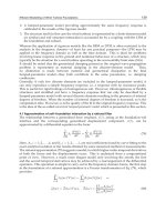
Figure 1
Pre-/post-1064 Nd:YAG (Laserscope Lyra) three treatments: blue vessels
3 mm spot size, F-200 J/cm
2
, PD 40 milliseconds; red vessels 1.5 mm spot
size, F-350 J/cm
2
, PD 20 milliseconds.
Laser Treatment for Leg Veins 203
A comparison of laser/IPL treatments for telangiectasia of less than
0.5 mm versus sclerotherapy is presented in Table 5.
CONTINUOUS WAVE ND:YAG LASERS
The Nd:YAG laser at 1064 nm has also been used to treat leg telangiec-
tasias in a continuous mode (13). Absorption by blood is relatively poor
at this wavelength (up to 3.7 nm) leading to much nonspecific damage.
Therefore, the continuous wave Nd:YAG laser has no role in the treat-
ment of leg vessels (14).
578 nm Copper Bromide (CuBr) Yellow Light Laser
A new yellow light laser utilizing a copper bromide medium ha s shown
efficacy in the treatment of red lower extremity telangiectasia of less than
2 mm. An average of 1.7 patient treatment sessions produced significant
clearing of 75% to 100% in 71.8% of patients. Positive results are confined
to the treatment of red vessels (1 mm) (15).
Figure 2
Pre-/post-diode laser/RF Syneron Polaris treatment of blue and red vessels
two treatments: l 915 nm; spot size 5 Â 8; LE 90 J/cm
2
; RF 90 J/cm
3
.
Table 3
Recent Technologic Advances in Laser/IPL Treatment of Lower Extremity Veins
Cooling technologies
Longer wavelengths
Extended pulse durations
Monomodal varying pulse duration/spot size/
fluence technology (1064 nm Nd:YAG)
Captured pulsing
Larger beam diameter (spot size)
Higher energy fluences
Abbreviation: Nd:YAG, neodymium–yttrium–aluminum–garnet.
204 Sadick
Table 4
Lasers for Leg Veins
Device
(company)
Wave
length
(nm)
Pulse
duration
(msec)
Spot size
(mm)
Maximum
fluence
(J/cm)
Maximum
speed (Hz)
Cooling
device Comments
E2000
(Palomar/
ConBio)
694 3 or 100 10 mm
hexagon;
20 mm
square
50 1 Sapphire
contact
Employs
photon
recycling
EpiTouch
Ruby
(Lumenis)
694 1.2 4–6 40 1–1 Gel Q-switched
mode
available
RubyStar
(Aesculap
Meditec)
694 2 3–14 35 1 Contact
plate
precooling
Q-switched
mode
available
GentleLASE
(Candela)
755 3 7–18 100 1 DCD 2Â7mm
spot for
leg veins
EpiTouch
Plus Alex
(Lumenis)
755 2–40 5–10 50 5 Gel Scanner
available
LPIR/
Apogee
(Cynosure)
755 5040 7–16 50 1 Cold air
flow
Scanner
available
LightSheer
(Lumenis)
800 5–30 9–9 60 1 Sapphire
contact
Diode
array in
handpiece
CoolGlide
(Cutera)
1064 10–100 9–9 100 2 Copper
contact
precooling
FDA
approval
pending,
scanner
Lyra
(Laser-
scope)
1064 10–50 3–5 100 4 Contact
cooling
Carbon
makes no
difference
Softlight
(Thermo-
lase)
1064 10–20 7 2–3 10 Not needed Sequence of
multiple
pulses
Flashlamp
(Lumenis)
Variable
from
550 to
1200
Variable 8–33 or
10–45
30–65 <1 Circulating
cooling
device
Combine
diode
laser þ
radio-
frequency
Polaris
(Syneron)
900 þ RF 250 5–8 RF: 100
J/cm
3
;
diode:
140 J/cm
1 Contact þ
cooling gel
Abbreviation: DCD, dynamic cooling device.
Laser Treatment for Leg Veins 205
CONTINUOUS WAVE LASERS
Argon and Continuous Wave Dye Lasers
Argon (488 and 514 nm) and continuous wave dye lasers (515 to 590 nm)
are well-absorbed by hemoglobin, and they penetrate to the depth of
mid-dermal vessels, more than 1 mm within the skin. Results with this
short-wave technology have overall been disappointing with improvement
reported in less than 50% of individuals in previous studies (17). Synergistic
treatment with sclerotherapy have yielded improved results (18).
PULSED LASERS AND LIGHT SOURCES
Potassium-Titanyl-Phosphate Lasers
Early attempts to treat vessels with the continuous wave potassium-
titanyl-phosphate (KTP) crystal laser were mostly unsuccessful. At
532 nm, hemoglobin ablation is excellent. However, the depth of penetra-
tion limits the use of this laser to superficial leg telangiectasias of less than
1 mm in diameter.
Although the results of treatment of facial vessels have been excel-
lent, the results of treatments utilizing small spot sizes and pulse dura-
tions of less than 10 milliseconds have been more variable (9,19–21).
More updated technologies including Versapulse KTP laser
(Lumenis, Santa Clara, California, U.S.) using larger spot sizes (3–5 mm)
and longer pulse durations (10–50 milliseconds) at fluences of 14 to 20
J/cm
2
have been more promising. A 4
C chilled tip provides epidermal
protection. In published studies, two to three treatments have yielded
maximal vessel improvement, although pigment dyschromia including
temporary hyperpigmentation has been reported in darker-or tanned-skin
individuals. Other technologies including the Aura (Laserscope, San Jose,
California, U.S.) have produced comparable results.
Table 5
Sclerotherapy vs. Laser/IPL for Treatment of Telangiectasia
Microsclerotherapy Laser flashlamp
Number of treatments ¼¼
Bruising Àþ
Discomfort Àþ
Clinical efficiency Àþ
Purpura þÀ
Pigmentation ¼¼
Ulceration Àþ
Cost þÀ
Patient satisfaction Àþ
Physical skill ¼¼
206 Sadick
Patient accep tance of this laser treatment is high with minimal
treatment discomfort of the longer penetrating wavelengths and a rela-
tively uncomplicated postoperative course (22).
Flashlamp-Pumped Pulsed Dye Laser
Newer innovations in flashlamp-pumped pulsed dye laser technology
have produce d improved treatment of leg telangiectasia (23). The tradi-
tional pulsed dye laser (POL)(585-nm, 450-microsecond pulse duration)
has been shown to be highly effective in the management of port wine
stains and facial telangiectasias. This technology was shown to be less
effective in the management of leg veins. Although 585-nm light can
penetrate 1.2 mm to reach the typical depth of leg telangiectasias, the
pulse duration is inadequate for effective damage of all but superficial
fine vessels approximately 0.1 mm or smaller in diameter (24).
Variable results, persistent purpura, and a high incidence of
both hyper- and hypopigmentation limited the widespread usage of this
technology.
Long-Pulsed Dye Lasers
Based on the theory of selective photothermolysis, the predicted pulse
duration ideally suited for thermal destruction of leg veins (0.1 to
several millimeters in diameter) is the 1- to 50-millisecond domain (25).
Four long-pulsed dye lasers, two with 1.5 millisecond pulse durations
(Sclero Plus, Candela, Wayland, Massachusetts, U.S., VLS, Cynosure,
Chelmsford, Massachusetts, U.S.) and two with variable pulse durations
as long as 40 millisecond s (V-beam, Candela, V-Star, Cynosure), are now
available. Each device uses a Rhodamine dye to produce wavelengths of
585, 590, 595, or 600 nm. These longer pulse durations and wavelengths
theoretically improve the ability to treat deeper, larger cali ber vessels (25).
More recent modifications to the pulsed dye laser have included the
addition of the dynamic cooling device (DCD) (Candela), a method
of cryogen spray cooling capable of generating higher fluences (up to
25 J/cm
2
).
Six studies reported in the literatu re have assessed the effectiveness
of these long-pulsed dye lasers in the treatment of leg veins, with variable
results (26–29). Most of these studies achieved 50% to 60% clearing of
treatment sites after three treatment sessions with an incidence of both
hyper- and hypopigmentation approaching 50%. The delivery of equiva-
lent laser fluences over extended pulse durations have helped to eliminate
posttreatment purpura.
Longer Wavelength Pulsed Lasers
Based upon the deeper penetration of longer wavelength visible and
near-infrared light and a small peak of hemoglobin absorption in the
Laser Treatment for Leg Veins 207
700-to 900-nm range, long-pulsed alexandrite and Nd:YAG lasers have
been developed to treat moderately deep, larger caliber spider and feeding
reticular veins of the lower extremities.
The alexandrite lasers have wavelength of 755 nm with pulse dura-
tions of 3 to 20 milliseconds. The Nd:YAG lasers have a wavelength of
1064 nm and pulse durations up to 100 milliseconds. Diode lasers with a
wavelength of 800, 810, and 930 nm and pulse durations of 10 to 250
milliseconds may also be used.
Long-Pulsed Alexandrite Lasers
Long-pulsed alexandrite lasers have recently been applied to the treatment
of leg telangiectasia and reticular veins, less than 3 mm in diameter, with
good results. The longer wavelength provides deeper tissue penetration
and an ability to treat larger diameter and more deeply situated vessels.
Although hemoglobin absorption of this wavelength is lower than that
of the 532 and 595 nm wavelengths, it is sufficient to achieve photocoagula-
tion of a wide range of vessel sizes with the use of higher fluences. To
penetrate tissue more deeply and to allow greater thermal diffusion time
to treat larger vessels, the alexandrite laser has been modified to provide
pulse duration of up to 20 milliseconds. Optimal treatment parameters
for long-pulsed alexandrite lasers seem to be 20 J/cm
2
, double pulsed at
a repetition rate of 1 Hz.
In two reported trials, the laser has been shown to be effective in the
treatment of mid-sized leg veins. Sixty-three percent clearance of leg veins
after three treatments (0–4 mm 0 1 mm) was reported (30). The best
response in this study was seen with sclerotherapy performed as a supple-
mental technique, confirming the importance of sclerotherapy for leg
veins.
In a second study, patients with Fitzpatrick skin types I and III
and leg veins measuring 0.3 to 2.0 mm in diameter were treated utiliz-
ing an 8-mm spot size and fluences of 60 to 80 J/cm
2
with concomi-
tant cryogen cooling. Seventy-five percent or greater clearance was
noted in treated site after a single treatment. Patient discomfort and
temporary hypopigmentation were reported in one-third of the treated
sites (31).
Diode Lasers
Diode lasers (800 nm at 5- to 250-millisecond pulse duration) have been
used to treat superficial telangiectasias and reticular veins. These devices
with near-infrared wavelengths allow deeper tissue penetration with
decreased absorption by melanin. In addition, their wavelength matches
a tertiary hemoglobin absorption peak at 915 nm. Two methods of deliv-
ery for diode lasers are available: filler optic transmission of an 810-nm
laser (gallium-arsenide) and an overlapping 800-nm diode array with a
fixed spot size of 9 mmÂ9mm up to 12mmÂ12 mm.
208 Sadick
The longer wavelength (940 nm) diode laser offers better vessel clear-
ance and fewer complications when compared to its predecessors. In one
study with short-term (16 weeks) follow up, the best results were obtained
in a subset of patients who had vessels ranging from 0.8 to 1.4 mm. Eighty
eight percent of these patients had greater than 75% vessel clearance, with
one-third of those patie nts obtaining complete vessel clearance (32). A
long-term (12 months) study showed even more improvement, with 75%
of all patients had greater than 75% vessel clearance (33).
Goldman describes the advantages of the 1064 nm Nd:YAG laser
for the treatment of leg veins are due to the longer wavelengths ability
to penetrate more deeply into the tissue and offer more effective thermo-
sclerosis of small to medium blood vessels. Another advantage is the
minimal melanin absorption at this wavelength, allowing for the treat-
ment of all skin types and patients with tanned skin (34).
In a study comparing the 1064 nm Nd:YAG, 810 nm diode and 755 nm
Alexandrite lasers for the treatment of 0.3–3.0 mm leg veins the overall best
results and fewest complications were obtained with the 1064 nm Nd:YAG
laser. Greater than 75% improvement was seen with 88% of patients treated
with this laser. The authors reported their results with the 810 nm diode
laser as ‘‘unpredictable’’ and the 755 nm Alexandrite laser induced too much
purpura, inflammation, and matting at the treatment sites (35).
In one study, using an 810-nm quasi-continuous diode laser with
vessel size of 0.2 to 0.5 mm, 60% mean vessel clearance was obtained after
a mean of 2.2 treatment sessions (36).
More recently, the introduction of higher fluence capability diode
lasers has occurred, providing enhanced efficacy in this treatment setting
(10,37).
IPL Sources
High IPL sources emanating from a filtered flashlamp (Photoderm VL,
Vasculight IPL, Lumenis, Palo Alto, California, U.S.) were developed
to treat leg veins. Other manufacturers of pulsed light devices include
Energis Technology (Energis Elite IPL)(Swansea, JK) and Danish
Dermatologic Development (Elipse)(Hoersholm, Denmark).
The Energis System is a low-output device with 5 to 19 J/cm
2
out-
put, a spot size of 10 mmÂ50 m, a pulse train of 15 to 40 milliseconds,
and four or five pulses per train with a delay of 1.5 milliseconds. The
Lumenis device is a high-output system with up to 90 J/cm
2
output, a
spot size of 8 mmÂ35 mm, variable pulse lengths of 2 to 40 milliseconds,
and a variable of 1 to 1000 milliseconds.
Selectivity for IPL is obtained by manipulating pulse widths to
match the thermal relaxation times of vessels larger than 0.2 mm and by
using a filter to remove lower wavelengths of visible light. High fluences
of up to 90 J/cm
2
can be delivered. Segmented pulsing of 1 to 25 milli-
second duration separated and synchronized with 1 to 100 millisecond test
intervals delivers wavelengths of 515 to 1000 nm. The IPL devices are most
Laser Treatment for Leg Veins 209
commonly used with the 550- and 570-nm filters to deliver primarily the
yellow and red wavelengths wi th a minor component of infrared.
The main advantages of IPL technology in the treatment of leg
veins has been the use of large spot sizes, causing minimal purpura.
The shorter wavelengths have not been shown to be effective in the treat-
ment of larger, deeper, and bluish-colored vessels.
IPL (515–1000 nm range) wi th various fluences from 5 to 90 J and
varied pulse durations of 2 to 25 milliseconds have been used to treat
venulectasias of 0.4 to 2.0 mm in diame ter. Clinical trials utilizing the
IPL with multiple pulses of variable duration have demonstrated
efficacy of up to 90% clearance in vessels of smaller than 0.2 mm in
diameter, 80% in vessels (0.2–0.5 mm), and 70% in vessels of 0.5 to
1 mm in diameter. Few studies have shown the 90% clearance rate in initi-
ally reported cases (38). In one study, 73.6% of patients with leg telangiec-
tasias up to 1 mm in diameter had 73.6% clearance immediately
posttreatment and 84.3% after one month. Hyperpigmentation was noted
in 3% to 4% of patients. The most successful treatment parameters were a
single 3 millisecond pulse at a flue nce of 22 J/cm
2
for vessels of less than
0.2 mm in diameter to a double pulse of 3j at a fluence of 40 J/cm
2
,
2.4/4.0 millisecond with a 10-millisecond delay. Vessels with 0.2 to
0.5 mm diameter were treated with the same double pulse parameters
or with a 3.0- to 6.0-millisecond pulse at a fluence of 35 to 45 J/cm
2
with
a 20-millisecond delay.
In a more recent study, the utilization of a short-pulse long-pulse
protocol using 2.4 or 3 millisecond and 6 to 7 millisecond pulses separated
by a 10- to 20- millisecond delay employment. The 570-nm filter has yielded
the best results using the IPL device in treating leg vessels. Seventy-four
percent clearance with 8% incidence of hyper- or hypopigmentation has
been reported (39). By combining short and long pulses, theoretically, both
superficial and larger diameter vessels should be targeted.
Newer contact epidermal cooling devices have allowed deliverances
of higher fluence with less epidermal absorption.
LONG-PULSED ND:YAG LASER (1064 nm)
Millisecond domain, 1064 nm, lasers have been utilized to treat both blue
venulectasias and large caliber subcutaneous reticular veins (40). The
deeper penetrating wavelength and the absence of absorption by melanin
allow treatmen t of dark skin phenotypes and larger diameter vessels
allowing uniform pan-vessel heating.
The newer pulsed 1064 nm lasers have pulse durations between 1
and 200 milliseconds [Vasculight Lum enis, (Palo Alto, California,
U.S.A.), Cool Touch Varian (San Jose, California, U.S.A.), Cool Glide
Altus (Burlingame, California, U.S.A.), and the Scion Profile, Sciton
(Palo Alto, California, U.S.A.)].
Penetrating wavelength (1064 nm) technologies are more painful,
requiring adequate cooling and sometimes topical anesthesia. Larger,
210 Sadick
bluer vessels greater than 0.5 mm in diameter respond best to treatment,
requiring lesser treatment sessions.
In consideration of the recently described bimodal approach to
treatment of lower extremity veins, smaller spot sizes and higher fluences
have been shown to be efficacious in the treatment of smaller red vessels
of less than 1 mm in diameter (41–43).
Vessels up to 3 to 4 mm in diameter can be treated with the long-
pulsed Nd:YAG laser. Minor effects of hydrostatic pressures may be
addressed by treating these larger vessels. Pain increases with treatment
of vessels of greater than 2 mm in diameter.
The Lyra uses contact cooling. Seventy-five percent improvement of
veins of all colors and sizes has been reported with this technology (44).
The Sciton Image has been utilized predominantly for treatment of
lower extremity telangiectasias and reticular veins up to 3 mm in diameter
(45). Its high energy fluence and large spot size have increased its efficacy
in treating both large diameter vessels (i.e., reticular veins) and small
capillary mats less than 1 mm in diameter. A static cooling device is
employed.
The Vasculight has also been utilized for treatment of both smaller
vessels and larger reticular veins up to 4 mm in diameter. The operator
applies a coupling cooling gel in addition to an internal DCD (1–4
C)
and applies the laser tip directly to the treatment vessel under considera-
tion. Superficial red telangiectasias less than 1 mm in diameter may be
treated with the handpiece coagulated and defocused off the skin and a
lower energy fluence of 90 to 100 J/cm
2
with a pulse duration of 10 to
12 milliseconds delivered as a single pulse.
Weiss et al. (35) achieved 75% improvement at three-month follow-
up of 0.3 to 3.0 mm vessels documented by Duplex closure. Settings in
this study including fluence of 80 to 120 J/cm
2
and single-pulse durations
of 10 to 30 milliseconds were utilized.
Sadick et al. (46) treated 20 Fitzpatrick skin type II to IV patients
with a similar technology. A mean of 25 treatments produced 10 0% clear-
ance in 88% of patients.
Mild purpura was noted in 20% of patients, and postlaser hyperpig-
mentation was noted in 10% of patients.
SKIN COOLING DEVICES
The CoolGlide utilizing a contact cooling tip has been found to produce
70% vessel clearance for vessels of all colors and sizes (150–250 J/cm
2
).
The only complication at three months was postinflammatory hyper-
pigmentation.
The Varia has been utilized exclusively for the treatment of leg veins
4. This technology has recently been shown to be particularly effective in
the treatment of lower extremity vessels in type V skin (47). Pulsed cooling
with the ‘‘Cool Tube’’ cryogen spray lowers epidermal skin temperature to
about 25
C and delivers a more precise specific cooling effect compared to
Laser Treatment for Leg Veins 211
the ‘‘continuous cooling’’ of topical gel and contact chill tips. Delivery of
active cooling immediately postlaser can quench any heat approaching the
surface through back scattering of conduction. Pos tcooling may improve
efficacy because one does not have to reheat targets affected by pre-
cooling. A brief precooling pulse can be added for additional protection,
if significant surface pigment is present.
The Role of Cooling
The cooling plays an integral role in the management of laser treatment for
leg veins in an effort to maintain epidermal protection, prevent damage to
adjacent vascular structures, and diminish patient discomfort (44).
Several approaches are presently utilized includi ng water-cooled
chambers ap plied directly to the skin through which the laser beam is
directed (Chess Chamber, VersaPulse, Chill Tip, and IPL Chiller), cool-
ing coupling gels, and refrigerated-spray cooling devices (e.g., DCD) or
cryogen spray.
Such cooling accomplishes two goals: it minimizes epidermal
damage and allows the laser surgeon to employ higher fluence thus creat-
ing more potential to produce pan-endothelial vascular destruction (48).
COMBINED LASER/RADIOFREQUENCY TECHNOLOGIES
Combined diode laser bipolar radiofrequency application (Fig. 3) has
recently been shown to be effective in the treatment of lower extremity
vessels.
This technology can generate light fluences from 50 to 100 J/cm
2
and radiofrequency energies of 10 to 100 J/cm
3
. Further studies are in
progress substantiating the efficacy. The theory behind this technology
is that lower amounts of light energy may be introduced into the target
chromophore, i.e., hemoglobin, which may have synergistic effects with
the bipolar radiofrequency component. This has been referred to as
electro-optical synergy.
Endpoint of Therapy
The endpoint of therapy utilizing all laser technologies for treatment of leg
veins is immediate contraction and late erythema. Overtreatment is to be
avoided as is prolonged blanching as this may eventuate in epidermal
necrosis leading to pigment dyschromia or epidermal irregularities.
Because of the high upregulation of cytokines after laser-endothelial
interaction, it is recommended that the laser surgeon wait for 8 to 12 weeks
between treatment sessions to assess results, and let the laser-induced
inflammatory mediator expression subside. Photoprotection both
pre- and posttreatment for at least three to four weeks is indicated.
212 Sadick
Complications
Prolonged erythema pigmentation and epiderm al surface irregularities
(scarring) are the main side effects associ ated with laser/IPL treatment
of lower extremity veins.
Overtreatment is the major etiologic factor. Immediate ve ssel
contraction and urticaria remain the endpoints of therapy and, if proper
guidelines are followed, will help to minimize these adverse sequelae.
Prolonged vessel contraction and whitening are signs of overtreatment.
Cooling technology should be an integral part of all laser/IPL vessel
treatment protocols. Conservative settings and ‘‘spot test’’ sessions in
dark skin phenotypical individuals as well as fastidious photoprotection
are other caveats of importance in minimizing side effects.
In the author’s experience with all the aforementioned systems,
postlaser hyperpigmentation has been noted in 15% to 30% of treated
individuals. It represents melanin versus post-sclerotherapy hemosiderin
as seen following sclerotherapy. Compassion has been shown to play
no role in this postlaser treatment of lower extremity vessels (45,49).
In the maj ority of case s, pigment dyschromia usually resolves in 8 to
12 weeks, but may last up to six months. Three to four percent hydr o-
quinone derivatives or 20% azelaic acid preparations may be employed
in this setting. In refractory cases, the Q-switche d ruby or alexandrite
laser, or IPL source, can improve this pigmentation. Persistent
erythema may be managed by twice -a-day a pplication of potent Class
I corticosteroids such as clobetasol proprionate and betamethasone
diproprionate (50).
Indications for Laser Treatment
Sclerotherapy remains the gold standard for treatment of lower extremity
telangiectasia and reticular veins; however, improved parameter lasers
and IPL sources have certainly gained a role in the phlebologist’s arma-
mentarium. Expense as well as variable response to previous technologies
remains an issue. Patients who are needle-phobic or have had poor clin-
ical outcomes or unusual complication profiles are good candidates for
the utilization of the above-described technologies for treatment of lower
extremity vessels.
When small linear new-arborizing vessels, which are difficult to can-
nulate, are present or the blushing associated with telangiectatic matting
is present, there are also primary indications for choosing a laser/IPL
technology for treating such vessels. A comparative assessment of micro-
sclerotherapy and laser/IPL treatment of lower extremity vessels in the
author’s experience is presented in Table 5.
CONCLUSIONS
Although sclerotherapy remains the gold standard for treatment of
lower extremity telangiectasia s, improved cooling technology, longer
Laser Treatment for Leg Veins 213
wavelength devices and variable pulsing modes have allowed more
consistent results in the management of lower extremity vessels of less
than 3 mm in diameter. Addressing hydrostatic forces and understanding
venous anatomy, appropriate wavelength pulse diameter considerations,
optimal treatment intervals, and emphasis upon strict photoprotection
will lead to improved clinical outcomes and greater patient satisfaction
in employing this evolving technology.
214 Sadick