Saladin anatomy and physiology unity of form and function 6th c2012 txtbk 2
Bạn đang xem bản rút gọn của tài liệu. Xem và tải ngay bản đầy đủ của tài liệu tại đây (24.63 MB, 400 trang )
CHAPTER 10
TABLE 10.15
The Muscular System
371
Muscles Acting on the Foot (continued)
Key a
Biceps femoris:
Long head
Short head
Anterior (extensor)
compartment
Medial (adductor)
compartment
Semitendinosus
Semimembranosus
Adductor magnus
Posterior (flexor) compartment
(hamstrings)
Femur
Gracilis
Adductor brevis
Adductor longus
Vastus lateralis
(a)
Vastus intermedius
Posterior
Lateral
Medial
Rectus femoris
Sartorius
Vastus medialis
(a)
(b)
Anterior
Gastrocnemius
(lateral head)
Gastrocnemius
(medial head)
Soleus
Key b
Fibula
Flexor hallucis longus
Anterior (extensor)
compartment
Fibularis longus
Lateral (fibular) compartment
Fibularis brevis
Tibialis posterior
Posterior (flexor)
compartment, superficial
Extensor hallucis longus
Tibia
Posterior (flexor)
compartment, deep
Flexor digitorum longus
Extensor digitorum longus
(b)
Tibialis anterior
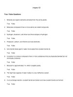
FIGURE 10.41 Serial Cross Sections Through the Lower Limb. Each section is taken at the correspondingly lettered level in the figure
at the left.
sal78259_ch10_312-378.indd 371
11/2/10 5:10 PM
372
PART TWO
Support and Movement
TABLE 10.16
Intrinsic Muscles of the Foot
The intrinsic muscles of the foot help to support the arches and act on the toes in ways that aid locomotion. Several of them are similar in name and location to the
intrinsic muscles of the hand.
Dorsal (Superior) Aspect of Foot. Only one of the intrinsic muscles, the extensor digitorum brevis, is on the dorsal (superior) side of the foot. The medial slip of
this muscle, serving the great toe, is sometimes called the extensor hallucis brevis.
Name
Action
Extensor Digitorum Brevis
Extends proximal phalanx I and all phalanges of
digits II–IV
O: Origin
I: Insertion
O: Calcaneus; inferior extensor retinaculum
of ankle
I: Proximal phalanx I, tendons of extensor
digitorum longus to middle and distal
phalanges II–IV
Innervation
Deep fibular (peroneal)
nerve
Ventral Layer 1 (most superficial). All remaining intrinsic muscles are on the ventral (inferior) aspect of the foot or between the metatarsal bones. They are
grouped in four layers (fig. 10.42). Dissecting into the foot from the plantar surface, one first encounters a tough fibrous sheet, the plantar aponeurosis, between
the skin and muscles. It diverges like a fan from the calcaneus to the bases of all the toes, and serves as an origin for several ventral muscles. The ventral muscles
include the stout flexor digitorum brevis on the midline of the foot, with four tendons that supply all digits except the hallux. It is flanked by the abductor digiti
minimi laterally and the abductor hallucis medially.
Flexor Digitorum Brevis
Flexes digits II–IV; supports arches of foot
O: Calcaneus; plantar aponeurosis
I: Middle phalanges II–V
Medial plantar nerve
Abductor Digiti Minimi89
Abducts and flexes little toe; supports arches of foot
O: Calcaneus; plantar aponeurosis
I: Proximal phalanx V
Lateral plantar nerve
Abductor Hallucis
Abducts great toe; supports arches of foot
O: Calcaneus; plantar aponeurosis; flexor
retinaculum
I: Proximal phalanx I
Medial plantar nerve
Ventral Layer 2. The next deeper layer consists of the thick quadratus plantae (flexor accessorius) in the middle of the foot and the four lumbrical muscles located
between the metatarsals.
Quadratus Plantae90
(quad-RAY-tus PLAN-tee)
Same as flexor digitorum longus (table 10.15); flexion
of digits II–V and associated locomotor functions
O: Two heads on the medial and lateral sides
of calcaneus
I: Distal phalanges II–V via flexor digitorum
longus tendons
Lateral plantar nerve
Four Lumbrical Muscles
(LUM-brih-cul)
Flex toes II–V
O: Tendon of flexor digitorum longus
I: Proximal phalanges II–V
Lateral and medial
plantar nerves
Ventral Layer 3. The muscles of this layer serve only the great and little toes. They are the flexor digiti minimi brevis, flexor hallucis brevis, and adductor hallucis.
The adductor hallucis has an oblique head that extends diagonally from the midplantar region to the base of the great toe, and a transverse head that passes
across the bases of digits II–IV and meets the long head at the base of the great toe.
Flexor Digiti Minimi Brevis
Flexes little toe
O: Metatarsal V, sheath of fibularis longus
I: Proximal phalanx V
Lateral plantar nerve
Flexor Hallucis Brevis
Flexes great toe
O: Cuboid; lateral cuneiform; tibialis
posterior tendon
I: Proximal phalanx I
Medial plantar nerve
Adductor Hallucis
Adducts great toe
O: Metatarsals II–IV; fibularis longus tendon;
ligaments at bases of digits III–V
I: Proximal phalanx I
Lateral plantar nerve
Ventral Layer 4 (deepest). This layer consists only of the small interosseous muscles located between the metatarsal bones—four dorsal and three plantar.
Each dorsal interosseous muscle is bipennate and originates on two adjacent metatarsals. The plantar interosseous muscles are unipennate and originate on
only one metatarsal each.
89
Four Dorsal Interosseous
Muscles
Abduct toes II–IV
O: Each with two heads arising from facing
surfaces of two adjacent metatarsals
I: Proximal phalanges II–IV
Lateral plantar nerve
Three Plantar Interosseous
Muscles
Adduct toes III–V
O: Medial aspect of metatarsals III–V
I: Proximal phalanges III–V
Lateral plantar nerve
digit = toe; minim = smallest
sal78259_ch10_312-378.indd 372
90
quadrat= four-sided; plantae= of the plantar region
11/2/10 5:10 PM
CHAPTER 10
TABLE 10.16
The Muscular System
373
Intrinsic Muscles of the Foot (continued)
Lumbricals
Flexor hallucis
longus tendon
Flexor digiti
minimi brevis
Flexor digitorum
longus tendon
Abductor hallucis
(cut)
Abductor hallucis
Abductor digiti
minimi
Flexor digitorum
brevis
Quadratus plantae
Plantar aponeurosis
(cut)
Flexor digitorum
brevis (cut)
Calcaneus
(a) Layer 1, plantar view
(b) Layer 2, plantar view
Adductor hallucis
Flexor hallucis brevis
Flexor digiti
minimi brevis
Plantar
interosseous
Dorsal
interosseous
Flexor hallucis
longus tendon (cut)
Abductor hallucis (cut)
Quadratus plantae
(cut)
(c) Layer 3, plantar view
Flexor digitorum
longus tendon (cut)
(d) Layer 4, plantar view
(e) Layer 4, dorsal view
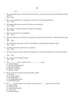
FIGURE 10.42 Intrinsic Muscles of the Foot. (a)–(d) First through fourth layers, respectively, in ventral (plantar) views. (e) Fourth
layer, dorsal view. The muscles belonging to each layer are shown in color and with boldface labels.
sal78259_ch10_312-378.indd 373
11/2/10 5:10 PM
374
PART TWO
Support and Movement
Apply What You Know
Not everyone has the same muscles. From the information
provided in this chapter, identify at least three muscles that
are lacking in some people.
Before You Go On
Answer the following questions to test your understanding of the
preceding section:
22. In the middle of a stride, you have one foot on the ground and
you are about to swing the other leg forward. What muscles
produce the movements of that leg?
23. Name the muscles that cross both the hip and knee joints
and produce actions at both.
24. List the major actions of the muscles of the anterior, medial,
and posterior compartments of the thigh.
25. Describe the role of plantar flexion and dorsiflexion in
walking. What muscles produce these actions?
DEEPER INSIGHT 10.5
Clinical Application
Common Athletic Injuries
Although the muscular system is subject to fewer diseases than most
organ systems, it is particularly vulnerable to injuries resulting from
sudden and intense stress placed on muscles and tendons. Each year,
thousands of athletes from the high school to professional level sustain
some type of injury to their muscles, as do the increasing numbers of
people who have taken up running and other forms of physical conditioning. Overzealous exertion without proper conditioning and warmup is frequently the cause. Compartment syndrome is one common
sports injury (see Deeper Insight 10.1). Others include:
Baseball finger—tears in the extensor tendons of the fingers resulting from the impact of a baseball with the extended fingertip.
Blocker’s arm—abnormal calcification in the lateral margin of the
forearm as a result of repeated impact with opposing players.
Charley horse—any painful tear, stiffness, and blood clotting in a
muscle. A charley horse of the quadriceps femoris is often caused
by football tackles.
Pitcher’s arm—inflammation at the origin of the flexor carpi muscles
resulting from hard wrist flexion in releasing a baseball.
Pulled groin—strain in the adductor muscles of the thigh; common in
gymnasts and dancers who perform splits and high kicks.
Pulled hamstrings—strained hamstring muscles or a partial tear in
their tendinous origins, often with a hematoma (blood clot) in the
fascia lata. This condition is frequently caused by repetitive kicking
(as in football and soccer) or long, hard running.
Rider’s bones—abnormal calcification in the tendons of the adductor
muscles of the medial thigh. It results from prolonged abduction
of the thighs when riding horses.
Rotator cuff injury—a tear in the tendon of any of the SITS (rotator
cuff) muscles, most often the tendon of the supraspinatus. Such
injuries are caused by strenuous circumduction of the arm, shoulder dislocation, hard falls or blows to the shoulder, or repetitive
use of the arm in a position above horizontal. They are common
among baseball pitchers and third basemen, bowlers, swimmers,
sal78259_ch10_312-378.indd 374
weight lifters, and in racquet sports. Recurrent inflammation of
a SITS tendon can cause a tendon to degenerate and then to
rupture in response to moderate stress. Injury causes pain and
makes the shoulder joint unstable and subject to dislocation.
Shinsplints—a general term embracing several kinds of injury with
pain in the crural region: tendinitis of the tibialis posterior muscle,
inflammation of the tibial periosteum, and anterior compartment
syndrome. Shinsplints may result from unaccustomed jogging,
walking on snowshoes, or any vigorous activity of the legs after a
period of relative inactivity.
Tennis elbow—inflammation at the origin of the extensor carpi
muscles on the lateral epicondyle of the humerus. It occurs when
these muscles are repeatedly tensed during backhand strokes
and then strained by sudden impact with the tennis ball. Any
activity that requires rotary movements of the forearm and a firm
grip of the hand (for example, using a screwdriver) can cause the
symptoms of tennis elbow.
Tennis leg—a partial tear in the lateral origin of the gastrocnemius
muscle. It results from repeated strains put on the muscle while
supporting the body weight on the toes.
Most athletic injuries can be prevented by proper conditioning.
A person who suddenly takes up vigorous exercise may not have sufficient muscle and bone mass to withstand the stresses such exercise
entails. These must be developed gradually. Stretching exercises keep
ligaments and joint capsules supple and therefore reduce injuries.
Warm-up exercises promote more efficient and less injurious musculoskeletal function in several ways, discussed in chapter 11. Most of all,
moderation is important, as most injuries simply result from overuse of
the muscles. “No pain, no gain” is a dangerous misconception.
Muscular injuries can be treated initially with “RICE”: rest, ice,
compression, and elevation. Rest prevents further injury and allows
repair processes to occur; ice reduces swelling; compression with an
elastic bandage helps to prevent fluid accumulation and swelling; and
elevation of an injured limb promotes drainage of blood from the
affected area and limits further swelling. If these measures are not
enough, anti-inflammatory drugs may be employed, including corticosteroids as well as aspirin and other nonsteroidal agents. Serious
injuries, such as compartment syndrome, require emergency attention
by a physician.
11/2/10 5:10 PM
CHAPTER 10
The Muscular System
375
STUDY GUIDE
Assess Your Learning Outcomes
To test your knowledge, discuss the
following topics with a study partner or
in writing, ideally from memory.
10.1 The Structural and Functional
Organization of Muscles (p. 313)
3.
1. Which muscles are included in the
muscular system and which ones
are not; the name of the science that
specializes in the muscular system
2. Functions of the muscular system
3. The relationship of muscle structure
to the endomysium, perimysium, and
epimysium; what constitutes a fascicle
of skeletal muscle and how it relates
to these connective tissues; and the
relationship of a fascia to a muscle
4. Classification of muscles according to
the orientation of their fascicles
5. Muscle compartments, interosseous
membranes, and intermuscular septa
6. The difference between direct and
indirect muscle attachments
7. The origin, belly, and insertion of a
muscle; the imperfection in origin–
insertion terminology
8. The action of a muscle; how it relates
to the classification of muscles as
prime movers, synergists, antagonists,
or fixators; why these terms are not
fixed for a given muscle but differ
from one joint movement to another,
and examples to illustrate this point
9. Intrinsic versus extrinsic muscles,
with examples
10. The innervation of muscles
11. Features to which the Latin names
of muscles commonly refer, with
examples
4.
10.2 Muscles of the Head and Neck
(p. 322)
Know the location, action, origin, insertion, and innervation of the named muscles in each of the following groups, and
be able to recognize them on laboratory
specimens or models to the extent
required in your course.
1. The frontalis and occipitalis muscles
of the scalp, eyebrows, and forehead
(table 10.1)
2. The orbicularis oculi, levator pal-
sal78259_ch10_312-378.indd 375
5.
6.
7.
8.
9.
10.
11.
12.
pebrae superioris, and corrugator
supercilii muscles, which move the
eyelid and other tissues around the
eye (table 10.1)
The nasalis muscle, which flares and
compresses the nostrils (table 10.1)
The orbicularis oris, levator labii
superioris, levator anguli oris, zygomaticus major and minor, risorius,
depressor anguli oris, depressor labii
inferioris, and mentalis muscles,
which act on the lips (table 10.1)
The buccinator muscles of the cheeks
(table 10.1)
The platysma, which acts upon the
mandible and the skin of the neck
(table 10.1)
The intrinsic muscles of the tongue
in general, and specific extrinsic
muscles: the genioglossus, hyoglossus, styloglossus, and palatoglossus
muscles (table 10.2)
The temporalis, masseter, medial
pterygoid, and lateral pterygoid
muscles of biting and chewing
(table 10.2)
The suprahyoid group: the digastric, geniohyoid, mylohyoid, and
stylohyoid muscles (table 10.2)
The infrahyoid group: the omohyoid,
sternohyoid, thyrohyoid, and
sternothyroid muscles (table 10.2)
The superior, middle, and inferior
pharyngeal constrictor muscles of the
throat (table 10.2)
The sternocleidomastoid and three
scalene muscles, which flex the neck,
and the trapezius, splenius capitis,
and semispinalis capitis muscles,
which extend it (table 10.3)
10.3 Muscles of the Trunk (p. 333)
For the following muscles, know the same
information as for section 10.2
1. The diaphragm and the external
intercostal, internal intercostal, and
innermost intercostal muscles of
respiration (table 10.4)
2. The external abdominal oblique,
internal abdominal oblique,
transverse abdominal, and rectus
abdominis muscles of the anterior
3.
4.
5.
6.
7.
and lateral abdominal wall
(table 10.5)
The superficial erector spinae muscle
(and its subdivisions) and the deep
semispinalis thoracis, quadratus
lumborum, and multifidus muscles
of the back (table 10.6)
The perineum, its two triangles, and
their skeletal landmarks (table 10.7)
The ischiocavernosus and bulbospongiosus muscles of the superficial
perineal space of the pelvic floor
(table 10.7)
The external urethral sphincter
and external anal sphincter, and in
females, the compressor urethrae, of
the middle compartment of the pelvic
floor (table 10.7)
The levator ani and coccygeus
muscles of the pelvic diaphragm, the
deepest compartment of the pelvic
floor (table 10.7)
10.4 Muscles Acting on the Shoulder and
Upper Limb (p. 343)
For the following muscles, know the same
information as for section 10.2.
1. The pectoralis minor, serratus
anterior, trapezius, levator scapulae,
rhomboideus major, and rhomboideus
minor muscles of scapular movement
(table 10.8)
2. Muscles that act on the humerus,
including the pectoralis major, latissimus dorsi, deltoid, teres major,
coracobrachialis, and four rotator cuff
(SITS) muscles—the supraspinatus,
infraspinatus, teres minor, and subscapularis (table 10.9)
3. The brachialis, biceps brachii, triceps
brachii, brachioradialis, anconeus,
pronator quadratus, pronator teres,
and supinator muscles of forearm
movement (table 10.10)
4. The relationship of the flexor
retinaculum, extensor retinaculum,
and carpal tunnel to the tendons of
the forearm muscles
5. The palmaris longus, flexor carpi
radialis, flexor carpi ulnaris, and flexor digitorum superficialis muscles of
the superficial anterior compartment
11/2/10 5:10 PM
376
6.
7.
8.
9.
10.
PART TWO
Support and Movement
of the forearm, and the flexor digitorum profundus and flexor pollicis
longus muscles of the deep anterior
compartment (table 10.11)
The extensor carpi radialis longus,
extensor carpi radialis brevis, extensor digitorum, extensor digiti minimi,
and extensor carpi ulnaris muscles of
the superficial posterior compartment
(table 10.11)
The abductor pollicis longus, extensor pollicis brevis, extensor pollicis
longus, and extensor indicis muscles
of the deep posterior compartment
(table 10.11)
The thenar group of intrinsic hand
muscles: adductor pollicis, abductor
pollicis brevis, flexor pollicis brevis,
and opponens pollicis (table 10.12)
The hypothenar group of intrinsic
hand muscles: abductor digiti minimi, flexor digiti minimi brevis, and
opponens digiti minimi (table 10.12)
The midpalmar group of intrinsic
hand muscles: four dorsal interosseous
muscles, three palmar interosseous
muscles, and four lumbrical muscles
(table 10.12)
10.5 Muscles Acting on the Hip and
Lower Limb (p. 359)
For the following muscles, know the same
information as for section 10.2.
1. The iliopsoas muscle of the hip, and
its two subdivisions, the iliacus and
psoas major (table 10.13)
2. The tensor fasciae latae, gluteus maximus, gluteus medius, and gluteus
minimus muscles of the hip and buttock, and the relationship of the first
two to the fascia lata and iliotibial
band (table 10.13)
3. The lateral rotators: gemellus
superior, gemellus inferior, obturator
externus, obturator internus, piriformis, and quadratus femoris muscles
(table 10.13)
4. The compartments of the thigh
muscles: anterior (extensor), medial
(adductor), and posterior (flexor)
compartments
5. Muscles of the medial compartment
of the thigh: adductor brevis, adductor longus, adductor magnus, gracilis,
and pectineus (table 10.13)
6. Muscles of the anterior compartment
of the thigh: sartorius and quadriceps
femoris, and the four heads of the
quadriceps (table 10.14)
7. The hamstring muscles of the posterior compartment of the thigh: biceps
femoris, semitendinosus, and semimembranosus (table 10.14)
8. The compartments of the leg muscles: anterior, posterior, and lateral
(table 10.15)
9. Muscles of the anterior compartment of the leg: fibularis tertius,
extensor digitorum longus, extensor
hallucis longus, and tibialis anterior
muscles of the anterior compartment
(table 10.15)
10. Muscles of the superficial posterior
11.
12.
13.
14.
compartment of the leg: popliteus
and triceps surae (gastrocnemius and
soleus), and the relationship of the
triceps surae to the calcaneal tendon
and calcaneus (table 10.15)
Muscles of the deep posterior compartment of the leg: flexor digitorum
longus, flexor hallucis longus, and
tibialis posterior muscles of the deep
posterior compartment
Muscles of the lateral compartment of
the leg: fibularis brevis and fibularis
longus (table 10.15)
The extensor digitorum brevis of the
dorsal aspect of the foot (table 10.16)
The four muscle compartments
(layers) of the ventral aspect of the
foot, and the muscles in each: the
flexor digitorum brevis, abductor
digiti minimi, and abductor hallucis
(layer 1); the quadratus plantae and
four lumbrical muscles (layer 2); the
flexor digiti minimi brevis, flexor
hallucis brevis, and adductor hallucis (layer 3); and the four dorsal
interosseous muscles and three
plantar interosseous muscles
(layer 4) (table 10.16)
Testing Your Recall
1. Which of the following muscles is the
prime mover in spitting out a mouthful of liquid?
a. platysma
b. buccinator
c. risorius
d. masseter
e. palatoglossus
2. Each muscle fiber has a sleeve of
areolar connective tissue around it
called
a. the fascia.
b. the endomysium.
c. the perimysium.
d. the epimysium.
e. the intermuscular septum.
sal78259_ch10_312-378.indd 376
3. Which of these is not a suprahyoid
muscle?
a. genioglossus
b. geniohyoid
c. stylohyoid
d. mylohyoid
e. digastric
5. Which of these muscles of the pelvic
floor is the deepest?
a. superficial transverse perineal
b. bulbospongiosus
c. ischiocavernosus
d. deep transverse perineal
e. levator ani
4. Which of these muscles is an extensor of the neck?
a. external oblique
b. sternocleidomastoid
c. splenius capitis
d. iliocostalis
e. latissimus dorsi
6. Which of these actions is not
performed by the trapezius?
a. extension of the neck
b. depression of the scapula
c. elevation of the scapula
d. rotation of the scapula
e. adduction of the humerus
11/2/10 5:10 PM
CHAPTER 10
7. Both the hands and feet are acted
upon by a muscle or muscles called
a. the extensor digitorum.
b. the abductor digiti minimi.
c. the flexor digitorum profundus.
d. the abductor hallucis.
e. the flexor digitorum longus.
10. Which of the following muscles raises the upper lip?
a. levator palpebrae superioris
b. orbicularis oris
c. zygomaticus minor
d. masseter
e. mentalis
8. Which of the following muscles does
not extend the hip joint?
a. quadriceps femoris
b. gluteus maximus
c. biceps femoris
d. semitendinosus
e. semimembranosus
11. The
of a muscle is the point
where it attaches to a relatively stationary bone.
9. Both the gastrocnemius and
muscles insert on the heel by way of
the calcaneal tendon.
a. semimembranosus
b. tibialis posterior
c. tibialis anterior
d. soleus
e. plantaris
12. A bundle of muscle fibers surrounded
by perimysium is called a/an
.
is the muscle that gener13. The
ates the most force in a given joint
movement.
The Muscular System
377
16. The anterior half of the perineum is a
region called the
.
17. The abdominal aponeuroses converge
on a median fibrous band on the
abdomen called the
.
18. A muscle that works with another to
produce the same or similar movement is called a/an
.
19. A muscle somewhat like a feather,
with fibers obliquely approaching its
tendon from both sides, is called a/an
muscle.
20. A circular muscle that closes a body
opening is called a/an
.
Answers in appendix B
14. The three large muscles on the posterior side of the thigh are commonly
known as the
muscles.
15. Connective tissue bands called
prevent flexor tendons of the forearm
and leg from rising like bowstrings.
Building Your Medical Vocabulary
State a medical meaning of each word
element below, and give a term in which
it or a slight variation of it is used.
1. capito2. ergo-
3. fasc-
7. mys-
4. labio-
8. omo-
5. lumbo-
9. penn-
6. mus-
10. tertAnswers in appendix B
True or False
Determine which five of the following statements are false, and briefly
explain why.
4. To push someone away from you, you
would use the serratus anterior more
than the trapezius.
1. Cutting the phrenic nerves would
paralyze the prime mover of
respiration.
5. Both the extensor digitorum and
extensor digiti minimi extend the
little finger.
2. The orbicularis oculi is a sphincter.
6. Curling the toes employs the quadratus plantae.
3. The origin of the sternocleidomastoid
muscle is the mastoid process of the
skull.
sal78259_ch10_312-378.indd 377
8. Exhaling requires contraction of the
internal intercostal muscles.
9. Hamstring injuries often result from
rapid flexion of the knee.
10. The tibialis anterior and tibialis
posterior are synergists.
Answers in appendix B
7. The scalenes are superficial to the
trapezius.
11/2/10 5:10 PM
378
PART TWO
Support and Movement
Testing Your Comprehension
1. Radical mastectomy, once a common
treatment for breast cancer, involved
removal of the pectoralis major along
with the breast. What functional
impairments would result from this?
What synergists could a physical
therapist train a patient to use to
recover some lost function?
2. Removal of cancerous lymph nodes
from the neck sometimes requires
removal of the sternocleidomastoid
on that side. How would this affect
a patient’s range of head movement?
3. Poorly conditioned, middle-aged
people may suffer a rupture of the
calcaneal tendon when the foot is
suddenly dorsiflexed. Explain each
of the following signs of a ruptured
calcaneal tendon: (a) a prominent
lump typically appears in the calf;
(b) the foot can be dorsiflexed farther
than usual; and (c) the patient cannot
plantar flex the foot very effectively.
wear flat shoes. What muscle(s) and
tendon(s) are involved? Explain.
5. A student moving out of a dormitory
kneels down, in correct fashion, to
lift a heavy box of books. What prime
movers are involved as he straightens
his legs to lift the box?
Answers at www.mhhe.com/saladin6
4. Women who habitually wear high
heels may suffer painful “high heel
syndrome” when they go barefoot or
Improve Your Grade at www.mhhe.com/saladin6
Download mp3 audio summaries and movies to study when it fits your schedule. Practice quizzes, labeling activities, games,
and flashcards offer fun ways to master the chapter concepts. Or, download image PowerPoint files for each chapter to create
a study guide or for taking notes during lecture.
sal78259_ch10_312-378.indd 378
11/2/10 5:10 PM
Atlas B
REGIONAL AND
SURFACE ANATOMY
ATLAS OUTLINE
B.1 Regional Anatomy 380
B.2 The Importance of Surface Anatomy 380
B.3 Learning Strategy 380
Figures B.1–B.2 The Head and Neck
Figures B.3–B.15 The Trunk
Figures B.16–B.19 The Upper Limb
Figures B.20–B.24 The Lower Limb
Figure B.25 Test of Muscle Recognition
Module 6: Muscular System
How many muscles can you identify from their surface
appearance?
379
sal78259_atlas_b_379-400.indd 379
11/3/10 2:38 PM
380
B.1
PART TWO
Support and Movement
Regional Anatomy
On the whole, this book takes a systems approach to anatomy, examining the structure and function of each organ
system, one at a time, regardless of which body regions
it may traverse. Physicians and surgeons, however, think
and act in terms of regional anatomy. If a patient presents with pain in the lower right quadrant (see fig. A.6a,
p. 33), the source may be the appendix, an ovary, or an
inguinal muscle, among other possibilities. The question
is to think not of an entire organ system (the esophagus is
probably irrelevant to that quadrant), but of what organs
are present in that region and what possibilities must be
considered as the cause of the pain. This atlas presents
several views of the body region by region so that you can
see some of the spatial relationships that exist among the
organ systems considered in their separate chapters.
B.2 The Importance of
Surface Anatomy
In the study of human anatomy, it is easy to become so
preoccupied with internal structure that we forget the
importance of what we can see and feel externally. Yet
external anatomy and appearance are major concerns in
giving a physical examination and in many aspects of
patient care. A knowledge of the body’s surface landmarks is essential to one’s competence in physical therapy,
cardiopulmonary resuscitation, surgery, making X-rays and
electrocardiograms, giving injections, drawing blood, listening to heart and respiratory sounds, measuring the pulse
and blood pressure, and finding pressure points to stop
arterial bleeding, among other procedures. A misguided
attempt to perform some of these procedures while disregarding or misunderstanding external anatomy can be
very harmful and even fatal to a patient.
Having just studied skeletal and muscular anatomy
in the preceding chapters, this is an opportune time for
you to study the body surface. Much of what we see there
reflects the underlying structure of the superficial bones
and muscles. A broad photographic overview of surface
anatomy is given in atlas A (see fig. A.5, p. 32), where it is
necessary for providing a vocabulary for reference in subsequent chapters. This atlas shows this surface anatomy
in closer detail so you can relate it to the musculoskeletal
anatomy of chapters 8 through 10.
sal78259_atlas_b_379-400.indd 380
B.3
Learning Strategy
To make the most profitable use of this atlas, refer back
to earlier chapters as you study these illustrations. Relate
drawings of the clavicles in chapter 8 to the photograph
in figure B.1, for example. Study the shape of the scapula
in chapter 8 and see how much of it you can trace on
the photographs in figure B.9. See if you can relate the
tendons visible on the hand (see fig. B.19) to the muscles
of the forearm illustrated in chapter 10, and the external
markings of the pelvic girdle (see fig. B.15) to bone structure in chapter 8.
For learning surface anatomy, there is a resource
available to you that is far more valuable than any laboratory model or textbook illustration—your own body. For
the best understanding of human structure, compare the
art and photographs in this book with your body or with
structures visible on a study partner. In addition to bones
and muscles, you can palpate a number of superficial
arteries, veins, tendons, ligaments, and cartilages, among
other structures. By palpating regions such as the shoulder, elbow, or ankle, you can develop a mental image of
the subsurface structures better than the image you can
obtain by looking at two-dimensional textbook images.
And the more you can study with other people, the more
you will appreciate the variations in human structure and
be able to apply your knowledge to your future patients or
clients, who will not look quite like any textbook diagram
or photograph you have ever seen. Through comparisons
of art, photography, and the living body, you will get a
much deeper understanding of the body than if you were
to study this atlas in isolation from the earlier chapters.
At the end of this atlas, you can test your knowledge
of externally visible muscle anatomy. The two photographs
in figure B.25 have 30 numbered muscles and a list of
26 names, some of which are shown more than once in
the photographs and some of which are not shown at all.
Identify the muscles to your best ability without looking
back at the previous illustrations, and then check your
answers in appendix B at the back of the book.
Throughout these illustrations, the following abbreviations apply: a. = artery; m. = muscle; n. = nerve; v. = vein.
Double letters such as mm. or vv. represent the plurals.
11/3/10 2:38 PM
ATLAS B
Regional and Surface Anatomy
381
Occipital
Frontal
Orbital
Temporal
Nasal
Auricular
Oral
Mental
Buccal (cheek)
Cervical
Nuchal (posterior cervical)
(a) Lateral view
Frons (forehead)
Root of nose
Bridge of nose
Superciliary
ridge
Superior palpebral
sulcus
Inferior palpebral
sulcus
Auricle (pinna)
of ear
Philtrum
Labia (lips)
Lateral commissure
Medial commissure
Dorsum nasi
Apex of nose
Ala nasi
Mentolabial sulcus
Mentum (chin)
Sternoclavicular
joints
Clavicle
Suprasternal notch
Supraclavicular
fossa
Sternum
(b) Anterior view
FIGURE B.1 The Head and Neck. (a) Anatomical regions of the head. (b) Features of the facial region and upper thorax.
● What muscle underlies the region of the philtrum? What muscle forms the slope of the shoulder?
sal78259_atlas_b_379-400.indd 381
11/3/10 2:38 PM
382
PART TWO
Support and Movement
Scalp
Cranium
Cerebrum
Frontal sinus
Nasal cavity
Brainstem
Cerebellum
Palate
Oral cavity
Tongue
Foramen magnum
of skull
Spinal cord
Epiglottis
Pharynx
Vertebral column
Vocal cord
Larynx
Trachea
Intervertebral discs
Esophagus
FIGURE B.2 Median Section of the Head. Shows contents of the cranial, nasal, and oral cavities.
sal78259_atlas_b_379-400.indd 382
11/3/10 2:38 PM
ATLAS B
Regional and Surface Anatomy
383
Platysma m.
Trapezius m.
Clavicle
Deltoid m.
Pectoralis major m.
Cephalic v.
Breast
Biceps brachii m.
Sheath of rectus
abdominis m.
External abdominal
oblique m.
Umbilicus
Anterior superior
spine of ilium
Inguinal ligament
Tensor fasciae latae m.
Mons pubis
Sartorius m.
Femoral v.
Adductor longus m.
Great saphenous v.
Gracilis m.
Vastus lateralis m.
Rectus femoris m.
FIGURE B.3 Superficial Anatomy of the Trunk (Female). Surface anatomy is shown on the anatomical left, and structures immediately deep to
the skin on the right.
sal78259_atlas_b_379-400.indd 383
11/3/10 2:38 PM
384
PART TWO
Support and Movement
Internal jugular v.
External jugular v.
Common
carotid a.
Omohyoid m.
Clavicle
Internal
intercostal mm.
External
intercostal mm.
Sternum
Subscapularis m.
Coracobrachialis m.
Lung
Costal
cartilages
Pericardium
Pleura
Diaphragm
Liver
Stomach
Gallbladder
External abdominal
oblique m.
Internal abdominal
oblique m.
Transverse abdominal m.
Large
intestine
Greater omentum
Urinary bladder
Penis
Femoral n.
Femoral a.
Scrotum
Femoral v.
FIGURE B.4 Anatomy at the Level of the Rib Cage and Greater Omentum (Male). The anterior body wall is removed, and the ribs, intercostal
muscles, and pleura are removed from the anatomical left.
sal78259_atlas_b_379-400.indd 384
11/3/10 2:38 PM
ATLAS B
Regional and Surface Anatomy
385
Thyroid cartilage of larynx
Brachiocephalic v.
Thyroid gland
Subclavian v.
Subclavian a.
Brachial nerve plexus
Aortic arch
Superior vena
cava
Axillary v.
Coracobrachialis m.
Axillary a.
Cephalic v.
Brachial v.
Humerus
Brachial a.
Heart
Lobes of lung
Spleen
Stomach
Large
intestine
Small intestine
Cecum
Appendix
Tensor fasciae latae m.
Penis (cut)
Pectineus m.
Ductus
deferens
Epididymis
Adductor longus m.
Testis
Gracilis m.
Scrotum
Adductor magnus m.
Rectus femoris m.
FIGURE B.5 Anatomy at the Level of the Lungs and Intestines (Male). The sternum, ribs, and greater omentum are removed.
● Name several viscera that are protected by the rib cage.
sal78259_atlas_b_379-400.indd 385
11/3/10 2:38 PM
386
PART TWO
Support and Movement
Trachea
Superior vena cava
Bronchus
Lung
(sectioned)
Esophagus
Thoracic aorta
Pleural cavity
Hepatic vv.
Spleen
Inferior vena cava
Splenic a.
Adrenal gland
Pancreas
Duodenum
Kidney
Superior mesenteric v.
Abdominal aorta
Superior
mesenteric a.
Inferior
mesenteric a.
Common iliac a.
Ureter
Ovary
Uterine tube
Tensor fasciae
latae m. (cut)
Uterus
Sartorius m. (cut)
Urinary bladder
Pectineus m.
Gracilis m.
Adductor longus m.
Rectus
femoris m. (cut)
Adductor brevis m.
Vastus intermedius
m.
Adductor
longus m. (cut)
Vastus lateralis m.
Vastus medialis m.
FIGURE B.6 Anatomy at the Level of the Retroperitoneal Viscera (Female). The heart is removed, the lungs are frontally sectioned, and the
viscera of the peritoneal cavity and the peritoneum itself are removed.
sal78259_atlas_b_379-400.indd 386
11/3/10 2:38 PM
ATLAS B
Regional and Surface Anatomy
387
Right common carotid a.
Left common
carotid a.
Right subclavian a.
Left subclavian a.
Brachiocephalic trunk
External
intercostal m.
Thoracic aorta
Ribs
Esophagus
Internal
intercostal m.
Diaphragm
Abdominal aorta
Intervertebral disc
Quadratus
lumborum m.
Lumbar vertebra
Iliac crest
Psoas major m.
Iliacus m.
Ilium
Sacrum
Anterior superior
spine of ilium
Gluteus medius m.
Brim of pelvis
Rectum
Vagina
Urethra
Adductor magnus m.
Femur
Adductor brevis m.
Gracilis m.
Adductor longus m.
FIGURE B.7 Anatomy at the Level of the Posterior Body Wall (Female). The lungs and retroperitoneal viscera are removed.
sal78259_atlas_b_379-400.indd 387
11/3/10 2:38 PM
388
PART TWO
Support and Movement
Sternocleidomastoid m.
Supraclavicular
fossa
Thyroid cartilage
Clavicle
Trapezius m.
Acromion
Sternum:
Suprasternal notch
Deltoid m.
Manubrium
Angle
Body
Pectoralis major m.
Xiphoid process
Nipple
Rectus
abdominis m.
Serratus anterior mm.
Tendinous
intersection of
rectus abdominis m.
Linea semilunaris
Anterior superior
spine of ilium
Umbilicus
Iliac crest
External abdominal
oblique m.
Linea alba
Inguinal ligament
(a) Male
Trapezius m.
Supraclavicular
fossa
Clavicle
Acromion
Sternum:
Suprasternal notch
Deltoid m.
Manubrium
Breast:
Angle
Axillary tail
Body
Nipple
Xiphoid process
Areola
Corpus (body)
Linea alba
Rectus
abdominis m.
Costal margin
Umbilicus
Linea semilunaris
External abdominal
oblique m.
Anterior superior
spine of ilium
(b) Female
FIGURE B.8 The Thorax and Abdomen, Anterior View. All of the features labeled are common to both sexes, though some are labeled only on
the photograph that shows them best.
● The V-shaped tendons on each side of the suprasternal notch in part (a) belong to what muscles?
sal78259_atlas_b_379-400.indd 388
11/3/10 2:38 PM
ATLAS B
Regional and Surface Anatomy
389
Flexor carpi ulnaris
Brachioradialis
Biceps brachii
Triceps brachii
Deltoid:
Anterior part
Middle part
Posterior part
Teres major
Infraspinatus
Medial border
of scapula
Trapezius
Vertebral furrow
Erector spinae
Latissimus dorsi
Iliac crest
(a) Male
Acromion
Medial border
of scapula
Infraspinatus
Trapezius
Inferior angle
of scapula
Latissimus
dorsi
Olecranon
Erector spinae
Iliac crest
Sacrum
Gluteus medius
Coccyx
Natal cleft
Gluteus maximus
Greater
trochanter
of femur
Gluteal fold
Hamstring muscles
(b) Female
FIGURE B.9 The Back and Gluteal Region. All of the features labeled are common to both sexes, though some are labeled only on the
photograph that shows them best.
sal78259_atlas_b_379-400.indd 389
11/3/10 2:38 PM
390
PART TWO
Support and Movement
Internal jugular v.
Subclavian v.
Nerves
Lungs
Ribs
Heart
Diaphragm
FIGURE B.10 Frontal View of the Thoracic Cavity.
Anterior
Pectoralis
major m.
Fat of breast
Sternum
Ventricles
of heart
Ribs
Pericardial
cavity
Right lung
Esophagus
Atria of heart
Aorta
Vertebra
Left lung
Spinal cord
Pleural cavity
Posterior
FIGURE B.11 Transverse Section of the Thorax. Section taken at the level shown by the inset and oriented the same as the reader’s body.
● In this section, which term best describes the position of the aorta relative to the heart: posterior, lateral, inferior, or proximal?
sal78259_atlas_b_379-400.indd 390
11/3/10 2:38 PM
ATLAS B
Regional and Surface Anatomy
391
Lung
Diaphragm
Transverse colon
Gallbladder
Small intestine
Mesenteric
arteries and veins
Mesentery
Descending colon
Cecum
Sigmoid colon
FIGURE B.12 Frontal View of the Abdominal Cavity.
Duodenum
Anterior
Stomach
Subcutaneous
fat
Rectus
abdominis m.
Large
intestine
Superior mesenteric
artery and vein
Pancreas
Inferior vena cava
Liver
Kidney
Peritoneal cavity
Perirenal
fat of
kidney
Peritoneum
Aorta
Erector
spinae m.
Vertebra
Posterior
Spinal cord
FIGURE B.13 Transverse Section of the Abdomen. Section taken at the level shown by the inset and oriented the same as the reader’s body.
● What tissue in this photograph is immediately superficial to the rectus abdominis muscle?
sal78259_atlas_b_379-400.indd 391
11/3/10 2:39 PM
Urinary bladder
Sigmoid colon
Pubic symphysis
Seminal vesicle
Prostate gland
Penis:
Root
Bulb
Rectum
Anal canal
Shaft:
Corpus
cavernosum
Anus
Corpus
spongiosum
Epididymis
Scrotum
Glans
Testis
(a) Male
Vertebra
Red bone marrow
Mesentery
Intervertebral disc
Small intestine
Sacrum
Sigmoid colon
Uterus
Cervix
Urinary bladder
Pubic symphysis
Urethra
Vagina
Rectum
Anal canal
Anus
Labium minus
Prepuce
Labium majus
(b) Female
FIGURE B.14 Median Sections of the Pelvic Cavity. Viewed from the left.
sal78259_atlas_b_379-400.indd 392
11/3/10 2:39 PM
ATLAS B
(a) Anterior view
Regional and Surface Anatomy
393
(b) Posterior view
FIGURE B.15 Pelvic Landmarks. (a) The anterior superior spines of the ilium are marked by anterolateral protuberances (arrows) at about the
location where the front pockets usually open on a pair of pants. (b) The posterior superior spines are marked in some people by dimples in the sacral
region (arrows).
Olecranon
Biceps brachii
Triceps brachii
Anterior axillary fold (pectoralis major)
Posterior axillary fold (latissimus dorsi)
Deltoid
Axilla (armpit)
Pectoralis major
Latissimus dorsi
FIGURE B.16 The Axillary Region.
sal78259_atlas_b_379-400.indd 393
11/3/10 2:39 PM
394
PART TWO
Support and Movement
Trapezius
Acromion
Deltoid
Interphalangeal
joints
Pectoralis major
Biceps brachii
Metacarpophalangeal
joints
Triceps brachii:
Lateral head
Long head
Styloid process
of radius
Brachioradialis
Extensor carpi
radialis longus
Lateral epicondyle
of humerus
Olecranon
Extensor digitorum
FIGURE B.17 The Upper Limb, Lateral View.
Triceps brachii
Biceps brachii
Medial epicondyle
of humerus
Cubital fossa
Cephalic vein
Olecranon
Median cubital vein
Head of radius
Brachioradialis
Brachioradialis
Flexor carpi radialis
Palmaris longus
Flexor carpi ulnaris
Flexor carpi ulnaris
Extensor carpi ulnaris
Extensor digitorum
Styloid process of
ulna
Styloid process of radius
Hypothenar eminence
Thenar eminence
Tendons of extensor digitorum
Flexion lines
Palmar surface of hand
Dorsum of hand
Pollex (thumb)
Volar surface of fingers
Flexion lines
(a) Anterior view
(b) Posterior view
FIGURE B.18 The Antebrachium (Forearm).
● Only two tendons of the extensor digitorum are labeled, but how many tendons does this muscle have in all?
sal78259_atlas_b_379-400.indd 394
11/3/10 2:39 PM
Palmaris longus tendon
Flexor carpi radialis tendon
Flexion lines
Thenar eminence
Hypothenar eminence
Pollex (thumb)
Flexion lines
I
Metacarpophalangeal
joint
Interphalangeal
joints
V
II
IV
III
(a) Anterior (palmar) view
Styloid process of radius
Styloid process of ulna
Extensor pollicis brevis tendon
Anatomical snuffbox
Extensor pollicis longus tendon
Extensor digiti minimi tendon
Extensor digitorum tendons
Adductor pollicis
FIGURE B.19 The Wrist and Hand.
● Mark the spot on one or both
photographs where a saddle joint can be
found.
(b) Posterior (dorsal) view
395
sal78259_atlas_b_379-400.indd 395
11/3/10 2:39 PM