Histoire naturelle des insectes 01
Bạn đang xem bản rút gọn của tài liệu. Xem và tải ngay bản đầy đủ của tài liệu tại đây (6.71 MB, 97 trang )
a, 17^1
[
811
]
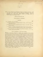
XVIII.
On the Muscular and Endoskeletal Systems of Limulus and Scorpio; with
some Notes on the Anatomy and Generic Characters of Scorpions. By E. Ray
Laxkester, M.A., LL.D., F.B.S., Jodrell Professor of Zoology, assisted by
W. B. S. Benham and Miss E. J. Beck.
Received and read June 19th, 1883.
[Plates
LXXII.
to
LXXXIIL]
CONTENTS.
Page
Part
„
I.
Introduction.
By
II. Description of tho
(Plates
Kay Lankestee
E.
311
Muscular and Endoskeletal Systems
of
Limulus.
By W.
B. S.
Benham.
LXXII.-LXXVI.)
„ III. Description of the Muscular
314
By
and Endoskeletal Systems of Scorpio.
E. J. Beck.
(Plates
LXXVII.-LXXIX.)
339
„ IV. Comparison of the Muscular and Endoskeletal Systems of Limulus and Scorpio, and Consideration of the Morphological Significance of the Facts recorded.
„
By E. Eat Lankestee 361
V. Notes on Certain Points in the Anatomy and Generic Characters of Scorpions.
Lankestee.
(Plates
Part
WHEN,
two years ago,
By
E.
Rat
LXXX.-LXXXIII.)
I.
Introduction.
372
By
E.
Rat Lanrester.
undertook to institute a close comparison of the structure
of Limulus, on the one hand with that of the Crustacea, and on the other hand with
I
that of the Scorpion and other Arachnida, in order to definitely and fully substantiate
the view which for
many
years had appeared to
me
plausible, viz. that
Limulus
is
no
Crustacean, but an Arachnid, I found considerable difficulty, owing to the fact that
details concerning the structure
critical points,
have found
it
were not
to
both of Limulus and of Scorpio, in reference to many
be met with in the literature of zoology.
necessary to undertake, in conjunction with
my
In consequence, I
pupils, investigations
upon various matters connected with the histology and coarser anatomy of both
Limulus and Scorpio, which have yielded remarkable results remarkable because
they were obtained in the attempt to verify a hypothesis, and have uniformly tended to
verify it.
Thus, I discovered in Scorpio an organ which represents the brick-red coxal
glands of Limulus (Proc. Roy. Soc. 1882), and in the remarkable microscopical structure
—
of these " vascular glands " I have detected a character which connects Limulus and the
Arachnids in the closest way whilst having no exact equivalent in any Crustacean
vol. xi.
part x. No. 1.
May, 1885.
3 b
PROF.
312
E.
Joum. Microsc.
(Quart.
EAT LANKESTER ON THE MUSCULAR AND
Sci.,
January 1884).
Further, 1 investigated the structure of
both the simple and the compound (or aggregated) eyes of Limulus and of Scorpio, and
again obtained from the minute microscopic structure evidence of the closest agreement
between these two genera and of
Micr.
Sci,,
total divergence
from the Crustacea (Quart. Joum.
January 1883).
Again, since the structure of the genital ducts in Crustacea
simple
is
any
or, in
male Apus), whilst both oviducts and sperm-ducts in
Scorpio and other Araclmida have the characteristic form of a mesh-work, I requested
my pupil Mr. W. B. S. Benham to investigate the structure of the spermatic duct and
case, non-reticulate (except in the
glands of Limulus, hitherto unexplored.
Mr. Benham found (and has described
in the
'Transactions of the Linnean Society,' 1883) a highly subdivided reticulum, or mesh-
work, constituting the spermatic duct, as in the Scorpions. The oviduct had previously
been shown by Owen to have essentially the form of a network.
Lastly, I have found (and
Sci.
am
about to explain in detail in the Quart. Journ. Micr.
the most intimate agreement between Limulus and Scorpio in respect of the
1
)
following points of minute structure
tissues
cases,
;
(3) the
:— (1) the blood-corpuscles
;
(2) the softer connective
entochondrite (internal sternum of Straus Durkheim), which
a mass of condensed connective tissue with
cells of
is,
in both
very characteristic appearance,
but so like in the two cases as to be practically indistinguishable
;
(3) the gastric caeca
and their lining epithelium.
is
Amongst the most important points of agreement between Limulus and the Arachnids
upon by Straus Durkheim, namely, the possession of an internal freely
that insisted
suspended sternum or plastron of connective tissue (cartilaginoid
numerous muscles are attached.
probably
is
more or
Such an entochondrite
less closely similar in
exists in
tissue),
to
which
no Crustacean 2
;
it
nature to the so-called " chorda " discovered
by Leydig in insects of the genus Sphinw.
In order to carry out fully the comparison of the entochondrite of Limulus with that
it became necessary to make an investigation of the muscles attached to this
of Scorpio,
organ in each case, and this has led on to a general investigation of the whole muscular
The investigation of Limulus has
its related supports in the two animals.
system and
been carried out by Mr. Benham, that of Scorpio by Miss Beck. No account of the
muscular system of either animal has before been given, although imperfect descriptions
of parts of the muscular system of Limulus are to be found both in the memoirs of
Owen and
of Alphonse Milne-Edwards.
As might be
expected,
we
find a considerable specialization of the
muscular system
two animals compared, resulting in a wide divergence as to certain muscles but
there remain, nevertheless, certain agreements which are of the most striking and
in the
;
important character.
1
Since published, in January 1884.
*
I have since found a rudimentary structure of the kind in
Apus
(Quart. Journ. Micr.
Sci.,
January 1884).
;
ENDOSKELETAL SYSTEMS OF LIMULUS AND SCOEPIO.
It will
be
sufficient
313
by way of introduction, that necessarily
to point out here,
in
Scorpio the muscles to the appendages of the mesosoma are almost entirely suppressed
(those of the last four pairs of appendages, which have
whilst,
entirely),
Again, in Scorpio the free articulation of the segments of the mesosoma
Limulus.
and of the metasoma
articulation
soma
become lung-books,
on the other hand, the same muscles are large and functionally important in
is
retained,
are ankylosed, and
and accordingly the musculature connected with that
In Limulus, on the other hand, the segments of the meso-
developed.
is
there
are
consequently no
intersegmental muscles.
great joint, however, that between prosoma and mesosoma,
and accordingly,
in
is
One
retained by Limulus
connexion with that one joint, we find an enormous and specialized
muscular development, differing from anything in Scorpio.
The most remarkable agreements to which
are in respect of (1) a large number of
hand
entochondrite
first
(2) certain of the
;
gill-bearing
cases
;
(3) the
the reader's attention
is
directed before-
the muscles attached to the prosomatic
muscles attached to the pectines of Scorpio and the
appendage of Limulus and
to the related small entochondrites in
both
muscles arising from the pericardium and inserted into the investment of
the great veinous sac, which in the one case
lies at
the base of a gill-book and in the other
case forms the investment of the in-sunken lung-book.
This
is
a most important agree-
ment, since in each case the muscle must have a very definite and peculiar action in
determining the flow of blood from the respiratory sinus to the heart. These muscles
were described as " brides transparentes " by A. Milne-EdwT ards, in his account of the
vascular system of Limulus.
in his drawing,
fig.
By Newport they were
seen in the Scorpion, and figured
27, pi. xiv. of the 'Philosophical Transactions' for
are not described or referred to by
him
in
1843
;
but they
any way, and their significance has never yet
been pointed out.
Lastly, the
agreement in the origin and insertion of the great dorso-ventral
muscles of the mesosoma
memoir
is
a prominent one.
vertical
In the fourth Chapter of the present
a further discussion of the agreements and differences of the muscular system
in Scojpio
and Limulus will be found.
3b2
ME. W.
314
Part
II.
B.
BENHAM ON THE MUSCULAK
S.
Description of the Muscular and EndosJceletal Systems o/Limulus.
By W.
B. S.
Hard
Some of
the
Hard Parts
in
I.
a.
Benham, B.Sc.
Parts.
Limulus
The
to
which Muscles are attached.
Tergites.
External View.
The Prosomatic Carapace has a horseshoe-shape, rounded and convex in front and
the sides, which latter are produced beyond the central portion, ending in a point
1.
at
behind.
anterior
(For a general description and figures of the segments fused to form the
and posterior carapaces of Limulus, see Lankester, "Limulus an Arachnid,"
Quart. Journ. Micr. Sci. 1882.)
The carapace
is
bounded behind by an almost straight
on each side of the middle
line.
that the posterior border
nearly vertical, but of
portion
is
is
line,
reaching about halfway
This straight portion bends sharply downwards, so
an arch, and on each side of
this is
little
depth.
In the middle of this
a slight depression running forwards
along the carapace to about half its length this depression produces a ridge on the
inner surface, at the posterior end of which an invagination of the chitin has taken
:
place, forming a pair of entapophyses (Pis.
LXXIIL, LXXV., and LXXVL, Ent
1
),
indicated externally by a shallow pit, on each side of this hinder arch, and situated in
the vertical border.
Outside these two parallel depressions, about two thirds from the middle line to the
edge of the carapace,
lateral eyes.
It
is
is
a slight longitudinal ridge
;
on
this ridge are
outside this ridge that the carapace
commences
situated the
its
downward
course.
2. Meso-metasomatic Carapace.—The hinder border of
the prosomatic carapace is
joined to the front edge of the abdominal (meso-metasomatic) carapace by a leathery
membrane extending right along the straight border on this border is an arch corre;
sponding to that in the prosomatic carapace. This front border is bent slightly
downwards, and at the end of the straight piece bends backwards and outwards,
parallel with the recurved portion of the sides of the prosomatic carapace.
halfway along this oblique border
At the edge
is
About
a ridge, ending in a point directed outwards.
the meso-metasomatic carapace is produced into six sharp recurved
between each consecutive pair of which is a rounded excavation in which is
articulated a movable spine
there are thus six pairs of movable spines to this
points,
:
AND ENDOSKELETAL SYSTEMS OF LIMULUS.
carapace.
Behind the
of these
last
the edge
315
continued into a point similar to
is
that of the recurved hinder portion of the prosomatic carapace.
the anterior border
by
Behind the arch in
a median arched portion of the carapace, transversely marked
is
six very slight depressions
;
between each of these, at the side of the arched
part,
pair of pits, the point of invagination of six pairs of " entapophyses " (Owen).
downwards
this line the carapace slopes
is
to the edge.
Behind the
a smoother part, which extends a short
form the posterior
way backwards, and
portion of the edge, which ends in a point.
abdominal carapace
is
scooped out
;
in the bay thus
is
a
Outside
last pit of invagination
is
continued outwards to
The hinder edge
formed a postanal spine
is
means of a strong membrane.
The Postanal Spine itself consists of a long tapering piece, triangular in
with the apex of the triangle upwards. It is the hinder portion of the
of the
articu
lated by
3.
"telsonic" segment, and
articulation with
is
the body
the exact equivalent of the Scorpion's "sting."
it
section,
typical
At
its
has a dorsal process, which curves slightly forwards,
and has the strong articulating membrane attached to it. The basal piece spreads
and is likewise continued slightly forwards, and has also the strong membrane
out,
attached (PL
LXXIII.
sp).
b.
Internal Aspect.
(Plate
LXXVI.
fig. 1.)
The Prosomatic Carapace is thus concave when seen from below and within,
running downwards in front and at the sides to join the sternite. Behind, from the
1.
vertical border, rise the entapophyses; these are strong processes, triangular in transverse section at their base, but flattened and broadened at their free ends
they are
directed forwards, downwards, and slightly inwards (enf).
To these structures various
muscles are attached.
;
From each of these entapophyses there runs forward a ridge (seen as a depression
from without) with slight minor ridges branching at the sides; outside this are attached
the main coxotergal muscles, each attachment being roughly separated by a slight ridge
from
neighbours (25, 26, 28, &c); within the ridge are attached other muscles
coxae, and from the plastron and from the abdominal appendages
(18, 51,
Lying along the posterior edge of the carapace is a curious network of
52, &c).
its
from the
chitin
2.
(PL
LXXVI. N)
The inner
extensive.
It
is
;
this is continued forwards
surface of the
in
front
along the line of the lateral eyes.
Abdominal (Meso-metasomatic) Carapace
continuous with the hinder portion
carapace, and thence backwards this surface narrows
till
behind
it
is
far
less
of the prosomatic
has only the width
of the postanal spine.
At the sides the floor of the mesosoma rises upwards, meeting it above the mesosomatic appendages just beyond the line of the entapophyses
thence the two, fused
together, continue outwards as a thin plate for a short way. This then rapidly thickens
;
ME. W.
316
B. S.
BENHAM ON THE MUSCULAK
a great deal, and becomes triangular in cross section, with
its
base horizontal
;
this is
pierced by a lateral canal, in which runs an artery supplying the movable spines
LXXVI.
(PI.
figs.
This canal
10, 11).
open behind into the metasoma, and in front
is
curves along the oblique anterior edge of the abdominal carapace and opens into the
prosomatic cavity.
On each side of the median arch mentioned above, are situated six entapophyses
LXXVI. Enf to JEnf), smaller than the pair in the prosomatic carapace, but with
(PI.
the same direction
;
each
smaller than
is
Five of these belong to the mesosoma, the
no entapophysis
:
of the six fused segments of which has
They
all
vary a good deal in shape,
Several muscles are attached to each of
laterally.
be seen later on.
these, as will
Along the
anterior edge
matic carapace.
This
is
is
a similar chitinous network to that found on the proso-
continuous along the line of the entapophyses, leaving spaces
attachment for muscles, and
II.
1.
predecessor, the last being very short.
the last belongs to the metasoma.
though in general they are flattened
for
its
first
is
found elsewhere.
The Sternites (seen from within).
Prosomatic Region.
LXXII.)
(Plate
— Outside the attachments of the limbs, whose basal
joints
form
the sides of the prosomatic region, the ventral hard chitinous portion of this region
curves outwards and downwards to join the dorsal portion (lateral convexity)
floor of the
prosoma, at the
sides, is
convex from within, and there
is
:
thus the
only a very shallow
space between tergite and sternite.
Anteriorly, in the median portion, there
is
a triangular
flat
portion, the subfrontal
much deeper space between tergite and sternite,
muscular stomach. The sides of this triangular space curve
area (Sfa), which forms the floor of a
in
which
is
lodged the
upwards and outwards, forming a continuation with the general convex sternal portion
outside the coxal attachments.
vertical wall,
which
rises for
The apex, which
a short distance and
is
median and
is
then continued as a chitinous
posterior, has an almost
mem-
brane backwards.
The median
portion of the floor of the prosoma, the real sternal region of this division
of the body, above which are lodged the various organs,
certain chitinous sclerites here
The mouth
is
and
is
principally
membranous, with
there.
situated in almost the centre of this part, between the bases of the
third pair of prosomatic limbs.
harder ridges along
it,
The oesophagus
which radiate along the
(«?) is
floor
of chitinous membrane, and has
of the prosoma towards the coxa?
of the limbs.
In front of the mouth, and between the coxae of the
an ovate piece of hard chitin, the
which forms a sort of upper lip (Cam).
appendages,
Latreille,
is
first
pair of prosomatic
sclerite of the "
camerostoma
"
of
AND ENDOSKELETAL SYSTEMS OF
In front of
this, in
the median line,
Behind the mouth
directed backwards
Behind
this
is
another
317
the subfrontal sclerite (Sf).
sclerite,
a large somewhat pear-shaped
this is
:
is
LIMULTJS.
sclerite,
with
the promeso-sternite (marked p.m.st in
broad end
into the chilaria {mist), identified
come the two apertures leading
Prof. Lankester as the metasternite.
its
PL LXXVL).
by
Mr. Packard has shown by their development
of appendages, and it is obvious enough that they
that they do not belong to the series
represent the pentagonal or triangular sternal sclerite of the Scorpions.
Slightly behind these,
and high up the sides of the membrane, behind the last entorises upwards to join the outward-sloping sternite of chitin,
coxite, where the membrane
is
a
on each
sclerite
The
side,
the lateral sclerite (PI.
LXXVI.
lat.scl).
formed simply by the basal joints of the
These basal joints are elongated dorso-ventrally, forming an entocoxite
sides of the prosomatic region are
appendages.
and, while the top of this portion, in each case,
is
attached to the " lateral convexity
"
(convex chitinous sternal portion of the prosoma), the lower part and the sides are
simply held in place by chitinous membrane, which extends all along the ventral
median region and up between the basal portion of the limbs
of prosomatic appendages
simply
lies
in the
;
but the
membrane.
it,
are
Each of the
two
sclerites
pair
is
side (near the
limbs
last five pairs of thoracic
with a
two rods of harder
little
chitin.
five pairs
not articulated to any hard part, but
only one bar to each
on each
(sternal chitinous portion) of the thorax
this articulates
is
Instead of having an anterior and posterior border to
the entocoxite (vide below) there
line with
first
to reach the lateral con-
This holds for the hinder
vexity (chitinous portion of the prosomatic floor).
is
:
and anterior to
word Cam
this, in a
LXXIL).
in PI.
attached to the lateral convexity
by means of a knob,
at the top of the entocoxite
;
hollow in a thickened portion of chitin, whence diverge
This structure, the knob and two rods of hard chitin,
continuous with and part of the lateral convexity (chitinous sternite),
coxal pivot or hyper-coxite (see
fig. 7,
PI.
may be
called the
LXXVL).
—
The Floor of the Abdomen (meso- and metasoma). This is continuous with the
It narrows posteriorly, and
floor of the prosoma, and, like it, is membranous.
median
is
interrupted by six transverse hollows (vn to xn), leading into the six mesosomatic
appendages,
viz.
the genital operculum and five gill-plates.
each of these hollows there rises on each
side,
From
near the middle
the hinder edge of
line,
a hollow tendon
continuous posteriorly with the stigmata on the base of the abdominal
appendages, and at their anterior ends having each a muscle inserted. These tendons
(ts
1
to ts
e
),
and their stigmata
will be
found described and figured in Prof. Lankester 's Memoir
" Limulus an Arachnid."
Between each pair of these " tendinous stigmata "
is
situated in the middle line, on
the posterior border of the transverse hollows in the floor, a small rectangular carti1
6
laginous " entochondrite," to which muscles are attached (s to s ).
318
ME. W.
Thus there
B. S.
BENHAM OX THE MUSCULAR
are six of these abdominal entochondrites, and six pairs of tendinous
stigmata.
The sides of the mesosoma rise up, and are continuous with a chitinous portion,
which continues outwards, and becomes fused with the tergite the two thus fused are
continued laterally for a short distance, then separate again and thicken out, containing
a canal, carrying an artery &c. to the movable abdominal spines.
The floor of this is
;
horizontal.
The membranous
of the
metasoma
floor of the
mesosoma
this is scooped out
;
hollow thus formed
is
on
continuous behind with the chitinous floor
is
its
anterior border, in the middle line
;
in the
situated the last entochondrite, and to the sides of this hollow
are attached the last pair of tendinous stigmata (see PI.
LXXIL). The metasomatic
bends sharply downwards, widens posteriorly, and curves upwards at the
sides to join the tergite
thus it is concave from within. This metasomatic cavity is
continuous with the lateral canal above mentioned.
floor itself
;
The hinder
border, which
the anus, surrounded by a
is
is
almost
membrane
flat, is
scooped out
similar to that
and in this bay is situated
round the mouth (R). Behind this
;
the postanal spine (sp).
Thus, if the abdominal region be looked at from below, supposing the appendages to
be removed, the sides curve upwards towards the observer (downwards, of course, in its
natural position), and outside this
is the flattened floor of the lateral canal
the concave sides are five transverse lines (see woodcut, fig. 3, in Lankester's
Limulus an Arachnid "), corresponding with those slight depressions seen on the
On
'•
abdominal
tergite, starting from between each pair of entapophyses.
From the last
upwards (downwards in natural position) the metasomatic sternite. This line
between the sixth and seventh entapophyses, so that the latter lies in the meta-
line rises
starts
soma, and, as will be seen by the muscles attached to
to this portion.
it, must be considered as belon«-in"
In the same way the muscles attached to the first pair of entapo-
physes, which arc invaginated from the posterior vertical border of the prosomatic
carapace, seem to show that these belong really to the mesosoma.
The microscopical
structure of the carapace shows
it
to consist of three layers
chitin of various thicknesses, the outermost being veiy thin
the
of
and remaining yellow, while
second remains almost colourless, and the innermost deeply stained under the
aeiion of borax-carmine.
The middle
as well as finer transverse striations.
transversely, but
layer shows fine
The inner
These layers are traversed by
some are attached
pit.
is
lines parallel to the surface,
more coarsely
striated,
mainly
sometimes obliquely, to the surface.
fine tubes,
which on reaching the outer layer contract
suddenly into an exceedingly fine capillary
small
wavy
layer
hairs,
;
these contain connective tissues, and to
around whose bases the external layer
is
depressed into a
—
—
;
AND ENDOSKELETAL SYSTEMS OF
Below the outermost
the cuticle
;
319
LIMTJLTJS.
layer of chitin are the flattened epidermic cells
which produced
these are surrounded by pigment, or contain pigment.
In the case of the network on the inner surface of the carapace, the layers of chitin,
except the outermost, are continued, surrounding spaces
The tubes
piercing the layers are
more or
III.
1.
Prosomatic Appendages
pairs are
more
with connective
filled
with connective
tissues.
tissue-cells.
Appendages.
six pairs of prosomatic
appendages, the
five
hinder
or less alike (the walking-legs), the last being used for digging as well
The first
The proximal joint
as walking.
pair
is
much
smaller and has fewer joints.
(coxa) of a walking-leg
end clorso-ventrally
distal
— Of the
less filled
is
a short piece, widening out from
becomes very wide
till it
Attached to the coxa of the third, fourth, and
at its
its
attachment to the body.
a small movable piece,
fifth pairs is
described by Lankester as the epicoxite, and directed towards the middle line.
The coxa
itself,
where
projects below the floor of the thorax,
it
this portion is the sterno-coxal process,
The
and
is
used
for
sterno-coxal process of the sixth proximal appendage
slightly
is
strongly toothed
;
manducatory purposes.
is
not toothed but
is
roughened.
When
the base of a walking-leg
and PL
LXXVI.
process,
two narrow chitinous
fig.
is
this portion
;
from the sterno-coxal
from one another, so as to form
bars, at first diverging
an anterior and posterior border to
LXXII.
looked at from within the body (see PI.
7) there are seen, rising almost vertically
to these borders various muscles are
After running nearly parallel for a short distance, and inclined outwards,
attached.
they converge and meet in a slightly thicker piece
;
from the posterior end of
short thick bar rises upwards and backwards, whilst from
its
this a
anterior end another piece
goes upwards and forwards to a knob, which articulates with the " coxal pivot
" sternal convexity."
the part where they meet
is
inserted.
first
is
this a rod goes
Other smaller bars go from the anterior border
Each of these
The
" on the
backwards to meet the anterior short bar
a rounded knob, into which the principal coxo-tergal muscle
From
sets of chitinous bars
may be termed an
to this articular "
knob."
" entocoxite."
thoracic appendage differs from this in that there
is
but a single chitinous
rod passing upwards, forwards, and outwards from the coxa along the membranous
sternal region in front of the camerostome, at the side of
which the coxa
is
situated.
This single rod probably represents the posterior border of the other entocoxites,
judging from the insertion of its muscles. The entocoxite is not fixed to any hard
structure at
2.
its
upper and anterior end, and
Mesosomatic Appendages,
a.
in a line
Gill-plates.
with
it
are two small sclerites.
— Of these there are
five pairs,
each pair
being united across the median plane.
The appendage
vol. xi.
part
x.
consists of a bag, flattened antero-posteriorly,
No.
2.
May, 1885.
open
to the
mesosomatic
3 c
MR. W.
320
cavity above
;
B. S.
BENHAM ON THE MUSCULAR
the sides of this bag
Across the middle
line, for
may be termed
the anterior and posterior lamellae.
a short distance on each side, these two lamellae are free
from one another and membranous, and are produced in the middle
membranous tongue-like appendix
called the sternal lobe (PI.
line ventrally as a
LXXI1I.
fig. 4,
ml),
containing a space continuous with that between the lamellae.
A
single branchiferous appendage, considered apart
joined across the middle
carries the gill-book
on
consists of a broad,
line,
its
From
posterior face.
from
fiat,
its
fellow to which
it
is
chitinous basal piece, which
this basal joint there springs a
broad
chitinous exite on the outer side, and on the inner side the limb continues in three
joints, the last of
which hangs pretty freely downwards
membranous
The gill-book is
at the side of the
tongue already spoken of as the sternal lobe (see PI. LXXIII.
fig. 4).
placed on the basal joint outside the posterior lamella, and consists of about 150 double
double leaf being a flattened bag of two plates opening into the space
between the anterior and posterior lamellae of the appendage. Of these the smallest is
leaves, the
placed anteriorly, and the largest posteriorly, each one overlying the succeeding lower
one.
The anterior lamella of the branchiferous limb is strengthened by two chitinous bars,
one going obliquely outwards, the other passing downwards along a flat chitinous plate,
which
is
situated just outside the sternal lobe.
muscles of the appendage are attached.
small sclerites (see
PL LXXIII.
On
To
these chitinous pieces
some of the
the posterior lamellae are also one or two
fig. 4).
Close to the base of the sternal lobe, on each side, and close to the middle line,
situated a stigma (stg)
;
this leads into a
is
hollow tendon, which passes upwards and
forwards for about f inch, and in its anterior end is inserted a muscle. The six muscles
from these tendinous stigmata on each side form the two large branchio-thoracic
muscles, which raise the floor of the
The
abdomen by
their contraction.
chitinous supports of the anterior lamellae have a similar structure to that of the
other chitinous parts, but bear some very curious large hairs inserted in cups situated
in the outer layer of chitin.
the large ones have a
These compressed hairs are of two
number of
which apparently a canal runs.
needle-shaped processes on
flat
sorts, large
and small
processes standing out from the sides, into each of
The
smaller kind of hair
is
narrower, and bears more
it.
—
The Genital Operculum. This is formed of a right and a left portion, which have
more completely across the middle line than have the lamelliferous appendages.
consists of an anterior and posterior lamella, which are separate and chitinous
b.
fused
It
right across, there being no
(PI.
LXXIV.
membranous "sternal lobe" nor tongue-like appendix
figs. 4, 5).
The posterior lamella bears no gill-book; but about one third of the way from the
base of the appendage, and near the middle line, are a pair of small chitinous papillae
;
AND ENDOSKELETAL SYSTEMS OF LIMULUS.
these are pierced by the genital apertures
and
up
;
321
each leads into a duct, which passes upwards
slightly outwards, lying parallel to the " posterior lamellar " muscle,
to the thoracic carapace, alongside the sixth coxotergal muscle
coming nearly
here
;
it
breaks up
into branches.
There are a pair of tendinous stigmata, and in
culum
other respects the genital oper-
all
similar to the succeeding appendages.
is
IV. Entochondrites.
1.
Prosomatic or Plastron.
5, 6) is a flat,
—This
internal skeletal structure (PI.
roughly rectangular, cartilaginous body, with
It lies in the centre of the
antero-posteriorly.
its
LXXVI.
figs. 3, 4,
longer axis directed
prosoma, above the mouth and nerve-
between the entocoxites, to which a large number of muscles pass from it.
Dorsal to it lies first the alimentary canal, and then the anterior aortic trunk. Muscles
collar,
pass from
It
it
to other parts.
convenient for subsequent use in the terminology of the muscles to apply the
is
name " plastron " to the prosomatic entochondrite.
The general flat surface may be called the " body " of the entochondrite
its
anterior border
stout process, to
comua"
The
is
concave anteriorly, and each side
which various muscles are attached
:
is
these
may be
called the " anterior
(Ac.en).
front edge
is
produced
laterally into a long slender bar of cartilage,
outwards and upwards, passes between the third and fourth entocoxites
end of
or plastron
produced forwards as a short
;
which, rising
to the distal
this process is inserted a short muscle, attaching it to the carapace outside the
coxotergal muscles.
Behind
this,
and springing close to
between the fourth and
fifth
it, is
a second long process
carapace by a muscle beyond the coxotergals.
comua" (l.c.en).
The hinder part
;
this passes
entocoxites, and like the front one
is
outwards
attached to the
These may be called the "
lateral
of the side of the " body " passes outwards, and with the produced
posterior edge of the entochondrite forms a " latero-posterior process" on each side
(Ip.c.en).
Posteriorly, in the middle line, is a "posterior process"
which
rises
very slightly
above the "body" (p.c.en).
From
cornu,
is
the dorsal face of this entochondrite, just behind the base of this hinder lateral
a short stout " dorsal process " (d.c.en) on each side, which rises backwards,
upwards, and slightly outwards.
To
all
these processes are attached muscles, some from the thoracic appendages,
others going to the carapace &c.
The microscopic
structure of this organ has been described by Prof. Lankester since
3c2
o22
MR. W.
this
Memoir was
Sci.,
Jan. 1884.
2.
in type
;
B. S.
BENHAM ON THE MUSCULAR
the reader
Mesosomatic Entochondrites.
is
referred to his paper in Quart. Journ. Micr.
—There
are six of these, lying on the floor of the
mesosoma on the hinder border of the bases of the appendages
The nerve-cord
their position).
as
is
in the case of the plastron.
it is
their long axis transversely directed
They
(PL
are
more or
LXXVT.
less
fig. 8).
PL LXXII. for
them
rectangular in shape, with
The
corners are slightly produced, giving attachment to muscles.
surface
(see
dorsal to these entochondrites, and not below
anterior and posterior
On
the median ventral
a ridge.
is
These have the same microscopic structure
as the plastron.
V. The Entapophyses.
There are seven pairs of these, one on the thoracic carapace on its hinder vertical
border, the rest in a line with these on the abdominal carapace.
Each consists of an
invagination of the chitin to form a strong process, directed forwards, downwards, and
slightly inwards
they are flattened from side to side.
Several muscles are attached to
;
each entapophysis
;
the oblique muscle
tergal (9)
thus,
e. g.,
on the inner
face, anteriorly, are
attached the bundles of
posteriorly, in the case of the last three, the ventral pygo-
(1, 2, 3);
to the ventral edge, the posterior lamellar muscle (23) from the abdominal
appendage of the same segment on the outer surface, ventrally, the posterior lamellar
muscle (22) from the succeeding abdominal appendage posteriorly, the dorsal pygal
muscle (6). To different entapophyses are attached different muscles.
;
;
;
To the outer edge of each entapophysis is attached a half-ring of hyaline cartilage
(capsuligenous tissue of Lankester) by the intervention of some fibro-cartilage (fibromassive tissue of Lankester) by means of this ring some of the muscles from the
;
abdominal appendages are attached.
The fibro-massive tissue is continuous from each entapophysis to the next one, and
forms a definite band-like structure on each side of the mesosoma, to which I give the
name
of " entapophysial ligament" (PL
LXXIII.
ec):
it
ends in the postabdominal
sternite.
Microscopic Structure.
—The entapophyses
are similar to the carapace
layer of chitin in the latter now, of course, lines the cavity
;
the outermost
within the
the layers are a good deal contorted, and are pierced by tubes in the
as is the carapace.
Some of these carry hairs, which project within the
entapophysis
same way
which
exists
:
cavity.
Below the
chitin are seen the epidermic cells
a good deal obscured by pigment.
which produce the
chitin.
These are
AND ENDOSKELETAL SYSTEMS OF LIMULUS.
323
VI. Tendinous Stigmata.
These are invaginations of the cuticle near the base of the abdominal appendages,
one on each side 6f the middle line on the posterior face of each appendage the
hollow invagination is as much as one inch in depth (PI. LXXXIV. figs. 4, 5, st).
:
The stigma is at first composed of two or three layers of epidermal chitin then,
we pass inwards, we find it invested by fibrous connective tissue forming a tendon,
;
which the branchio-thoracic muscle
is
as
to
attached.
Muscles.
I.
The Longitudinal Muscles.
—
as the principal muscles
The Prosoma and the Mesosoma. These cannot be separated,
prosoma, but mainly lie in the mesosoma.
rise in the
No.
The Dorsal Entapophysio-plastral.
1.
—This
rises
on the dorsal face of the
posterior process of the plastron or prosomatic entochondrite, beneath 54, and passes
over the base or attachment of the dorsal plastro-tergal muscle (55), directly back-
wards into the mesosoma, below the intestine, just on each side of the median plane
(PI.
LXXIII.
figs. 1, 2,
and PL
LXXVI.
fig. 4).
On
reaching the mesosoma
it
gives
a bundle (83) to the third entapophysis, to which structure it is attached on the
The main muscle then passes on, giving off a bundle successively, to
anterior edge.
off
each of the following entapophyscs (84, 85, 86), nos.
running on to the
last
entapophysis
4, 5, 6,
—the metasomatic.
In
the main bundle (87)
its
course
it
includes the
mesosomatic muscles (12) between the main bundle and each branch to the
vertical
entapophyses.
No.
2.
The Ventral Entapophysio-plastral.
— This
rises
from the dorsal face of the
plastron, nearly covering this structure, passing beneath no. 54
just below which
it
passes, it runs into the
go to the entapophyses
entapophysis.
3, 4, 5,
Just outside
its
and
6.
branches
;
like the preceding,
mesosoma, breaking up into bundles, which
It likewise
rise
ends in the metasomatic (seventh)
the veno-pericardiac muscles (68).
The branch to the third entapophysis is lettered 103 in the Plates.
The branches to the fourth, fifth, and sixth entapophyses are lettered
104, 105, 106
respectively.
The terminal
slip (107) is inserted into
—
seventh entapophysis.
The Ventral Longitudinal. This
preceding, lying on the abdominal floor.
No.
3.
is
a
much
It is
smaller muscle than either of the
shown
in PI.
LXXV.
fig. 3.
It rises
from the dorsal face of the plastron underneath the origin of no. 2. On reaching the
mesosoma it gives off a bundle (no. 69) to the second mesosomatic entochondrite. It
ME.
324
"W. B.
S.
BENHAM ON THE MUSCULAE
gives off similar bundles (70, 71) to the next
75, 76, 77) to the fourth, fifth, sixth,
It is continued
and
is
as well as
bundles (74,
floor of the
abdomen,
after giving off its last slip,
inserted into the metasomatic sternite.
No.
PI.
backwards on the
two entochondrites,
and seventh entapophyses.
The Inter-entapophysial Muscles.
4.
LXXV.
4
fig.
A
a.
2)
—Of
these there are four (best
seen
in
:—
small one running from the hinder edge of the
entapophysis to the
first
anterior inner face of the second.
b.
4
c.
4
d.
These
No.
A
4
lie
78.
larger one from the
From
From
first
to the third.
the
first to
the fourth.
the
first to
the
fifth.
successively lower, no. 4 a being uppermost.
The Arthrotergal Muscle (PL
LXXV.
fig.
2).— This large muscle passes from
the tergum of the prosoma to the tergum of the mesosoma, across the joint
in flexing the
No.
5.
plastron
;
it assists
prosoma on the mesosoma.
Intersternal or Longitudinal Muscle.
close to
—Rising
from the dorsal face of the
the posterior process of this structure,
it passes from segment to
segment of the mesosoma, being attached in each case to the mesosomatic entochondrites, and ending in the metasomatic sternite, being fixed near its anterior edge
(PL
LXXV.
fig. 3).
The Longitudinal Muscles of the Metasoma.
No.
6.
Internal Pygo-tergal Muscle.
membrane (mb) attached
directly forwards, being attached
it
is
fifth
—Arising
close to the
median plane, from the
to the dorsal process of the post-anal spine, it passes almost
partly to the carapace (6)
;
then passing forwards
attached successively to the inner faces of the metasomatic, the sixth, and the
entapophyses, by
No.
more
7.
its
branches 91, 92, and 93 respectively.
Middle Pygo-sternal.
laterally
than no.
splitting into branches 94, 95,
sternite (p, ah, st)
No.
more
8.
—Arising
also
6, it passes laterally
which
rises
from the membrane above mentioned
forwards, to be inserted into the carapace,
which are inserted
into that part of the metasomatic
sharply upwards to join the carapace.
The External Pygo-tergal has the same arrangement as no. 7, but is placed
and slightly ventrally its slips to the metasomatic sternite are lettered
laterally,
;
96, 97.
No.
9.
Tlie
Ventral Entapophysio-pygal.
membrane of the main
—This
muscle
arises
part of the spine, runs forwards, and
below no. 6 in the
is
inserted into the
seventh, sixth, and fifth entapophyses on their outer faces by branches 90, 88, and 89
respectively.
No.
10.
The Inner Sterno-pygal
arises in
membrane
at the basal portion of the spine,
AND ENDOSKELETAL SYSTEMS OF LIMULUS.
and runs
slightly
sternite, to
which
outwards and forwards to the uprising portion of the post-abdominal
it is
attached.
It is
No. 11. The Outer Sterno-pygal
laterally
than no. 10.
No. 61.
run to the
more
arises
and thence
fibres, rising in
is
9,
and below no.
and end in the metasomatic
I) or so-ventral
Muscles.
(Plate
—Of
LXXV.)
these there are six on each side.
inserted into one of the six mesosomatic (or abdominal) entochondrites on
branches of which
it
the entapophyses,
its
first
sternite,
5.
outer edge, and rising slightly obliquely across the bundles of no.
the
7.
attached to the floor of the metasoma.
it is
No. 12. The Vertical Mesosomatic Muscles.
Each
than no.
the floor of the fourth mesosomatic segment,
to the sixth segment,
a good deal mixed with nos. 3 and
II.
laterally placed
from the basal membrane of the spine more
Passing below no. 8
A few muscular
fifth
325
passes, is attached to the
tergum anteriorly
general course being vertical.
pair of this series passes
to the base of
[It is especially
its
between the
1,
each of
noteworthy that
from the entochondrite of the genital operculum
to
the prosomatic tergum, being attached just in front of the great entapophysis on each
side.— E. R. L.]
No. 13. The Oblique Entapophysio-sternals.
—A muscle from the second mesosomatic
entochondrite passes backwards, upwards, and outwards above no.
branches of no.
No. 14.
A
2, to its
attachment to the deep
(free)
and below the
3,
end of the fourth entapophysis.
muscle, with a similar course, from the third mesosomatic entochondrite
to the fifth entapophysis.
No. 15.
A
similar muscle from the fourth mesosomatic entochondrite to the sixth
entapophysis.
No. 16.
A
similar muscle from the fourth mesosomatic entochondrite to the metaso-
matic (seventh) entapophysis.
No. 17.
A
similar muscle from the fifth abdominal entochondrite to the metasomatic
(seventh) entapophysis.
No. 18. The Branchio-thoracic Muscles.
—A
series of
muscles pass from the hollow
tendons which open at the stigmata of the mesosomatic appendages.
pairs of these.
in its passage
Each tendon has attached
to it
upwards and forwards passes outside no. 12 muscle
entapophyses, beneath the dorsal lateral
to the carapace in the
There are
six
a thickening bundle of muscle which
p] astro-tergal
to the inside of the
muscle (52) to
its
attachment
prosoma, alongside that of the coxo-tergal muscles, but nearer
the median plane.
No. 19. From the bundle attached
it
to the last stigma (see
PL LXXV.
fig. 1),
before
has joined those arising from the anterior stigmata, there rises a muscle which
passes
more
directly
upwards than the mass of the branchio-thoracic, and
to the inner postezior face of the second entapophysis.
is
attached
326
ME. W.
No.
20.
BENHAM ON THE MUSCULAE
The External Branchial Muscles.
genital operculum
when
then,
B. S.
leaves the appendage,
it
—Inserted
a large muscle which passes at
is
side the other muscles of the
it
metasoma
to its
LXXIV.
There
is
and PL
fig. 1,
LXXVI.
is
fig.
1);
and upwards, out-
attachment on the anterior border of the
by the bases of the entapophyses
fig. 1).
a similar pair of muscles in each of the other five mesosomatic appendages.
[These are attached at their
(PL
LXXV.
upwards (PL
passes outwards, backwards,
mesosomatic carapace, outside the line formed
(PL
the anterior lamella of the
in
first
LXXV.
There
fig. 7).
is
tergal
origins
no entapophysis
near the corresponding entapophysis
to the
tergum of the genital segment,
rendered obvious by the position of this muscular attachment, unless
we may
as
consider
the great prosomatic entapophyses as originally belonging to that segment, but transferred and ankylosed to the prosoma, just in the
vertebra of
mammals
is
No. 21. Anterior Entapophysio-hranchial
no. 20
is
same way
as the
—E. R. L.]
Muscles. — Nearer
body of the
middle line than
the
a smaller muscle inserted in the anterior lamella of the genital operculum,
and passing nearly directly upwards, and slightly outwards, outside
muscles of the mesosoma but no. 20, to
physis (see
A
atlas
transferred to the axis.
PL LXXIV.
its
all
the
other
attachment to the great prosomatic entapo-
fig. 1).
similar muscle occurs in each of the five succeeding mesosomatic appendages,
each attached to the entapophysis of
its
own segment.
—
No. 22. Posterior Entapophysio-hranchial Muscles. From the posterior lamella of the
genital operculum there goes a muscle upwards and forwards, beneath the muscle 1,
to its
attachment on the prosomatic carapace, alongside the sixth coxo-tergal muscle,
nearer the median plane.
In each of the
is
five
succeeding mesosomatic appendages
attached to the entapophysis of the preceding segment
second mesosomatic appendage
is
is
a similar muscle, but each
;
thus this muscle from the
attached to the great prosomatic or
first
entapophysis,
that from the third to the second entapophysis, and so on.
No. 23. Pre-entapophysio-branchial Muscles.
matic appendages
is
— In the
second and following mesoso-
a second muscle, attached in each case to the entapophysis of the
preceding segment, but inserted into the anterior instead of the posterior lamella of
the appendage.
No. 65. Chilarial Muscles.
—A small
muscle passes from the posterior process of the
prosomatic entochondrite into the chilaria.
III.
a.
Muscles of the Appendages.
—
No. 24. The Tergo-coxal Muscles of the first Pair.
prosomatic appendage a small muscle rises nearly vertically,
Prosomatic Appendages.
From
the coxa of the
first
passing just across the inner border of the anterior cornu of the prosomatic entochondrite,
and between the muscles of
this process to the carapace, to
which
it is
attached
327
AND ENDOSKELETAL SYSTEMS OF LIMULUS.
on the level of the third coxo-tcrgal (the second large muscle seen on opening the
animal), but nearer the median line.
No. 25. The Tergo-coxals of the second, third, fourth, fifth, and sixth Pairs.—& large
muscle, short, but thickening rapidly, is attached to the rounded knob at the top of the
ring formed by the two borders of the entocoxite of each of the five following prosomatic limbs (25 a, 25 b, 25 c, 25 d, 25 e). They arise from the carapace in order one
behind the other
LXXVI.
(PI.
fig.
1
and
fig. 7,
also
PL LXXIIL).
No. 26. Antero-superior Tergo-coxal Muscles.
No. 27. Anteroinferior Tergo-coxal Muscles.
No. 28. Postero-superior Tergo-coxal Muscles.
No. 29. Postero-inferior Tergo-coxal Muscles.
sets of muscles are found in connexion with each of the
These four
somatic appendages succeeding the
shown
entocoxite of each limb, as
They
first.
in
five pairs
PL LXXVI.
fig. 7,
carapace surrounding the origin of the muscles 25 a, 25
b,
and
25
c,
No. 30. Anterior Plastro-coxal Muscle.— A muscle attached
arise
from
25
and 25
d,
area?
into the anterior face of the rod-like entocoxite of the
—A muscle
entochondrite, also goes to the entocoxite of the
no. 30.
No. 32. Superior Plastro-coxal Muscle.
first
—A muscle
first
on the
e.
to the inner ventral face
of the anterior cornu of the prosomatic entochondrite, and passing forwards
No. 31. Posterior Plastro-coxal Muscle.
of pro-
are inserted into different parts of the
is
inserted
prosomatic appendage.
arising
behind no. 30, from the
is inserted below
appendage, but
arising from the outer face of the
anterior cornu of the prosomatic entochondrite, passes slightly forwards, enters the space
bounded by the two borders of the entocoxite of the second limb it here breaks up
m going to the inner face of the anterior, n to the inner face of the posterior
into two
they each spread out, passing upwards; they do not go far into the
Here
border.
For this and nos. 33, 34, see PL LXXVI. fig. 7.
coxa.
No. 33. Mid Plastro-coxal Muscle. This muscle rises below no. 32 from the ento;
—
chondrite,
and passes
slightly forwards
;
entocoxite of the second limb.
it is
inserted into the posterior border of the
—
No. 34. Inferior Plastro-coxal Muscle. Rises behind and below no. 33 from the
under-surface of the body of the entochondrite, and passing forwards, below no. 33, is
inserted into the anterior border of the entocoxite of the second prosomatic limb.
No. 35.
I
o, its
anterior
;
p, its posterior branch.
No. 36. I These muscles go
No. 37.
to the third prosomatic appendage,
and have a similar
[ course to nos. 32, 33, 34.
No. 38. This muscle
fourth appendage
:
q, its
is
similar to 32
anterior
;
and 35,
r, its posterior
rises
behind them, and goes to the
branch.
No. 39. This muscle passes in a more backward direction from
its
origin in the edge
of the entochondrite to the fourth entocoxite (corresponds to 33).
vol. xi.
pakt
x.
No.
3.
May, 1885.
3d
—
328
ME. W.
BENHAM ON THE MUSCULAR
B. S.
No. 40. This muscle passes beneath 39, and goes to the fourth entocoxite (corresponds to 34).
No. 41. This muscle
appendage:
s, its
and
similar to 32, 35,
is
anterior;
t,
38,
and goes to the
fifth
prosomatic
posterior branch.
No. 42. This muscle lies parallel to but behind 39, and passes backwards to the
appendage (corresponds to 33, 36, and 39).
No. 43. This muscle rises under and behind 40, passes beneath 42, and goes to
limb.
It
fifth
fifth
corresponds to 34, 37, and 40.
No. 44. This muscle
its anterior, z, its
is
similar to 32, 35, 38,
posterior branch.
It rises
and 41, and goes
to sixth
appendage:
y,
from the postero-lateral process of the ento-
chondrite.
No. 45. Rises behind 42, runs parallel
to
it,
and goes
to the sixth
appendage, passing
under 44.
No. 46. This
rising
is
much
a
from the middle
larger muscle than the corresponding ones 34, 37, 40, 43,
line of the ventral surface of the
body of the prosomatic entochondrite, beneath and behind the previous muscles of the limbs, and, passing rather
backwards, goes to the sixth appendage.
No. 47. Is smaller than 46,
rises
behind
it,
and goes outwards to the entocoxite of
the sixth appendage.
No. 60. Rises from the postero-lateral process of the plastron behind 44, and goes
to
the posterior border of the entocoxite.
b.
Mesosomatic Appendages.
—The muscles connected with these have nearly
described under the heading " Dorso-ventral Muscles."
No. 20. The external branchials six pairs.
No. 21. The anterior entapophysio-branchials:
all
been
These are:
:
No. 22. The posterior entapophysio-branchials.
No. 23. The pre-entapophysio-branchials: five
six pairs.
pairs.
—
No. 48. The Internal Branchial Muscles. Rising from the ventral ridge of the first
mesosomatic entochondrite a small muscle dips into the genital operculum, and is
distributed partly to
figs.
4
&
its
anterior
A similar muscle occurs in
No.
and partly
to its posterior lamella (see PI.
LXXIV.
5).
the five succeeding appendages (PI.
LXXIV.
fig. 3).
The Branchio-thoracic Muscles have been described above they rise from
the tendinous portion of the stigmata, which lie at the base of the posterior lamella of
18.
;
each of the six pairs of mesosomatic appendages, near the middle line.
Nos. 112, 113. These muscles appear to be branches from no. 20 to the lobes of
appendages (see
fig.
3,
PL LXXIV.).
—
No. 114. Muscle of the Inner Lobe. This muscle passes from the
half of each appendage, to the extremity of the internal lobe.
No. 115. Branch of 48,
to sclerite (p)
on anterior
face.
sclerite (a), in
each
AND ENDOSKELETAL SYSTEMS OF LIMULUS.
329
IV. Muscles connected with the Plastron or Prosomatic Entochondrite not described
in the preceding Sections.
No. 49. The Tergo-proplastral Muscles (Anterior Plastro-tergals).
is
—This muscle (r&ndl)
inserted into the inner side of the anterior cornu of the plastron,
wards, upwards, and forwards to
mediad of the muscle
A
No. 50.
its
and passes out-
attachment to the carapace, slightly in front of and
25.
smaller muscle, inserted behind 49, passes slightly forwards and upwards,
in front of 24, to its
attachment
prosomatic carapace.
to the
No. 51. Similarly inserted, passing behind 24 to
behind and mediad of the third coxotergal (25
No. 52. The Borso-lateral Plastro-tergal.
its
attachment to the carapace,
b).
—This
(r
and
inserted into the dorsal
I) is
process of the entochondrite, passes backwards, upwards, and outwards across no. 18,
and
is
attached to the carapace in the same line with this latter muscle.
No. 53. The Borso-lateral Entapophysio-plastral.
—This
muscle
(r
and
is
I)
inserted
on the hinder edge, near the base, of the dorsal process of the plastron, and passes
upwards and backwards, and only very
no. 2, to the
first
slightly outwards, crossing the attachments of
entapophysis, to the anterior inner edge of which
it is
attached.
—
No. 54. The Anterior Entapophysio-mesoplastral Muscles. Each of these (r and I)
rises along the middle line of the dorsal surface of the " body " of the plastron, and
passing across the attachment of no.
is
attached to the
first
2,
outwards, and slightly backwards and upwards,
entapophysis alongside 53.
No. 55. The Posterior Entapophysio-mesoplastral Muscles.
—Each
(r
and
/)
rises
from
the base of the posterior process of the plastron, and passing outwards, across no. 2,
goes to the
first
entapophysis, to which
it is
attached close to 54.
No. 56. The Posterior Entapophysio-metaplastral Muscles.
—Each
and
(r
I)
rises
below 55, from the side of the posterior process, and passing outwards and backwards
and
crosses no. 2,
is
attached to the second entapophysis.
No. 57. The Lateral Tergo-proplastral Muscles.
into the distal
end of the anterior
carapace between nos. 25
No. 58.
A
(b
and
similar muscle (r
c),
and
lateral
No. 59.
A
small muscle
is
and
is
/) is
inserted
attached to the
inserted into the posterior lateral cornu, and is
d.
inserted a short
attached to the carapace close to 25
No. 72.
(r
but outside the line formed by these muscles.
I) is
attached behind 57, between 25, c and
is
—A short muscle
cornu of the plastron, and
way down the
posterior lateral cornu, and
(d).
The Vertical Entapophysio-metaplastral Muscles.
—Each
(r
and
below 56, from the edge of the posterior process of the plastron, and passes
I)
rises
to the third
entapophysis.
No. 67. Plastro-buccal Muscle.
muscular
fibres
—From the under-surface of the entochondrite, a few
go to the oesophagus close to the mouth.
3d2
MR. W.
330
B. S.
BENHAM ON THE MUSCULAR
other muscles attached to the plastron which have been mentioned in previous
The
sections are
No.
1,
No.
2,
:
from
its
posterior process;
from the dorsal face of the body
from the dorsal face of the body
No. 3,
and those from the under-surface, 30
to 47,
which go
to the prosomatic appendages.
V. Muscles connected with the six Mesosomatic Entochondrites.
First
Muscle No. 5 from the prosomatic entochondrite is inserted here.
No. 12 (first pair of vertical mesosomatic muscles) is inserted here and passes
:
vertically to the carapace.
Second: No. 5 passes on from
its
connexion with the first mesosomatic entochondrite.
No. 12 (second pair of vertical mesosomatic muscles).
No. 13, the
first
pair of oblique entapophysio-sternal muscles, passes from this
entochondrite to the fourth entapophysis.
No. 62. The first pair of mesosomatic intersternal muscles passing from this to
the fourth entochondrite.
Third
:
No. 63.
A similar muscle
No.
A slip from the ventral longitudinal muscle (no. 3) is also inserted here.
69.
passing from this to the fifth entochondrite.
No. 5 continues from the second entochondrite. Also a third pair of vertical
mesosomatic muscles, no. 12, and a slip (70) from no. 3 are inserted here.
No. 14, the second pair of oblique entapophysio-sternal muscles, goes hence to
the fifth entapophysis.
arises here and
Fourth: No. 64, the second pair of mesosomatic intersternal muscles,
passes to the fifth entochondrite.
No. 5
is
continued.
present as the fourth pair of vertical mesosomatic muscles.
No. 71 is a slip attached here from the great ventral longitudinal muscle,
No. 12
is
similar to the slips 69 and 70.
No. 15, the third pair of oblique entapophysio-sternal muscles,
arising from the sixth pair of entapophyses.
is
inserted here,
from the
No. 16, a fourth pair of oblique entapophysio-sternal muscles, arising
mesosomatic
fourth
the
into
inserted
is
also
entapophyses,
of
pair
seventh
In fig. 1, PI. LXXIV., it is
entochondrite, as shown in fig. 3, PL LXXV.
represented as inserted into the
fifth
entochondrite.
The drawings have
difficult to letter accurately.
been somewhat complicated and
the muscle no. 16 is omitted in fig.
reference 16
No. 62
is
arising
1, PI.
LXXIV., and
Apparently
that to which the
is attached should properly be lettered 17.
from the second mesosomatic entochondrite, and noted above,
inserted here.
AND ENDOSKELETAL SYSTEMS OF LIMULUS.
Fifth
No. 5
:
is
No. 12
331
continued.
is
present as the
fifth pair
No. 17, a
of vertical mesosomatic muscles.
fifth pair
of oblique entapophysio-sternal
No. 16) from the seventh pair of entapophyses,
is
muscles, arising (like
inserted into this fifth
mesosomatic entochondrite.
No. 63 arising from the second mesosomatic entochondrite is inserted here.
No. 64 arising from the third mesosomatic entochondrite is inserted here.
Sixth
No. 5
:
is
continued from the preceding entochondrite to this one, and passes
on from
metasomatic sternite
this to its final insertion in the solid
dermal chitin forming the
(epi-
hind part of the meso-metasomatic
floor of the
carapace).
No. 12
is
present as the sixth and last pair of vertical mesosomatic muscles.
VI. Pharyngeal.
No. 66. The Sterno-pharyngeal Muscles.
—From
the ventral surface of the anterior
a number of muscular bands
curvature of the alimentary canal
pass,
though not
in
regular distinct bands, to the subfrontal area of the chitinous prosomatic carapace.
No. 67. The Plastro-buccal Muscle.
—A few muscular
fibres
go from the oesophagus
to the ventral surface of the entochondrite.
VII. Pericardiac.
—
No. 68. The Veno-pericardiac Muscles. On the floor of the mesosoma (as in the
Scorpion), on each side, near the middle line, is a blood-sinus, the " venous collectinghere the blood collects from the body, on its way
sinus " as M. Milne-Edwards calls it
;
From the floor of the sinus a vessel goes
to be aerated in the leaves of the gill-books.
into each of the abdominal appendages; from the space between the anterior and
posterior lamella? of these
From
which
it
reaches the gill-books.
the roof of this collecting-sinus in each abdominal segment a muscle arises,
is
it is a narrow, flat, almost trans;
by M. Milne-Edwards " brides transparentes," and
inserted into the floor of the pericardium
parent mass of muscular
fibre, called
not recognized by him as composed of muscular
muscles.
tissue.
These are the veno-pericardiac
Besides the six pairs in the abdomen, two pairs occur on each side of the
thoracic entochondrite (PI.
LXXV.
fig. 2).
DESCRIPTION OF PLATES LXXII.
to
LXXVI.
(illustrating Limulus).
References.
a.
Chitinous sclerite on the anterior plate
of a mesosomatic appendage.
Ab. Anterior thickened border of the meso-
metasomatic carapace.
ab. Anterior border of
A.c.en. Anterior
plastron
cornu of
:
an eutocoxite.
the
cntosternite
the proplastral process.
AT. Alimentary canal.
or
332
ME. W.
Ap. Aperture leading
B. S.
to the space
BEXHAM ON THE MUSCULAR
N
between
the two plates forming a mesosomatic
Anterior
No
process
of
mesosomatic
the
in
mesosomatic appen-
into the venous collecting sinus.
ce
Ca. Canal in the side wall of the mesosoma.
Cam. Camerostome
01
(chitinized upper lip).
1>-
prosoma.
ec.
JEct.
Postabdominal, or metasomatie sternite.
Pb
Posterior border of the thoracic carapace.
Dorsal process of the plastron, or dorsal
pb. Posterior border of an entocoxite.
Entapophysial ligament.
Pc. Pericardium.
Cut portion of the same ligament.
p.c.en. Posterior
Genital duct.
prs. Prosomatic sternite.
PT. Metasomatie
entocoxite, to
tergo-coxal muscle
is
attached.
r.
Bi. Ridge on the inner surface of the thoracic carapace.
Lateral sclerite.
S.
6
Lateral cornu of plastron, or proplastral
coruu.
l.p.e.en.
.^-s
Sf or
Latero-postorior process of plastron, or
n, o,
p,
q, r, s,
t,
of attachment of the pygal
y, z.
Portions
of
the
plastro-
sclerite.
pyge "
(vvyii).
Aperture (stigma) leading into the hollow
tendon of a brauchio-thoracic muscle.
ts
Yl
-ts
:
dage (part of the sternal wall produced).
mst. Mesosomatic tcrgite.
mtgt. Mctastornite or " chilaria."
1.
Dorsal entapophysio-plastral.
2.
Ventral entapophysio-plastral.
3.
Ventral longitudinal.
The hollow tendons
of
the brauchio-
thoracic muscle.
Median lobe of a mesosomatic appen-
4. Inter-entapophy.sial.
Subfrontal
T. Tergite.
coxal muscles 32-14.
ml.
Mesosomatic entochondrites.
sp. Postanal spine or "
stg.
muscles.
m,
.
sf.
Muscular stomach.
sfa. Subfrontal area.
lateral metaplastral procoss.
M. Mouth.
mb. Membrane
Ventral ridge of a mesosomatic entochondrite.
Lamella;, forming the "gill-book " of a
mesosomatic appendage.
l.c.en.
tcrgite.
B. Eectum.
which the
L. Liver and genital organ.
lat. gel.
or
chondrite.
Inner lobe of a mesosomatic appendage.
Int. Intestine.
I.
plastron
pr. Posterior process of a mesosomatic ento-
gp. Genital pore.
" of
the
of
p.m.st. Pro-sternite.
.
K. " Knob
process
lateral metaplastral process.
EnP-Ent1 The seven eutapophyses.
il.
Chitinous sclerite on mesosomatic appen-
p.ab.st
Enc.e. Entocoxite.
gel.
Outer lobe of a mesosomatic appendage.
dage.
Coxal pivot.
metaplastral process.
Ec or
(Esophagus.
P. Plastron or thoracic entosternite.
Con. Convexity of the lateral region of the
d.c.en.
of the posterior carapace belong-
Aperture of the branchial blood-vessel
Chitinous bar
dage.
cp.
Anglo
ing to motasomatic area.
sternite.
c.
on the inner surface
chitin
of the carapace.
appendage.
ar.
Network of
Ve or
ve.
Venous
W. Wall
of
collecting sinus.
mesosoma.
X. Portion of mesosomatic
X. Point
of
sternite.
attachment of mesosomatic
appendage.
References to Muscles.
4 a. Muscle from first to second entapophy sis.
4 b. From first to third entapophysis.
4 c. From first to fourth entapophysis.
4 d. From first to fifth entapophysis.
AND ENDOSKELETAL SYSTEMS OP
LIMTJLUS.
mid
Intersfcernal.
39. Fourth
Internal tergo-pygal.
40. Fourth inferior plastro-coxal.
41. Fifth superior plastro-coxal.
7.
Middle tergo-pygal.
8.
External tergo-pygal.
s, its
9.
Ventral entapophysio-pygal.
t,
its
333
plastro-coxal.
anterior slip.
posterior slip.
10. Internal storno-pygal.
42. Fifth
11. External sterno-pygal.
43. Fifth inferior plastro-coxal.
12. Vertical mesosomatic (" transverse abdominal
44. Sixth antero-superior plastro-coxal.
of
Milne-Edwards).
45. Sixth
13.
plastro-coxal.
47. Sixth postero-inferior plastro-coxal.
Mesosomatic oblique entapophysio-sternal.
16.
48. Internal branchial.
49.
17.
50.
18. Branchio-thoracic (Hilne-Edwards).
51.
19.
mid
46. Sixth anteroinferior plastro-coxal.
14.
15.
mid plastro-coxal.
A slip
from 18 to the second entapophysis.
i-
Three tergo-proplastrals.
52. Dorso-lateral plastro-tergal.
20. External branchial.
53. Dorso-lateral plastro-entapbphysial.
21. Anterior entapophysio-branchial.
54. Anterior mesoplastro-entapophysial.
22. Posterior entapophysio-branchial.
55. Posterior mesoplastro-entapophysial.
23. Pre-entapophysio-branehial.
56. Dorsal metaplastro-entapophysial.
24. Tergo-coxal of the
57. First lateral proplastro-tergal.
first
prosomatic appendage.
25. Tergo-coxals of the succeeding appendages.
58. Superior second lateral proplastro-tergal.
25
a.
Tergo-coxal of second appendage.
25
b.
Tergo-coxal of third appendage.
60. Sixth postero-superior plastro-coxal.
25
e.
Tergo-coxal of fourth appendage.
61. Meso-mctasomatic sternal.
62.
25
d.
Tergo-coxal of
25
e.
Tergo-coxal of sixth appendage.
fifth
appendage.
59. Inferior second lateral proplastro-tergal.
63. V Mesosomatic inter-sternals.
Antero-superior tergo-coxal.
64.
Anteroinferior torgo-coxal.
65. Chilarial.
Postero-superior tergo-coxal.
66. Gastro-sternal.
Postero-inferior tergo-coxal.
Anterior plastro-coxal of
first
67. Plastro-buccal.
appendage.
Posterior plastro-coxal of first appendage.
68. Veno-pericardiac (pericardio-vcutrals).
69.
A
Second superior plastro-coxal.
m,
its
anterior slip (to anterior border of entocoxite).
n, its posterior slip (to posterior
coxite).
slip
from the ventral longitudinal (3) to the
second abdominal sternite.
border of ento-
70.
A
slip
from 3
71.
A
slip
from 3 to fourth abdominal
to third
abdominal
sternite.
sternite.
72. Vertical entapophysio-metaplastral.
73. Middle entapophysio-plastral (part of 2).
Second mid plastro-coxal.
74. Slip from 3 to fourth entapophysis.
Second inferior plastro-coxal.
75. Slip from 3 to
Third superior plastro-coxal.
76. Slip from 3 to sixth entapophysis.
o, its
p,
its
36. Third
anterior slip.
posterior slip.
mid
plastro-coxal.
fifth
entapophysis.
77. Slip from 3 to seventh entapophysis.
78. Intertergal.
83. Slip from 1 to third entapophysis.
37. Third iaferior plastro-coxal.
84. Slip from 1 to fourth entapophysis.
38. Fourth superior plastro-coxal.
85. Slip from 1 to fifth entapophysis.
q, its anterior slip.
r, its
posterior slip.
86. Slip from 1 to sixth entapophysis.
Termination of
]
in seventh entapophysis.
ME. W.
334
B.
S.
BENHAM ON THE MUSCULAR
from a sclerite (p) on mesosomatic
appendage to the outer lobe of the same.
112. Muselo
88. Mid-ventral entapophysio-pygal.
89. Extcrno-ventral entapophysio-pygal.
113.
90. Interno-ventral entapophysio-pygal.
92.
Mid
A
slip
dorsal entapophysio-pygal.
114.
A
115.
A
93. Interno-dorsal entapophysio-pygal.
94. Internal sternal slip from 7 (a tergo-pygal).
slip
vii.-xii.
104.
from 73
slip,
106.
2 to
2
fifth
(p) on the meso-
spine belonging to the
first
meso-
somatic segment.
partly from 2 and partly from 73 to
A slip from
A slip from
to sclerite
Mesosomatic appendages.
vn a. Immovable
to the third entapophysis.
vm a.-xm «.
fourth entapophysis.
105.
from 48
Prosomatic appendages.
i.-vi.
97. External sternal slip from 8 (a tergo-pygal).
slip
on the meso-
muscle from sclerite (p) on mesosomatic
appendage to tho inner lobe of the same.
96. Internal sternal slip from 8 (a tergo pygal).
A
A
sclerite (a)
somatic appendage.
95. External sternal slip from 7 (a tergo-pygal).
103.
from 20 to
somatic appendage.
91. Externo-dorsal entapophysio-pygal.
Movable spines corresponding to the
last five
entapophysis.
first of
to sixth entapophysis.
segments of the mesosoma and the
the metasoma.
107. Terminal portion of 2 (the ventral entapophysioplastral).
PLATE LXXII.
The
of muscles
of the animal (natural size) after removal
floor
shows
floor at the sides (cen), whilst along the line of
there
and
is
a nearly vertical dip
It
viscera.
the raised subfrontal area (sfa), the great arching of the
chitinous
attachment of the appendage
the walls of this hollow being formed by
;
the entocoxites and the intervening membrane.
In the
middle of the
seen the cut oesophagus («), in front of this the
behind it the " promesosternite " behind these
and
camerostome (cam),
prosoma or thorax
is
;
again are seen a pair of cavities leading into the chilaria (metasternite).
The transverse hollows (vii-xii) in the mesosomatic floor are the entrances
to the
spaces between
hollow tendons
(ts)
the plates of
the
mesosomatic appendages;
the
On
the
of the branchio-thoracic muscles are seen.
post-abdominal sternal region (p.ab.st) are seen the areas of attachment of
Behind
some of the pygal muscles.
post-anal spine or " pyge."
On
this is the cut rectum,
the left the wall of the
cut away to show the canal (ca), which runs forwards
mesosoma, and holds blood-vessels and nerves.
and then the
mesosoma has been
to the front part of the
PLATE LXXIIL
The carapace and
more
heart have been removed, and the alimentary canal.
superficial dissection than
Fig. 1.
Fig. 1
is
a
fig. 2.
the right side the dissection is more superficial than on the left: the tergoThat
coxals (24, 25) are seen attached to the " knob " of the entocoxites.
On
of the
first
appendage (24)
is
seen nearer the middle line than the rest.
AND ENDOSKELETAL SYSTEMS OF LIMULUS.
335
Various plastro-entapophysial muscles are shown attached to the
One
apophysis.
entapophysis to the other, the tergo-pygals
(6, 7, 8)
first
ent-
from one
sees the inter-entapophysial muscles (4) passing
and entapophysio-pygals
(91, 92, 93), the external branchials (20), the small veno-pericardiacs (68)
passing outwards and upwards across the intersternals.
The
larger vertical
abdominals (12) also pass across the intersternals. Besides these the cut
bases of the entapophyses are shown, and connecting these the inter-entapophysial cartilages (ec)
;
and
at the sides of the anterior part of the figure are
the lateral cornua of the plastron with their muscles 57, 58.
On the left side of the figure the liver and genital organ have been
removed, so as to expose the body of the entosternite or " plastron " (P) and
its
The
dorsal process.
dorsal entapophysio-plastral (1)
across the vertical muscles,
The pygal muscles and
Fig. 2.
On
the
left side
and giving
is
off various slips to
now
seen passing
the entapophyses.
the cartilaginous band have been removed.
the dissection
is
more
superficial
of the dorsal entapophysio-plastral muscle
inter-entapophysials having been removed.
than on the right
tron (A.c. en), with their muscles (49, 50, 51), are also seen
;
(sW)
the course
and posteriorly
The abdo-
the sterno-pygals (10, 11) and entapophysio-pygals (88, 89, 90).
minal sternites
:
more completely shown, the
The anterior cornua of the plasis
lying on the floor of the
mesosoma
are also exposed.
On the right
so as to
side of the figure the above-named muscles have been removed,
show the course of the ventral entapophysio-plastral muscles (2& 73)
with their
been
slips to
cut, so as to
The
the entapophyses.
show the
posterior
entapophysio-metaplastrals have
median process of the plastron (pcen).
PLATE LXX1V.
Fig.
1.
All the overlying muscles have been removed, and thus the floor of the animal
is
On
exposed.
and the
the right the distribution of the ventral longitudinal (3)
intersternal muscles (5)
sternites to the entapophyses,
abdominal
On
is
seen.
and the
The
slips
passing from the various
vertical muscles (12) inserted in the
sternites are also seen.
the left the aforenamed muscles have been removed in order to show
the venous-collecting sinus (Vc) lying at the side of the floor of the animal,
passing anteriorly above the plastron, where
pericardiac muscles (68) are
towards the middle
line.
it
has been cut.
shown springing from
this canal,
The veno-
and are turned
Passing up at the side of the canal are shown the
hollow tendons of the branchio-thoracic muscles (18), each ending anteriorly
Attached to the side of each
in a muscular portion, which has been cut.
abdominal entochondrite
vol. xi.
part
x.
No.
4.
is
seen a small muscle (48), the internal branchial.
May, 1885.
3 e