Nghiên cứu đặc điểm lâm sàng, hình ảnh cắt lớp vi tính và hiệu quả điều trị đột quỵ thiếu máu não cấp được tái thông mạch bằng dụng cụ cơ học tt tiếng anh
Bạn đang xem bản rút gọn của tài liệu. Xem và tải ngay bản đầy đủ của tài liệu tại đây (800.3 KB, 27 trang )
MINISTRY OF EDUCATION AND TRANING
MINISTRY OF NATIONAL DEFENCE
108 INSTITUTE OF CLINICAL MEDICAL AND
PHARMACEUTICAL SCIENCES
NGUYEN VAN PHUONG
STUDY ON CLINICAL CHARACTERISTICS, COMPUTED
TOMOGRAPHY IMAGING AND EFFICIENCY OF
MECHANICAL THROMBECTOMY IN PATIENTS WITH
ACUTE ISCHEMIC STROKE
SPECIALIZED : ANESTHETICS AND RESUSCITATION
CODE
: 62.72.01.22
THE SUMMARY OF MEDICAL PHILOSOPHIC THESIS
Ha Noi - 2019
THIS STUDY HAD BEEN IMPLEMENTED IN THE 108 INSTITUTE
OF CLINICAL MEDICAL AND PHARMACEUTICAL SCIENCES
The supervisors:
Associate Professor Ph.D TRAN DUY ANH
Associate Professor Ph.D LE VAN TRUONG
Reviewer No.1:
Associate Professor Ph.D Cong Quyet Thang
Reviewer No.2:
Associate Professor Ph.D Nguyen Hoang Ngoc
Reviewer No. 3:
Associate Professor Ph.D Pham Dinh Dai
The thesis was defended in Institute Committee Council at The 108
Institute of Clinical Medical and Pharmaceutical Sciences at …. .m,
……….., 2019.
Can find full text document of this thesis in:
National Library.
108 Institute of Clinical Medical and Pharmaceutical Sciences Library.
1
INTRODUCTION
Stroke is the third leading cause of death and the leading cause
of serious, long-term disability. In which, the ischemic stroke were
accounts for 80% of stroke. Large vessel occlusion stroke had
severe clinical events and causes high disability rates. Mechanical
thrombectomy has been approved by American Heart
Association/American Stroke Association with level IA in 2015 as
standard treatment for acute anterior circulation stroke due to
occlusions of the internal carotid artery (ICA) or the M1 segment of
the middle cerebral artery (MCA) and improvement of functional
independence compared with standard medical care.
However, the selection of patients with acute ischemic stroke
(AIS) are appropriate, whoes are still difficult, especially in many
stroke centers in Viet Nam. So that the study on clinical
characteristics and computerized tomography imaging of AIS
patients due to large cerebral vessels occlusion were necessary and
meaningful in clinical practice. The effectiveness of mechanical
revascularization, which were reported on many international studies,
but there are not many in Vietnam. From that fact, we performed "
Study on clinical characteristics, computed tomography imaging and
efficiency of mechanical thrombectomy in patients with acute
ischemic stroke", the thesis had two main purposes:
1. Clinical characteristics, computed tomography imaging of acute
ischemic stroke due to large vessel of the anterior cerebral artery
system occlusion have been had endovascular mechanical
revascularization.
2.
Evaluated
the
effectiveness
and
safety
of
the
endovascular mechanical revascularization method to treated acute
ischemic strokes due to large vessel of the anterior cerebral artery
system occlusion.
THE NEW POINTS OF THESIS
- The research results provide data on clinical characteristics and
computed tomography imaging of acute ischemic stroke due to large
vessel of the anterior cerebral artery system occlusion.
2
- The effectiveness of the endovascular mechanical revascularization
method to treated acute ischemic strokes due to large vessel in
Vietnam.
- Understand the influence factors on good outcome and mortality
after mechanical thrombectomy in patients with acute large vessel
occlusion stroke.
THE STRUCTURE OF THESIS
The thesis consists of 116 pages, including the questions (2
pages), the overview (36 pages), the subjects and methods (19
pages), the research results (25 pages), the discussions (31 pages), the
conclusions (2 pages) and the recommendations (1 page).
There are 31 tables, 16 charts, 2 graph and 13 figures. The
reference has 19 Vietnamese and 131 foreign references.
Five articles related to the subject have been published.
ABBREVIATIONS
AIS: Acute ischemic stroke
CT: computed tomography
ASPECTS: Alberta Stroke Program Early Computed Tomography Score
CTA: computed tomography angiography
ICA: internal carotid artery
LVO: large vessel occlusion
MCA: middle cerebral artery
MT: Mechanical thrombectomy
mRS: modified Rankin scale
n: Number of patients
NIHSS: The National Institutes of Health Stroke Scale
TICI: The thrombolysis in cerebral infarction
CHAPTER 1 – OVERVIEW
1.1. Diagnosis of ischemic stroke
1.1.1. Clinical diagnosis of ischemic stroke
The clinical symptoms of AIS were very diverse, they
depend on the location of the infaction, but there were the following
common clinical symptoms: Paralysis, facial paralysis, language
disorders, visual disturbances, double vision, forced gaze deviation.
In addition, there were also sensory disorders, and unconscious.
3
The National Institutes of Health Stroke Scale (NIHSS) is a
tool used by healthcare providers to objectively quantify the
impairment caused by a stroke. The NIHSS is composed of 11 items,
each of which scores a specific ability between a 0 and 4. For each
item, a score of 0 typically indicates normal function in that specific
ability, while a higher score is indicative of some level of
impairment. The individual scores from each item are summed in
order to calculate a patient's total NIHSS score. The maximum
possible score is 42, with the minimum score being 0.
1.1.2. Clinical diagnosis of the location of acute ischemic stroke
due to large vessel of the anterior cerebral artery system
The cortical signs such as aphasia and neglect are sensitive
indicators for large vessel occlusion (LVO) stroke.
Middle cerebral artery (MCA) occlusion stroke had signs:
aphasia, neglect, motor deficits, loss of sensation in any part of the
body and Conjugate Eye Deviation (CED -prévost's sign).
Internal carotid artery (ICA) occlusion stroke, there are
manifestations of MCA occlusion stroke signs and anterior cerebral
artery (ACA) occlusion stroke signs.
1.1.3. Computerized tomography diagnosis of acute ischemic stroke
Non-contrast computed tomography (NCCT) remains a widely
used imaging technique and plays an important role in the evaluation
of patients with acute ischaemic stroke. NCCT had helped identify
early ischemic changes signs include changes in brain parenchyma
that reflect either decreased attenuation (eg, loss of definition of the
lentiform nucleus) or tissue swelling (eg, hemispheric sulcal
effacement, effacement of the lateral ventricle). Systematic
approaches to recognition of early ischemic changes such as the
Alberta Stroke Program Early CT Score (ASPECTS) system improve
the detection of early ischemic changes.
Computed tomography angiography (CTA) uses an injection
of contrast material into your blood vessels and CT scanning to help
diagnose and evaluate blood vessel disease or related conditions,
4
such as aneurysms or blockages. In Thesis, CTA helped determining
the occlusive of cerebral location, collateral flow.
1.2. Endovascular mechanical revascularization method to
treated acute ischemic strokes due to large vessel occlusion.
1.2.1. Mechanical thrombectomy systems
- The first generation with Merci (Merci Retrieval System) device of
Concentric Medical and Penumbra system (Penumbra system) of
Penumbra Inc.
- The second generation has stent Solitaire (Solitaire FR stentriever)
of Covidien, stent Trevo (Trevo ProVue stentriever) of the Stryker
company and A Direct Aspiration First Pass Technique (ADAPT) of
Penumbra Inc.
1.2.2. Complications of endovascular mechanical revascularization
- Complications related to contrast drugs: Hypersensitivity reactions,
acute kidney injury.
- Complications related to the intervention process: Intracranial
haemorrhage, cerebral arterial dissection, embolization to new or
target vessel territory, access-site problems, reocclusion after
thrombectomy.
- Complications related to the process of care and treatment
1.2.3. Studies on endovascular mechanical revascularization to
treated acute ischemic strokes due to large vessel occlusion.
- In the world, typical and famous are 5 studies using DCCH of 2nd
generation, showing high revascularization rate (up to 88%), patients
with good neurological outcomes from 45 to 72%, windows
treatment is extended 6-8 hours. That is research MR CLEAN,
ESCAPE, SWIFT PRIME, EXTEND-IA, REVASCAT.
- In Vietnam, studies at Bach Mai Hospital, Ho Chi Minh City
University of Medicine and Pharmacy, People's Hospital 115,
Hospital 108 reported results of revusculation from 71 to 89% and
results good neurological recovery from 42 to 63%.
5
CHAPTER 2
SUBJECTS AND METHODS
2.1.
Subjects
- Patients with acute ischemic stroke were examined and treated at
the 108 Military Central Hospital.
- Study time: from June 2016 to March 2018.
2.1.1. Criteria for selecting patients
- Criteria for diagnosis of acute ischemic stroke due to large vessel
occlusion:
+ Clinically (based on WHO): Sudden facial drooping, sudden
arm weakness, sudden speech difficulties.
+ Used noncontrast CT scan was taken to exclude presence of
intracranial hermorrhage, determine ASPECTS score
+ Used CTA to identify located artery occlusion. The large
vessel include: Internal carotid artery, Middle cerebral artery
(segment M1, M2).
- Criteria for selecting patients to apply mechanical thrombectomy
based on 2018 guidelines for management of acute ischemic stroke of
American Heart Association/American Stroke Association: (1)
prestroke mRS score of 0 to 1; (2) causative occlusion of the internal
carotid artery or MCA segment 1 (M1); (3) age ≥18 years; (4) NIHSS
score of ≥6; (5) treatment can be initiated (groin puncture) within 6
hours of symptom onset and (6) relatives of patients agree to apply
the technique and participate in study.
2.1.2. Criteria for exclusion of patients
Relative contraindications follow the 2018 guidelines for
management of acute ischemic stroke of American Heart
Association/American Stroke Association:
- Patients with severe systemic diseases such as liver failure, severe
renal failure, coagulopathy, late stage cancer
- There is a history of allergy contrast drug.
- History of severe head trauma, myocardial infarction or cranial
surgery in the last 3 months.
6
- Risk of high bleeding: Platelet count <100,000/mm3; Treatment of
recent anticoagulants at INR ≥ 3.0; Use heparin within 48 hours and
activated partial thromboplastin time (APTT)> 2 times normal.
2.2.
Material and ethods
2.2.1. Type of study
A prospective, descriptive and comparative before-after study.
2.2.2. Sample size
Sample size: This is before-after study, so we calculate the
sample size for the study using the following formula:
n=
Including:
n: sample size.
Z: coefficient of confidence, α = 0.05 and Z (1- α/2) = 1.96.
p: The ratio of patients with good neurological recovery results
after 90 days
p = (p1 + p2)/2
p1 = 0.36: the rate of patients with good neurological recovery
(mRS: 0-2) after 90 days when apply mechanical thrombectomy with
MERCI, in the Multi-MERCI study was 36%;
p2 = 0.59: The proportion of patients with good neurological
recovery results after 90 days, in ADAPT study was 59% in the study
of Vargas J. in 2017.
So the result of p = (0.36 + 0.59) / 2 = 0.47.
D: The difference between the two ratios:
D = p2 - p1 = 0.59 - 0.36 = 0.23.
F = 7.85: Sample force of 80% corresponds to a level of p
meaning 0.05.
In conclusion:
n=
The minimum study object was 72 patients.
2.3. Evaluation criteria
2.3.1. Purpose 1
2.3.1.1. Clinical evaluation
= 72
7
- Clinical manifestations confirmed by neurological and stroke
examination.
- Glasgow score based assessments by Graham Teasdale and Bryan J.
Jennett (1974).
- Assessment of muscle strength according to the British Medical
Research Council in 1994 (Medical research coucil - MRC grade).
- Evaluation of NIHSS scores: Stroke scale of the National Institutes
of Health Stroke Scale (NIHSS) was introduced in 1989.
0 point ……………. .. No stroke symptoms
1-4 points ………… .. Minor stroke
5-15 points …………..Moderate stroke
16-20 points ………....Moderate to severe stroke
21-42 points …………Severe stroke
2.3.1.2. Evaluate the results of computed tomography images
- Early signs of ischemic stroke on nonconstrast CT scan
+ The hyperdense artery sign: Caused by new blood clots in the
vessels, often observed in the middle cerebral artery.
+ Hypo attennuating brain tisue including: Cortical sulcal
effacement, loss of the insular ribbon, obscuration of the lentiform
nucleus, loss of gray-white matter differentiation in the basal ganglia.
- Evaluating ASPECTS score on noncontrast CT. Alberta Stroke
Program Early CT score (ASPECTS) is a 10-point quantitative score
used to assess early ischemic changes on non-contrast CT head.
ASPECTS is intended to provide a reliable and reproducible grading
system on non-contrast CT examinations of the head for detection of
early ischemic changes in patients suspected of having acute
large vessel anterior circulation occlusion.
- Determine the location of occlusion vessel on CTA: where the
contrast drug does not pass or pass less than the opposite side, which
is the obstruction or narrowing of the artery .
- Collaterals and clot burden were determined on baseline CTA. The
collateral score grades distal arteries filling with a 4-point scale with
(+) 0 constituting absent collaterals (0% filling of the occluded
8
territory), (+) 1for poor collaterals (>0% and ≤50% filling of the
occluded territory), (+) 2 for moderate collaterals (>50% and <100%
filling of the occluded territory), and (+) 3 for good collaterals (100%
filling of the occluded territory).
- Evaluate the collaterals
flow scale on digital subtraction
angiography (DSA) based on the guidance of the Neurological
Intervention Association/American Radiology Associates. (+) Grade
0: No collaterals visible to the ischemic site. (+) Grade 1: Slow
collaterals to the periphery of the ischemic site with persistence of
some of the defect. (+) Grade 2: Rapid collaterals to the periphery of
ischemic site with persistence of some of the defect and to only a
portion of the ischemic territory. (+) Grade 3: Collaterals with slow
but complete angiographic blood flow of the ischemic bed by the late
venous phase. (+) Grade 4: Complete and rapid collateral blood flow
to the vascular bed in the entire ischemic territory by retrograde
perfusion.
2.3.2. Purpose 2
2.3.2.1. Evaluate the effectiveness of endovascular recanalization
- Evaluate the effectiveness of reperfusion after mechanical
thrombectomy according to modified TICI classification (modifiel
Thrombolysis in cerebral infarction score - mTICI). Good reperfusion
(mTICI 2b - 3). Bad reperfusion or there is no reperfusion (mTICI
<2b).
- The reocclusion was the status of previous good reperfusion
(mTICI 2b-3), but the results on CTA after 24 hours, there were
MORI score of 0 or 1. The MORI score had assessed
revascularization based on the results of the CTA.
- Recanalization was evaluated according to the modified Mori grade:
Grade 0, no reperfusion; Grade 1, movement of thrombus not
associated with any flow improvement; Grade 2, partial (branch)
recanalization in < 50% of the branches in the occluded arterial
territory; Grade 3, nearly complete recanalization with reperfusion in
≥ 50% of the branches in the occluded-arterial territory.
2.3.2.2. Evaluate clinical effectiveness
9
Evaluate clinical efficacy at discharge, after 90 days and
mortality by disability modified Rankin scale (mRS).
2.3.2.3. Evaluate the safety of treatment
- Identify side effects due to contrast: allergy, anaphylactic shock,
acute kidney injury according to the most up-to-date standards.
- Determine procedural complications: access-site problems
(hematoma), cerebral arterial dissection, embolization to new or
target vessel territory.
+ Embolization to new or target vessel territory. Evaluating
during the thrombectomy.
+ Cerebral arterial dissection: Artery tear and bleeding of the
brain tissue on digital subtraction angiography (DSA).
- Determining levels intracerebral hemorrhage (ICH) include
hemorrhagic transformation, subarachnoid hemorrhage (SAH).
+ Evaluate the hemorrhagic transformation level after
thrombectomy on CT scan based on ECASS II study including 4
types: Hemorrhage infarction type 1 (HI1): Small hyperdense
petechiae. Hemorrhage infarction type 2 (HI2): More confluent
hyperdensity throughout the infarct zone; without mass effect.
Parenchymal hematoma type 1 (PH1): Homogeneous hyperdensity
occupying <30% of the infarct zone; some mass effect. Parenchymal
hematoma
type
2
(PH2):
Homogeneous
hyperdensity
occupying>30% of the infarct zone; significant mass effect.
+ Evaluation of subarachnoid hemorrhage based on CT scan.
- Symptomatic intracranial hemorrhage (sICH) is defined as follows:
+ Clinical: change NIHSS score ≥ 4 within 24 hours after
thrombectomy.
+ Computed tomography image: There was an image
hyperattenuation in the brain tissue, infarct or subarachnoid cavity.
With the hemorrhagic transformation were usually parenchymal
hematoma type 2 (PH2) or subarachnoid hemorrhage.
- Identify complications related to treatment: hospital-acquired
pneumonia, tracheotomy, malignant cerebral infarction.
2.2. Statistical Analysis
10
Data processing with SPSS 22.0 with algorithms: ratio
comparison (χ2 or exact Fisher test); Univariate analysis;
Multivariate logistic regression analysis. Quantitative data are
expressed as mean ± SD. P < .05 was considered statistically
significant.
CHAPTER 3 - RESULTS
3.1. General characteristics
3.1.1. Characteristics of the studies objects
- The total number of studies objects were 103
- Age and sex: The average age was 64.7 ± 12.6 years (from 32 to
84). The age group over 60 accounts of 68.9%. Men account 61.2%,
women 38.8%
- History: the most common was hypertension (57.7%), atrial
fibrillation (32.5%), valvular heart disease (22.8%) and other factors
such as heart failure (14.6%) , pre-stroke (13.8%), diabetes (13%).
3.1.2. Time characteristics
- The mean onset-to-door (OTD) time was 201.2 ± 100.5 (11 – 360)
minutes.
- The mean door to puncture (DTP) time 62.2 ± 29 (10-18 minutes).
- The mean puncture to recanalization (PTR) time is 53.9 ± 35.2
minutes (13 -170 minutes).
3.2. Clinical characteristics and computed tomography images of
acute ischemic stroke due to large vessel occlusion of anterior system
3.2.1. Clinical characteristics
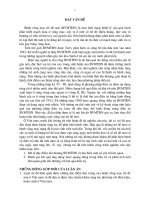
Conjugate Eye Deviation
Receptive Aphasia
22.3%
34.0%
7th Central facial palsy
Hemiparesis
Expressive aphasia
90.3%
97.1%
75.7%
0
20
40
60
80
Chart 3.3. Clinical characteristics at admission
100
11
Common signs such as hemiparesis 97.1%; 7th central facial palsy
90.3%; expressive aphasia accounts for 75.7%. Conjugate eye
deviation accounted for 22.3%.
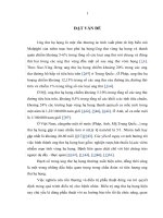
Glassgow
MEAN
20
NIHSS
17.7
17.1
14.6
15
10
11.8
11.2
12.9
12.3
9.3
5
At admission
After
After
At discharge
recanalization recanalization
24 hour
TIME
Chart 3.6. Mean Glasgow and NIHSS change
The mean Glasgow score at admission was 11.4 and at
discharge was 12.8. The mean NIHSS sacle at admission was 17.1
and at discharge was 9.3
3.2.2. Characteristics of computed tomography images
Table 3.11. Characteristics of computed tomography images at
admission
Group
Total
≤ 3 hour
> 3 hour
p
OR
Signs
n=103 % n=103
%
n=103 %
Normal
30
29.1
19
44.2
11
18.3
Ischemic
73
65.9
24
55.8
49
81.7
Cerebral
atrophy
15
14.6
9
20.9
6
Pre-stroke
20
19.4
8
18.6
12
<0.01
3.5
10.0
>0.05
0.4
20.0
>0.05
1.1
With AIS due to LVO of anterior system, CT images normal
was 29.1%. The rate of patients with ischemic after 3 hours (81.7%)
was higher than before 3 hours (55.8%), the difference was
statistically significant with p<0.01, odds ratio OR = 3.5.
12
Table 3.12. Early signs of infarction on non-contrast computed
tomography
≤ 3 hour
Total
Signs
> 3 hour
p
OR
40.0
<0.05
3.4
26
43.3
<0.05
3.3
16.3
36
60.0
<0.01
7.7
1
2.3
12
20.0
<0.05
10.5
24.3
3
7.0
22
36.7
<0.01
7.7
34
33.0
8
18.6
26
43.3
<0.05
3.3
Hyperdense MCA
sign
20
19.4
7
16.3
13
21.7
>0.05
1.42
Hypoattenuation
involving
onethird or more of
the MCA territory
9
8.7
1
2.3
8
13.3
<0.05
6.5
n=103
%
n=103
%
n=103
%
Cortical sulcal
effacement
31
30.1
7
16.3
24
Loss of graywhite matter
differentiation
in the basal
ganglia
34
33.0
8
18.6
Loss of the insular
ribbon
43
41.7
7
Obscuration of the
lentiform nucleus
13
12.7
Obscuration of the
Sylvian fissure
25
Hypoattenuation o
f internal capsule
- Early signs of infarction: cortical sulcal effacement (30.1%), loss of
the insular ribbon (41.7%), obscuration of the lentiform nucleus
13
(12.7%), loss of gray-white matter differentiation in the basal ganglia
(33%) and hyperdense MCA sign (19.4%).
- Most early signs of infarction on non-contrast CT that patients who
came after 3 hours were more significant with p <0.05, with odds
ratio (OR) of 3.3 to 10.5.
Table 3.14. Change ASPECTS on CT scan between before and
after thrombectomy
Time
After 24 hour
Before (n=103)
p
ASPECTS
(n=96)
SD)
7.88 1.76
6.22 2.34
<0.01
0-5 (n,%)
8 (7.8)
38 (39.6)
<0.01
6-7 (n,%)
34 (33.0)
31 (32.3)
0.38
8-10 (n,%)
61 (59.2)
27 (28.1)
<0.01
(
X
- The mean ASPECTS before was 7.88 1.76, after 24 hours of
thrombectomy was 6.222.34, the difference is statistically
significant with p <0.01.
- The grade ASPECTS: before thrombectomy, the rate of patients
with ASPECTS < 6 and > 7 was, the difference has statistical
meaning with p <0.01.
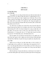
ICA occlusion
M1-MCA occlusion
M2-MCA occlusion
6.8%
42.7%
50.5%
Chart 3.8. Arterial occlusion
M1-MCA accounts for 50.5%, M2 accounts 6.8% and ICA
occlusion 42.7%.
14
Table 3.15. The collateral flow scale on CTA
Collateral flow scale
Score
n=103
%
Good
3
10
9.7
Intermidiate
2
41
39.8
Poor
0 or 1
52
50.5
The collateral flow scale on CTA were mainly poor and no
collateral flow (50.5%), intermidiate (39.8%), good (9.7%)
3.3. The effectiveness and safety of the endovascular mechanical
revascularization to treated acute ischemic strokes due to large
vessel of the anterior cerebral artery system occlusion and
related factors
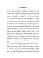
% 100%
100
Before thrombectomy
After thrombectomy
74.8%
80
60
40
17.5%
20
0%
0 1.0%
6.8%
0
0
0
0
TICI 0
TICI 1
TICI 2a
TICI 2b
TICI 3
Chart 3.12. Changes in cerebral perfusion before and after
thrombectomy on digital subtraction angiography
After thrombectomy, TICI 2b was 17.5% and TICI 3 was
74.8%, so that successful revascularization (TICI > 2b) accounts for
92.3% (17.5% + 74.8% ). Only 1 patient (0.8%) failed (TICI 1).
15
%
100%
Before thrombectomy
100
After thrombectomy
71.8%
80
60
40
12.6%
20
0
5.8%
3.9%
0
0
0
MORI O
MORI 1
MORI 2
MORI 3
Chart 3.13. Changes in cerebral perfusion before&after
thrombectomy on CTA
After 24 hour thrombectomy, there are 71.8% good flow (MORI
3) and 12.6% not flow (MORI =0) due to failure or re-occlusion.
mRS 0-2
mRS3
mRS4-5
20.4%
41.7%
mRS6
30.1%
7.8%
%
mRS discharge
16.5% 5.8%
62.1%
15.5%
%
mRS after 90 days
0%
20%
40%
60%
80%
100%
Chart 3.15. Results of treatment by mRS
mRS 0-2 at discharge was 41.7%, after 3 month was 62.1%.
Mortality rate (mRS = 6) at discharge was 7.8%, after 3 month was
15.5%.
16
Table 3.16. Side effects due to contrast
Side effects
n=103
%
Allergic reactions
3
2.9
Acute kidney injury
5
4.8
Allergic reactions (2.9%) and acute kidney injury (4.8%).
Table 3.17. Complications during the thrombectomy
Complication
n=103
%
Cerebral arterial dissection
6
5.8
Embolization to new or target vessel territory
30
29.1
Aspiration again emboli
25
24.3
Arterial occlusions in other locations
5
4.8
Access-site hematoma
5
4.8
Procedural complications: access-site problems (hematoma)
were detected in 5 patients (4.8 %); device-related complications:
cerebral arterial dissection were 5.8% and embolization to new or
target vessel territory were 29.1%, but aspiration again emboli were
24.3%, arterial occlusions in other locations were 4.8%.
Table 3.18. intracerebral hemorrhage after thrombectomy
Intracerebral hemorrhage
n=103
%
HI 1
5
4.8
Non-symptomatic
HI 2
1
1.0
intracerebral
PH 1
9
8.7
hemorrhage
Total
15
14.5
PH 2
4
3.9
Symptomatic
subarachnoid
intracerebral
4
3.9
hemorrhage
hemorrhage
Total
8
7.7
Non-symptomatic intracerebral hemorrhage were 14.5%.
Symptomatic intracerebral hemorrhage include: subarachnoid
hemorrhage (3.9%) and parenchymal hemorrhage typ 2 (PH2 –
3.9%) were 7.7%.
17
Bảng 3.23. Logistic regression analysis of predictors of good
outcome after 3 months
Odds ratio
Confidence
Factors
(OR)
interval (CI) 95%
Age
1.014
0.98 – 1.05
p
>0.05
The onset-to-door time
1
0.99 – 1.01
>0.05
The puncture to recanalization time
Conjugate eye deviation
Glasgow score >8
NIHSS score <15
ASPECTS score >7
3.4
2.79
0.9
1.17
0.97
1.4-8.1
0.14-0.95
0.73-1.30
1.01-1.34
0.71-1.32
0.024
0.04
>0.05
>0.05
>0.05
Good collateral flow on CTA
3.35
0.12-0.72
0.007
Good collateral flow on DSA
0.62
0.27-1.35
>0.05
In multivariate analysis, there were 3 factors: conjugate eye
deviation (OR=2.79; 95% CI: 0.14-0.95; p=0.04); good collateral
flow on CTA (OR = 3.35; 95% CI: 0.12-0.72; p= 0.007); puncture to
recanalization time < 30 minutes (OR=3.4; 95% CI: 1.4-8.1;
p=0.005) were independent predictors of good outcome at 3 months.
Bảng 3.27. Logistic regression analysis of predictors of mortality
after 3 months
Odds ratio
Confidence
Factors
p
(OR)
interval (CI) 95%
Age
0.9
0.9 – 1.0
>0.05
The onset-to-door time
1.1
0.2 – 6.1
>0.05
The puncture to recanalization time
1.2
1.1 – 16.4
>0.05
Conjugate Eye Deviation
Glasgow score < 8
NIHSS score >15
ASPECTS score <7
1.7
2.9
1.1
0.9
0.3 – 7.8
1.4 – 3.9
0.9 – 1.3
0.7 – 1.4
>0.05
0.01
>0.05
>0.05
Poor collateral flow on CTA
2,4
1,6 - 3,1
< 0.05
There were 2 factors: Glasgow score < 8 (OR= 2.9; 95% CI: 1.43.9; p=0.01) and poor collateral flow on CTA (OR=4.2; 95% CI: 1.63.1; p<0.05) were independent predictors of mortality at 3 months.
18
CHAPTER 4 - DISCUSSIONS
4.1. Clinical symptom characteristics
4.1.1. Symptoms at admission
Signs that account for a high percentage such as hemiparesis
(97.1%); 7th central facial palsy (90.3%); expressive aphasia
(75.7%). Conjugate eye deviation (22.3%). Do Duc Thuan et al.
(2017) found that the highest severity of hemiparesis in AIS patients
was highest at 79.24%, followed by language disorder accounting for
75.47% and unconcious disorder rate lower than 35.85%. Peter
Vanacker (2014) found that hemiparesis accounted for the highest
percentage of 96% with 80% before and after the brain.
4.1.2. NIHSS score
When at admission, the mean NIHSS score was 17.6, after the
thrombectomy change not significantly, but after 24 hours of, the
NIHSS score was decrease and when the discharge, the mea NIHSS
score was significantly reduced, it was 9.3. The studies at 103, or 108
military hospitals recorded the mean NIHSS score in the study of 17.
Previous studies about thrombectomy in large vessel occlusion stroke
as REVASCAT, EXTEND-IA trials had the median NIHSS of 17.
4.2. Characteristics of computed tomography images
4.2.1. Characteristics of brain tissue injury
For patients with large vessel occlusion stroke of the anterior
cerebral artery system, no brain tissue injury on CT was 29.1%. The
proportion of patients with brain tissue injury on non-contrast CT
were 65.9%. The patients, who arrived after 3 hours had brain tissue
injury (81.7%) was higher than patients, who arrived before 3 hours
(55.8%), the difference was significant. Statistical meaning with p
<0.01, odds ratio OR = 3.5.
Joanna M. Wardlaw and Orell Mielke had overviewed for 13
years (from 1990 to 2003) from 15 studies with 3468 patients AIS,
who arrived before 6 hours, The sensitivity non-contrast CT was 66%
(20% - 87%), the specificity was 87% (56% -100%).
4.2.2. The early signs of ischemic
19
The early signs of ischemic were detected in 65.9%. Early signs
of infarction: cortical sulcal effacement (30.1%), loss of the insular
ribbon (41.7%), obscuration of the lentiform nucleus (12.7%), loss of
gray-white matter differentiation in the basal ganglia (33%) and
hyperdense MCA sign (19.4%). The most early signs on non-contrast
CT recorded that patients who came after 3 hours were more
detection than before 3 hour, significant with p <0.05, odds ratio
(OR) of 3.3 to 10, 5.
In the study of Mai.D.T, the early signs were: cortical sulcal
effacement (13.89%); cortical sulcal effacement (8.33%); loss of
gray-white matter differentiation in the basal ganglia and loss of the
insular ribbon (5.56%). These signs are much lower than our results
because the author had used non-contrast CT with small number
patients and small vessel occlusion.
4.2.4. ASPECTS score
The mean ASPECTS score was 7.8 ± 1.7. ASPECTS score ≥ 8
was observed to be 59.2%, ASPECTS score 6-7 was 33%. The
difference was statistically significant between patients before and
after 3 hours in the mean ASPECTS and ASPECTS group.
Our results are quite similar to previous trial of N.H. Ngoc at
Hospital 108. The average ASPECTS score is 7.8 ± 1.4, the
percentage of patients with ASPECTS ≥ 8 was observed to be 61.6%.
Large studies on thrombectomy as IMS III selected patients with
ASPECTS ≥ 8 was 56.9% and 59%. REVASCAT trial has a median
ASPECTS score of 7 (6-10), or ESCAPE trial was 9.
4.2.5. Arterial collateral flow
The level of collateral flow on CTA were mainly with poor level
(0 and 1 point) was observed to be 42.7% and intermidiate level
(39.8%). The level of collateral flow on CTA of MR CLEAN trial
was observed: 0 points (5.3%), 1 point (27.5%), 2 point (40.2% accounts for the highest) and 27% of 3 points (good collateral flow).
4.3. The effectiveness and safety of the endovascular mechanical
revascularization to treated acute ischemic strokes due to large
vessel of the anterior cerebral artery system occlusion
20
4.3.1. The effectiveness of treatment
4.3.1.1. Revascularization results
After thrombectomy, the rate of reperfusion were TICI 2b
(17.5%) and TICI 3 (74.8%), successful revascularization was
observed to be 92.3% (17.5% + 74.8% ). Only 1 patient (0.8%) failed
(TICI 1). The successful revascularization result was a high, when
compared to other studies in Vietnam and the world.
4.3.1.2. Clinical outcome
The rate of mRS from 0-2 at discharge was 41.7%, after 3
months was 62.1%. The mortality rate (mRS = 6) at discharge was
7.8%, after 3 months was 15.5%. Our results are similar to that of
Nguyen Quang Anh: 0-2 mRS after 3 months 64.3%, 3-5 mRS
accounts for 21.4%, 14.3% of mortality. Compared to other results
such as N. H. Ngoc and T. L. T. Anh, we have higher neurological
recovery results, but the difference is not statistically significant.
4.3.2. Safety of the method
4.3.2.1. Side effects due to contrast
Side effects related to contrast include allergic reactions (2.9%)
and acute kidney injury (4.8%). Previous studies reported 3.2%
allergic reactions and 5.2% acute kidney injury.
4.3.2.2. Complications during the thrombectomy
Procedural complications: access-site problems (hematoma)
were detected in 5 patients (4.8 %); device-related complications:
cerebral arterial dissection were 5.8% and embolization to new or
target vessel territory were 29.1%, but aspiration again emboli were
24.3%, arterial occlusions in other locations were 4.8%. The review
of studies by Dutch authors in 2017 shows that the rate cerebral
arterial dissection were from 1.5% to 4.3%.
4.3.2.3. The intracerebral hemorrhage after thrombectomy
21
Non-symptomatic intracerebral hemorrhage were 14.5%.
Symptomatic intracerebral hemorrhage include: subarachnoid
hemorrhage (3.9%) and parenchymal hemorrhage typ 2 (PH2 –
3.9%) were 7.7%. According to author N. Q. Anh, the prevalence of
symptomatic intracerebral hemorrhage after thrombectomy accounted
for 13.4%, in SWIFT trial was 9%, in TREVO trial was 7% and in
IMS III trial was 6.2%.
4.3.3. The influences on treatment results
In multivariate analysis, there were 3 factors: conjugate eye
deviation (OR=2.79; 95% CI: 0.14-0.95; p=0.04); good collateral
flow on CTA (OR = 3.35; 95% CI: 0.12-0.72; p= 0.007); puncture to
recanalization time < 30 minutes (OR=3.4; 95% CI: 1.4-8.1;
p=0.005) were independent predictors of good outcome at 3 months.
Spiotta, A.M. found that the thrombectomy time less than 60 minutes
had better neurological outcomes than over 60 minutes. The MR
CLEAN trial concluded that the level of cerebral collateral flow on
CTA was related to good outcome after 3 months, but no cerebral
collateral flow on DSA.
When analyzing multivariate factors of patients to mortality
after 3 months, there were 2 factors: Glasgow score < 8 (OR= 2.9;
95% CI: 1.4-3.9; p=0.01) and poor collateral flow on CTA (OR=4.2;
95% CI: 1.6-3.1; p<0.05) were independent predictors of mortality at
3 months. The study by Korean authors show that age (OR, 1,043;
95% CI, 1,002-1,086; p = 0,041), pre-stroke or transient ischemic
attack (OR, 3,124; 95% CI , 1,340-7,281; p = 0.008) are factors
related to mortality. Our results of the addition of Glasgow below 8
are factors that affect mortality.
22
CONCLUSIONS
1. Clinical characteristics, computed tomography imaging of acute
ischemic stroke due to large vessel of the anterior cerebral artery
system occlusion
- The symptoms onset of acute ischemic stroke were: hemiparesis
97.1%; 7th central facial palsy 90.3%; expressive aphasia accounts
for 75.7%; conjugate eye deviation accounted for 22.3%.
- The mean Glasgow score, NIHSS sacle at admission were 11.4;
17.1 and at discharge were 12.8; 9.3.
- Computed tomography imaging of acute ischemic stroke due to
large vessel of the anterior cerebral artery system occlusion:
+ The signs of ischemic were detected in 65.9%. Early signs
of infarction: cortical sulcal effacement (30.1%), loss of the insular
ribbon (41.7%), obscuration of the lentiform nucleus (12.7%), loss of
gray-white matter differentiation in the basal ganglia (33%) and
hyperdense MCA sign (19.4%).
+ The rate of cerebral artery occlusion: động mạch não giữa
47,9%; M1- middle cerebral artery was 50.5%, M2 accounts 6.8%
and internal carotid artery was 42.7%.
+ The mean ASPECTS was 7.881.76, mostly 8-10 points
accounted 59.2%. The collateral flow scale on computed tomography
angiography were mainly poor and no collateral flow (50.5%),
intermidiate (39.8%), good (9.7%)
2. The effectiveness and safety of the endovascular mechanical
thrombectomy treated acute ischemic strokes due to large vessel
of anterior cerebral occlusion.
2.1. The effectiveness of revascularization
23
The successful revascularization (TICI > 2b) accounts for
92.3%. There are 71.8% good flow (MORI 3) on computed
tomography angiography
2.2. The clinical effectiveness: The good function rate, mortality rate
after 3 months were 62.1%; 15.5%
2.3. The safety of the endovascular mechanical thrombectomy
- Side effects due to contrast: Allergic reactions was 2.9% and acute
kidney injury was 4.8%.
- Procedural complications include: access-site problems (hematoma)
were detected in 5 patients (4.8 %); device-related complications:
Cerebral arterial dissection were 5.8% and embolization to new or
target vessel territory were 29.1%, but aspiration again emboli were
24.3%, arterial occlusions in other locations were 4.8%.
- Non-symptomatic intracerebral hemorrhage were 14.5%.
Symptomatic intracerebral hemorrhage include: subarachnoid
hemorrhage (3.9%) and parenchymal hemorrhage typ 2 (PH2 –
3.9%) were 7.7%.
2.4. The factors influence on treatment results.
- There were 3 factors: conjugate eye Deviation, good collateral flow
on computed tomography angiography and the puncture to
recanalization time < 30 minutes were independent predictors of
good outcome at 3 months.
- There were 2 factors: Glasgow score < 8 and poor collateral flow on
computed tomography angiography were independent predictors of
mortality at 3 months