Nghiên cứu đánh giá kết quả phẫu thuật thay van hai lá bằng van cơ học st jude tại bệnh viện trung ương quân đội 108 tt tiếng anh
Bạn đang xem bản rút gọn của tài liệu. Xem và tải ngay bản đầy đủ của tài liệu tại đây (231.36 KB, 24 trang )
1
INTRODUCTION
Mitral valve disease is the most common valvular heart disease
in our country, mainly due to rheumatic heart disease. Most patients
with this valvular heart disease will get heart failure at labor age and
become a burden for families and society. The appearance of the
extracorporeal circulation machine in 1953 allows safe access and
regeneration of heart chambers while cardiovascular surgery has been
strongly developed, along with the design and improvement of
prosthetic cardiac valve. The first valve generation was Starr - Edwards
(1961); the single-wing disc valve (1969), double-wing disc valve
(1977), bio-artificial valves were also developed. In our country in
1958, Professor Ton That Tung made a replacement of mitral valve and
since 1971, there have been many domestic cardiac surgery centers
where almost the cardiac surgeries have been carried out.
The St.Jude mechanical prosthetic heart valves has been still
commonly used thanks to high strength and good hemodynamics.
However, improvements in mechanical valves, technical changes and
differences in disease structure, management organization, care, surgery
and postoperative follow-up in each hospital have not been fully
studied. 108 Military Central Hospital is the first unit in the Army to
perform cardiac surgery, therefore, it is very necessary to make such
evaluation. For the above reasons, we carry out the topic with following
objectives:
1. Commenting on clinical and subclinical characteristics of
patients under the surgery of mitral valve replacement with St.Jude
mechanical valve.
2. Evaluating the surgical results for replacement of mitral
valve with St.Jude mechanical valve at 108 Military Central Hospital
CONTRIBUTIONS OF THE THESIS
- The findings enrich the study results on clinical and hemodynamic
characteristics, contribute to the application into clinical cardiovascular
practice in and out of the Army.
2
- The study has provided basic information about the surgical results
for mitral valve replacement with St.Jude mechanical valve at the 108
Hospital - the Army’s first hospital that performs cardiac surgery as a
basis for evaluating the efficiency of the procedures of preparations for
surgery, resuscitation and post-operative follow-up at the hospital.
THESIS STRUCTURE
The thesis consists of 122 pages (excluding appendices and
references), including 4 main chapters: Introduction: 2 pages, chapter 1
– Overview: 33 pages, chapter 2 - Objects and study methodology: 21
pages, chapter 3 – Findings: 29 pages, chapter 4 – Discussion: 34 pages,
conclusions and recommendations: 3 pages. The thesis has 50 tables, 20
charts, 18 illustrations, 1 diagram, 146 references, including 37
Vietnamese documents and 109 English documents.
CHAPTER 1: LITERATURA REVIEW
1.1. Applied surgical anatomy of mitral valve
The mitral valve (MV) is also called the left atrioventricular valve,
consisting of valve rings, valve leaflet, ligaments, and muscle chains:
- The valve-atrial junction: the location where the valve leaflet is
connected with the left atrium. In surgery, it is based on the difference
in color between the left atrium - LA (light pink) and valve leaflets
(light yellow) to locate valve position. The anterior valve ring is very
thick and firm, which is the insertion for the anterior leaflet of mitral
valve, however the contiguous area between mitral valve and the aorta
valve has almost no valve rings. The posterior valve ring is the location
to which the posterior leaflet of MV adheres and it is a weak and easily
relaxed position in MV cases. In MV surgery, it should be noted: (1)
The circumflex artery: running between the background of the left
auricle and the anterior valve edge for about 3-4 mm from the joint of
the valve - atrium, then running away from the posterior valve ring. (2)
Coronary sinuses: running along the posterior valve ring, initially
outside the artery, then crossing the artery to go inside, about 5mm from
the valve ring. (3) His bundle: located near the right fiber triangle (4)
The non-coronary leaflets and left coronary leaflets of aortic valve
3
(AV): closely related to the background of anterior leaflets of mitral
valve before; the root of these valves is 6 – 10 mm from the VHL ring.
- Valve leaflets: the anterior leaflet is big while the posterior leaflet
is small. Carpentier divided each valve leaflet into three sections, which
is very meaningful in surgery.
- Valve suspension system: muscle chain with shrinkage function
and ligaments with elastic properties to hold the valve leaflets and
ventricle's contraction function. Especially when cutting mitral valve, it
should be noted to avoid excessive pulling, which causes cracking of
the papillary muscles, which can cause ventricle type 2 (according to
Treasure’s classification).
- Ligament system: including anterior leaflet ligament and posterior
leaflet ligament. One point should be noted is that the anterior leaflet
ligament cling to the rough area, the background of the posterior leaflet,
therefore, it should be noted for preservation in mitral valve surgery.
1.2. Diagnosis of mitral valve disease
- Causes: mainly due to rheumatic heart disease (RF); the frequency
of RF and heart valve disease due to RF at the rate of 0.2 to 18.6 ‰ in
the number of schooling children.
- Clinical developments: Moderate double-leaflet stenosis usually
does not show symptoms for about 10 years. The first and common
symptoms are difficult breathing, then right heart failure. Listening to
the heart: T1 sound is strong at the promontory; diastole vibrates; if the
pressure of pulmonary artery (PAP) increases from moderate to high
level, T2 sound seems strong and split, anterior systolic murmurs, clắc
sounds, Graham-steel murmurs, Untreated double-leaflet complications:
60-70% pulmonary edema, 20-30% systemic embolism, 10%
pulmonary infarction, 1-5% infection. ECG: P wave is wide with 2
peaks, cardiopulmonary x-ray: See big heart; echocardiography is a
basic test to diagnose and assess the severity of the disease.
Mitral incompetence (MI) is divided into acute MI and chronic MI
with causes, clinical course and different settlement methods. Acute MI:
usually due to ligament rupture or post-traumatic papillary muscle,
infection, infection, anemia or idiopathic situation, clinical expressions
4
are severe due to sudden ventricle failure; the mortality rate is 6.3% per
year. Chronic MI: Usually not showing functional symptoms, heart
failure after 6-10 years, therefore, when there are functional symptoms,
the ventricle dysfunction has not recovered, increasing complications
such as heart failure, death. Determining the MI mechanism is very
important: If the central MI line accompanied by a normal MV
structure, it shows that the mechanical MI flow is often due to the
dilatation in valve ring, ventricle or the limitation in movement of
posterior leaflet of MV due to dyskinesia in the ventricle in. patients
with CAD and if MI line has the eccentricity accompanied with
abnormal MV structure show an open line due to physical injury. Other
authors: open line width through openings is a reliable indicator; If this
indicator is ≥ 0.7 cm in width, it is evidence of severe MI.
1.3. History of development of mechanical mitral valve and St.Jude valve
1.3.1. Classification, some prosthetic heart valve indicators
Prosthetic heart valves include mechanical and biological valves.
The area indicators of artificial valves include: GOA, COA, EOA in
which EOA is most concerned because this is the amount of blood
through the valv ; This indicator is usually taken over m² of body skin
area (IEOA). For the MV, this indicator is 1.2 cm²/m²BSA and the
inappropriate valve will be replaced if IEOA is < 0.9 cm² / m².
1.3.2 St.Jude prosthetic mechanical valve
It is the preferred double-wing valve in North America, Europe and
our country has the superiority in durability, large EOA, minimum flow
resistance and few side effects. Teh structure include: Valve frame:
Graphite and carbon coated. Valve wing: Semicircle and straight, made
of graphite mixed with tungsten for optical resistance and also covered.
The valve wing coupling protrudes from the valve rin, therefore, it is
convenient to select in small ventricle cases while still ensuring IEOA
index. Valve rings: very thick and firm. When it is open: creating an
opening angle of 85o (central), therefore, the EOA is big and
significantly reduces eddy current and diastolic pressure difference
through the valve. When it closes: creating a closed angle of 30-35º
compared to the valve ring plane.
5
1.3.3. The determinants of choosing the prosthetic heart valves instead of MV
Selection of prosthetic heart valves needs to ensure the factors:
good hemodynamics, high durability, avoiding the risk of thrombosis
and convenience for surgeons. Age is the first factor to select biological
valves or mechanical valves, compliance with the use of anticoagulants,
size and damages to valve rings.
1.4 Current mechanical mitral valve replacement surgery
1.4.1. Recommendations and indications for treatment of MV disease
Cardiovascular associations offer 3 types of promotion: Type I
recommendation: should be implemented because the benefit is much
higher than the risk (proven evidence). Type II recommendation: Type
IIa should be implemented because the benefit is higher than risk,
however, more researches are needed for affirmation (evidence proved
from a multi-center study but limited, with or without randomized test).
Type IIb should be considered because benefits may be higher than risk,
however, the big multi-center studies are needed. (Evidence has been
proven but the level is still very limited, only in small studies or expert
opinions). Type III recommendation: should not be implemented
because the risk of more benefits.
Indications is based on ACC / AHA recommendations.
1.4.2. Some new methods of mechanical mitral valve replacement
Endoscopic surgery for mitral valve replacement, transcutaneous
mitral valve replacement and robot surgery have been studied and
developed with modern equipment will be a gradual trend to replace
open heart surgery. Short-term results well improves in hemodynamics,
clinical aspects and long-term results are still under investigation and no
relevant published results have been published. However, a research
report at 32 centers: premature death rate (30 days): 12.5%, ventricle
entry blockage: for transcutaneous mitral valve replacement: 9.3% .
1.4.3. Complications and treatment follow-up after surgery
- Complications related to open heart surgery (related to CEC,
bleeding, bacterial infection) and related to mechanical MV (ventricle
rupture, coronary injury, obstruction of ventricle exit line, valve edge
opening, embolism, antifreeze aspect, etc). Control of complications
6
depends on the experience, skills of the surgeon, the professionalism of
nurses and management coordination, periodic folow-up and
examination, anti-coagulation compliance.
- Long-term monitoring and treatment for the patient with
mechanical valve: good coordination is needed between patients and
physicians such as regular examination; nutrient-rich diet; regime of
physical activity, proper working and strict adherence to anticoagulant
medicines, prophylaxis, heart rate control and Dressler syndrome, etc.
1.5. Research results of mechanical mitral valve replacement
- Mortality rate: author Ikonomidis J.S has made the St.Jude mitral
valve replacement at 5.3% for 20 years, 1.3%. by Kandemir O. for the
first 30 days. Mortality rate is closely related to preoperative cardiac
function, age, and coronary disease and may be related to the
preservation technique of leaf ligament after MV because when the
whole apparatus is removed below the valve, the rate of the ventricle
rupture is 14% (1990).
- Other complications have a low rate; Emery states that:
thrombosis rate is 0.5%, infective endocarditis is 0.2% and pannus is
0%. Author Ikonomidis J.S. have treated 359 patients over the past 20
years: Valve-related mortality rate of 0.2%, embolism of 3%,
hemorrhage of 2% and infective endocarditis of 2%. Similarly, Remadi
J., over the past 19 years of treatment, thrombosis is 0.69%/ year,
hemorrhage is 0.2%/year. Author Do Kim Que did not see hemorrhage
or heart valve thrombosis.
1.5.2. Postoperative hemodynamic after mechanical MV replacement
There are many studies on teh eraly hemodynamic changes after
mechanical MV replacement, however, there are very few studies on
clinical and hemodynamic changes in the medium and long term: Saad
Bader has shown a significant improvement in NYHA levels, RN rate,
NT size; the decerasing PAP is very meaningful at 6 months after
surgery. The longest study by Emery R.W. during 25 years (1,498
patients): SJM valve brings the best hemodynamic results, improves the
clinical condition and patient's quality of life. Author Nguyen Duc Hien:
Heart failure condition and PAP improve well, which is similar to the
7
comments of author Rodrigues A.J. Author Do Kim Que, after 2 years
of study, shows that St.Jude MV works well, which is similar to teh
study results by Remadi J.P over 440 patients and author Emery R.W.
has not recognized any mechanical damage of St.Jude valve for 25
years of research. Author Ikonomidis J.S shows that the SJM
mechanical valve is a reliable and durable mechanical valve.
Many domestic authors have studied the post-operative results
to replace the prosthetic mitral valve. However, these results are still not
homogeneous in terms of time; follow-up time is not long, and a followup and evaluation procedure needs to be more systematic. In addition,
there are different characteristics such as: external apperance of disease,
treatment according to route (Army and civic scope), difference in
cardiac surgery and resuscitation, etc and structural characteristics of
St .Jude valve with the surgeon's skills in surgery; the studies evaluating
the results will be necessary to help evaluate the process, build up the
team and serve the Army and the people.
CHAPTER 2: SUBJECTS AND STUDY METHODS
2.1. Study object
2.1.1. Criteria for selecting patients
All patients have St.Jude mechanical MV replaced with or without
VBL shaping at 108 Hospital from 05/2010 to 12/2014. Standard for
patient grouping: MV is main, MI ≤ 1/4, called MS group; simple MI;
The coordination between MS and MI /4 2/4, is called MSI group.
2.1.2. Exclusion criteria
+ The patients that have MV replaced with St.Jude mechanical
valve but with other diseases such aortic valve disease under the
requirement of surgery treatment or congenital heart disease.
+ Patients with MI after myocardial infarction.
+ Patients who do not have sufficient information in the record or
do not agree to participate in the study.
2.2. Study Methods
2.2.1. Study design: Study, describe series of cases, intervention,
follow-up and non-control.
2.2.2. Study criteria:
8
- Pre-operative study criteria: General characteristics such as age
(> 60 and ≤ 60 years); urban and countryside areas; RF (according to
Jones 1988 criteria); prehistory of MV intervention (MV dilatation,
closed heart surgery); ASA incision infection risk scale; Favorable
prognosis for placing Mallampati intubation and prognosis of mortality
rate in heart surgery through Euro Score scale. Clinical characteristics:
Degree of heart failure according to NYHA. Subclinical characteristics:
ECG: (sinus rhythm-SR, atrial fibrillation-AF), echocardiography: LA
diameter (measured on TM type ultrasound from the section along the
axis of the standard chest, through the orgin of o the aorta when the
aortic valve closes, normally: 28 ± 4mm), Dd, Ds, PAPs (PAPs = Gmax
(HoBL) + 10(mmHg), other vulnerability levels: MS, MI, tricuspid
incompetence (based on 2008 standard of the Vietnam National Heart
Association) Wilkins scale and evaluation of the prosthetic valve
operation is based on the American Society of echocardiography.
Table 2.1: Function parameters of prosthetic mitral valve
Parameters
Normal
Possible stenosis Severe stenosis suggestion
Gmean (mmHg)
≤5
5 – 10
> 10
2
MVA (cm )
≥2
1–2
<1
PHT (ms)
< 130
130 – 200
> 200
- Surgical procedure and research criteria:
+ Pre-operative preparations: Within 01 day before surgery, it is
necessary to guide for disinfection bath with Chlorhexidine 4% solution
and use sterile clothes of hospital, abstain from food for 12 hours before
surgery, enemas and stop anticoagulants
+ Step 1: Open the chest: Open into the mediastinum along the
vertical line between the breastbone, open the pericardium, reveal the
heart. Anticoagulation with systemic Heparin (3 mg/kg body weight)
reaches the ACT > 400 'standard to run the extracorporeal circulation
machine, tighten the cardinal veins, repair the aorta. Transmission of
cardiac paralysis solution through the original of aorta
* Regarding on the domestic studt criteria, this surgery include:
protecting heart muscle hypothermia with cold crystal solution or not
making hypothermia with warm blood.
9
+ Step 2: Approaching to MV: Path of opening classic atrial and
RA - septum atriorum- LA.
* The study criteria include: incision, LA thrombosis, left auricle,
and MV sytem damage.
+ Step 3: Replace MV with St. Jude: Cutting off the MV,
preserving part or all of the MV apparatus, posterior leaflet and
ligament of posterior leaflet. Hospital 108 uses St. Jude valve, type of
Saint Jude Masters introduced in 1995 with valve sizes of 27, 29, 31.
Measuring artificial valve size with a specialized rule set of Saint Jude
firm. Sew to fix the St. Jude valve ring with MV ring with U-shape
sutures, checking the tightness between artificial valve ring and mitral
valve ring, seam tightness and operation of artificial St. Jude valve
wing. Combination surgery: repair TV
* The research criteria in this surgery include: (1) Preserving the
posterior leaflet, preserving the ligament of posterior leaflet: yes or no.
(2) Size of St. Jude artificial two-leaf valve used. (3) Correction of TV:
correction method. (4) The result of heartbeat beating again after
clamping the aorta: natural, electric shock, Pace machine
+ Step 4: Heart beats again: Close the heart opening lines (closing
LA and RA). Drawing canuyn of veins, aorta, drawing the left heart
drain to neutralize heparin with protamin sulfate.
* The research criteria in this surgery include: (1) Heart beats again
naturally, supports Pace or electric shock (2) Heart rate when beating:
sinus or AF. (3) Time for circulation outside the body (minutes): from
the time of starting the machine to stopping the machine. (4) Aortic
clamping time (minutes): from the start of clamping aorta to the time of
removing the aortic clamp. (5) Number and location of drains: on the
heart, behind the sternum, pleural cavity.
+ Step 5: Close the incision: Close the heart membrane, sternum.
* Research criteria include: (1) Time of surgery (minutes): starting
from the incision to the end of skin suture.
- Monitoring process and early evaluation criteria after surgery:
+ Treating according to cardiovascular resuscitation regimen,
tracking live functions on Monitor Philips and recording monitoring
sheet) with indexes: blood gas, electrolytes, liver and kidney function,
blood formula... immediately after returning to the recovery room for
10
15-30 minutes. Anticoagulant (heparin) immediately after 6-8 hours
after surgery if the risk of bleeding is over. Combining heparin with
vitamin K resistance in the first 1-3 days after surgery, then maintaining
vitamin K and adjusting dose according to INR, TP, APTT test results
* Research targets: (1) vasomotor (dose); (2) Time of mechanical
ventilation (hours): Calculated from the beginning of the use of the
ventilator until the removal of the ventilator: <12 hours; 12-24 hours; >
24 hours. (3) Active resuscitation period (days): from the time when the
patient is moved to recovery room until returned to treatment ward (<5
days; 5 - 7 days; > 7 days)
(4) Complications: Early death after surgery; Repeat
operation due to bleeding, bleeding location: When blood flow through
chest drain > 5ml/kg/ hour lasts for the first 3 hours after surgery; Low
cardiac output syndrome (LCOS); Infectious complications: pulmonary
infection due to mechanical ventilation often appear after 48 hours and
based on the standards of the American Thoracic Society;
Complications related to artificial mitral valve: valve blockage due to
thrombosis (according to ACC/AHA guidelines), disproportionate
between artificial valve size compared to patients (IEOA < 0.9cm²/m²
BSA) , hemolysis (appear jaundice - mucosa, dark urine and reduced
red blood cell and Hb, patients without a history of hemolysis, broken
ventricles...; Complications related to surgery: coronary artery
lesions...; Temporary oliguria: urine < 1ml/kg/hour, respond well to
high-dose diuretics, stable hemodynamic patients and no pre-operative
renal disease history; Renal failure requiring dialysis after surgery:
determined by biochemical index: blood urea above 8.3mmol/l; Blood
creatinine above 115 mmol/l. Internal treatment, peritoneal dialysis or
hemodialysis. Liver failure after surgery: determined if after surgery,
there is a sign of jaundice, rapid increase mucosa, dark urine and
hepatic cerebral syndrome division of I to IV) and high liver enzymes:
GOT, GPT, Bilirubin, time Prothrombin prolonged ≥ 1,5;
Gastrointestinal bleeding after surgery: is determined if there is blood
clots, black and pasty feces with ill-smell in the vomitting fluid and
anaemia. In addition, complications of pleural effusion, pleural cavity;
11
complications of pericardial effusion; Acute heart failure, acute cardiac
tamponade (BECK trisomy) was also monitored during this period.
- Indicators and evaluation results after surgery: Patients were
instructed to monitor and treat after surgery according to a unified
procedure at 108 Hospital, including: Re-examination time: usually recheck on Friday every week as recorded in the examination book every
week of the first month after surgery, every 3 months of the following
months and every 6 months of the following years. The examination
and subclinical content needed to be recorded in the monitoring book:
(1) Symptoms: dyspnea, chest pain, arrhythmia, discomfort due to
mechanical valve noise, symptoms of right heart failure, hemorrhage,
embolism; (2) Degree graduation of heart failure according to NYHA;
(3) Status of sternum incision; (4) Subclinical: INR test.
Electrocardiogram (determination of arrhythmia: sinus or non-sinus)
and cardiopulmonary x-ray (with manifestations of lung disease such as
inflammation, pleural effusion ...) and cardiac Doppler ultrasound:
indicated at least once a year. The indicators to focus on are MVA, NT,
Dd, Ds, PAPs, pressure difference through St. Jude MV and the degree
of TV opening. Other tests such as biochemistry, hematology
(designated at least 01 time/year). (5) Complications after surgery.
* Research targets include: (1) Heart rate at the time of follow-up:
heart rate at times through heart examination and hearing, suspected
cases of arrhythmia will conduct an ECG to determine: SR; AF and
other rhythms. (2) Clinical symptoms: heart failure degree graduation
according to NYHA. (3) Transthoracic Echocardiogram: working status
of St. Jude valve; Ventricular size (Dd, Ds); Ventricular function (EF%
index); Pressure is different through St. Jude mechanical MV; Pressure
on systolic right artery; Status of TV open; Area of St. Jude. (4)
Postoperative drug use status: anticoagulants, cardiac drugs, diuretic,
beta blockers, tranquillizer, antihypertensive...). (5) Complications at
the time of follow-up after surgery: Complications due to the use of
anticoagulants: cerebral hemorrhage; gastrointestinal bleeding;
subcutaneous, external meatus hemorrhage or tooth root bleeding.
Complications from thrombosis: cerebral infarction stroke; Peripheral
vascular occlusion complications, artificial valve thrombosis (abnormal
dyspnea, unclear mechanical heart valve, echocardiography found the
movement of leaflet is restricted). Complications related to artificial
12
valves: mechanical failure; hemolysis; disproportionate mechanical
valve and heart (IEOA < 0.9cm²). Other complications: infection;
hemodynamic disorders, conduction; bacterial endocarditis; pannus.
+ Evaluation on artificial MV echocardiography is unusual if: The
valve wing restricts movement or uneven opening - closing; Appeared
edge valve; Differential pressure through valves > 10mmHg; Valve hole
area < 1.8cm²; Some other indicators: top speed of blood flow through
the valve > 2.5msec; Part-time pressure (PHT) > 18msec (if available).
+ Operation of artificial MV: Good: Valve is in the right position and
operates normally (with clinical and subclinical parameters within normal
limits). Not good: The movement of the two leaflets is restricted.
+ Artificial valve thrombosis (according of AHA/ACC):
2.3. Data analysis
Data collection is done in a unified form, entered and analyzed by SPSS
16.0 software to calculate parameters: average, variance, standard deviation.
CHAPTER 3: RESEARCH RESULTS
3.1. General characteristics of the research group
- Average age: 48.1 ± 9.2 years old, male: 47.0 ± 10.5 years old and
female: 48.7 ± 8.3 years old. Female is more than male, female/male =
1.7. Rural area is more than urban, a history of low heart rate accounts
for 68.9%, of which 33.6% is treated, 16.4% of patients is conducted
percutaneous mitral valve dilatation, 5.7% of closed heart surgery
- Mallampati I index mainly with 92%, without Mallampati IV. The
risk of high incision infection: ASA 3 is 92.6% and ASA 4 is 2.5%.
Stratified risk of death after surgery 1% -2% accounts for 77.1%.
3.2. Clinical and subclinical characteristics
- Clinical characteristics: Heart failure level according to NYHA:
NYHA III accounts for 34.4%, NYHA II accounts for 65.6%. The first
and common symptoms is dyspnea (96.7%), chest pain (92.6%), right
heart failure (44.3%)
- Subclinical features: On the ECG, AF (65.6%), SR (34.4%). X-ray
images: heart-chest index (> 65%) accounts for 82.8%. 4.1% of patients
have pleural effusion.
Results on echocardiography: MV disease: Mainly with MS,
accounting for 81.2%, 11 (9%) patients with MI and 13 (10.7%) with
MS.
13
+ Image of MV lesions: valve edge: thick and sticky (92.6%),
calcified (33.9%), and defective (0.8%). Ligaments: thick (87.6%),
contracted (81.1%), broken (2.5%). MV: thick and sticky (95.9%),
calcified (74.6%), contracted (69.7%), valve prolapse (4.1%), swell
with pustules (1.6%).
+ Some ultrasonic indicators:
Table 3.1. Preoperative echocardiographic indicators (n = 122)
Characteristics
Min Max
Average
Left atrial diameter (NT) (mm)
Left ventricular end diastolic dimension (Dd)
(mm)
Left ventricular diameter systole (Ds) (mm)
Right arterial pressure systole (PAPs) (mmHg)
EF index (%)
Right ventricle (TP) (mm)
28
31
83
92
53.7 ± 10.6
49.3 ± 9.4
24
58
32.9 ± 6.7
20
27
18
102
79
40
45.8 ± 15.9
60.1 ± 9.8
21.5 ± 4.6
Ultrasound results: dilated left atrium, lightly dilated left ventricle.
The left ventricular systolic function preserved with EF (%) is 60.1 ±
9.8. Pressure of systolic right artery increase moderately and severely.
+ Wilkins lesions: The valve shape is preserved with Wilkins 8-10
points, accounting for 86.5%.
+ Average difference in MV: The difference in MV pressure >
10mmHg, accounting for 93.7%.
+ TV injury: Medium-severe TV lesions in MS account for 51.6%.
The table above shows that no incompetence and light incompetence of
TV accounts for 48.4%, major tricuspid incompetence combined with
the MS - incompetence: 80.3%.
+ Some other lesions: LA and LA appendage thrombus (31.9%),
moderate PAPs (23%), severe (13.1%), aortic valve opening 2/4 (5.6%).
3.3. Results of surgical parameters
- Protection of heart muscle in surgery: hypothermia 32-33ºC with
cold crystal solution (50.8%) and warm blood solution (49.2%).
- Suitable mechanical MV replacement when IEOA> 0.9cm²/m²
BSA (normally 1.2cm/m² BSA), the appropriate valve size ratio is
100%.
14
Table 3.2. Use of St. Jude mechanical mitral valve size (n = 122)
Size of St. Jude valve
n
Ratio (%)
27
3
2.5
29
63
51.6
31
56
45.9
Total
122
100
Table 3.3. Appropriate ratio of valve size with body skin area (n = 122)
Size of St.
Jude valve
Average skin
area (m²)
Theoretic
EOA (cm²)
IEOA
(cm²/m²)
Appropriate
ratio n (%)
27
1.42 ± 0.10
2.43 ± 0.63 1.71 ± 0.23
3 (100)
29
1.47 ± 0.17
2.66 ± 0.26 1.80 ± 0.15
63 (100)
31
1.50 ± 0.14
3.08 ± 1.09 2.50 ± 0.78
56 (100)
- The shortest time of aortic clamping is 30 minutes, the longest
time is 180 minutes and the average time is 60.3 ± 26.5 minutes. The
circulation time is 95.6 ± 32.1 minutes.
- The incision line approaches the MV: Atrial drain - LA (91.8%),
RA - atrial septum - LA (8.2%).
- LA thrombosis: ratio (18%), lower than preoperative ultrasound.
- Preserving part or all of the posterior leaflet and ligament of
posterior leaflet: 88.5%.
- TV formation: 108 Hospital mainly uses PTFE strip in forming
TV, accounting for 76.0% (57/75), uses autologous cardiac strip
accounting for 14.7% (11/75). No TV formation accounts for 38.5%.
- Heart rate: heart self-bear again (97.5%), SR (60.7%), comparison
of rhythm with before cardioversion surgery with significance.
- Comparison of ultrasound results: valve damage, thrombosis, TV
found that the difference was not significant (p > 0.05).
3.4. Early results after surgery of St. Jude mitral valve replacement
- Results of heart rate: Comparison of preoperative heart rate with
postoperative and early postoperative periods: There is an increase in
cardioversion, significant differences with (p < 0.05). SR (45.9%).
- Hemodynamic status: assessed on sinus heart rate < 100ck/min
and maximum blood pressure > 100mmHg with vasomotor in the
unstable group 100% with combination of 2 drugs, hemodynamic
stabilization after surgery with 25 % unused, light dose (41%).
15
- Mechanical ventilation time is recommended < 12 hours (86.9%).
Resuscitation period < 5 days (76.2%).
- The level of heart failure according to NYHA: Before heart failure
surgery class II and III is 100%, monitoring the early results the
patient's effort capacity is significantly improved p < 0.05, NYHA
transferred to class I is 80.3%.
Table 3.4. Early Doppler echocardiography results after surgery (n = 122)
Echocardiography index
Min Max
Average
Left atrial diameter (mm)
20
77
43.4 ± 9.4
Left ventricular end diastolic dimension(mm) 31
93
47.5 ± 8.2
Left ventricular diameter systole (mm)
20
61
32.6 ± 7.3
Right arterial pressure systole (mmHg)
15
69
32.8 ± 9.3
EF index (%)
28
81
63.8 ± 5.6
TP (mm)
15
34
20.9 ± 3.3
LA is dilated, sizes of LV and ventricle vary slightly. RA pressure
increase slightly and LV systolic function is preserved. Difference of
LA size, right arterial pressure systole with p < 0.05. EF index increases
after surgery with p > 0.05. Dd and Ds indexes are not different (p >
0.05) when compared with preoperative echocardiography.
- Early tricuspid incompetence after surgery: 76.0% (57/75)
forming TV with PTFE strip, moderate and severe incompetence after
surgery accounts for 9.3% (7/75). 38.5% of patients did not repair
tricuspid valves in St. Jude mechanical MV replacement surgery,
moderate and severe tricuspid valve incompetence accounts for 10.6%.
- Average difference through St. Jude 6.3 ± 3.4mmHg, 5-10 mmHg
accounts for 87.7%.
- The rate of early complications is low with 4.1% of bleeding,
2.5% of valve edge openness, 2.5% of bacterial contamination, 9.8%
of heart failure, 3.3% of multiple organ failure.
- The re-surgery rate is 4.9%, mainly sternal bleeding, 01 case of
bleeding in the left atrial appendage, 01 case of placing drain in the RV.
3.5. Results of periodic follow-up (54 months)
The patient is made an appointment for examination as scheduled in
the patient's monitoring book every Friday, the shortest follow-up time
is 7 months and the longest is 54 months.
16
- Heart rate at the time of follow-up: SR increased slightly in the
first 24 months after surgery: 6 months (44.6%), 12 months (45.5%), 24
months (47 %) and the rate of reduction or instability at the time of
follow-up is longer, but the difference was not statistically significant
(p> 0.05). In contrast, the AF rate decreases after surgery and increases
the rate or instability of follow-up from the second year after surgery.
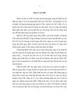
- The level of heart failure according to NYHA:
Figure 3.1. Degree graduation of heart failure according to NYHA
Before surgery, 100% of NYHA II and III is converted to NYHA 0
and I, after surgery > 64%, NYHA II rate is increased from the 24 th.
- SJM's activities: SJM is in the right position and operates with
normal parameters, valve edge is not open, the pressure difference
through the valve St. Jude is good and normal right arterial pressure is
rated as good and accounts for 100%.
- The operation sound of St. Jude mechanical mitral valve: pleasant
feeling, not affecting sleep of patients, accounting for 99.2%
- Echocardiography at different times
Table 3.5a. Doppler echocardiography index at different times after surgery
Echo
-cardiography
index
NT (mm)
Dd (mm)
Ds (mm)
PAPs (mmHg)
EF (%)
TP (mm)
Before
surgery
53.7 ± 10.6
49.3 ± 9.4
32.9 ± 6.7
45.8 ± 15.9
60.1 ± 9.8
21.4 ± 4.8
Average value
1 month 6 months 12 months
n = 122
n = 121 n = 121
41.8 ± 8.6 40.2 ± 8.1 39.6 ± 7.2
46.6 ± 6.9 45.7 ± 6.0 45.3 ± 6.8
31.2 ± 5.8 29.3 ± 5.7 29.1 ± 5.3
31.5 ± 6.4 29.5 ± 4.9 29.1 ± 4.8
62.2 ± 7.9 63.9 ± 8.4 64.9 ± 7.1
20.6 ± 2.3 20.9 ± 4.4 20.9 ± 4.3
24 months
n = 115
38.2 ± 7.3
45.9 ± 6.3
29.2 ± 5.3
29.5 ± 4.8
64.3 ± 9.2
21.3 ± 6.8
LA size; Dd; Ds; PAPs decreases gradually over time in 24 months.
17
Table 3.5b. Doppler echocardiography index at different times after surgery
EchoAverage value
cardiography
36 months
48 months 54 months
Before
index
surgery
n = 82
n = 34
n = 19
NT (mm)
53.7 ± 10.6
37.8 ± 5.6
39.9 ± 5.8
39.3 ± 4.9
Dd (mm)
49.3 ± 9.4
45.8 ± 5.7
46.8 ± 7.5
46.6 ± 8.8
Ds (mm)
32.9 ± 6.7
28.3 ± 4.5
28.5 ± 4.9
28.8± 5.2
PAPs (mmHg) 45.8 ± 15.9
29.6 ± 4.9
31.0 ± 8.2
29.7 ± 3.4
EF (%)
60.1 ± 9.8
66.4 ± 7.5
65.3 ± 6.2
65.2 ± 4.7
TP (mm)
21.4 ± 4.8
21.6 ± 6.5
21.5± 4.4
20.7 ± 1.1
Average echocardiographic index: LA size; Dd; Ds and PAPs
increase gradually from the 36th to the 54th month, EF% index is stable.
Differential pressure across St. Jude mechanical mitral valve:
1 MONTH
6 MONTHS
12 MONTHS
24 MONTHS
36 MONTHS
48 MONTHS
54 MONTHS
Chart 3.2. The level of differential pressure across St. Jude artificial valve
Differential pressure through St. Jude artificial valve mainly from 5
- 10mmHg accounts for > 89% at the time of monitoring.
- Moderate - severe tricuspid valve incompetence after surgery:
Ratio of tricuspid incompetence class II and III
1 month
6 months 12 months 24 months 36 months 48 months 54 months
____ Group with tricuspid valve repair
____ Group without tricuspid valve repair
Chart 3.3. The level of tricuspid incompetence at the time of follow-up
18
The group that did not repair the TV had an increase in tricuspid
incompetence, it’s different when compared with the group with TV
repair at > 24 months (p < 0.05).
- Complications at the times of follow-up: mild hemorrhage of 1.6%
-5.3% at the time of follow-up, embolism at 12 months (0.8%), artificial
valve thrombosis from 0.9% -6.1% limits the valve wing operation from
2.4% -6.1%. Artificial valve edge incompetence appears 24 months
after surgery at a rate of 0.9% -3.4%.
CHAPTER 4: DISCUSSION
4.1. General characteristics of the research team
Age average: 48.1 ± 9.2, this is the age for performing the main
work of the human and society, so if the St.Jude mechanical MV
replacement surgery has good results, it will help patients improve
symptoms and enhance the quality of life.
Gender and geography: the ratio of women/men is 1.7 according to
WHO's meeting in Genevo: The frequency of HF does not depend on
gender, race, or geography, but depends very much on the age, season,
living conditions, socio-cultural levels and the role of Leukocyte
Antigens DR4, which is more common in Asian women. The 3-point
ASA scale score (92.5%) indicates that there are severe systemic
disorders, the risk of surgical wound infection, high complications and
mortality (Wolters study). Stratify risk factors according to Euro Score,
the risk of mortality after surgery in the study is estimated at 1-2%.
4.2. Clinical and paraclinical characteristics
100% of patients have heart failure according to NYHA II and III,
of which NYHA III is 34.4%, lower than other studies in the country.
The most common symptom is dyspnea, 96.7% with NYHA /4 2/4 at a
rate of 100%, this result is similar to many domestic studies. Syndromes
of right heart failure and arrhythmia are also common in this group.
Paraclinical characteristics: AF (65.6%), according to ACC/AHA,
the ratio of AF gradually increases with age, in some domestic reports,
this rate is about 47.3 - 80%.
Echocardiography: MV regurgitation and stenosis account for 82%,
suitable with studies showing that regurgitation and stenosis are
common in the over-30 group. The LA stretches, LV stretches lightly
and PAP increase moderately, while the RV remains relatively normal
19
and the LV systolic function in most patients is preserved.. This result is
similar to other studies. Typical injury of the MV apparatus due to
rheumatic heart disease with thickening, calcification and shrinkage
accounts for a high ratio of over 69%, the synthesis of the lesion and
mobile factors of the clinical valve often uses Willkins score, the results
show that the valve morphology is preserved (86.5%), differential
pressure across the valve > 10mmHg accounts for a high ratio (93.7%).
TI (51,6%). The combined lesion such as moderate and severe TV
regurgitation accounts for 51.6%. The results of the foreign studies
show that the actual frequency of secondary TV regurgitation is
unknown, according to author Dreyfus G.D., the TV rings are
abnormally stretched at about 50%.
4.3. Surgical results
At 108 Hospital, mainly using SJM in the position of MV, with
valve size of 29 mainly, there is one author thinking that this valve size
is suitable with MV size of Vietnamese adults. Through IEOA analysis
with valve size of 29 (1.80 ± 0.15) and valve size of 31 (2.50 ± 0.78),
the studies show the suitability of mechanical valve size at the mitral
valve position with patient's body is when IEOA is about 1.2cm²,
unsuitable when IEOA is < 0.9cm². A retrospective analysis shows that
this unsuitablity is associated with congestive heart failure, PAP
increase and survival rate decrease after MV replacement.
The technique of maximally preserving the valve tissue of the
apparatus will help reduce the complication of left ventricular rupture,
help stabilize the heart morphology and structure after the surgery. Most
authors have demonstrated that if posterior leaflet ligaments are
preserved, ventricular function is better than cases which all ligaments
are cut. Creating a tricuspid valve when the valve ring size is > 35mm,
at present most authors consider fixing the tricuspid valve very
systematically from the point of view of Capentier A.
The heart beats itself again and turn rhythm into sinus with a rate of
significant difference increase with p < 0.05 compared to the
preoperative rhythm. Author Le Ngoc Thanh: Predictors for successful
rhythm turning include: age < 50 years old; time with AF < 2 years;
cardiac/chest index < 0.65; diameter of LA/longitudinal section on
20
echocardiography < 45mm; area of LA/4-chamber section area on
echocardiography < 45cm².
4.4. Results after surgery of MV replacement with St.Jude
mechanical valve
Result on heatbeat: in the resuscitation period, SR after surgery is
56.6% compared with 34.4% before surgery with p <0.05. The process
on turning rhythm to sinus after surgery in patients with AF before
surgery depends on many factors, not only depends on pathological
resolution at MV. Therefore, all studies have suggested that AF should
be treated systematically because of AF associated with life and the risk
of thrombosis.
At the time of follow-up, SR tends to increase in the first 24 months
after surgery, the rate of atrial fibrillation decreases, the difference have
no statistical meaning (p> 0.05). Some studies found that the rate of AF
has not changed but other studies found a significant improvement in
the rate of AF. Following up heart rate at different times found that AF
decreases early after surgery and is relatively stable at later times,
coordinates treatment with internal Cardiologists for better effectiveness
after surgery. Therefore, many opinions suggest that AF intervention
with MV surgery will have good results, reducing the rate of stroke after
surgery. The results of the study are also synonymous with improving
heart failure and patient's exertion ability after surgery as commented by
author Nguyen Hong Hanh.
The level of heart failure according to NYHA after surgery:
markedly improved after surgery: NYHA I accounts for 80.3%, NYHA
II accounts for 19.7%, marked difference in the level of heart failure
before and after surgery with p < 0.05. Author Doan Quoc Hung has
studied 243 postoperative patients, NYHA III - IV has improved from
68.2% to 10.2%, According to Kouchoukos N.T. heart functions will
improve markedly after surgery, the higher the rate of severe heart
failure III, IV before surgery of any study results is, the lower the
postoperative function is. At follow-up times, NYHA tended to decrease
immediately after surgery and lasted for 2 years afterwards, however,
the results showed that there was an increase in heart failure levels
according to NYHA in the following follow-up months. In the longer
follow-up, the rate of NYHA II increased gradually from 20% to 50%
21
compared to the first 2 years after surgery. In 4 years, NYHA I and II
after surgery reached about 90%, equivalent to Remadi author's study
on following-up after replacing St.Jude mechanical MV for 19 years,
89.4% NYHA I and II. When comparing heart failure levels at the times
of follow-up with the group with and without TR, it is found that there
is an increase in the rate of heart failure levels in the group without TR,
difference with statistical meaning with p < 0,05.
On echocardiography: LA horizontal diameter and PAP were
averagely decreased than before surgery, meaning with p <0.05. The
result of this study is EF% = 63.8 ± 5.6%, indicating a very good
improvement in systolic function after surgery. The ejection fraction
will help determine the situation of systolic dysfunction or assess
tdegree of LV decreased function in patients with heart failure, EF index
- besides the ability to predict diseases, it is also very important because
its significance in assessing the effectiveness of therapies, therefore the
improvement in the patient's EF index shows whether treatment is
actually effective for that patient or not.
Results of echocardiography at the follow-up time in the first 2
years after surgery showed that the average echocardiography indicators
of LA, Dd, Ds, and PAPs decreased gradually over time, this result is
consistent with studies in the country and the world, the EF% index
increased after surgery and remained stable at 64%, indicating that
surgery has improvedheart morphology and functions. Follwing-up at
the next 3 years, the indicators of LA, Dd, Ds, PAP tend to increase
slightly, systolic function of LV does not change about 65%, according
to many studies, Dd is a good factor to predict and follow-up
postoperative results. PAPs: significantly reduced after surgery with
14.3mmHg, longer followed in this study, PAPs fluctuated within
normal limits, thereby improving the quality of life after surgery.
Differential pressure across SJM valve: from 5 -10mmHg is 87.7%
to help ensure the function of artificial valve after surgery and stabilize
the heart structure. Follow-up longer after surgery, differential pressure
> 10mmHg, about 5% and there is no difference with the corresponding
number measured immediately after surgery showing that this result
ensures cardiac function during the ventricular filling period, the
diastolic and operation of SJM seems to have no changes over time.
22
The sound from SJM's greatly affects the feeling and quality of life
of patients with mechanical valves, there are 99% of patients with
mechanical MV satisfied.
In the group without TV repair, the appearance of moderate - severe
TV regurgitation after surgery (because TV are not regurgitated or
slightly regurgitated before surgery) is 10.6% and in the group with
PTFE strip, the re-regurgitation rate is 12.3%, on the other hand, the
early cardiac echocardiography results after surgery see that there is an
increase in the average diameter of LA, RV and PAP, this may indicate
an unpredictable result after TV repair, some authors share our views,
author Abdelfattah I. said that PTFE have high durability when forming
TV. At 108 Hospital, PTFE strips are used as follows: (2) Create the
rear pillar with Kay-padged PTFE method; (2) Create the front pillar
with a stich using Ethibond 2.0 with PTFE padding on the right bundle
of yarn at the front edge of the TV ring; (3) Bridge the rear pillar and
front pillar with a piece of PTFE (the size is measured by the leaf-wall
valve ring). Author Matsunaga followed-up for 5 years with patients
received TV repair and he found that the rate of tricuspid regurgitation
increased gradually with time, 25% after 1 year, 53% after 3 years, 74%
after 5 years. The degree of TV regurgitation at the time of follow-up
after surgery: tends to increase gradually from the second year after
surgery. In the group received TV repair, the TV re-regurgitation
appears but such rate is relatively stable at about 20% and stable when
following-up more than 24 months. The difference is significant when
comparing with and without TV repair at the time > 24 months after
surgery with p < 0.05. Author Navia: the level of TV regurgitation
increases gradually over time. Therefore, author Correia said that "a
valve not to be forgotten" in intervention of MV disease in the study on
MV. Our study results contribute to the suggestion to use PTFE strips in
in TV formation to overcome fibrosis and non-softness caused by
artificial valve rings.
Early complications after surgery and re-operation: Death 0%, acute
heart failure 9.8%, bleeding 4.1%, Valve edge regurgitation 2.5%,
infection 2.5%. Results of Baumgartner J.F, this rate in 5 years is about
5%. The rate of postoperative bleeding in the research has changed
quite a lot in cardiovascular surgery centers from 1% to 26.2%/year.
23
Although the rate of postoperative bleeding is easy to see in patients
using anticoagulant, this is a very unfortunate complication and can be
limited by good hemostatic techniques during and after surgery.
Adjusting anticoagulant to achieve the right effect for each patient,
however, the goal of achieving effective threshold is not simple.
Table 4.1. Compare the rate of complications associated with anticoagulation
Studies
Bleeding
Vascular occlusion
(% patients-year)
(% patients-year)
Takanabu Mori
25.5
0
Remadi J.P.
1
0,69
Doan Quoc Hung
2.18
0
This study
4.1
0
The surgical rate accounted for 4.9% of which postoperative
bleeding accounted for 83.3% such as auricle bleeding, post-sternal
bleeding ... Surgical complications occur in 01 case of the drainage of
the pericardium into the right ventricle causing acute cardiac tamponade
promptly detected, this is a serious complications, high mortality rate.
Bui Duc Phu author bleeds 4.1% and needs an operation. Most reports
do not see bleeding from the heart's stitches but we met a patient who
blew at the auricle.
Serious complications such as left ventricular rupture were not seen
in this research, the average rate was 1.2% and the mortality rate was
75%. According to Garcia-Fuster the protection of the posterior leaflet,
part or all of the ligaments will reduce the risk of left ventricular rupture
and mortality after surgery. Valve thrombosis, thrombosis, limiting
artificial valve movement occur at any time: the rate of artificial MV
regurgitation decreases with the research months from 5.2% (January)
to 0 % (4th year), mechanical mitral thrombosis appears scattered and
may limit or limit SJM movement. Author Remadi J.P. 19-year study of
440 patients replaced van St.Jude: blood clot rate of 0.2% /year.
CONCLUSION
Studying 122 patients received MV replacement with SJM from 5/2010
to 12/2014, we would like to draw the following conclusions:
1. Clinical and paraclinical characteristics before the surgery
24
Patients in working age, female/male = 1.7, rheumatic heart disease
(68.9%). Received a surgery in late stage with clear clinical symptoms:
breathing difficulty (96.7%), right heart failure (44.3%). NYHA II and
III account for 100%, AF (65.6%), LA dilatation, PAPs: (45.8 ± 15.9
mmHg). TR at moderate-severe level (51.6%), left atrial thrombus
(31.9%), stenosis-regurgitation combination (80.3%), typical images of
MV lesions due to RF: adhesive thickening ( 95.9%), calcification
(74.6%), shrinkage (69.7%), Willkins 8-10 points (86.5%).
2. Results after the surgery of MV replacement with St.Jude valves
The valve size of St.Jude 29 is mainly used (51.6%), the rate
suitable for the area of skin is 100% and the preservation of posterior
leaflet ligaments (88.5%). Fix TV using PTFE strip (76.0%). The
average time of the aortic and CEC pair is not significantly longer than
other authors, averaging 60.3 ± 26.5 minutes and 95.6 ± 32.1 minutes.
Heart rate beats naturally with SR (60.7%).
Clinical and paraclinical symptoms are improved significantly after
surgery: NYHA I (80.3%), stable hemodynamics (59%), SR (45.9%)
echocardiographic indicators such as LA size, LV diameter, PAP,
differential pressure across the valve are good and the difference with
preoperative ultrasound results have meaning of p <0.05 when
comparing LA and PAP. The rate of TR for group received valve repair
is 12.2% and stable in the follow-up period.
Early complications after surgery have a low rate (<6%). Thus, it
shows that St.Jude valve is a safe mechanical valve that improves
symptoms well and enhances the quality of life for patients.
RECOMMENDATIONS
1. Postoperative patients should closely contact the cardiologist for
controling abnormalities and complications.
2. More long-term studies are necessary to evaluate hemodynamic
changes, cardiac morphology, and the suitability of St.Jude artificial
valves of each patient over the time as well as evaluate the
postoperative follow-up procedures and long-term complications after
the surgery.