Nghiên cứu đặc điểm lâm sàng, cận lâm sàng, siêu âm doppler tim ở bệnh nhân KNT bán phần trước và sau phẫu thuật tt tiếng ang
Bạn đang xem bản rút gọn của tài liệu. Xem và tải ngay bản đầy đủ của tài liệu tại đây (308.32 KB, 27 trang )
MINISTRY OF EDUCATION
MINISTRY OF DEFENSE
AND TRAINING
108 INSTITUTE OF CLINICAL MEDICAL AND
PHARMACEUTICAL SCIENCES
TRAN THI AN
STUDY ON CLINICAL, SUBCLINICAL,
ECHOCARDIOGRAPHIC CHARACTERISTICS OF
PATIENTS WITH PARTIAL ATRIOVENTRICULAR
SEPTAL DEFECT BEFORE AND AFTER SURGERY
Specialized: Internal Cardiology
Code: 62.72.01.41
SUMMARY OF DOCTORAL DISSERTATION
Ha Noi – 2019
THIS DISSERTATION WAS DONE AT 108 INSTITUTE OF
CLINICAL MEDICAL AND PHARMACEUTICAL SCIENCES
Science supervisor:
1. Associate Professor, PhD: Pham Nguyen Son
Reviewer 1: Associate Professor, PhD Pham Thi Hong Thi
Reviewer 2: Associate Professor, PhD Hoang Dinh Anh
Reviewer 3: Associate Professor, PhD Luong Cong Thuc
The dissertation will be defended in front of the Council Evaluation at:
The dissertation can be found at library of:
1. Vietnam National Library
2. 108 Institute of clinica l medical and pharmaceutical sciences
1
INTRODUCTION OF THE DISSERTATIONPREAMBLE
AVSD (atrioventricular septal defect) is an anomal
characterized by a lack of atrioventricular septal wall with a variety
of abnormalities of the atrioventricular valves. The cause of this
abnormality is the incomplete connection of the endothelium during
pregnancy.AVSD accounts for to 3–5% of CHD (congenital heart
defects), and 60% of these cases are partial AVSD.
The AVSD repair surgery was first performed in 1951 by
Clarence Dennis at the University of Minnesota and
cardiopulmonary technology was also first applied in the
world.There are many categories of AVSD, but currently AVSD is
classified into two groups: complete and partial form. The
appropriate time for surgical treatment as well as long-term results
are issues that have been interested and studied by many authors
around the world.
The rate of reoperation is still high of 10-25%, depending on
each the center, mainly due to the progression of MR (mitral valve
regurgitation) or LVOTO (left ventricular ouflow tract obstruction).
Therefore, long-term follow-up after surgerywith echocardiography
is a mandatory indication for patients with AVSD.
There are many major cardiac surgery centers performed
partial AVSD surgery in Vietnam however there have not been yet
many general studies on the diagnosis, the diagnostic means, the role
of echocardiography in diagnosis, prognosis and indications for
surgery, treatment methods as well as preoperative characteristics
affecting treatment results, changes in cardiac morphology and
function after surgery of Vietnamese patients.
2
Therefore, we performed the study "Study on clinical,
subclinica l and echocardiographic characteristis of patients with
partial AVSD before and after the surgery".
1.
Objectives of the study
a. Investigate the clinical, subclinical characteristics and Doppler
echocardiography of patients with partial AVSD.
b. Evaluate clinical, subclinical and morphologica l, fuctional
cardiac changes after surgery in patients with partial AVSD
2.
Scientific and practical significance and new contributions
of the study
This study is a significant scientific and practical research,
provides new contributions to the cardiovascular profession in
general and to echocardiography in particular:
– This study gives a relatively comprehensive view of partial
AVSD in Vietnamese in the following aspects:
+ Clinical: the main symptoms are dyspnea (NYHA II 56,7%),
systolic murmur of MR and TR (88.1% and 53.7%, respectively).
+ Chest X-ray: increased cardiothoracic ratio and increased
pulmonary circulation suggestive of left to right shunt flow.
+ ECG: there are some typical signs such as left axis (62.7%),
incomplete right bundle branch block (67.2%).
+ Echocardiography: characterized by the presence of the primum
ASD (100%) in combination with cleft of anterior mitral valve
(97%). The increase in pulmonary pressure was proportional to
the diameter of the ASD and the degree of pulmonary pressure
was closely related to the time of mechanical ventilation after
surgery. The percentage of moderate to severe mitral valve
rergurgitation was 86.6% and that ofmoderate to severe tricuspid
3
valve regurgitation was 79,1%. The degree of valve regurgitation
is proportional to number of valve repair techniques.
– The study also showed that the efficacy and safety of pAVSD
repair surgery vary due to patient’s age, weight as well as the
generalcondition.The efficacy and safety of the surgery revealed
through the improvement of clinical indexes, the assessment of
morphology and cardiac function by echocardiography (decreased
pulmonary pressure, decreased MR grade, decreased TR grade,
preserved systolic function after surgery, reduce the diameter of
RV...). Transthoracic echocardiography is a simple, inexpensive,
easy-to-use diagnostic tool to evaluate treatment results and longterm follow-up.
3.
The layout of the disse rtation
– The dissertation has 136 pages including sections: Introduction (3
pages), chapter I: Overview (33 pages), chapter II: Objects and research
methods (26 pages), chapter III: Results (39 pages), Chapter IV:
Discussion (32 pages), Conclusion (2 pages), Recommendations (1 page).
– The dissertation has 52 tables, 8 charts, 31 pictures, 2
diagrams. Use 123 references (20 Vietnamese documents, 97 English
documents, 6 French documents).
CHAPTER I
OVERVIEW
1.1 Basic knowledge about partial AVSD
1.1.1 History of research and embryology, anatomical
abnormalities of partial AVSD
In 1846, AVSD was first described by Peacock, the lesion
identification was incomplete atrial and ventricular septal wall. In
1875, Rokitansky was the one who used the term "complete" and
"partial" to describe this pathology.
4
The anatomical standard of partial AVSD is primum ASD and
cleft of anterior leaf mitral valve (few cases do not have). Partial
AVSD has separated mitral valve and tricuspide valve with separated
and complete valve rings.
1.1.3 Pathophysiology of partial AVSD
Because of anatomical abnormalities, many patients with
AVSD have one or more of the following disorders: shunt via ASD,
left and right atrioventricular valve regurgitation. Without surgery,
about 15% of untreated patients will develop pulmonary vascular
disease and atrial fibrillation in adolescence.
1.1.4 Diagnosis of partial AVSD
1.1.4.1 Diagnosis of partial AVSD
The clinical manifestations of the partial AVSD change and
are related to hemodynamic changes.
Clinical symptoms often appear late with the symptoms such
as shortness of breath, palpitations, and fatigue.
Physical signs: a systolic murmur due to increased flow
through the pulmonary valve, the seconde sound of pulmonary valve
is loud and splited (prolonging the pulmonary component of the T2).
In addition, the systolic murmur of MR or TR can be heard.
1.1.4.2 Paraclinical partial AVSD
Chest X ray
Right ventricular and pulmonary arterylobes are usuallydilated
and there is signs of increased pulmonary perfusion.
ECG
Classically, the ECG has a left axis with angles from 0 to –
0
90 . Signs of right ventricular hypertrophy with rsR'in the precordial
leads. Left precordial leads or qRs or qRS reflect the degree of right
ventricular hypertrophy. Right bundle branch block is also common.
5
Doppler echocardiography
Echocardiography allows to identify and classify the AVSD
morphology. In addition to assess morphological changes,
echocardiography also evaluates changes in hemodynamic adn
functional parameters.
Atrioventricular valve morphology: mitral valve and tricuspide
valve are on the same plane, mitral valve leaves and tricuspide leaves
cling to the tip of the ventricular septum, with 2 separate
atrioventricular valve holes.
Cleft of atrioventricular valve: the subcostal view, the
parasternal short axis view and apical four-chamber view provide a
clear view of the atrioventricular valves. Cleft of anterior mitral
valve directly toward to the inlet ventricular septum.
Variation in the left ventricular outlet:the anteriorly aortic
shift, not “wedged” between the MV and TV loop, causes the aorta
anterior to the atrioventricular junction which may cause LVOTO.
Characteristics of the primum ASD: Focal are seen extending
to the atrioventricular valve, no atrioventricular segment, size varies
but often is wide.
Several other combined characteristics:
The extension of the LVOT with the ratio of outlet/inlet > 1.
Counter-clockwise displacement of the MV chordare. The
balance/imbalance of the two ventricles and the two atriums. There
might have inlet VSD without shunt or trivial flow. And some other
abnormalities can be seen (ventricular dysplasia, stenosis of the RVOT)
Hemodynamic and functional parameters
Echocardiographic parameters include: left ventricular size
and function, right ventricular size, degree of MR, TR, ASD shunt,
PAP and pulmonary flow (Qp), aortic flow (Qs).
6
The above parameters can be assessed simply and accurately
by Doppler echocardiography and can be repeated many times,
safely and inexpensively.
In the world, the basic knowledge about the disease as well as
the treatment of surgery have been studied for a long time. In 1954,
Lillehei and co-workers successfully carried out the first partial
AVSD repair surgery with the good results.
The study of Hani K. Najm collected data of 180 childrens
who had surgery to repair of partial AVSD from 7/1982 to 12/1996
in Canada, the average age was 3.6 years (1 month - 16.4 years). The
short term death rate is 1.6%. Other complications: atrial arrhythmia,
transient atrioventricular block soon after surgery. The average
postoperative follow up time with echocardiography was 4.6 ± 3.6
years (2 months - 13.7 years) showed that ASD residual shunts
accounted for 1%, mild (or no), moderate and severe MR were 85%,
14% and 1% respectively.
Research of Krupickova et al. (2000 – 2015) on 51
symptomatic patients with partial and transitional AVSD with mean
age of 179 days (0 - 357 days), of which 31% of patients had severe
valve anomalies. The in hospital death rate was 5.9%, 22% of
patients had to undergo re-surgery (4 days - 5.1 years), 1 patient had
to replace mechanical valve. Multivariate analysis showed that
unfavorable anatomical status of MV is an independent risk factor
for reoperation MV.
Besides, the study of Barnett and colleagues on adult patients
(from 13 - 65 years old, the average age is 48 years old), with a
Qp/Qs ratio of 3.9 (from 2.4 to 4.4) showed no deaths during hospital
stay, improved heart failure through NYHA postoperative evaluation
of patients. This suggests the safety and the effect of partial AVSD
7
surgeryand should be recommended for all patients to prevent
changes in morphology and cardiac function.
1.2.2 Studies in Vietnam
In Vietnam, there is a lot of difficulty in early
diagnosistherefore many patients come for treatment at high age
compared to the recommended age of operation.
Le Thi Thanh Xuan and Nguyen Tan Vien published research
results on ehocardiography of morphology and hemodynamics in
children with AVSD. The results showed that the complete AVSD
accounted for 71.6%, the rest was partial AVSD; 44% had
atrioventricular valve regurgitation, of which none had severe
atrioventricular valve regurgitation, 48% had pulmonary hypertesion,
11% had other combined heart defects.
Research of Bui Duc Phu and Le Ba Minh Du at Hue Central
Hospital on surgical results of 17 cases of AVSD from 1/2000 to
6/2005. There are no death related surgery, the atrioventricular valve
regurgitation improved.
Most recently (in 2015), Dao Quang Vinh conducted a study
to evaluate the results of partial AVSD surgery. The study included
89 patients, the early and first 6-month mortality rate accounted for
1.1%, 1.1% severe MR need to be reoperated. The severity of MR
decreased and heart failure improved.
CHAPTER 2
SUBJECTS AND METHODS OF THE STUDY
2.1 Object of research
Including 67 patients, diagnosed with partial AVSD and had
indication for operation at Hanoi Heart Hospital. The period was
from January 2011 to December 2014.
8
Inclusion criteria: Patients were recruited when the following
criteria were met:
a. The patient was diagnosed of partial AVSD based on
echocardiography results in Ha Noi Heart Hospital:
+ Primum atrial septal atrial (or unique atrial form).
+ MV and TV are separate and located on the same plane.
+ There are cleft(s) of anterior MV leaflet (few do not have).
b. The patient was indicated surgery and had surgery to repair
partial AVSD at Hanoi Heart Hospital.
c. Patients agreed to participate in the study.
Exclusion criteria:
a. The patient was accompanied by another complex CHD.
b. Partial AVSD with manifestations of Eisenmenger syndrome
(patients with frequent cyanosis, echocardiogrphy showing
bidirectional or right to left shunt
mainly, cardiac
catheterization with pulmonary resistance > 10 Wood).
c. The patient was operated.
d. Patients with severe medical illness accompanied.
e. Patient and family members did not agree to participate in the
study.
f. Patients did not come for follow-up visits or later than 2 weeks.
Sample size se lection method: Due to the low proportion of
patients with partial AVSD, we selected a convenient method.
2.2 Research methodology
2.2.2 Research design: prospective
2.2.3 Steps to conduct research: We conducted data on patient's
medical history, clinica l examination, subclinical tests, etc. according
to the pre-designed study sample. The patient evaluation follow up
times included: before surgery (time M-1), after surgery and before
9
discharge (usually about 1 week after surgery - time M0), 1 month
after surgery (time M1), 3 months after surgery (time of M3) and 6
months after surgery (time of M6).
2.2.3.1 Clinical parameters
– General characteristics
– Clinical characteristics: general and local signs
2.2.3.2 Subclinical parameters
Chest X ray: measured cardiothoracic ratio and evaluate
status of pulmonary circulation.
ECG: analyzed by standard ECG reading.
2.2.3.3 Echocardiography: performed at all the times of
examination, according to ESC 2010 guideline.
The diagnostic criteria for partial AVSD and morphological,
functional and hemodynamic parameters.
2.2.3.4 Surgical parameters and surgical techniques: recorded
parameters related to surgica l procedures (identification of structural
abnormalities), performed surgical techniques, time-based
parameters surgery and complications.
We also offered a number of criteria to evaluate short-term
treatment results: early mortality after surgery, the rate of severe
patients discharge, the proportion of patients requiring permanent
pacemaker implant, the rate of early reoperated within 30 days, the
reduction of MR and PAP degree and some other parameters.
2.2.4 Data processing
Data entry: information cards of subjects were extracted from
medical records, encoded with passcodes to ensure confidential
information. The answers were cleaned manually, then entered using
Microsoft Excel software.
Data analysis
10
The data was processed, converted and analyzed by Stata 12.0
software.
In the process of processing, cleaning the missing values,
entered incorrectly, unreasonably, less clearly than comparing
with paper questionnaire.
Descriptive statistics are performed by calculating frequencies,
averages, and ratios to find the distribution of demographic
variables (age, gender), clinica l and subclinica l characteristics.
Inference statistics are shown by the Fisher - Exact test
(because there are> 20% of cells have expected frequency <5)
when testing the difference between 4 patient groups by 4 age
groups in proportion Clinica l and subclinical characteristics. Use
ANOVA statistical tests (normal distribution and uniform
variance) or Krusal - Wallis test (if non-standard distribution) to
compare the differences between quantitative indicators by 4 age
groups.
Student Use the Student’s t – test paired test (with standard
distribution) or Wilcoxon signed - rank test (without standard
distribution) to compare the difference before and after in terms
of quantitative indicators from time to time. For qualitative
variables, compare the ratios before and after using the Chi square
test of McNemar (with table 2x2) and McNemar - Bowker test
(with table 2xn) to evaluate at the above times compared to the
time of admission.
Statistical significance level α = 0.05 is applied.
11
Calculate the value of echocardiography in diagnosis:
Diagnosis of surgery
Total
(+)
(–)
Diagnosis of
echocardiography
Total
(+)
a
c
a+c
(–)
b
d
b+d
a+b
c+d
a +b+c+d
Sensitivity = a/(a+b); Specificity = d/(c+d)
Positive predictive value = a / (a + c);
Negative predictive value = d / (b + d).
The results were presented in tables and charts
2.3.
Research ethics
The study did not violate ethical regulations when studying
biomedical research. Before recruited in this study, patients were
fully explained about the purpose, requirements and content of the
study. After that, those patients who voluntarily participated would
be included in the research, had full corrective surgery when
indicated and consulted with the whole hospital, the report of the
consultation and the patients agree to surgery. The patient's condition
and other personal information is kept confidential. The study was
approved by the hospital-level ethics committee. Do not take patients
to test unrecognized treatments. The purpose of the study is to
protect and improve public health.
12
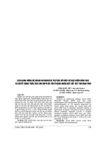
RESEARCH CHART
13
CHAPTER 3
RESEARCH RESULTS
3.1 General characteristics of the study patient group
The median age was 192 months (16 years), the youngest of 4
months, the oldest of 64 years. We divided patients into 4 age
groups, from 2 years old and younger (22.4%), from 2 to 5 years old
(14.9%), from 5 to 16 years old (13.4%) and over 16 years old
(49,3%). The distribution of patients by gender male/ female is
46.3% and 53.7%.
3.2 Clinical and subclinical characteristics of the subjects
3.2.1 Clinical characteristics of research subjects
– Reasons for detecting the disease: various, dyspnea accounted
for 22.4% and other reasons 29.9%.
– Functional characteristics: the most common symptom is
shortness of breath with 56.7% of patients at NYHA II, 1.5% at
NYHA III, no patients at NYHA IV.
– Physical characteristics:the splitted S2 at pulmonary valve
location were 46.3% and 23.9%, respectively, systolic murmur of
MR and TR were 88.1% and 53.7% respectively.
– Patients with Down syndrome were 7.5%.
– Children get often recurrent bronchitis and delayed weight
(40% and 26.7% in children under 2 years, respectively).
3.2.2 Subclinical characteristics of research subjects
3.2.2.1 Some subclinical characteristics of the research subjects
Chest X-ray: 94.0% with cardiothoracic ratio > 50%, 49.3% with
signs of increased pulmonary circulation.
ECG:
– Some basic parameters: sinus rhythm was 91%, 5 patients with
atrial fibrillation (7.5%) and 1 patient with BAV III (1.5%). ECG
14
axis was mainly left axis (62.7%). 01 case of WPW (1.5%), no other
arrhythmias.
– Some characteristics of conduction system: incomplete right
bundle branch block was primary (67.2%). BAV I was also common
(34.3%).
3.2.2.2 Some characteristics of the Doppler echocardiography of the
research subjects
Some basic parameters
– The majority of patients had good systolic left ventricular (EF)
function before surgery and there was no difference between age
groups. There were 16.4% of cases with left ventricular dilatation,
but up to 92.5% with right ventricular dilatation.
– Heart valve anatomy characteristics: 97% with "cleft" on
anterior leaf of MV. 89.6% of patients had 2 balanced papillary
muscle (10.4% had 2 muscle columns but unbalanced). 22.4% had
"cleft" on septal leaf of TV. 22.4% had dysplasia TV.
– Valve regurgitation characteristics: 65,7% were severe
regurgitation of mitral valve, and 47,8% were severe regurgitation of
tricuspid valve.
– Heart septal perforation: large primium ASD (100%) with
median diameter of 22 mm, 94% left-right shunt, 6% had
bidirectional shunt but not often.
– Some hemodynamic characteristics: 13.6% of patients did not
have pre-operated HTAP, the severity of HTAP was: 18.2% mild,
39.4% moderate and 28.8% severe. The highest PAP group (≥ 60
mmHg) was the oldest (median is 20 years old).
3.3 Clinical, subclinical and morphological changes, cardiac
function after surgery in the study patient group
3.3.1 Clinical changes after surgery
15
The change of functional signs: 58,2% patients was dyspnea before
surgery with NYHA II, III however, 100% patients had NYH I at the
follow-up time of 1 month, 3 months, 6 months.
Changes of physical signs
– The rate of systolic murmur of MR and TR postop were much
lower than preop (before surgery and after 6 months, MR
murmur reduced from88.1% to 15.4%, TR murmur from
53.7% to 0%).
– A strong and splited T2 sound is almost non-existent in patients
after surgery.
3.3.2 Subclinical changes after surgery
3.3.2.1 Changes of some subclinical characteristics
Chest x-ray: 94% patients hadcardiothoracicratio ≥ 50%
before surgery which reduced to 32.7% one month after surgery,
signs of increased pulmonary circulation decreased from 49.4% to
1.9%.
ECG: There was no significant change with parameters such
as heart rate pattern, ECG axis, bundle branch block, atrioventricular
block.
3.3.2.2 Changes in echocardiography characteristics aftersurgery
Some basic parameters: increased LV end – diastolic
diameter, in contrast, decreased RV end – diastolic diameter
compared to before surgery and no significant change in the EF
index.
Changes in regurgitation of atrioventricular valve: there was a
significant improvement in the degree of MR and TR over time.
16
Table 3.31. The degree of ventricular valve regurgitation over
time
M-1 (1)
Mo (2)
M1 (3)
M3 (4)
M6 (5)
n (%)
n (%)
n (%)
n (%)
n (%)
Characteristics
p
Mitral regurgitation
No - Mild
9 (13,4)
39 (58,2)
36 (69,2)
29 (70,7)
29 (74,4)
Moderate
14 (20,9)
25 (37,3)
15 (28,9)
9 (22,0)
7 (17,9)
Severe
44 (65,7)
3 (4,5)
1 (1,9)
3 (7,3)
3 (7,7)
p5-1: <0,001
p2-1: <0,001
p2-1: <0,001
p3-1: <0,001
p4-1: <0,001
Tricuspide regurgitation
No - Mild
14 (20,9)
46 (68,7)
48 (92,3)
37 (90,2)
34 (87,2)
Moderate
21 (31,3)
17 (25,4)
4 (7,7)
4 (9,8)
5 (12,8)
Severe
32 (47,8)
4 (6,0)
0
0
0
p3-1: <0,001
p4-1: <0,001
p5-1: <0,001
Evaluation of changes in PAP over time: Preop mean systolic
PAP was 43.3 mmHg, 1 month - 3 months - 6 months after surgery
were 25-26 and 25 mmHg respectively.
3.3.3 Surgical parameters and related to the preoperative
condition
Reconstruct some assessment of atrioventricular valve abnormalities
at surgery compared with preoperative echocardiography
– Surgeons agreed with the diagnosis of partial AVSD: 100%.
– There was a high agreement on the rate of diagnosis of cleft of
MR (97% ultrasound and 94% surgeon).
The value of echocardiography in diagnosing some atrioventricular
valve abnormalities
17
Table 3.36. Value of echocardiogram in the diagnosis of
atrioventricular valve abnormalities
Cleft of Dilatation Cleft of Hypoplasia
Parameter
MV
ring of MV
TV
of septal TV
Sensitivity
98
2,4
66.6
29,2
Specificity
25
96
79,7
88,5
Predictive positive value
98,4
50
13,3
80
Negative predictive value
50
38
98
44
The results show that: assessing MV abnormalities had high
sensitivity and positive predictive value.
The techniques used for repairing MV and repairing TV:
close of cleft MV was the most common (94%), for TV, the most
used techniquewas the De Vega method (49.3%). The relationship
assessment showed that the degree of MR, TR before surgery was
closely related to the number of methods used to repair valves.
Analysis of the relationship of preoperative systolic PAP with
the perioperative: there was a close relationship with the time of
mechanical ventilation, the higher of pre-operative PAPs, the longer
the mechanical ventilation would last. But no association was found
between the degree of preoperative MR and the surgical period.
Table 3.44. Summary of some short-term treatment res ults
Parameters
Patients (n)
Percent (%)
Discharge
67
100
Reoperation
2
3,0
18
Permanent pacemaker implant
1
1,5
Temporary pacemaker implant
4
6,0
Discharge to die at home
0
0
Death
0
0
Evaluation of treatment results based on criteria of reducing
MR, PAPs or both: there was a clear improvement (comparing the
time 1 week - 1 month after surgery with preoperation): the degree of
MR well reduced after 1 week - 1 month surgery was 73.1% and
82.7%, respectively; similar to the reduction in systolic PAPs 1 week
- 1 month of 89.4% and 90.4%, respectively; combining these two
criteria, the ratio was 61.2% and 73.1%, respectively.
Some complications during and after surgery: no premature
death, BAV III rate was 11.9% but 6/8 cases recovered to sinus
before discharge, in addition to the rate of pneumonia bronchopneumonia accounting for 17.9%, heart failure 7.5% and
some other complications.
CHAPTER 4
DISCUSS
4.1 General characteristics of the study patient group
Patient age: the late detection of congenital heart disease was
a feature of our current socio-economic conditions. A study of 40
years of partial AVSD surgery at the Mayo Clinic, the median age of
patients was 9.6 years, 6.1 years and 7.2 years, respectively during
the 50s, 70s and 80s.
4.3 Clinical, subclinical and morphological changes, cardiac
function after surgery in the study patient group
4.3.1 Clinical changes after surgery
19
4.3.1.1 Changes in functional signs: There was a statistically
significant change in functional signs before and after surgery
(dyspnea NYHA II, III before surgery accounted for 58.2%, 100%
at NYHA I at any time postoperative), this result was similar to the
result of the author Dao Quang Vinh and some other authors. This
showed an improvement in patients after surgery regardless of age.
4.3.2.2 Change of physical signs:
– There was a clear change in heart auscultation, the rate of
systolic murmur of MR and TR were recorded to decrease much
compared to before surgery. MR after surgery was the leading cause
of the re-surgery of patients with partial AVSD, the new appearance
or the increase of MR murmur would be a sign that suggested the
next indications to be done for evaluation (echocardiography).
– A strong, splitting T2 sound did not appear after surgery
(showing a significant decrease in blood flow to the lungs, a
significant reduction in PAP).
4.3.2 Subclinical changes after surgery
4.3.2.1 Change of some subclinical characteristics
Chest X Ray
Cardiothoracic ratio, signs of increased pulmonary circulation and
significant decreased PAP after surgery indirectly showed that pulmonary
vascular disease was not a problem of the patient in this study.
ECG
There were no significant changes with most of the basic
parameters except for a significantly lower heart rate compared to
before surgery.
Particularly 5 cases of atrial fibrillation before surgery were
over 40 years old, 2 patients had sinus rhythm after surgery. This
wasreally important for the patient, as atrial fibrillation was the
20
premise of the risk of stroke. There were 2 cases of BAV III with
permanent pacemaker (3%), lower than some other studies reported
by Di Mambro et al. 7.5%.
4.3.2.2 Changes in echocardiography characteristics after surgery
Some basic parameters
Longitudinal follow-up showed a significant change in the LV
end-diastolic diameter (Dd) which was greater than before surgery,
in contrast, the RV end-diastolic diameter was smaller before surgery
and there was no significant change in the EF index. Our results were
similar to those reported by Nguyen Thi Mai Ngoc and author Dao
Quang Vinh. There was no left-right shunt after surgery to help
reduce the volume and pressure RV, so it did not affect ventricular
septal and left ventricle.
Left ventricular systolic function remained within normal
range before surgery and along the time of follow-up, as the study of
author Dao Quang Vinh showed that surgery did not adversely affect
heart function.
Characteristics of MR and TR after surgery
The rate of MR severe was high before surgery and decreased
significantly after surgery. Three patients with severe MR at 3
months, 6 months were> 50 years old and 2 of them had atrial
fibrillation before surgery. This showed the relationship between age
of surgery and the ability to succeed in terms of valve repair, author
Sarisoy and colleagues also reached the same conclusion.
The rate of reoperation through many studies ranged from 1116% (author El-Najdawi et al., O'Sullivan et al.) with the reason of
MR, stenosis of LVOT. Stulak et al's study showed that the
reoperation time was about 10 years. We therefore need to continue
21
monitoring and evaluating the progression of MR with
echocardiography.
Repair of TR also achieved good results, similar to the
research results of author Dao Quang Vinh, author Waqar et al.
Results in terms of hemodynamics
In our study, it showed a significant decrease of PAP
compared to before surgery at all times with median PAPs value at
the time before surgery, 1 week, 1 month, 3 months after surgery and
6 months, respectively, 43.4 - 25 - 26 and 25 mmHg and at all ages.
Thus the surgical effect was seen in patients of all age groups, young
or old.
Limitations of the study:
– Small sample size: 67 patients, the age of patients scattered.
– Short-term vertical follow up.
– Some other diagnostic imaging methods have not been applied
(such as real-time 3D esophageal echocardiography).
CONCLUSION
1
Clinical, subclinical and echocardiography characte ristics
in patients with partial AVSD
1.1 Clinical characteristics
– Median age: 192 months (male/female ratio: 1/1.16), patients
over 16 years accounted for 49.3%. Common function signs
were shortness of breath with NYHA II accounting for 56.7%.
– Physical signs: detected murmur of MR was 88.1% and
murmur of TR was 53.7%. The splitted S2 at pulmonary valve
location were 46.3% and 23.9%, respectively.
– Patients with Down syndrome were 7.5%.
22
– Children get often recurrent bronchitis and delayed weight in
history (90.3% and 80.0% in children under 2 years,
respectively).
1.2 Subclinical characteristics
– Chest X-ray: 94.0% with cardiothoracic ratio ≥ 50%, 49.3%
with increased pulmonary circulation.
– ECG: 91% of sinus rhythm, 7.5% of atrial fibrillation, 1.5% of
BAV III. The left axis accounted for 62.7%, uncomplete right
bundle branch block was 67.2% and BAV I was 34.3%.
1.3 Doppler echocardiography characteristics:unified the
diagnosis to identify a partial AVSD with the diagnosis
of 100% surgeons.
- 100% had the large primum ASD (unique atrial ASD
type rate is 9%).
- 97% had cleft of MV.
- 86.6% had moderate – severe MR. The degree of MR was
related to the techniques used to repair valve.
- 79.1% had moderate – severe TR. The degree of TR is related
to the techniques used to repair valve.
- The diagnostic value of cleft MV was highly sensitive (98%)
as well as the positive predictive value (98.4%)
- HTAP with PAPs median was 43.3 mmHg. The level of
HTAP was related to the age and diameter of ASD and was
closely related to the duration of mechanical ventilation.
2.
Evaluation of clinical, subclinical, morphological and
cardiac function changes after surgery in patients with partial
AVSD
2.1 Changes in clinical features: There is a clear improvement
after surgery and maintained up to 6 months:
23
– Symptoms of dyspnea improved clearly in 100% of patients
with NHYA I after surgery.
– Murmur of MR was only 15.4 %. No more murmur of TR
recorded.
2.2 Subclinical and morphological, function cardiac changes after
surgery
– Chest X-ray showed a clear improvement: cardiothoracic ratio
≥ 50% accounting for 94% pre surgery 1 month after surgery
accounted for 32.7%.
– ECG: 2 cases transferred atrial fibrillation to sinus, 2 cases
BAV III irreversibly (3%).
2.3 Changes in morphology and cardiac function according to
Doppler echocardiography parameters after surgery
There was a clear improvement immediately after surgery and
maintained up to 6 months with some key parameters as follows:
- 100% no residual ASD.
- Severe MR decreased from 65.7% 7.7%. No more severe TR.
- PAPs significantly decreased from 43.3 25 mmHg.
2.4 Some early results after surgery
Some early results after surgery: 0% mortality rate, 3% reoperated not related to MR, 3% implanted permanent pacemaker
due to BAV III.
REQUEST
Based on the research results, we have the following
recommendations:
1. Many patients in the study were found to be above the age of
16, much higher than the age recommended for surgery for a variety
of reasons. When patients have signs of suspected CHD with
increased pulmonaryperfusion diagnostic echocardiography is