Ebook Immunology at a glance (10th edition): Part 2
Bạn đang xem bản rút gọn của tài liệu. Xem và tải ngay bản đầy đủ của tài liệu tại đây (6.52 MB, 59 trang )
26
Antimicrobial immunity: a general scheme
Entry
acute
s'
'natural antibiotic
Surface barriers
infl
am
C5
C6 C7 C8 C9
C3
bl o c
k in g
C3
TH
C2
C4
COMPLEMENT
ion
C3
PHAGOCYTIC
CELLS
m
at
C3
lysis
C1
B
phagocytosis
MAC
r
int
lar
llu
NK
e
ac
intracellular
killing
su
rvi
val
TH
s
per
CELLMEDIATED
IMMUNITY
ANTIBODY
chronic
inflammation
nce
iste
TC
extracellular killing
killing
Spread
intracellular
killing
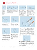
At this point the reader will appreciate that the immune system is
highly efficient at recognizing foreign substances by their shape but
has no infallible way of distinguishing whether they are dangerous
(‘pathogenic’). By and large, this approach works well to control
infection, but it does have its unfortunate side, e.g. the violent immune
response against foreign but harmless structures such as pollen grains,
etc. (see Fig. 35).
Would-be parasitic microorganisms that penetrate the barriers of
skin or mucous membranes (top) have to run the gauntlet of four
main recognition systems: complement (top right), phagocytic cells
(centre), antibody (right) and cell-mediated immunity (bottom),
together with their often interacting effector mechanisms. Unless
primed by previous contact with the appropriate antigen, antibody and
cell-mediated (adaptive) responses do not come into action for several
days, whereas complement and phagocytic cells (innate), being ever-
sequestration
present, act within minutes. There are also (top centre) specialized
innate elements, such as lysozyme, interferons, etc., which act more
or less non-specifically, much as antibiotics do. Innate molecules that
have evolved to block virus infection are sometimes called restriction
factors.
Generally speaking, complement and antibody are most active
against microorganisms free in the blood or tissues, while cell-mediated
responses are most active against those that seek refuge in cells (left).
But which mechanism, if any, is actually effective depends largely on
the tactics of the microorganism itself. Successful parasites are those
able to evade, resist or inhibit the relevant immune mechanisms, as
illustrated in the following five figures. Evasion molecules, together
with those that directly damage the host, are known as virulence
factors. With increased knowledge of the host and pathogen genomes,
identification of virulence factors has become a top priority.
60 Immunology at a Glance, Tenth Edition. J.H.L. Playfair and B.M. Chain. © 2013 John Wiley & Sons, Ltd. Published 2013 by John Wiley & Sons, Ltd.
Entry Many microorganisms enter the body through wounds or bites,
but others live on the skin or mucous membranes of the intestine,
respiratory tract, etc., and are thus technically outside the body.
Surface barriers Skin and mucous membranes are to some extent
protected by acid pH, enzymes, mucus and other antimicrobial secretions, as well as IgA antibody (see below). The lungs, intestine, genitourinary tract and eye each have their own specialized combination
of protection mechanisms.
Natural antibiotics The antibacterial enzyme lysozyme (produced
largely by macrophages; see Fig. 29) and defensins, a family of
polypeptides with broad antimicrobial properties, produced especially
at mucosal surfaces, provide protection against many bacteria. Recent
research has also discovered a whole range of molecules blocking
viruses from becoming established in cells. These ‘restriction factors’
are regulated by the antiviral interferons (see Figs 24 and 27), soluble
proteins released at sites of viral entry.
C3 Complement is activated directly (‘alternative pathway’) by many
microorganisms, particularly bacteria, leading to their lysis or phagocytosis. The same effect can also be achieved when C3 is activated by
antibody (‘classic pathway’; see Fig. 6) or by mannose-binding
protein.
TH Helper T cells perform several distinct functions in the immune
response to microbes. Some respond to ‘carrier’ determinants and
stimulate antibody synthesis by B cells. Viruses, bacteria, protozoa
and worms have all been shown to function as fairly strong carriers,
although there are a few organisms to which the antibody response
appears to be T-independent. Others secrete cytokines that attract
and activate macrophages, eosinophils, etc. (see Figs 21 and 24), or
enhance the activity of cytotoxic T cells. The central role of T helper
cells in many infections is shown by the serious effects of their
destruction, e.g. in AIDS (see Fig. 28).
B Antibody formation by B lymphocytes is an almost universal
feature of infection, of great diagnostic as well as protective value. As
a general rule, IgM antibodies come first, then IgG and the other
classes; IgM is therefore often a sign of recent infection. At mucous
surfaces, IgA is the most effective antibody (see Figs 14 and 17).
Blocking Where microorganisms or their toxins need to enter cells,
antibody may block this by combining with their specific attachment
site. Antibody able to do this effectively is termed ‘neutralizing’. Vaccines against tetanus, diphtheria and polio all work via this mechanism, as does IgA in the intestine.
Phagocytosis by polymorphonuclear leucocytes or macrophages is
the ultimate fate of the majority of unsuccessful pathogens. Both C3
and antibody improve this tremendously by attaching the microbe to
the phagocytic cell through C3 or Fc receptors on the latter; this is
known as ‘opsonization’ (see Fig. 9).
Intracellular killing Once inside the phagocytic cell, most organisms
are killed and degraded by reactive oxygen species, lysosomal
enzymes, etc. (see Fig. 8). In certain cases, ‘activation’ of macro-
phages by T cells may be needed to trigger the killing process (see
Fig. 21).
Extracellular killing Monocytes, polymorphs and other killer (K)
cells can kill antibody-coated cells in vitro, without phagocytosis;
however, it is not clear how much this actually happens in vivo.
NK Natural killer cells are able to kill many virus-infected cells
rapidly, but without the specificity characteristic of lymphocytes. NK
cells are activated by cells that lose expression of MHC class I molecules, a frequent characteristic of virus-infected cells and tumours
that attempt to evade adaptive immune recognition in this way.
Intracellular survival Several important viruses, bacteria and protozoa can survive inside macrophages, where they resist killing. Other
organisms survive within cells of muscle, liver, brain, etc. In such
cases, antibody cannot attack them and cell-mediated responses are
the only hope.
T C Cytotoxic T cell, specialized for killing of cells harbouring virus,
also allogeneic (e.g. grafted) cells (see Figs 21 and 39), and sometimes
tumours (see Fig. 42).
Sequestration Microorganisms that cannot be killed (e.g. some
mycobacteria) or products that cannot be degraded (e.g. streptococcal
cell walls) can be walled off by the formation of a granuloma by
macrophages and fibroblasts, aided by TH-mediated immune responses
(see Figs 21 and 37).
Spread Successful microorganisms must be able to leave the body
and infect another one. Coughs and sneezes, faeces and insect bites
are the most common modes of spread.
Persistence Some very successful parasites are able to escape all the
above-mentioned immunological destruction mechanisms by sophisticated protective devices of their own. Needless to say, these constitute some of the most chronic and intractable infectious diseases.
Major strategies for immune evasion include resistance to phagocytosis and/or intracellular killing, antigenic variation, immunosuppression and various forms of concealment.
Inflammation Although some microorganisms cause tissue damage
directly (e.g. cytopathic viruses or the toxins of staphylococci), it is
unfortunately true that much of the tissue damage resulting from infection is due to the response of the host. Acute and chronic inflammation
are discussed in detail elsewhere (see Figs 7 and 37), but it is worth
noting here that infectious organisms frequently place the host in a
real dilemma: whether to eliminate the infection at all costs or to limit
tissue damage and allow some of the organisms to survive. Given
enough time, natural selection should arrive at the balance that is most
favourable for both parasite and host survival.
Virulence factors include toxins, adhesion factors, resistance factors
for antibiotics, enzymes that destroy immunological molecules,
cytokine inhibitors, antigenic variation. Successful pathogens often
possess many of these.
Antimicrobial immunity: a general scheme Potentially useful immunity 61
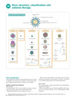
Immunity to viruses
27
in secretions
IgA
ENTRY VIA
RECEPTOR
INTERFERON
DIRECT
SPREAD
protection
MAC
BUDDING
DNA or
RNA
lysis
BLOOD
SPREAD
capsid
TH
B
PHAGOCYTOSIS
ANTIBODY
NK
envelope
MHC I
KILLING BY
NK & T CELLS
TC
IL- 2
TH
complexes
autoantibody
CYTOTOXICITY
LATENCY
DTH
TISSUE DAMAGE
Viruses differ from all other infectious organisms in being much
smaller (see Appendix I) and lacking cell walls and independent metabolic activity, so that they are unable to replicate outside the cells of
their host. The key process in virus infection is therefore intracellular
replication, which may or may not lead to cell death. In the figure,
viruses are depicted as hexagons, but in fact their size and shape are
extremely varied.
For rapid protection, interferon (top) activates a large number of
innate mechanisms that can block viruses entering or replicating
within cells. These molecules, collectively known as restriction
factors, have the same ‘natural antibiotic’ role as lysozyme in bacterial infection, although the mechanisms are quite different. Antibody
(right) is valuable in preventing entry and blood-borne spread of
some viruses, but is often limited by the remarkable ability of viruses
to alter their outer shape, and thus escape detection by existing antibody (the epidemics of influenza that occur each year are good examples of this mechanism at work). Other viruses escape immune
surveillance by antibody by spreading from cell to cell (left). For
these viruses the burden of adaptive immunity falls to the cytotoxic
T-cell system, which specializes in recognizing MHC class I antigens
carrying viral peptides from within the cell (see Fig. 18). However,
many viruses (such as the herpes family) have evolved ways to
escape cytotoxic T-cell recognition, by downregulating MHC expression, secreting ‘decoy’ molecules or inhibiting antigen processing.
NK cells, which kill best when there is little or no MHC on the
infected cell and come into action more rapidly than TC cells, therefore have an important role.
Note that tissue damage may result from either the virus itself or
the host immune response to it. In the long run, no parasite that seriously damages or kills its host can count on its own survival, so that
adaptation, which can be very rapid in viruses, generally tends to be
in the direction of decreased virulence. But infections that are well
adapted to their normal animal host can occasionally be highly virulent
to humans; rabies (dogs) and Marburg virus (monkeys) are examples
of this (‘zoonosis’).
Intermediate between viruses and bacteria are those obligatory
intracellular organisms that do possess cell walls (Rickettsia, Chlamydia) and others without walls but capable of extracellular replication
(Mycoplasma). Immunologically, the former are closer to viruses, the
latter to bacteria.
62 Immunology at a Glance, Tenth Edition. J.H.L. Playfair and B.M. Chain. © 2013 John Wiley & Sons, Ltd. Published 2013 by John Wiley & Sons, Ltd.
Receptors All viruses need to interact with specific receptors on the
cell surface; examples include Epstein–Barr virus (EBV; CR2 on cells),
rabies (acetylcholine receptor on neurones), measles (CD46 on cells)
and HIV (CD4 and chemokine receptors on T cells and macrophages).
Interferon A group of proteins (see Figs 23 and 24) produced in
response to virus infection, which stimulate cells to make proteins that
block viral transcription, and thus protect them from infection.
C
T , NK, cytotoxicity As described in Figs 11, 18 and 21, cytotoxic T
cells ‘learn’ to recognize class I MHC antigens, and then respond to
these in association with virus antigens on the cell surface. It was
during the study of antiviral immunity in mice that the central role of
the MHC in T-cell responses was discovered. In contrast, NK cells
destroy cells with low or absent MHC, a common consequence of viral
infection.
Antibody Specific antibody can bind to virus and thus block its ability
to bind to its specific receptor and hence infect cells. This is called
neutralization and is an important part of protection against many
viruses, including such common infections as influenza. Sometimes,
viruses are able to enter cells still bound to antibody: within the cytoplasm, a molecule called TRIM21 binds antibody, and activates mechanisms that lead to rapid degradation of the virus–antibody complex.
Viruses
There is no proper taxonomy for viruses, which can be classified
according to size, shape, the nature of their genome (DNA or RNA),
how they spread (budding, cytolysis or directly; all are illustrated) and
– of special interest here – whether they are eliminated or merely
driven into hiding by the immune response. Brief details of a selection
of important groups of viruses are given below.
Poxviruses (smallpox, vaccinia) Large; DNA; spread locally, avoiding antibody, as well as in blood leucocytes; express antigens on the
infected cell, attracting CMI. The antigenic cross-reaction between
these two viruses is the basis for the use of vaccinia to protect against
smallpox (Jenner, 1798). Thanks to this vaccine, smallpox is the first
disease ever to have been eliminated from the entire globe. However,
stocks of vaccine against smallpox are once again being stockpiled in
case this organism is spread deliberately as a form of bioterrorism.
Herpesviruses (herpes simplex, varicella, EBV, CMV [cytomegalovirus], KSHV [Kaposi sarcoma-associated herpes virus]) Medium;
DNA; tend to persist and cause different symptoms when reactivated:
thus, varicella (chickenpox) reappears as zoster (shingles); EBV (infectious mononucleosis) may initiate malignancy (Burkitt’s lymphoma;
see Fig. 42); CMV has become important as an opportunistic infection
in immunosuppressed patients; and KSHV causes Kaposi’s sarcoma in
patients with AIDS (see Fig. 28). Some herpes viruses have apparently
acquired host genes such as cytokines or Fc receptors during evolution,
modifying them so as to interfere with proper immune function.
Adenoviruses (throat and eye infections) Medium; DNA. Numerous
antigenically different types make immunity very inefficient and vaccination a problem. However, modified adenoviruses and adenoassociated viruses are being explored as possible gene therapy vectors,
because they infect many cell types very efficiently.
Myxoviruses (influenza, mumps, measles) Large; RNA; spread by
budding. Influenza is the classic example of attachment by specific receptor (neuraminic acid) and also of antigenic variation, which limits the
usefulness of adaptive immunity. In fact the size of the yearly epidemics
of influenza can be directly related to the extent by which each year’s
virus strain differs from its predecessor. Mumps, by spreading in the
testis, can initiate autoimmune damage. Measles infects lymphocytes and
antigen-presenting cells, causes non-specific suppression of CMI and can
persist to cause SSPE (subacute sclerosing panencephalitis); some
workers feel that multiple sclerosis may also be a disease of this type.
Rubella (‘German measles’) Medium; RNA. A mild disease feared
for its ability to damage the fetus in the first 4 months of pregnancy.
An attenuated vaccine gives good immunity.
Rabies Large; RNA. Spreads via nerves to the central nervous system,
usually following an infected dog bite. Passive antibody combined
with a vaccine can be life-saving.
Arboviruses (yellow fever, dengue) Arthropod-borne; small; RNA.
Blood spread to the liver leads to jaundice.
Enteroviruses (polio) Small; RNA. Polio enters the body via the gut
and then travels to the central nervous system where it causes paralysis
and death. Within the blood it is susceptible to antibody neutralization,
the basis for effective vaccines (see Fig. 41).
Rhinoviruses (common cold) Small; RNA. As with adenoviruses
there are too many serotypes for antibody-mediated immunity to be
effective across the whole population.
Hepatitis can be caused by at least six viruses, including A (infective;
RNA), B (serum-transmitted; DNA) and C (previously known as ‘non-A
non-B’; RNA). In hepatitis B and C, immune complexes and autoantibodies are found, and virus persists in ‘carriers’, particularly in tropical
countries and China, where it is strongly associated with cirrhosis and
cancer of the liver. Treatment with IFNα or other antivirals can sometimes induce immunity and result in viral control. Very effective vaccines are now available for uninfected adults against hepatitis A and B.
Arenaviruses (Lassa fever) Medium; RNA. A haemorrhagic disease
of rats, often fatal in humans. A somewhat similar zoonosis is Marburg
disease of monkeys.
Retroviruses (tumours, immune deficiency) RNA. Contain reverse
transcriptase, which allows insertion into the DNA of the infected cell.
The human T-cell leukaemia viruses (HTLV) and the AIDS virus
(HIV) belong to this group and are discussed separately (for details
see Fig. 28).
Atypical organisms
Trachoma An organism of the psittacosis group (Chlamydia). The
frightful scarring of the conjunctiva may be due to over-vigorous CMI.
Typhus and other Rickettsia may survive in macrophages, like the
tubercle bacillus.
Prions These are host proteins which under certain circumstances can
be induced to polymerize spontaneously to form particles called
‘prions’. They are found predominantly in brain, and can cause progressive brain damage (hence their original classification as ‘slow
viruses’). The first example of a ‘prion’ disease was kuru, a fatal brain
disease spread only by cannibalism. However, prion diseases are now
thought to be responsible for scrapie and, most notoriously, for the UK
epidemic of bovine spongiform encephalopathy (BSE or ‘mad cow
disease’) and the human equivalent, Creutzfeldt–Jakob disease (CJD).
Many aspects of prion disease remain poorly understood and there is
no known treatment. There appears to be little or no immune response
to prions, perhaps because they are ‘self’ molecules.
Immunity to viruses Potentially useful immunity 63
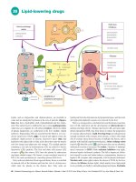
HIV and AIDS
28
VIRUS
envelope
THERAPY
reverse transcriptase
p51, p66
HAART
p15
p17
p24
RNA
uncoating
penetration
gp120 gp160
gp41
core
INFECTION
OF CELLS
reverse transcription
DNA
integration into
host DNA
eptor
corec
new viral RNA
CD 4
RNA
gag
pol
env
tat
ANTIBODY
p24
gp41
gp120
nucleus
cytoplasm
AC
UT
E
lipid
bilayer
MAC
SPREAD
sexual
viral
blood
budding proteins
mother
INF
release
ECT
ION
child
fever
Cytokines ?
lysis ?
ARC
weight loss
microglia ?
TH
TC
P GL
CMI DEFECT
IN
B RA
AIDS
dementia
OPPORTUNISTIC INFECTIONS
IMMUNITY
Pneumocystis
Tox oplasma
Histoplasma
Cryptosporidium
Strongyloides
When in the summer of 1981 the Centers for Disease Control in the
USA noticed an unusual demand for a drug used to treat Pneumocystis
pneumonia, a rare infection except in severely immunosuppressed
patients, and cases began to be increasingly reported in homosexual
men, haemophiliacs receiving certain batches of blood products and
drug users sharing needles, it became clear that a potentially terrible
new epidemic had hit mankind, more insidious than the plague, more
deadly than leprosy. The disease was baptized acquired immune deficiency syndrome (AIDS), and has become the most widely studied
infectious disease of all time.
By 1984 the cause had been traced to a virus, now named HIV
(human immunodeficiency virus), an RNA lentivirus (a subfamily of
the retroviruses) that possesses the enzyme reverse transcriptase. This
allows it to copy its RNA into DNA which is then integrated into the
nucleus of the cells it infects, principally T-helper cells and macrophages. By processes still not fully understood, this leads to a slow
disappearance of T-helper cells, with derangement of the whole
immune system and the development of life-threatening opportunistic
infections and tumours. The origin of HIV continues to be debated.
mycobacteria
Cryptococcus
Candida
CMV
herpes
Kaposi's sarcoma
B lymphoma
DISEASE
Attempts to link the epidemic to contaminated polio vaccine, or even
to a political conspiracy have been totally discredited. The most
likely hypothesis is that it spread from chimpanzees at some time
during the twentieth century, perhaps due to human consumption
of infected meat. Enormous effort has gone into trying to develop
vaccines against HIV. HIV infection stimulates strong cellular immunity and antibody responses, but these responses never seem to be
able to completely eliminate the virus, or even stop it dividing. In
part, this may be because the virus infects T-helper cells, and hence
blocks the development of full immunity. But the properties of HIV
reverse transcriptase also give it an unusual ability to vary its antigens, which makes protective immunity or vaccination very difficult
to attain.
HIV I and II, the AIDS viruses, closely related to the simian (monkey)
virus SIV and more distantly to retroviruses such as HTLV I and II,
which are rare causes of T-cell leukaemias. Their genome consists of
double-stranded RNA. HIV II causes a much slower and less aggressive disease, and is predominantly found in Africa.
64 Immunology at a Glance, Tenth Edition. J.H.L. Playfair and B.M. Chain. © 2013 John Wiley & Sons, Ltd. Published 2013 by John Wiley & Sons, Ltd.
Gag The gene for the core proteins p17, p24 and p15. Like many
viruses, HIV uses single genes to make long polyproteins which are
then cut up by the virus’s own enzyme (a protease) into a number of
different functional units. Drugs that block this protease are an important class of HIV inhibitors.
Pol The gene for various enzymes, including the all-important reverse
transcriptase.
Env The gene for the envelope protein gp160, which is cleaved during
viral assembly to make gp120, the major structural protein of the viral
envelope. Interaction with the CD4 molecule found on T cells and
macrophages, and a second interaction with a chemokine receptor
(usually CCR5 or CXCR4), allows the virus to infect cells. About 1
in 10 000 Caucasian individuals have a homozygous deletion in CCR5,
and these individuals are highly resistant to infection with HIV. Gag,
pol and env genes are found in all lentiviruses.
Tat, rev, nef, vif, vpu Genes unique to HIV, which can either enhance
or inhibit viral synthesis. Several of these molecules also antagonize
cellular defence systems. For example, nef downregulates MHC class
I and hence helps the virus escape immune detection, while vif blocks
the enzyme APOBEC which destroys the viral RNA.
Reverse transcriptase is required to make a DNA copy of the viral
RNA. This may then be integrated into the cell’s own nuclear DNA,
from which further copies of viral RNA can be made, leading to the
assembly of complete virus particles which bud from the surface to
infect other cells. A key feature of this enzyme is that it allows errors
in transcription to occur (on average there is one base pair mutation for
every round of viral replication). This feature allows the rapid evolution of new variants of virus during the course of an infection.
Acute infection A few weeks after HIV infection some patients
develop a flu-like or glandular fever-like illness, although many
remain symptomless. This is associated with a rapid rise in the level
of virus in blood. During these weeks infected individuals rapidly
develop antibody to HIV, which is routinely used for diagnosis. A very
strong cellular TC response also develops, which decreases the amount
of virus in blood (‘viral load’) to a much lower, and sometimes undetectable, level. However, during this early phase there is also massive
destruction of CD4 cells, predominantly in gut tissue. The mechanisms
remain unclear.
Asymptomatic period Virus levels remain low for variable periods
between a few months and more than 20 years. During this period
infected individuals show few symptoms, although the number of
CD4+ T cells falls gradually. Despite this apparent ‘latency’, virus is
in fact replicating rapidly and continuously, mainly within lymph
nodes, and there is an enormous turnover of CD4+ T cells, as infected
cells die and are replaced. There may be a stage of progressive generalized lymphadenopathy (PGL).
Symptomatic period Patients develop a variety of symptoms, including recurrent Candida infections, night sweats, oral hairy leukoplakia
and peripheral neuropathy (AIDS-related complex; ARC).
AIDS The full pattern includes the above plus severe life-threatening
opportunistic infections and/or tumours. In some patients cerebral
symptoms predominate. Almost every HIV-infected patient eventually
progresses to AIDS. In 2009 there were estimated to be 33 million
individuals infected with HIV worldwide, and over 2 million deaths
from the disease, although the numbers of infected people appear to
have reached a plateau. The vast majority of infected individuals are
in sub-Saharan Africa, but there are expanding epidemics in many
countries in the Far East. There are an estimated 1.5 million infected
people in North America, 600 000–800 000 in western Europe and
around 86 000 in the UK (many of them undiagnosed).
Kaposi’s sarcoma A disseminated skin tumour thought to originate
from the endothelium of lymphatics. It is caused by human herpes
virus-8 (HHV-8, also known as KSHV), although it is still not clear why
it is more common in AIDS than in other immunodeficient conditions.
T cells are the most strikingly affected cells, the numbers of CD4+
(helper) T cells falling steadily as AIDS progresses, which leads to a
failure of all types of T-dependent immunity. Although only 1% or less
of T cells are actually infected, the virus preferentially targets memory
cells.
MAC Macrophages and the related antigen-presenting cells, brain
microglia, etc. are probably a main reservoir of HIV and are usually
the initial cell type to become infected.
Transmission is still mainly by intercourse (heterosexual as well as
homosexual), although in some areas infected blood from drug needles
is more common. HIV can also be transmitted from mother to child
at birth (vertical transmission) giving rise to neonatal AIDS. Not every
exposure to HIV leads to infection, but as few as 10 virus particles are
thought to be able to do so.
Pathology HIV is not a lytic virus, and calculations suggest that
uninfected as well as infected T cells die. Many mechanisms have been
proposed (including autoimmunity) but none is generally accepted.
Immunity The major antibody responses to HIV are against p24, p41
and gp120. Some antibody against gp120 is neutralizing but is very
specific to the immunizing strain of virus. A strong CD8 T response
against HIV-infected cells persists throughout the asymptomatic phase
of HIV infection, suggesting that these cells are the major effector
mechanism keeping HIV replication in check. Several innate mechanisms that may have a role in limiting lentivirus replication have been
described (the molecules involved are often referred to as restriction
factors). An RNA/DNA-modifying enzyme related to the one believed
to be involved in somatic hypermutation (see Fig. 13) can provide
protection by causing lethal mutations in viral nucleic acids. A cellular
protein called TRIM5 acts at the stage of viral uncoating, while a
membrane protein called tetherin inhibits the ability of newly formed
virus to bud off from the cell surface. But HIV appears to have evolved
ways of escaping all of them!
Therapy Early drugs used for treatment against HIV were inhibitors
of viral reverse transcriptase, such as zidovudine (AZT). Treatment
with a single drug provides only very short-term benefit as the virus
mutates so fast that resistant strains soon emerge. However, the
development of new families of drugs, e.g. against the HIV-specific
protease, allowed the introduction of multidrug therapy, known as
HAART (highly active antiretroviral therapy). Patients are treated with
three, four or even more different antivirals simultaneously. These
regimens have seen some spectacular successes in the clinic, leading
to disappearance of AIDS-associated infections, and undetectable
levels of virus for several years. However, this approach never results
in permanent elimination of virus, and resistant strains eventually
emerge. In any case the cost is prohibitive in most of the countries
where HIV is common. Thus, the requirement for an effective HIV
vaccine remains acute, and several trials aimed especially at stimulating a strong cellular response are under way.
HIV and AIDS Potentially useful immunity 65
Immunity to bacteria
29
Chromosome
Flagella
Pili
e
z ym
o
s
Ly
CAPSULE
COMPLEMENT
c
blo
TH
k
AGGRESSINS
EXOTOXINS
lysis
B
damage
PHAGOCYTOSIS
ANTIBODY
TH
M
M PG
M
PG
GRAM+
e.g. Staphylococcus
Streptococcus
intracellular
survival
LPS
GRAM–
e.g. Salmonella
Neisseria
immune complexes
autoantibody
CMI
granuloma
Cell wall
TISSUE DAMAGE
Unlike viruses, bacteria are cellular organisms, mostly capable of fully
independent life, but some live on or in larger animals some or all of
the time. Indeed, it is estimated that each human is colonized by some
1014 bacteria, equivalent to 10 bacteria for every cell of the body. This
microbiome is made up of several thousand different species, most of
which are innocuous and may even have a beneficial role in enhancing
human health. However, a few species can cause disease and, together
with viruses, these now constitute the major infectious threat to health
in developed countries. Since the discovery of antibiotics, bacterial
infection has been controlled largely by chemotherapy. However, with
the recent rise in antibiotic-resistant strains of bacteria, there is
renewed interest in developing new or improved vaccines against the
bacteria responsible for such diseases as tuberculosis, meningitis and
food poisoning.
The usual destiny of unsuccessful bacteria is death by phagocytosis;
survival therefore entails avoidance of this fate. The main ways in which
a bacterium (top left) can achieve this lie in the capsule (affecting attachment), the cell wall (affecting digestion) and the release of exotoxins
(which damage phagocytic and other cells). Fortunately, most capsules
and toxins are strongly antigenic and antibody overcomes many of their
effects; this is the basis of the majority of antibacterial vaccines. In the
figure, processes beneficial to the bacteria or harmful to the host are shown
in broken lines. Bacteria living on body surfaces (e.g. teeth) can form
colonies (‘biofilms’) which protect them against both immunity and antibiotics. As with viruses, some of the most virulent and obstinate bacterial
infections are zoonoses – plague (rats) and brucellosis (cattle) being examples. Bacteria that manage to survive in macrophages (e.g. tuberculosis
[TB]) can induce severe immune-mediated tissue damage (see Fig. 37).
66 Immunology at a Glance, Tenth Edition. J.H.L. Playfair and B.M. Chain. © 2013 John Wiley & Sons, Ltd. Published 2013 by John Wiley & Sons, Ltd.
Cell wall Outside their plasma membrane (M in the figure) bacteria
have a cell wall composed of a mucopeptide called peptidoglycan
(PG); it is here that lysozyme acts by attacking the N-acetylmuramic
acid–N-acetylglucosamine links. In addition, Gram-negative bacteria
have a second membrane with lipopolysaccharides (LPS, also called
endotoxin) inserted in it. Bacterial cell walls are powerful inducers of
inflammation, largely through their ability to activate the Toll-like
receptors of innate immunity (see Figs 3 and 5).
Flagella, the main agent of bacterial motility, contain highly antigenic
proteins (the ‘H antigens’ of typhoid, etc.), which give rise to immobilizing antibody. Some flagellar proteins activate the Toll-like receptor TLR5 (see Fig. 5).
Pili are used by bacteria to adhere to cells; antibody can prevent this
(e.g. IgA against gonococcus).
Capsule Many bacteria owe their virulence to capsules, which protect
them from contact with phagocytes. Most are large, branched, polysaccharide molecules, but some are protein. Many of these capsular
polysaccharides, and also some proteins from flagella, are T-independent
antigens (see Fig. 19). Examples of capsulated bacteria are pneumococcus, meningococcus and Haemophilus spp.
Exotoxins (as distinct from the endotoxin [LPS] of cell walls) Grampositive bacteria often secrete proteins with destructive effects on
phagocytes, local tissues, the CNS, etc.; frequently, these are the cause
of death. In addition there are proteins collectively known as aggressins
that help the bacteria to spread by dissolving host tissue.
Sepsis Occasionally, uncontrolled systemic responses to bacterial
infection develop, which can lead to rapid life-threatening disease
(‘toxic shock’). Such responses are still an important cause of death
after major surgery. Over-production of TNF-α, especially by macrophages, has a major role in these reactions.
Bacteria
Here, bacteria are given their popular rather than their proper taxonomic names. Some individual aspects of interest are listed below:
Strep Streptococcus, classified either by haemolytic exotoxins (α, β,
γ) or cell wall antigens (groups A–Q). Group A β-haemolytic are the
most pathogenic, possessing capsules (M protein) that attach to mucous
membranes but that resist phagocytosis, numerous exotoxins (whence
scarlet fever), indigestible cell walls causing severe cell-mediated
reactions, antigens that cross-react with cardiac muscle (rheumatic
fever) and a tendency to kidney-damaging immune complexes.
Staph Staphylococcus. Antiphagocytic factors include the fibrinforming enzyme coagulase and protein A, which binds to the Fc
portion of IgG, blocking opsonization. Numerous other toxins make
staphylococci highly destructive, abscess-forming organisms. Largescale use of antibiotics has caused the emergence of bacterial strains
resistant to many antibiotics (methicillin-resistant Staphyloccus aureus
[MRSA]), which are now proving a serious threat, particularly as
hospital-acquired infections.
Pneumococcus (now S. pneumoniae), meningococcus Typed by
the polysaccharides of their capsules, and especially virulent in the
tropics, where vaccines made from capsular polysaccharides are
proving highly effective in preventing epidemics. Also more common
in patients with deficient antibody responses (see Fig. 33). Chemical
coupling of the capsular polysaccharides to a protein, such as diphtheria toxoid, converts these antigens from T-cell independent to T-cell
dependent, thus greatly increasing memory and potency. Such conjugate vaccines have proven highly effective at preventing childhood
meningitis and Haemophilus infection.
Gonococcus IgA may block attachment to mucous surfaces, but the
bacteria secrete a protease that destroys the IgA; thus, the infection is
seldom eliminated, leading to a ‘carrier’ state. Gonococci and meningococci are the only bacteria definitely shown to be disposed of by
complement-mediated lysis.
Tuberculosis and leprosy bacilli These mycobacteria have very tough
cell walls, rich in lipids, which resist intracellular killing; they can also
inhibit phagosome–lysosome fusion. Chronic cell-mediated immunity
results in the formation of granuloma, tissue destruction and scarring
(see Fig. 37). In leprosy, a ‘spectrum’ between localization and dissemination corresponds to the predominance of cell-mediated immunity and of antibody, respectively. Tuberculosis is once again on the
rise, partly as a result of increased travel, partly because of increased
drug resistance and partly as a consequence of AIDS, and better vaccines to replace the only partially effective BCG (bacille Calmette–
Guérin) are urgently being sought.
Escherichia coli is now perhaps the best-known bacterial species in
the world, because of its ubiquitous use as a tool in all molecular biology
laboratories. However, the species is a made up of an enormous number
of different strains. Most are harmless inhabitants of the intestine of
many mammals including humans, and may even be beneficial in supplying some vitamins and in suppressing the growth of other pathogenic
bacteria. But a few strains produce exotoxins and have been responsible
for major outbreaks of food poisoning. Shigella (causing dysentery)
and cholera are two other examples of bacteria that grow only in the
intestine, and are responsible for important human diseases.
Salmonella (e.g. S. typhi) also infects the intestine but can survive
and spread to other parts of the body within macrophages. Recovery
after infections may lead to a ‘carrier’ state.
Tetanus owes its severity to the rapid action of its exotoxin on the
CNS. Antibody (‘antitoxin’) is highly effective at blocking toxin
action, an example where neither complement nor phagocytic cells are
needed.
Diphtheria also secretes powerful neurotoxins, but death can be due
to local tissue damage in the larynx (‘false membrane’).
Syphilis is an example of bacteria surviving all forms of immune
attack without sheltering inside cells. The commonly found autoantibody to mitochondrial cardiolipin is the basis of the diagnostic Wasserman reaction. Cross-reactions of this type, due presumably to bacterial
attempts to mimic host antigens and thus escape the attentions of the
immune system, are clearly a problem to the host, which has to choose
between ignoring the infection and making autoantibodies (see Fig.
38) that may be damaging to its own tissues. Borrelia, another spirochaete, has the property (found also with some viruses and protozoa)
of varying its surface antigens to confuse the host’s antibody-forming
system. As a result, waves of infection are seen (‘relapsing fever’).
Brucella may do the same.
Immunity to bacteria Potentially useful immunity 67
30
Immunity to fungi and ectoparasites
Dermatophytes
Skin
secretions
Candida albicans
brain
PMN
Complement
Cryptococcus
Actinomycetes
Aspergillus,
etc.
ANTIBODY
T
Histoplasma
Coccidioides
Blastomyces
Pneumocystis
Lung
GRANULOMAS
The vast majority of fungi are free-living, but a few can infect larger
animals, colonizing the skin or entering via the lung in the form of
spores (centre left). Fungal infections are normally only a superficial
nuisance (e.g. ringworm, top), but a few fungi can cause serious systemic disease, particularly if exposure is intense (e.g. farmers) or the
immune system is in some way compromised (e.g. AIDS); the outcome
depends on the degree and type of immune response, and may range
from an unnoticed respiratory episode to rapid fatal dissemination or
a violent hypersensitivity reaction.
In general, the survival mechanisms of successful fungi are similar to
those of bacteria: antiphagocytic capsules (e.g. Cryptococcus), resistance
to digestion within macrophages (e.g. Histoplasma) and destruction of
polymorphs (e.g. Coccidioides). Some yeasts activate complement via the
alternative pathway, but it is not known if this has any effect on survival.
Perhaps the most interesting fungus from the immunological point
of view is Candida albicans (upper left), a common and harmless
CMI
HYPERSENSITIVITY
inhabitant of skin and mucous membranes which readily takes
advantage of any weakening of host resistance. This is most strikingly seen when polymorphs (PMN) or T cells are defective, but
it also occurs in patients who are undernourished, immunosuppressed,
iron deficient, alcoholic, diabetic, aged or simply ‘run down’ (see
Fig. 33). Organisms that thrive only in the presence of immunodeficiency are called ‘opportunists’ and they include not only fungi, but
also several viruses (e.g. CMV), bacteria (e.g. Pseudomonas), protozoa (e.g. Toxoplasma) and worms (e.g. Strongyloides), and their
existence testifies to the unobtrusive efficiency of the normal immune
system.
The most important ectoparasites (‘outside living’; skin dwelling)
are mites, ticks, lice and fleas. The last three are vectors for
several major viral and bacterial diseases. The evidence for immunity, and the feasibility of a vaccine, are currently under intense
study.
68 Immunology at a Glance, Tenth Edition. J.H.L. Playfair and B.M. Chain. © 2013 John Wiley & Sons, Ltd. Published 2013 by John Wiley & Sons, Ltd.
PMN Polymorphonuclear leucocyte (‘neutrophil’), an important
phagocytic cell. Recurrent fungal as well as bacterial infections may
be due to defects in PMN numbers or function, which may in turn be
genetic or drug-induced (steroids, antibiotics). Functional defects may
affect chemotaxis (‘lazy leucocyte’), phagolysosome formation (Chédiak–Higashi syndrome), peroxide production (chronic granulomatous
disease), myeloperoxidase and other enzymes. Deficiencies in complement or antibody will of course also compromise phagocytosis (see
also Fig. 33).
Histoplasma (histoplasmosis), Coccidioides (coccidioidomycosis)
and Blastomyces (blastomycosis) spp. are similar in causing pulmonary disease, particularly in America, which may either heal spontaneously, disseminate body-wide or progress to chronic granulomatosis
and fibrosis, depending on the immunological status of the patient.
The obvious resemblance to tuberculosis and leprosy emphasizes the
point that it is microbial survival mechanisms (in this case, resistance
to digestion in macrophages) rather than taxonomic relationships that
determine the pattern of disease.
T As severe fungal infection in both the skin and mucous membranes
(Candida spp.) and in the lung (Pneumocystis spp.) are common in
T-cell deficiencies, T cells evidently have antifungal properties, but
the precise mechanism is not clear. Some fungi (see below) can apparently also be destroyed by NK cells.
Pneumocystis jirovecii (formerly P. carinii) is mentioned here because
although it was originally assumed to be a protozoan, studies of its
RNA suggest that it is nearer to the fungi. Pneumocystis pneumonia
has become one of the most feared complications of AIDS (see Fig.
28), which suggests that T cells normally prevent its proliferation,
although the mechanism is so far unknown.
Hypersensitivity reactions are a feature of many fungal infections,
especially those infecting the lung. They are mainly of type I or IV
(for an explanation of what this means see Fig. 34).
Dermatophytes Filamentous fungi that metabolize keratin and therefore live off skin, hair and nails (ringworm). Sebaceous secretions help
to control them, but CMI may also play an ill-defined part.
Candida albicans (formerly Monilia) A yeast-like fungus that causes
severe spreading infections of the skin, mouth, etc. in patients with
immunodeficiency, especially T-cell defects, but the precise role of T
cells in controlling this infection is not understood. Dissemination may
occur to the heart and eye.
Cryptococcus A capsulated yeast able to resist phagocytosis unless
opsonized by antibody and/or complement (compare pneumococcus,
etc.). In immunodeficient patients, spread to the brain and meninges
is a serious complication. The organisms can be killed, at least in vitro,
by NK cells.
Actinomycetes spp. and other sporing fungi from mouldy hay, etc.
can reach the lung alveoli, stimulate antibody production and subsequently induce severe hypersensitivity (‘farmer’s lung’). Both IgG and
IgE may be involved. Aspergillus sp. is particularly prone to cause
trouble in patients with tuberculosis or cellular immunodeficiency.
Dissemination may occur to almost any organ. The toxin (aflatoxin)
is a risk factor for liver cancer.
Ectoparasites
Mites are related to spiders. Sarcoptes scabei (scabies) burrows and
lays eggs in the skin and induces antibody, but such protective immunity as there is appears to be cell-mediated (TH1). The house dust mite
Dermatophagoides pteronyssinus is an important cause of asthma. It
induces high levels of IgE, and sublingual desensitization has had
some success, probably by switching the T-cell response away from
TH2 and towards the TH1 pattern. A DNA-based vaccine has been tried
in mice.
Ticks, like mites, are arachnids, living on the skin and feeding on
blood. They are vectors of several diseases, including Lyme disease,
typhus and relapsing fever. A vaccine has had some success in cattle.
Lice (Pediculosis spp.) feed on skin, clinging to hairs. There are three
main species, P. capitis (head lice), Phthirius pubis (pubic lice) and P.
corporis (body lice). A vaccine has proved successful in salmon.
Fleas Pulex irritans is an important vector for plague, tularemia and
brucellosis.
Mosquitoes and other vectors. Although not strictly parasites, mosquitoes should be mentioned as vectors for malaria, dengue, yellow
fever and some forms of filariasis. Other important vectors are the
sandfly (leishmaniasis), tsetse fly (trypanosomiasis), simulium fly
(onchocerciasis) and reduviid bug (Chagas’ disease).
Immunity to fungi and ectoparasites Potentially useful immunity 69
31
Immunity to protozoa
Entry/spread via bite
Insect
borne
BLOOD
Antigenic
variation
African tryps.
malaria
Leishmania
Tryp.cruzi
trypanosomes
malaria
C3
LIVER
Food/water
borne
Entamoeba
Tox oplasma
Giardia
Isospora, etc.
H
MACROP
complexes
S
AGE
CMI
cross
reaction
GUT
Spread
Immunosuppression
ANTIBODY
MUSCLE
TISSUE DAMAGE
Relatively few (less than 20) species of protozoa infect humans, but
among these are four of the most formidable parasites of all, in terms of
numbers affected and severity of disease: malaria, the African and American trypanosomes, and Leishmania (top left). These owe their success
to combinations of the strategies found among bacteria and viruses:
long-distance spread by insect vectors (compare plague, typhus, yellow
fever), intracellular habitat (compare tuberculosis, viruses), antigenic
variation (compare influenza) and immunosuppression (compare
HIV). However, these strategies are so highly developed that complete
acquired resistance to protozoal infections is quite exceptional, and what
trypanosomes
malaria
Tox oplasma
Polyclonal Ig
trypanosomes (IgM)
malaria (IgG)
Leishmania (IgM,G)
AUTOIMMUNITY
immunity there is often serves merely to keep parasite numbers down
(‘premunition’) and the host alive, to the advantage of the parasite. The
rationale for vaccination is correspondingly weak, especially because
some of the symptoms of these diseases appear to be brought about by
the immune response rather than the parasite itself.
In contrast, the intestinal protozoa (bottom left) generally cause
fairly mild disease, except when immunity is deficient or suppressed.
Nevertheless, together with the intestinal worm infections described
on the next page, they add up to a tremendous health burden on the
inhabitants of tropical countries.
70 Immunology at a Glance, Tenth Edition. J.H.L. Playfair and B.M. Chain. © 2013 John Wiley & Sons, Ltd. Published 2013 by John Wiley & Sons, Ltd.
African trypanosomes Trypanosoma gambiense and T. rhodesiense,
carried by tsetse flies, cause sleeping sickness in West and East Africa,
respectively. The blood form, although susceptible to antibody and
complement, survives by repeatedly replacing its surface coat of glycoprotein ‘variant antigen’ by a gene-switching mechanism; the number
of variants is unknown but large (perhaps as many as 1000). High
levels of non-specific IgM, including autoantibodies, coexist with
suppressed antibody responses to other antigens such as vaccines; this
may be due to polyclonal activation of B cells by a parasite product
(compare bacterial lipopolysaccharides). Humans are resistant to the
trypanosomes of rodents because of a normal serum factor (highdensity lipoprotein [HDL]) that agglutinates them – a striking example
of innate immunity.
Malaria Malaria kills more than one million people each year, most
of them children, and most of them in the world’s poorest countries.
Plasmodium falciparum (the most serious species), P. malariae, P.
vivax and P. ovale are transmitted by female Anopheles mosquitoes.
There is a brief liver stage, against which some immunity can be
induced, probably via cytotoxic T cells, followed by a cyclical invasion of red cells, against which antibody is partially effective; antigenic variation, polymorphism and polyclonal IgG production may
account for the slow development of immunity. Despite over 40 years
of research, there is still no 100% effective vaccine (but see below).
Vaccination protects against the red cell stage in certain animal models,
and also against the sexual gamete state. Recently, a recombinant
vaccine consisting of a sporozoite antigen fused to hepatits B surface
antigen has shown real promise in African children. Human red cells
lacking the Duffy blood group, or containing fetal haemoglobin (sickle
cell disease), are ‘naturally’ resistant to P. vivax and P. falciparum,
respectively. P. malariae is specially prone to induce immune complex
deposition in the kidney. High levels of the cytokine TNF (see Fig.
24) are found in severe cases of malaria, and this may represent overstimulation of macrophages by a parasite product – a form of pathology also seen in Gram-negative bacterial septicaemia (see Fig. 34).
Malaria was one of the first diseases to be experimentally treated by
the use of anti-TNF antibody, although without success so far; in fact
TNF may also have a role in protective immunity.
Babesia spp., or piroplasms, are tick-borne cattle parasites resembling malaria which occasionally infect humans, particularly following removal of the spleen or immunosuppressive therapy. In cattle and
dogs an attenuated vaccine has been strikingly successful.
Leishmania A confusing variety of parasites, carried by sandflies,
which cause an even more bewildering array of diseases in different
parts of the tropics, although only in about 5% of exposed individuals.
The organisms inhabit macrophages, and the pathology (mainly in the
skin and viscera) seems to depend on the strength of cell-mediated
immunity and/or its balance with antibody (compare leprosy). Cutaneous leishmaniasis in Africa is unusual in stimulating self-cure and
subsequent resistance. This example of protection has apparently been
known and applied in the Middle East for many centuries (‘leishmanization’). There is evidence from mouse experiments that resistance is
mediated by TH1 cells and can be compromised by TH2 cells, and also
that nitric oxide (see Fig. 9) may be a major killing element.
Trypanosoma cruzi, the cause of Chagas’ disease in Central and
South America, is transmitted from animal reservoirs by reduviid
bugs. It infects many cells, notably cardiac muscle and autonomic
nervous ganglia. There is some suggestion that cell-mediated autoimmunity against normal cardiac muscle may be responsible for the
chronic heart failure, and similarly with the nervous system, where
uptake of parasite antigens by neurones and actual similarity between
host and parasite have both been shown to occur. The organism
has been killed in vitro by antibody and eosinophils, but the only
prospect for vaccination seems to be against the blood stage. A better
prospect would be to get rid of the poor housing in which the vector
flourishes.
Toxoplasma spp. T. gondii is particularly virulent in the fetus and
immunosuppressed patients, chiefly affecting the brain and eye. It can
survive inside macrophages by preventing phagolysosome formation
(compare tuberculosis), but cell-mediated immunity can overcome
this. Toxoplasma stimulates macrophages and suppresses T cells,
leading to varied effects on resistance to other infections.
Entamoeba histolytica normally causes disease in the colon (amoebic
dysentery), but can move via the blood to the liver, etc., and cause
dangerous abscesses by direct lysis of host cells. Some animals, and
perhaps humans, may develop a degree of immunity to these tissue
stages but not to the intestinal disease.
Giardia, Balantidium, Cryptosporidium, Isospora spp., etc. normally restrict their effects to the gut, causing dysentery and occa
sionally malabsorption, but can be a severe complication of AIDS (see
Fig. 28).
Theileria (East Coast fever), a cattle infection resembling malaria,
except that the ‘liver’ stage occurs in lymphocytes, is unusual in being
killed by cytotoxic T cells, i.e. it behaves essentially like a virus.
Immunity to protozoa Potentially useful immunity 71
32
Immunity to worms
Filarial
roundworms
Entry/spread via bite
Onchocerca
Loa loa
W .bancrofti
B.malayi
BLOOD
microfilaria
?
Flukes
schistosomes
Fasciola
Clonorchis
ANTIBODY
LYMPHATICS
BLADDER
complexes
Tapeworms
CMI
LIVER
Echinococcus
Taenia
IgE
LUNG
Intestinal
roundworms
Ascaris, etc.
Trichinella
guinea worm
hookworms
EYE
eggs
EOSINOPHILS
+
–
GUT
Spread
Parasitic worms of all three classes (roundworms, tapeworms and
flukes) are responsible for numerous human diseases, including three
of the most unpleasant (upper left): onchocerciasis, elephantiasis and
schistosomiasis. These worms are transmitted with the aid of specific
insect or snail vectors, and are restricted to the tropics, while the
remainder (lower left) can be picked up anywhere by eating food contaminated with their eggs, larvae or cysts. A feature of many worm infections is their complex life cycles and circuitous migratory patterns, during
which they often take up residence in a particular organ (see figure).
Another striking feature is the predominance of eosinophils and
of IgE; as a result, hypersensitivity reactions in skin, lung, etc. are
common, but whether they are ever protective is still controversial. As
they do not replicate in the human host (unlike protozoa, bacteria and
MUSCLE
?
MAST CELLS
BASOPHILS
INFLAMMATION/ HYPERSENSITIVITY
viruses), individual worms must resist the immune response particularly well in order to survive and, as with the best-adapted protozoa
(compare malaria), immunity operates, if at all, to keep down the
numbers of worms rather than to eliminate them. The outlook for vaccination might seem very dim, but it is surprisingly effective in certain
dog and cattle infections.
Mystifying, but provocative, is the finding that several drugs originally used against worms (niridazole, levamisole, hetrazan) turn out
to have suppressive or stimulatory effects on T cells, inflammation and
other immunological elements, bringing out the point that worms are
highly developed animals and share many structures and pathways
with their hosts. Some very effective drugs against worms act against
their nervous system.
72 Immunology at a Glance, Tenth Edition. J.H.L. Playfair and B.M. Chain. © 2013 John Wiley & Sons, Ltd. Published 2013 by John Wiley & Sons, Ltd.
Eosinophils may have three effects in worm infections: phagocytosis
of the copious antigen–antibody complexes, modulation of hypersensitivity by inactivation of mediators and (in vitro at least) killing of
certain worms with the aid of IgG antibody. Eosinophilia is partly due
to mast-cell and T-cell chemotactic factors; T cells may also stimulate
output from the bone marrow via cytokines such as IL-5.
IgE Worms, and even some worm extracts, stimulate specific and
non-specific IgE production; it has been suggested but not proved that
the resulting inflammatory response (e.g. in the gut) may hinder worm
attachment or entry. There is also a belief that the high IgE levels, by
blocking mast cells, can prevent allergy to pollen, etc. Production of
IgE is considered to reflect the activity of TH2 helper cells.
Roundworms (nematodes)
Nematodes may be filarial (in which the first-stage larva, or microfilaria, can only develop in an insect, and only the third stage is infective to humans) or intestinal (in which full development can occur in
the patient).
Filarial nematodes Onchocerca volvulus is spread by Simulium flies,
which deposit larvae and collect microfilariae in the skin. Microfilariae
also inhabit the eye, causing ‘river blindness’, which may be largely
due to immune responses. In the Middle East, pathology is restricted
to the skin; parasitologists and immunologists disagree as to whether
this reflects different species or a disease spectrum (compare leprosy).
Loa loa (loasis) is somewhat similar but less severe. Wuchereria bancrofti and Brugia malayi are spread by mosquitoes, which suck microfilariae from the blood. The larvae inhabit lymphatics, causing
enormously enlaged limbs and/or scrotum (elephantiasis), partly by
blockage and partly by inducing cell-mediated immune responses; soil
elements (e.g. silicates) may also be involved. In some animal models,
microfilaraemia can be controlled by antibody.
Intestinal nematodes (Ascaris, Strongyloides, Toxocara spp.). Travelling through the lung, larvae may cause asthma, etc., associated with
eosinophilia. Trichinella spiralis larvae encyst in muscles. In some
animal models, worms of this type stimulate good protective immunity. Strongyloides sp. has become an important cause of disease in
immunosuppressed patients, suggesting that in normal individuals it
is controlled immunologically. Toxocara sp., picked up from dogs or
cats, is an important cause of widespread disease in young children,
and eye damage in older ones.
Guinea worms (Dracunculus) live under the skin and can be up to
1.2 m long. Hookworms (Ancylostoma, Necator spp.) enter through
the skin and live in the small intestine on blood, causing severe
anaemia. None of these worms appear to stimulate useful immunity.
Flukes (trematodes)
Trematodes spend part of their life cycle in a snail, from which the
cercariae infect humans either by penetrating the skin (Schistosoma
sp.) or by being eaten (Fasciola, Clonorchis spp.). The latter (‘liver
flukes’) inhabit the liver but do not induce protective immunity.
Schistosomes (‘blood flukes’) live and mate harmlessly in venous
blood (Schistosoma mansoni, S. japonicum: mesenteric; S. haematobium: bladder), causing trouble only when their eggs are trapped in
the liver or bladder, where strong granulomatous T-cell-mediated reactions lead to fibrosis in the liver and nodules and sometimes cancer in
the bladder. The adult worms evade immune attack by covering their
surface with antigens derived from host cells, at the same time stimulating antibody that may destroy subsequent infections at an early
stage. Eosinophils, macrophages, IgG , IgE and the TH2 cytokines IL-4,
IL-5 and IL-13, have all been implicated. Schistosomes also secrete a
variety of molecules that destroy host antibodies and inhibit macrophages, etc., making the adult worm virtually indestructible. Nevertheless, there is evidence for the development of partial immunity, mainly
directed at the skin and lung stages of the cycle. The combination of
adult survival with killing of young forms is referred to as ‘concomitant immunity’. An irradiated cercarial vaccine is effective in animals,
but purified antigens are also being tried.
Fasciola spp. are chiefly a problem in farm animals, where they live
in the bile duct. What immunity there is appears to lead mainly to liver
damage and vaccines have been disappointing.
Clonorchis sp. infects humans but otherwise resembles Fasciola spp.
It may lead to cancer of the bile duct.
Tapeworms (cestodes)
Cestodes may live harmlessly in the intestine (e.g. Taenia spp.), occasionally invading, and dying in, the brain (‘cysticercosis’), or establish
cystic colonies in the liver, etc. (e.g. the hydatid cysts of Echinococcus
spp.), where the worms are shielded from the effects of antibody.
Antigen from the latter, if released (e.g. at surgery) can cause severe
immediate hypersensitivity reactions (see Fig. 35). An experimental
vaccine has proved effective in dogs and sheep, the primary and intermediate hosts.
Immunity to worms Potentially useful immunity 73
Immunodeficiency
33
3, 4
pouch
CNS
PARA.
Thymus
EFFECTS OF DEFICIENCY
PNP
DiGeorge
+
Nezelof
Atax. tel.
ADA
SCID
SCID
ret.
dys.
T
T
T cells
TC
CYTOKINES
TH
HIV
(MAC, B etc.)
TS
?
IgE
IgA
Bruton
LS
agam
ma
B
– +
PLATELETS
IgM
IgD
IgG
B
globulinaemia
S
irrad.
drugs
?
?
?
RBC
C2
MAC
?
CGD
myeloperox
G6PD
PK
Ched. Higashi
MBP
C3
opsonization
MS
C1
C4
MONO
HS
Antibody, complement
ANTIBODY
Wisk. Ald
COMPLEMENT
PMN
C5
GS
chemotaxis
Satisfactory immunity depends on the interaction of such an enormous
variety of cells and molecules that inevitably a corresponding variety
of different defects can reduce its efficiency, all with much the same
end result: increased susceptibility to infection (right). There is a
tendency for somewhat different patterns of disease according to
whether the defect predominantly affects T cells (top), antibody and/
or complement (centre) or myeloid cells (bottom).
Immunodeficiency may be secondary to other conditions (e.g.
drugs, malnutrition or infection itself) or, less commonly, a result of
primary genetic defects. It is remarkable how many of the latter are
‘X-linked’ (i.e. inherited by boys from their mothers; top left in
figure), suggesting that the unpaired part of the X chromosome carries
several immunologically important genes (see Fig. 47). In some cases
it appears that cell differentiation is interrupted at a particular stage
(black arrows), but much more often there is a variable mixture
of partial and apparently disconnected defects. The remarkable
advances in genetics, and especially the ability to sequence enormous
Viruses: vaccinia
measles, CMV
TB, BCG
Fungi: Candida
Pneumocystis
tumours
autoimmunity
Bacteria: staph., strep.
pneumococcus
Neisseria
Pneumocystis
tumours, arthritis
autoimmunity
(IgA)
allergy
Myeloid cells
C6
C7
C8
C9
Bacteria: staph., E.coli
Klebsiella
Fungi: Candida
granuloma (CGD)
amounts of DNA, have resulted in a rapid increase in the number of
diseases for which the missing gene product has now been identified
(e.g. individual complement components, polymorph or lymphocyte
enzymes (black circles), or cytokine receptor and adhesion molecules).
Treatments being developed focus on replacement therapy, using
either genes (gene therapy) or proteins. Although generally rare, these
diseases have taught immunologists an enormous amount about the
human immune system, providing ‘experiments of nature’ which complement and expand the many experimental genetic models developed
in animals, especially rodents (for further details see Fig. 47).
The incidence of primary immunodeficiency depends on the definition of normality. Some scientists would argue that any manifestation
of disease caused by infection reflects some level of immunodeficiency.
Certainly both the frequency with which ‘normal’ people succumb to
colds, sore throats and food poisoning etc. and the severity of the
ensuing illnesses varies enormously between individuals. However,
serious deficiency is found only in about one person per 1000.
74 Immunology at a Glance, Tenth Edition. J.H.L. Playfair and B.M. Chain. © 2013 John Wiley & Sons, Ltd. Published 2013 by John Wiley & Sons, Ltd.
Defects affecting several types of cell
Ret. dys. Reticular dysgenesis, a complete failure of stem cells, not
compatible with survival for more than a few days after birth.
SCID Severe combined immunodeficiency, in which both T and B
cells are defective. Some cases appear to be caused by deficiency of
an enzyme, adenosine deaminase (ADA), which can be replaced by
blood or marrow transfusion. Others result from a mutation in a
cytokine receptor (the shared γ chain of the IL-2, IL-4 and IL-7 receptor). Recent gene therapy trials have used recombinant retroviruses to
introduce the missing gene into bone marrow stem cells and have
resulted in reconstitution of fully functional immune system. In a small
number of children, however, tumours apparently caused by retroviral
insertion have been reported. In some cases, HLA class I or II molecules are absent from lymphocytes (‘bare lymphocyte syndrome’).
defects of C1, C4 and C2 predispose to immune complex disease,
particularly SLE, and of C5–9 to neisserial infection (meningococcal,
gonococcal). C3 deficiency, as expected (see Fig. 6), is the most
serious of all, and seldom compatible with survival. Low levels of
mannose-binding protein (MBP) predispose to severe infections in
children.
Defects affecting myeloid cells
CGD Chronic granulomatous disease, an X-linked defect of the
oxygen breakdown pathway (see Fig. 9) usually involving a cytochrome, leads to chronic infection with bacteria that do not themselves
produce peroxide (catalase positive) and with fungi such as Aspergillus spp. Gene therapy trials are in progress to try and replace the
missing enzyme subunit. In a minority of cases there is another, nonX-linked, defect.
Atax. tel. Ataxia telangiectasia, a combination of defects in brain,
skin, T cells and immunoglobulin (especially IgA), apparently resulting from a deficiency of DNA repair.
Myeloperoxidase, G6PD (glucose-6-phosphate dehydrogenase), PK
(pyruvate kinase) and other polymorph enzymes may be genetically
deficient, causing recurrent bacterial and fungal infection.
Wisk. Ald Wiskott-Aldrich syndrome, a combination of eczema, platelet
deficiency, and absent antibody response to polysaccharides. The
genetic defect for this disease lies in a protein regulating cytoskeleton
formation, but how this results in the pathology remains unclear.
Ched. Higashi In the Chédiak–Higashi syndrome, the polymorphs
contain large granules but do not form proper phagolysosomes. In other
cases the response to chemotaxis is impaired (‘lazy leucocyte’).
Defects predominantly affecting T cells
Genetic defects in several of these receptors (see Fig. 5) have now
been reported, and more will undoubtedly be discovered. Some examples are Toll-like receptor 5 deficiency associated with susceptibility
to Legionnaires’ disease, NOD-2 deficiency associated with Crohn’s
disease, and variations in the mannose receptor associated with susceptibility to leprosy and tuberculosis. Mutations in the interferon
signalling pathway are associated with increased severity of common
viral infections.
DiGeorge syndrome: absence of thymus and parathyroids, with maldevelopment of other third and fourth pharyngeal pouch derivatives.
Serious but very rare; it may respond to thymus grafting.
Nezelof syndrome: somewhat similar to DiGeorge syndrome but with
normal parathyroids and sometimes B-cell defects.
PNP Purine nucleoside phosphorylase, a purine salvage enzyme
found in T cells. Deficiency causes nucleosides, particularly deoxyguanosine, to accumulate and damage the T cell.
Cytokine defects, or defects in their receptors, appear to be rare, but
IL-2 and IFNγ deficiency have been reported, as have individuals with
deficiencies in the IL-12 receptor, and hence an inability to mount TH1
responses. Deficiencies in TH17 cells may lead to increased susceptibility to common and normally harmless fungal infections. There are also
rare defects in several of the leucocyte adhesion molecules.
Defects predominantly affecting B cells
Agammaglobulinaemia or hypogammaglobulinaemia may reflect the
absence of B cells (Bruton type), their failure to differentiate into
plasma cells (variable types) or selective inability to make one class
of immunoglobulin – most commonly IgA, but sometimes IgG or IgM.
In X-linked hyper-IgM syndrome, there is a genetic defect in the CD40
ligand molecule on T-helper cells, which results in an inability to
switch from making IgM to IgG.
Autoimmunity, allergies and polyarthritis are remarkably common in
patients with antibody deficiencies, while both T- and B-cell defects
appear to increase the risk of some tumours, especially those of the
haemopoietic system.
Defects of complement
Virtually all the complement components may be genetically deficient;
sometimes there is complete absence, sometimes a reduced level, suggesting a regulatory rather than a structural gene defect. In addition,
deficiency of inactivators may cause trouble, e.g. C1 inhibitor (hereditary angio-oedema), C3b inhibitor (very low C3 levels). In general,
Receptors of innate immunity
Secondary immunodeficiency
Age Immunity tends to be weaker in infancy and old age, the former
being partly compensated by passively transferred maternal antibody.
In the industrialized world, infection has become an important cause
of illness and death in the elderly.
Malnutrition is associated with defects in antibody and, in severe
cases, T cells; this may explain the more serious course of diseases
(e.g. measles) in tropical countries. Both calorie and protein intake are
important, as well as vitamins and minerals e.g. iron, copper and zinc.
Drugs can cause immunodeficiency, either intentionally (see Fig. 40)
or unintentionally.
Infections Immunosuppression is found in a great variety of infections, being one of the major parasite ‘escape’ mechanisms (see Figs
27–32). HIV infection, by progressively destroying CD4 T cells,
weakens the whole immune system (for more about AIDS see Fig.
28). Other viruses, such as measles, can temporarily depress T-cell
function. Although this transitory effect may be of little consequence
in the industrialized world, the increased susceptibility to common
environmental pathogens, especially in food and water, is a major
danger and cause of death to many children living in conditions of
poor sanitation and hygiene in many other parts of the world. In all
cases of T-cell deficiency, cell-mediated responses are of course
reduced, but there are often secondary effects on antibody as well.
Tumours are often associated with immunodeficiency, notably Hodgkin’s disease, myeloma and leukaemias; it is sometimes hard to be
sure which is cause and which effect.
Immunodeficiency Undesirable effects of immunity 75
34
Harmful immunity: a general scheme
TRANSPLANT
stimulation
Rejection
Antibody
Complexes
B
Tolerance
SELF
ANTIGEN
TH
Mast
cell
TYPE I
ACUTE
INFLAMMATION
COMPLEMENT
Autoimmunity
TYPE V (II)
TYPE III
{ phagocytosis
cytotoxicity
TYPE II
CHRONIC
INFLAMMATION
TYPE IV
PMN
MICROBIAL
INFECTION
TC
Elimination
So far we have been considering the successful side of the immune
system – its defence role against microbial infection (bottom left). The
effectiveness of this is due to two main features: (i) the wide range of
pathogens it can specifically recognize and remember, and (ii) the
strong non-specific mechanisms it can mobilize to eliminate them.
Unfortunately, both of these abilities can also operate against their
possessor.
1 Wide-ranging specificity necessitates an efficient mechanism for
avoiding action against ‘self’ determinants (the problem of autoimmunity; centre). Also there are cases where the elimination of non-self
material may not be desirable (the problem of transplant rejection; top).
2 Strong non-specific weapons (e.g. complement, polymorphs, macrophages and other inflammatory agents; centre) cannot always be
trained precisely on the proper target, but may spill over to damage
neighbouring tissues (the problem of hypersensitivity: right).
The nomenclature of these immunopathological reactions has
never been very tidy. Originally, any evidence of altered reactivity to
MAC
HYPERSENSITIVITY
an antigen following prior contact was called ‘allergy’, while ‘hypersensitivity’ was defined as ‘acute’, ‘immediate’ or ‘delayed’ on the
basis of the time taken for changes – often quite harmless skin test
reactions – to appear. In fact ‘harmful immunity’ can arise as a result
of inappropriate or excessive responses to foreign antigens (innocuous
ones as in many common allergies and allogeneic transplants or as a
by-product of the response to pathogens) or to self antigens (giving
rise to autoimmunity). In all these cases the basic mechanisms are
often shared and can be usefully classified according to the very influential scheme of Gell and Coombs (extreme right). However, this
classification only covers hypersensitivities involving adaptive immunity, and it is becoming increasingly clear that many of the most
common degenerative diseases, such as atherosclerosis and Alzheimer’s disease are caused by chronic activation of innate immunity,
especially macrophages, independently of adaptive immunity. A modified classification that includes ‘innate hypersensitivities’ is therefore
probably needed.
76 Immunology at a Glance, Tenth Edition. J.H.L. Playfair and B.M. Chain. © 2013 John Wiley & Sons, Ltd. Published 2013 by John Wiley & Sons, Ltd.
TH Helper T cell, which by the recognition of carrier determinants
permits antibody responses by B cells and the activation of macrophages. T cells recognizing self antigens probably exist in every
person but are normally kept in check by a variety of mechanisms (see
Figs 22 and 38).
B B lymphocyte, the potential antibody-forming cell. B lymphocytes
that recognize many, although probably not all, ‘self’ determinants are
found in normal animals; they can be switched on to make autoantibody by ‘part-self’ (or ‘cross-reacting’) antigens if a helper T cell can
recognize a ‘non-self’ determinant on the same antigen (e.g. a drug or
a virus; for further details see Fig. 38).
T C Cytotoxic T cells against ‘self’ cells have been demonstrated in
some autoimmune diseases (e.g. Hashimoto’s thyroiditis).
Mast cell A tissue cell with basophilic granules containing vasoactive
amines, etc., which can be released following interaction of antigen
with passively acquired surface antibody (IgE), resulting in rapid
inflammation – local (‘allergy’) or systemic (‘anaphylaxis’) (see Fig.
35).
Complexes Combination with antigen is, of course, the basis of all
effects of antibody. When there is excess formation of antibody–
antigen complexes, some of these settle out of the blood onto the walls
of the blood vessels (especially in the skin and kidneys). Tissue
damage may then occur from the activation of complement, PMN or
platelets (see Fig. 36). Platelet aggregation is a prominent feature of
kidney graft rejection. Alternatively, antibodies can form complexes
with self antigens on the surface of cells (type II hypersensitivity),
activating complement and damaging tissue.
Innate immune damage
Complement is responsible for many of the tissue-damaging effects
of antigen–antibody interactions, as well as their useful function
against microorganisms. The inflammatory effects are mostly due to
the anaphylatoxins (C3a and C5a) which act on mast cells, while
opsonization (by C3b) and lysis (by C5–9) are important in the destruction of transplanted cells and (via autoantibody) of autoantigens.
PMN Polymorphonuclear leucocytes are attracted rapidly to sites of
inflammation by complement-mediated chemotaxis, where they
phagocytose antigen–antibody complexes; their lysosomal enzymes
can cause tissue destruction, as in the classic Arthus reaction. Paradoxically, impaired function of these cells such as occurs in chronic
granulomatous disease and perhaps also Crohn’s disease may lead to
chronic bacterial infections becoming established, which in turn lead
to chronic inflammation and tissue damage.
MAC Macrophages are important in phagocytosis, but may also be
attracted to and activated at the site of antigen persistence, resulting
in both tissue necrosis and granuloma formation (see Fig. 37). The
slower arrival of monocytes and macrophages in the skin following
antigen injection gave rise to the name ‘delayed hypersensitivity’.
Bacterial lipopolysaccharide (LPS) and several other microbial molecules can activate macrophages directly, causing TNF and IL-1
release. When this occurs on a large scale, it can result in vascular
collapse and damage to several organs. This ‘endotoxin shock’ (a type
of hypersensitivity of ‘innate’ immunity) is a feature of infections with
meningococci and other Gram-negative bacteria (see Fig. 29). LPS
can also directly activate the complement (alternative) and clotting
pathways. Macrophages can also be activated by some non-infectious
stimuli. Uric acid crystals activate macrophage IL-1 secretion and give
rise to the painful symptoms of gout. Chronic macrophage activation
by oxidized lipoproteins in blood vessels or the β amyloid protein
in brain may underly atherosclerosis and Alzheimer’s disease,
respectively.
Types of hypersensitivity (Gell and
Coombs’ classification)
I Acute (allergic; anaphylactic; immediate; reaginic): mediated by
IgE antibody together with mast cells (e.g. hay fever). Can also give
rise to eosinophil activation, most notably in asthma.
II Antibody mediated (cytotoxic): mediated by IgG or IgM together
with complement or phagocytic cells (e.g. blood transfusion reactions,
rheumatic fever, many autoimmune diseases).
III Antigen–antibody complex mediated: inflammation involving
complement, polymorphs, etc. (e.g. Arthus reaction, serum sickness,
SLE, chronic glomerulonephritis).
IV Cell mediated (delayed; tuberculin-type): T-cell dependent recruitment of macrophages, eosinophils, etc. (e.g. tuberculoid leprosy, schistosomal cirrhosis, viral skin rashes, skin graft rejection).
V Stimulatory: a proposal to split off from type II those cases where
antibody directly stimulates a cell function (e.g. stimulation of the
thyroid TSH receptor in thyrotoxicosis).
Harmful immunity: a general scheme Undesirable effects of immunity 77
35
Allergy and anaphylaxis
Skin test
Allergen
10 min.
TH1
IgE
synthesis
TH2
B
–
Non-IgE triggers
noradrenaline
ACH
IL-4
Oedema
IgE
+
+
Attachment
to mast
cell
+ +
Mucus
secretion
cGMP
cAMP
+
MEDIATORS
_
Cross-linking
by allergen
Calcium
influx
C3a
C5a
Ca2+
DSCG
adrenaline steroids
PG, LT
Bronchial
constriction
Chemotaxis
antihistamines
Inhibitors
Degranulation and
mediator release
By far the most common form of hypersensitivity is Gell and Coombs’
type I, which embraces such everyday allergic conditions as hay fever,
eczema and urticaria but also the rare and terrifying anaphylactic reactions to bee stings, peanuts, penicillin, etc. In both cases the underlying
mechanism is a sudden degranulation of mast cells (centre) with the
release of inflammatory mediators, triggered by specific antibodies of
the IgE class. It is therefore an example of acute inflammation (as
already described in Fig. 7) but induced by the presence of a particular
antigen rather than by injury or infection. With systemic release (anaphylaxis) there is bronchospasm, vomiting, skin rashes, oedema of the
nose and throat, and vascular collapse, sometimes fatal, while with
more localized release one or other of these symptoms predominates,
depending on the site of exposure to the antigen. Type I hypersensitivity also underlies many cases of asthma, where continuous triggering
of local inflammation leads to hypersensitivity of the lung wall and
consequent prolonged bronchoconstriction and airway obstruction.
Antigens that can trigger these reactions are known as ‘allergens’.
Allergens are often small molecular weight proteins (e.g. insect
enzymes) or molecules that bind to host proteins (e.g. penicillins).
People who suffer unduly from allergy usually have raised levels of
IgE in their blood and are called ‘atopic’, a trait that is usually inher-
Vascular
permeability
ited, at least 12 genes being involved. As worm antigens are among
the most powerful allergens, the existence of this unpleasant and
apparently useless form of immune response has been assumed to date
from a time when worm infections were a serious evolutionary threat.
Inflammation itself, of course, is an invaluable part of the response to
injury and infection, and where injury is minimal (e.g. worms in the
gut), IgE offers a rapid and specific trigger for increasing access of
blood cells, etc. to the area. It is important to note that the term
‘allergy’ is sometimes used more loosely to describe any adverse
response to environmental stimuli, such as allergy to fungal spores
experienced by some farmers, which has a totally different immunological basis, or food ‘allergies’, some of which do not involve the
immune system at all.
There is a close link between inflammation and the emotions via the
autonomic nervous system, through the influence of the sympathetic (α
and β) and parasympathetic (γ) receptors on intracellular levels of the
cyclic nucleotides adenosine monophosphate (AMP) and guanosine
monophosphate (GMP), which in turn regulate cell function – in the
case of mast cells, mediator release (see Fig. 25). Note also that mast
cell degranulation can be triggered directly by tissue injury (see Fig. 7)
and complement activation (see Fig. 6), and by some bacteria.
78 Immunology at a Glance, Tenth Edition. J.H.L. Playfair and B.M. Chain. © 2013 John Wiley & Sons, Ltd. Published 2013 by John Wiley & Sons, Ltd.
IgE The major class of reaginic (skin sensitizing; homocytotropic)
antibody. Normally less than 1/10 000 of total Ig, its level can be up
to 30 times higher, and specific antibody levels 100 times higher, in
allergic or worm-infested patients. Binding of its Fc portion to receptors (Fcε) on mast cells and basophils, followed by cross-linking of
adjacent molecules by antigen, triggers degranulation. Injection of
antigen into the skin of allergic individuals causes inflammation within
minutes – the ‘immediate skin response’. A humanized monoclonal
antibody against IgE (see Fig. 14) has recently been approved for
the treatment of severe allergic asthma. IgG antibody, by efficiently
removing antigens, can protect against mast cell degranulation.
T H Helper T cell. IgE production by B cells is dependent on the
cytokine IL-4, released by TH2 cells. In atopic patients, allergens tend
to induce an unbalanced production of the ‘TH2 type’ cytokines IL-4,
IL-5, IL-13, etc., but very little of the TH1 cytokines such as IFNγ
which downregulate IgE production. Drugs that inhibit these cytokines
are being tested for treatment of these diseases.
Mast cells in the tissues and blood basophils are broadly similar, but
there are differences in the content of mediators. There are also important differences between the mast cells in the lung and gut (‘mucosal’)
and those around blood vessels elsewhere (‘connective tissue’). Mast
cells are regulated by T lymphocytes via cytokine production.
Eosinophils have an important role in inflammation in the lung,
which can lead to asthma, and perhaps also to gut inflammatory diseases, including those that may underlie some food allergies. Similar
to mast cells they release a variety of inflammatory mediators, and
they too are regulated by T-cell-derived cytokines, especially IL-5.
They are prominent with PMN, in the ‘late phase’ reaction that follows
up to 24 hours after the immediate response.
Ca2+ Following the cross-linking of IgE receptors, membrane lipid
changes lead to the entry of calcium, and an increase in adenylate
cyclase, which in turn raises cyclic AMP (cAMP) levels.
cAMP, cGMP Cyclic adenosine/guanosine monophosphates, the relative levels of which regulate cell activity. A fall in the cAMP : cGMP
ratio is favoured by Ca2+ entry and by activation of α and γ receptors,
and results in degranulation. Activation of the β receptor (e.g. by
adrenaline) has the opposite effect; atopic patients may have a partial
defect of β-receptor function, permitting excessive mediator release.
Atopy is a condition characterized by high levels of circulating IgE
antibodies, which predisposes the individual to the development of
allergy. This is regulated by both genetic and environmental factors,
which are currently the object of intense study. The genetic regulation
of atopy is complex and multigenic, involving polymorphisms at 20
or more loci. These include polymorphisms in the Fcε receptor, but
also non-immunological components such as the receptor for the neurotransmitter 5HT. Interestingly, the prevalence of atopy has increased
over the past three decades. This has been variously attributed to
increased levels of pollutants in the environment or, more convincingly, to decreased exposure to bacterial infection during early childhood, and hence an imbalance in the developing TH1/ TH2 balance of
the immune system (the so-called hygiene hypothesis).
infection, or by allergens, dust or even changes in air temperature),
which causes them to constrict, resulting in obstruction of the airways
and shortness of breath; this can be severe and even fatal. Constriction
is thought to be triggered initially by mast cell degranulation (the early
phase). Mediators released by the mast cells activate muscle constriction and mucus secretion, but also recruit eosinophils to the lung wall,
which in turn degranulate, causing a second delayed episode several
hours later. Asthma has a strong genetic predisposition, and there has
been an intensive search for gene polymorphisms associated with this
disease. Over 25 candidate genes have been identified, and there are
probably more. Treatment is still predominantly symptomatic by
administering bronchodilators, often delivered by ‘inhalers’.
Mediators
Many of these are preformed in the mast cell granules, including
histamine, which increases vascular permeability and constricts
bronchi, chemotactic factors for neutrophils and eosinophils, and a
factor that activates platelets to release their own mediators. Others
are newly formed after the mast cell is triggered, such as prostaglandins (PG) and leukotrienes (LT; for details see Fig. 7), which have
similar effects to histamine but act less rapidly.
Inhibitors
Sodium cromoglycate (DSCG; Intal) and steroids (e.g. betamethasone) are thought to inhibit mediator release by stabilizing lysosomal
membranes. Other drugs used in allergy include antihistamines
(which do not, however, counteract the other mediators, and are not
helpful in asthma); adrenaline, isoprenaline, etc., which stimulate β
receptors; anticholinergics (e.g. atropine), which block γ receptors;
and theophylline, which raises cAMP levels. It has been gratifying to
physicians to see the molecular pharmacology of cell regulation confirming so many of their empirical observations on the control of
allergic disease.
Non-IgE triggering
The complement products C3a and C5a can cause mast cells to degranulate, and so can some chemicals and insect toxins. Such non-IgEmediated reactions are called ‘anaphylactoid’.
Allergic diseases
The term ‘allergy’ is often used to cover a whole range of different
disorders. Originally, the term ‘atopy’ referred only to hay fever and
asthma, which are usually due to plant or animal ‘allergens’ in the air,
such as pollens, fungi and mites. However, similar allergens may also
cause skin reactions (urticaria), either from local contact or following
absorption. Urticaria after eating shellfish, strawberries, cows’ milk,
etc. is a clear case where the site of entry and the site of reaction are
quite different, due to the ability of IgE antibodies to attach to mast
cells anywhere in the body.
Some allergies do not result from type I hypersensitivity. The allergic reaction of some farmers to hay (farmer’s lung) or some individuals to their pets (e.g. pigeon fancier’s disease) seem to be due to
immune complex formation (type III hypersensitivity). Allergy to
wheat gluten (coeliac disease) is probably mediated predominantly by
T cells, and may therefore be classified as type IV hypersensitivity.
Some food ‘allergies’, e.g. to milk, do not have an immunological
basis at all and are more properly termed ‘food intolerance’.
Asthma is a chronic condition in which the airways become thickened and hypersensitive to environmental stimulation (e.g. during viral
Allergy and anaphylaxis Undesirable effects of immunity 79
36
Immune complexes, complement and disease
Formation of immune complexes
ANTIGEN
Deposition in tissues (e.g. skin)
antigen
pre-existing
antibody
B
T
C3
C3a
C5a
PC
C3
MAST
CELL
BLOOD
VESSEL
VAS
CUL
AR
C3 etc.
Antibody
C3
PMN
phagocytosis
lysosomal
enzymes
PE
RM
EA
B
ILIT
Y
tissue damage
C3
attachment via FcR, CR
basement membrane
epithelial cell
macrophage
phagocytosis
C3
endothelial cell
C3
small
complexes
C3a
C5a
BASO
Pl
Pl
C3
sinusoid
vascular
permeability
C3
PMN
PMN degranulation
damage to endothelium
Phagocytosis (e.g. in liver)
All the useful functions of antibody depend on its ability to combine
with the corresponding antigen to form an immune complex (glance
back at Fig. 20 to be reminded of the forces that bring this about). The
normal fate of these complexes is phagocytosis (bottom left), which
is greatly enhanced if complement becomes attached to the complex;
thus, complex formation is an essential prelude to antigen disposal.
However, there are circumstances when this fails to happen, particularly if the complexes are small (e.g. with proportions such as Ag2 : Ab1
or Ag3 : Ab2). This can occur if there is an excess of antigen, as in
persistent infections and in autoimmunity, where the antibody is of
very low affinity or where there are defects of the phagocytic or the
complement systems.
If not rapidly phagocytosed, complexes can induce serious inflammatory changes in either the tissues (top right) or in the walls of small
Deposition in blood vessel (e.g. kidney)
blood vessels (bottom right), depending on the site of formation. In
both cases it is activation of complement and enzyme release by
polymorphs that do the damage. The renal glomerular capillaries are
particularly vulnerable, and immune complex disease is the most
common cause of chronic glomerulonephritis, which is itself the most
frequent cause of kidney failure.
Note that increased vascular permeability plays a preparatory role
both for complex deposition in vessels and for exudation of complement and PMN into the tissues, underlining the close links between
type I and type III hypersensitivity. Likewise there is an overlap with
type II, in that some cases of glomerulonephritis are caused by antibody against the basement membrane itself, but produce virtually
identical damage.
80 Immunology at a Glance, Tenth Edition. J.H.L. Playfair and B.M. Chain. © 2013 John Wiley & Sons, Ltd. Published 2013 by John Wiley & Sons, Ltd.
Complexes of small size are formed in antigen excess, as occurs early
in the antibody response to a large dose of antigen, or with persistent
exposure to drugs or chronic infections (e.g. streptococci, hepatitis,
malaria), or associated with autoantibodies.
Fc receptors (FcR) A family of receptors found at the surface of
many cell types that bind to the constant (known historically as the
Fc) region of antibodies (see Fig. 14). Fc receptors on macrophages
and neutrophils facilitate phagocytosis, and are responsible for the
opsonizing effects of antibody. Most Fc receptors bind much more
efficiently to antibodies that form part of an antigen–antibody complex,
thus ensuring that free antibody in serum does not fill up the receptors
and interfere with their function.
PC Plasma cells are the last stage of differentiation of activated B
cells. Plasma cells are long-lived cells that settle in the medulla of
lymph nodes, or in the bone marrow, and produce extraordinarily large
amounts of specific antibody until they die.
Macrophages lining the liver (Kupffer cells) or spleen sinusoids
remove particles from the blood, including large complexes.
PMN Polymorphonuclear leucocyte, the principal phagocyte of
blood, with granules (lysosomes) that contain numerous antibacterial
enzymes. When these are released neighbouring cells are often damaged.
This is particularly likely to happen when PMNs attempt to phagocytose complexes that are fixed to other tissues.
C3 The central component of complement, a series of serum proteins
involved in inflammation and antibacterial immunity. When complexes
bind C1, C4 and C2, C3 is split into a small fragment, C3a, which
activates mast cells and basophils, and a larger one, C3b, which promotes phagocytosis by attaching to receptors on PMNs and macrophages (CR in figure). Subsequent components generate chemotactic
factors that attract PMNs to the site. C3 can also be split via the ‘alternative’ pathway initiated by bacterial endotoxins, etc. Complement is
also responsible for preventing the formation of large precipitates and
solubilizing precipitates once they have formed (see also Fig. 6).
Mast cells, basophils, and platelets contribute to increased vascular
permeability by releasing histamine, etc. (see Fig. 35).
The glomerular basement membrane (GBM), together with endothelial cells and external epithelial ‘podocytes’, separates blood from
urine. Immune complexes are usually trapped on the blood side of the
basement membrane, except when antibody is directed specifically
against the GBM itself (as in the autoimmune disease Goodpasture’s
syndrome) but small complexes can pass through the basement membrane to accumulate in the urinary space. Mesangial cells may proliferate into the subendothelial space, presumably in an attempt to remove
complexes. Endothelial proliferation may occur too, resulting in
glomerular thickening and loss of function.
Immune complex diseases
The classic types of immune complex disease, neither of which is much
seen nowadays, are the Arthus reaction, in which antigen injected into
the skin of animals with high levels of antibody induces local tissue
necrosis (top right in figure), and serum sickness, in which passively
injected serum, e.g. a horse antiserum used to treat pneumonia, induces
an antibody response, early in the course of which small complexes are
deposited in various blood vessels, causing a fever with skin and joint
symptoms about a week later. However, certain diseases are thought to
represent essentially the same type of pathological reactions.
SLE Systemic lupus erythematosus, a disease of unknown origin in
which autoantibodies to nuclear antigens (which include DNA, RNA
and DNA/RNA-associated proteins) are deposited, with complement,
in the kidney, skin, joints, brain, etc. The immune complexes also
stimulate plasmacytoid dendritic cells to produce very high levels
of type I interferons which contribute to inflammation (see Fig. 24).
Treatment is by immunosuppression or, in severe cases, exchange
transfusion to deplete autoantibody.
Polyarteritis nodosa An inflammatory disease of small arteries
affecting numerous organs. Some cases may be due to complexes of
hepatitis B antigen with antibody and complement.
RA Rheumatoid arthritis features both local (Arthus-type) damage to
joint surfaces and systemic vasculitis. The cause is unknown but
complexes between autoantibodies and IgG (rheumatoid factor) are a
constant finding. Immune complexes bind to macrophages within
joints inducing the release of tumour necrosis fact (see Fig. 24) and
RA in many patients can be effectively treated by administering antibodies to TNF-α. The symptoms of RA are also alleviated by removing circulating B cells by administering an antibody to the B-cell
marker CD20.
Alveolitis caused by Actinomyces and other fungi (see Fig. 30) may
be due to an Arthus-type reaction in the lung (e.g. farmer’s lung).
Similar immune complex disease reactions occur in some individuals
who keep pigeons or other birds.
Thyroiditis, Goodpasture’s syndrome, and other autoimmune diseases can be caused by antibodies binding to ‘self’ antigens on these
tissues (a ‘type II’ hypersensitivity reaction), hence causing damage
to the organ.
Infectious diseases The skin rashes, joint pains and renal complications of several infections can be caused by type III reactions. Very
high levels of antibody (most of it non-specific) are also associated
with some parasitic diseases such as malaria. In addition, widespread
activation of complement can occur in septic shock, induced by LPS
from Gram-negative bacteria, and in the haemorrhagic shock of
viruses such as dengue, in both of which it is associated with cytokines
such as TNF. Complement, neutrophils and cytokines are also thought
to be involved in the pulmonary vascular leakage of the adult respiratory distress syndrome (ARDS) that follows massive trauma.
Haemolytic disease of the newborn
In general, mothers are tolerant to the antigens carried by their fetus.
However, women who do not carry the red blood cell Rhesus antigen
D (Rh negative) can sometimes become immunized against this
antigen by a Rh-positive fetus at birth, when blood cells of the fetus
can enter the mother’s circulation due to damage to the placenta. The
antibodies cross the placenta in a subsequent pregnancy and cause
serious anaemia in the fetus. This danger can be substantially reduced
by administering anti-Rh antibodies to the mother at the time of birth,
thus rapidly removing the circulating fetal blood cells from the mother’s circulation and preventing the initial immunization.
Note that this is not really an immune complex disease, but would
be classified as Gell and Coombs’ type II.
Immune complexes, complement and disease Undesirable effects of immunity 81
Chronic and cell-mediated inflammation
37
Immunological (cell-mediated immunity)
contact 2–3 da
ys
sensitivity
chronic
infection
2–3 days
Non-immunological
DTH SKIN TEST
non-degradable
material
II
TH
MAF
TH
phagocytosis
persistence
activation
II
TH
CK
fibrosis
MAC
BASO
TH
VAS
CUL
AR
TH
tissue damage
recruitment
PE
RM
EA
MONO
BIL
IT
giant cell
EOS
TH
Y
EOS
epithelioid cell
calcification
Granuloma
Following the changes in permeability, the activation of complement
and the influx of polymorphs, the last arrivals at sites of inflammation
are the ‘mononuclear cells’: lymphocytes and monocytes (bottom
left). Lymphocytes are usually specific in their attack, and only cause
harm when attack is not called for (i.e. when the target is ‘self’ or a
transplant), but monocytes and macrophages are equipped with
enzymes that they normally use in the process of mopping up dead
tissue cells and polymorphs, but which can also damage healthy
cells, including other macrophages. When the stimulus is persistent,
the result may be a growing mass of macrophages, or granuloma
(bottom right), the hallmark of chronic inflammation.
These changes can occur in the absence of any specific immune
response (e.g. reactions to foreign bodies; top right), but they are often
greatly augmented by the activity of specific T lymphocytes (left)
which, by secreting cytokines, attract and immobilize monocytes and
activate macrophages. When this process is predominantly beneficial
(as in healed tuberculosis) we speak of ‘cell-mediated immunity’
(CMI); when it is harmful (as in contact sensitivity or schistosomal
cirrhosis) it is termed ‘type IV hypersensitivity’, the underlying
mechanism being the same and the difference one of emphasis
(compare with Fig. 21). Confusingly, direct killing by cytotoxic T cells
is also called ‘cell-mediated immunity’, although because it mainly
affects virus-containing cells, a better name would be ‘cell-mediated
autoimmunity’ or, in the case of organ grafts, ‘cell-mediated transplant
rejection’.
In any case, it is rare for one type of tissue damage to occur in isolation, interaction of cells and sharing of biochemical pathways being a
feature of immune mechanisms, useful and harmful alike.
82 Immunology at a Glance, Tenth Edition. J.H.L. Playfair and B.M. Chain. © 2013 John Wiley & Sons, Ltd. Published 2013 by John Wiley & Sons, Ltd.
Cell-mediated immunity (CMI) Contact between recirculating T cells
and antigen leads to cytokine secretion with attraction and activation
of monocytes and other myeloid cells (for further details see Fig. 21).
In the case of persistent antigens, particularly with intracellular infections such as tuberculosis, leprosy, brucellosis, leishmaniasis, schistosomiasis (the egg granuloma), trichinosis and fungi such as Histoplasma
spp., chronic inflammation may result. The principal cell type associated with CMI has long been thought to be the TH1 cell, via the release
of IFNγ and other macrophage activating factors. However, more
recently, attention has focused on the T17 cell (see Fig. 21), which seems
to play a key part in mediating tissue damage in several infectious and
autoimmune diseases, principally via recruitment of granulocytes.
Delayed-type hypersensitivity (DTH) One of the key features of
CMI, antigen-specific memory, can be tested in vitro by measuring
lymphocyte proliferation or the release of cytokines such as IFNγ, or
in vivo by the response to antigen injected into the skin. A positive
DTH response consists of a reddened swelling 2–3 days later, the
Mantoux or Heaf tests for tuberculosis being typical examples. While
DTH frequently correlates with protective immunity, this is not invariably the case. Sometimes basophils are prominent, giving a quicker
response known as ‘Jones Mote’ hypersensitivity.
Contact sensitivity In this variant of DTH, antigens (usually plant or
chemical molecules) react with proteins in the skin and stimulate a
TH and TC cell response. The result is an eczema-like reaction with
oedema and mononuclear cell infiltration 1–2 days later. Contact sensitivity to nickel in watches or jewellery is one of the most common
forms of contact allergic dermatitis.
Chronic non-immunological inflammation Materials that are phagocytosed but cannot be degraded, or that are toxic to macrophages, such
as talc, silica, asbestos, and the cell wall peptidoglycan of group A
streptococci, will give rise to granulomas even in T-cell-deprived
animals, and are therefore considered to be able to activate macrophages without the aid of T cells. A number of chronic degenerative
diseases (e.g Alzheimer’s disease in brain, and atherosclerosis in
vessels) are associated with T-independent macrophage inflammatory
responses, although it remains unclear whether the inflammatory
response is a primary cause of disease, or a secondary response to
some other underlying pathology. The controversial reports that antioxidants increase lifespan may perhaps be due to their ability to
dampen down macrophage-mediated tissue damage.
Cancer Chronic inflammation associated with infection is strongly
associated with the development of cancer. Examples include Helicobacter pylori, which gives rises to ulcers and strongly increases the
risk of developing stomach cancer. Similarly, chronic infection with
hepatitis B or C viruses often leads to liver cancer. The mechanisms
that link inflammation and cancer include increased angiogenesis, the
formation of new blood vessels that provide nutrients and oxygen for
tumour cells to grow.
Granulomas
Granulomas, aggregates of macrophages, lymphocytes, and a variety
of other cell types, are an important feature of several chronic infections, most notably tuberculosis. They are initiated and maintained
principally by the recruitment of macrophages by T cells into a site of
persistent antigen or toxic material. Immune complexes are also a
stimulus for granuloma formation.
Tissue damage within a granuloma is caused principally by lysosomal
enzymes released by macrophages, and by reactive oxygen species
produced by the oxidative burst (see Fig. 9). The centre of older
granulomas therefore often consists of necrotic (dying) tissue.
However, as granulomas grow, they frequently damage the surrounding organ, e.g. by obstructing and rupturing blood vessels, or airways
in the lung in tuberculosis.
Epithelioid cells are large cells found in palisades around areas of
necrotic tissue. They are thought to derive from macrophages, specialized for enzyme secretion rather than phagocytosis.
Giant cells are formed by fusion of macrophages; they are particularly prominent in ‘foreign-body’ granulomas.
Eosinophils are often found in granulomas, perhaps attracted by
antigen–antibody complexes, but also under the influence of T cells.
Fibrosis around a granuloma represents an attempt at ‘healing’. Longstanding granulomas, e.g. healed tuberculosis, may eventually calcify,
e.g. the well-known Ghon focus in the lung X-ray of many healthy
people.
Granulomatous diseases
Granulomas are found in several diseases, some of known and some
of unknown aetiology, suggesting an irritant or immunological origin.
A few of the better known are listed below.
Sarcoidosis is characterized by granulomas in the lung, skin, eye, etc.
An interesting but paradoxical feature is a profound deficiency of other
cell-mediated T-cell immunity (e.g. a loss of Mantoux test responses)
and often an increased Ig level and antibody responsiveness.
Crohn’s disease (regional ileitis) is somewhat similar to sarcoidosis,
but usually restricted to the intestine. It is associated with pronounced
T-cell infiltration into the intestinal wall, and hence was thought to be
due to autoimmunity against gut proteins, perhaps stimulated by crossreacting bacteria. However, Crohn’s disease is associated with a
genetic defect in the bacterial-sensing NOD proteins (see Fig. 5), and
may be more similar to chronic granulomatous disease in deriving
from a failure to effectively clear chronic bacterial infection from the
gut. Ulcerative colitis may have a similar aetiology.
Temporal arteritis is a chronic inflammatory disease of arteries, with
granulomas in which giant cells are prominent.
Primary biliary cirrhosis In this rare autoimmune disease (see also
Fig. 38), granulomas form around the bile ducts. The disease is
believed to result from cross-reaction between a bacterial antigen and
a mitochondrial ‘self antigen’.
Eosinophilic granuloma Sometimes eosinophils outnumber the other
cells in a granuloma; this is particularly seen in worm infections and
in rare bone conditions.
Chronic granulomatous disease (CGD) An immunodeficiency
disease, characterized by a defect in granulocyte function, which leads
to chronic bacterial infection and granuloma development (see
Fig. 33).
Chronic and cell-mediated inflammation Undesirable effects of immunity 83
Autoimmune disease
38
CENTRAL
failure to
delete
PERIPHERAL
autoreactive
T cell
T
T-CELL
deletion
TOLERANCE
IMMUNITY
pre – T
Cytotoxicity
Cytokines
failure
T REG
B REG
antivirus
cytotoxicity
TC
cell damage
polyclonal
activation
T-cell
bypass
B-CELL
deletion
TOLERANCE
B
failure to
delete
organ
damage
MAC
TH
pre – B
AUTOIMMUNITY
autoreactive
B cell
Autoimmunity represents the failure of self-tolerance. Before proceeding, the reader is recommended to glance back at Fig. 22, which summarizes the mechanisms by which the immune system normally
safeguards its lymphocytes against self-reactivity. This is essentially
a problem for the adaptive immune system, since both B and T cells
generate their antigen-binding receptors by random gene rearrangement (see Figs 12 and 13) and receptors recognizing self antigens are
bound to be generated in the process.
The main mechanisms by which these are prevented from causing
harm are shown in Fig. 22. The figure above highlights some of the
points at which they can break down or be induced to fail. These are
numerous, but two influences are particularly significant: genetics and
infection. Identical twins show concordance rates around 30% for
many autoimmune diseases (concordance is the frequency of disease
in one twin occurring in the other). The association of autoimmune
diseases with individual HLA genes, especially class II, implies a
crucial role for CD4+ T cells, although the association is fairly weak
antireceptor
hormone
neurotransmitter
immune
complexes
AUTOIMMUNE
DISEASE
diabetes
thyroiditis
RA
hepatitis B
haemolytic
anaemia
thyrotoxicosis
PA
myasthenia
SLE
‘non-organ-specific’
AUTOANTIBODY
(relative risk 4–14; relative risk is the chance of developing the disease
compared with people without the gene) except for ankylosing spondylitis, where the very strong link (over 90) is with a class I gene, B 27.
The role of infection in autoimmunity is suggestive but seldom clearcut: autoimmune disease frequently follows infection, but no autoimmune disease has yet been convincingly shown to be due to a specific
pathogen. The killing of virus-infected cells by cytotoxic T cells could
be regarded as an exception, but here the autodestruction is a beneficial
part of recovery, although it may cause excessive damage, e.g. hepatitis
B and the myocarditis of coxsackie virus infection.
It is important to realize that autoimmunity (centre of figure) does not
necessarily mean autoimmune disease (right), the latter term being
restricted to conditions where there is reasonable evidence that the symptoms are in fact due to autoantibodies and/or autoreactive T cells (see
opposite page). The finding of autoantibodies in the absence of obvious
disease, or even in healthy people, emphasizes the fact that the precise
aetiology of most autoimmune diseases is still not fully understood.
84 Immunology at a Glance, Tenth Edition. J.H.L. Playfair and B.M. Chain. © 2013 John Wiley & Sons, Ltd. Published 2013 by John Wiley & Sons, Ltd.