Ebook Transplantation at a glance: Part 1
Bạn đang xem bản rút gọn của tài liệu. Xem và tải ngay bản đầy đủ của tài liệu tại đây (4.1 MB, 49 trang )
Transplantation at a Glance
Transplantation
at a Glance
Menna Clatworthy
University Lecturer in Renal Medicine
University of Cambridge
Cambridge, UK
Christopher Watson
Professor of Transplantation
University of Cambridge
Cambridge, UK
Michael Allison
Consultant Hepatologist
Addenbrooke’s Hospital
Cambridge, UK
John Dark
Professor of Cardiothoracic Surgery
The Freeman Hospital
Newcastle-upon-Tyne, UK
A John Wiley & Sons, Ltd., Publication
This edition first published 2012 © 2012 by John Wiley & Sons, Ltd.
Wiley-Blackwell is an imprint of John Wiley & Sons, formed by the merger of Wiley’s global
Scientific, Technical and Medical business with Blackwell Publishing.
Registered office: John Wiley & Sons, Ltd, The Atrium, Southern Gate, Chichester, West Sussex,
PO19 8SQ, UK
Editorial offices:
9600 Garsington Road, Oxford, OX4 2DQ, UK
The Atrium, Southern Gate, Chichester, West Sussex, PO19 8SQ, UK
111 River Street, Hoboken, NJ 07030-5774, USA
For details of our global editorial offices, for customer services and for information about how to
apply for permission to reuse the copyright material in this book please see our website at
www.wiley.com/wiley-blackwell.
The right of the author to be identified as the author of this work has been asserted in accordance
with the UK Copyright, Designs and Patents Act 1988.
All rights reserved. No part of this publication may be reproduced, stored in a retrieval system, or
transmitted, in any form or by any means, electronic, mechanical, photocopying, recording or
otherwise, except as permitted by the UK Copyright, Designs and Patents Act 1988, without the
prior permission of the publisher.
Designations used by companies to distinguish their products are often claimed as trademarks. All
brand names and product names used in this book are trade names, service marks, trademarks or
registered trademarks of their respective owners. The publisher is not associated with any product
or vendor mentioned in this book. This publication is designed to provide accurate and
authoritative information in regard to the subject matter covered. It is sold on the understanding
that the publisher is not engaged in rendering professional services. If professional advice or other
expert assistance is required, the services of a competent professional should be sought.
Library of Congress Cataloging-in-Publication Data
Transplantation at a glance / Menna Clatworthy . . . [et al.].
p. ; cm. – (At a glance)
Includes bibliographical references and index.
ISBN 978-0-470-65842-0 (pbk. : alk. paper)
I. Clatworthy, Menna. II. Series: At a glance series (Oxford, England).
[DNLM: 1. Organ Transplantation. 2. Transplantation Immunology. 3. Transplants. WO
660]
617.9'54–dc23
A catalogue record for this book is available from the British Library.
Wiley also publishes its books in a variety of electronic formats. Some content that appears in
print may not be available in electronic books.
Cover image: Science Photo Library
Set in 9/11.5 pt Times by Toppan Best-set Premedia Limited
1 2012
Contents
Preface 7
List of abbreviations 8
1 History of transplantation 10
Organ donors
2 Diagnosis of death and its physiology 12
3 Deceased organ donation 14
4 Live donor kidney transplantation 16
5 Live donor liver transplantation 18
Organ preservation
6 Organ preservation 20
Immunology of organ transplantation
7 Innate immunity 22
8 Adaptive immunity and antigen presentation 24
9 Humoral and cellular immunity 26
Histocompatibility in transplantation
10 Tissue typing and HLA matching 28
11 Detecting HLA antibodies 30
12 Antibody-incompatible transplantation 32
Organ allocation
13 Organ allocation 34
Immunosuppression
14 Immunosuppression: induction vs maintenance 36
15 Biological agents 37
16 T cell-targeted immunosuppression 38
Complications of immunosuppression
17 Side effects of immunosuppressive agents 40
18 Post-transplant infection 42
19 CMV infection 44
20 Post-transplant malignancy 46
Kidney transplantation
21 End-stage renal failure 48
22 Complications of ESRF 50
23 Dialysis and its complications 52
24 Assessment for kidney transplantion 54
25 Kidney transplantation: the operation 56
26 Surgical complications of kidney transplantation 58
27 Delayed graft function 60
28 Transplant rejection 62
29 Chronic renal allograft dysfunction 64
Pancreas and islet transplantation
30 Transplantation for diabetes mellitus 66
31 Pancreas transplantation 68
32 Islet transplantation 70
Liver transplantation
33 Causes of liver failure 72
34 Assessment for liver transplantation 74
35 Liver transplantation: the operation 76
36 Complications of liver transplantation 78
Intestinal transplantation
37 Intestinal failure and assessment 80
38 Intestinal transplantation 82
Heart transplantation
39 Assessment for heart transplantation 84
40 Heart transplantation: the operation 86
41 Complications of heart transplantation 88
Lung transplantation
42 Assessment for lung transplantation 90
43 Lung transplantation: the operation 92
44 Complications of lung transplantation 94
Composite tissue transplantation
45 Composite tissue transplantation 96
Xenotransplantation
46 Xenotransplantation 98
Index 100
Contents 5
Preface
The early attempts at transplantation in the first half of the 20th
century were limited by technical challenges and ignorance of the
immune response. Half a century later, with an appreciation of
some aspects of human immunology, the first successful renal
transplant was performed between identical twins. From these
beginnings transplantation has progressed from being an experimental treatment available to a few, to a thriving discipline providing life-changing treatment for many. Its power to dramatically
transform the quality and quantity of life continues to capture and
inspire those involved at all levels of care. Transplantation is a
truly multidisciplinary specialty where input from physicians, surgeons, tissue-typists, nurses, coordinators and many others is
required in the provision of optimal care. It is also a rapidly
moving discipline in which advances in surgical technique and
immunological knowledge are constantly being used to improve
outcomes. As a newcomer to the field, the breadth of knowledge
required can appear bewildering, and it is with this in mind that
we have written Transplantation at a Glance. We hope that in this
short, illustrated text we have provided the reader with a succinct,
yet comprehensive overview of the most important aspects of
transplantation. The book is designed to be easily read and to
rapidly illuminate this exciting subject. We have long felt that
many aspects of transplantation are best conveyed by diagrammatic or pictorial representation, and it was this conviction that
led to the creation of Transplantation at a Glance. In particular,
the two fundamentals of transplantation, basic immunology and
surgical technique, are best learned through pictures. For those
approaching transplantation without a significant background in
immunology or the manifestations of organ failure, we have provided an up-to-date, crash course that allows the understanding of
concepts important in transplantation so that subsequent chapters
can be easily mastered. For those without a surgical background,
the essential operative principles are simply summarised. Most
importantly, throughout the text we have aimed to provide a practical and clinically relevant guide to transplantation which we hope
will assist those wishing to rapidly familiarise themselves with the
field, regardless of background knowledge.
MRC
CJEW
Preface 7
List of abbreviations
6-MP
ACR
ADCC
ADH
AKI
ALD
ALG
ALP
ALT
AMR
ANCA
APC
APD
APKD
ARB
AST
ATG
ATN
AV
AVF
BAL
BCR
BMI
BOS
BP
CABG
CAPD
CAV
CD
CDC
CDR
CF
CKD
CMV
CNI
CO
COPD
CPET
CPP
cRF
CRP
CSF
CT
CTA
CXR
DAMP
DBD
DC
DCD
DGF
DLCO
DSA
DTT
EBV
ECG
6-mercaptopurine
acute cellular rejection; albumin–creatinine ratio
antibody-dependent cellular cytotoxicity
antidiuretic hormone
acute kidney injury
alcohol-related liver disease
anti-lymphocyte globulin
alkaline phosphatase
alanine transaminase
antibody-mediated rejection
antineutrophil cytoplasmic antibody
antigen-presenting cell
automated peritoneal dialysis
adult polycystic kidney disease
angiotensin receptor blocker
aspartate transaminase
anti-thymocyte globulin
acute tubular necrosis
atrioventricular
arteriovenous fistula
bronchoalveolar lavage
B cell receptor
body mass index
bronchiolitis obliterans syndrome
blood pressure
coronary artery bypass graft
continuous ambulatory peritoneal dialysis
cardiac allograft vasculopathy
cluster of differentiation
complement-dependent cytotoxicity
complementarity-determining region
cystic fibrosis
chronic kidney disease
cytomegalovirus
calcineurin inhibitor
carbon monoxide; cardiac output
chronic obstructive pulmonary disease
cardiopulmonary exercise testing
cerebral perfusion pressure
calculated reaction frequency
C-reactive protein
cerebrospinal fluid
computed tomography
composite tissue allotransplantation
chest X-ray
danger/damage-associated molecular pattern
donation after brain death
dendritic cell
donation after circulatory death
delayed graft function
diffusing capacity of the lung for carbon monoxide
donor-specific antibodies
dithiothreitol
Epstein-Barr virus
electrocardiogram
8 List of abbreviations
ECMO
EEG
ELISA
EPO
EPS
ERCP
ESRF
EVLP
FcγR
FEV1
FFP
FGF
FP
FSGS
FVC
GDM
GERD
GFR
GN
HAI
HAS
HBIG
HBV
HCV
HD
HLA
HSP
HSV
IAK
ICP
IF
IFALD
IFN
IL
IMPDH
IMV
INR
IPF
ITA
ITU
IVC
JVP
KIR
KS
LV
LVAD
LVEDP
LVH
mAb
MAC
MAP
MELD
MHC
MI
MMF
extra-corporeal membrane oxygenator
electroencephalogram
enzyme-linked immunosorbent assay
erythropoietin
encapsulating peritoneal sclerosis
endoscopic retrograde cholangio-pancreatography
end-stage renal failure
ex vivo lung perfusion
Fc-gamma receptor
forced expiratory volume in 1 second
fresh frozen plasma
fibroblast growth factor
fusion protein
focal segmental glomerulosclerosis
forced vital capacity
gestational diabetes mellitus
gastro-oesophageal reflux disease
glomerular filtration rate
glomerulonephritis
healthcare-associated infection
human albumin solution
hepatitis B immune globulin
hepatitis B virus
hepatitis C virus
haemodialysis
human leucocyte antigen
heat shock protein
herpes simplex virus
islet after kidney
intracranial pressure
interstitial fibrosis
intestinal failure-associated liver disease
interferon
interleukin
inosine monophosphate dehydrogenase
inferior mesenteric vein
international normalised ratio
idiopathic pulmonary fibrosis
islet transplantation alone
intensive therapy unit
inferior vena cava
jugular venous pressure
killer-cell immunoglobulin-like receptor
Kaposi’s sarcoma
left ventricular
left ventricular assist device
left ventricular end diastolic pressure
left ventricular hypertrophy
monoclonal antibody
membrane attack complex
mean arterial pressure
model for end-stage liver disease
major histocompatibility complex
myocardial infarction
mycophenolate mofetil
MODY
MPA
MPAP
MPS
MR
MRSA
NAFLD
NK
NODAT
NSAID
ODR
PA
PAK
PAMP
PCR
PD
PN
PRA
PTA
PTC
PTH
PTLD
PVD
PVR
maturity onset diabetes of the young
mycophenolic acid
mean pulmonary arterial pressure
mycophenolate sodium
magnetic resonance
methicillin-resistant Staphylococcus aureus
non-alcoholic fatty liver disease
natural killer
new onset diabetes after transplant
non-steroidal anti-inflammatory drug
organ donor register
pulmonary artery
pancreas after kidney
pathogen-associated molecular pattern
polymerase chain reaction; protein–creatinine ratio
peritoneal dialysis
parenteral nutrition
panel reactive antibodies
pancreas transplant alone
peritubular capillary
parathyroid hormone
post-transplant lymphoproliferative disease
peripheral vascular disease
pulmonary vascular resistance
RFA
RRT
SAP
SMA
SMV
SPK
T3
TA
TACE
TCR
TGF
TIA
TIN
TLR
TMR
TNF
TPG
TPMT
TPR
US
VAD
VRE
VZV
radiofrequency ablation
renal replacement therapy
serum amyloid protein
superior mesenteric artery
superior mesenteric vein
simultaneous pancreas and kidney
triiodothyronine
tubular atrophy
trans-arterial chemo-embolisation
T cell receptor
transforming growth factor
transient ischaemic attack
tubulointerstitial nephritis
toll-like receptor
T cell-mediated rejection
tumour necrosis factor
transpulmonary pressure gradient
thiopurine S-methyltransferase
total peripheral resistance
ultrasound
ventricular assist device
vancomycin-resistant enterococci
varicella zoster virus
List of abbreviations 9
1
History of transplantation
2000
2005: Devauchelle & Dubernard perform the first
face transplant
1998: Dubernard performs the first hand transplant
1990
1988: Grant & Wall perform successful first liver and small
bowel transplant
1988: Winter & Waldmann produce Campath 1H
(alemtuzumab), the first humanised monoclonal
antibody
1988: OKT3 (muromonab-CD3) – first monoclonal
antibody licensed in transplantation
1975: Kohler & Milstein discover technique to make
monoclonal antibodies
1990s: Tacrolimus, sirolimus and mycophenolate
immunosuppressants introduced
1980
1987: Reitz performs the first heart-lung transplant
in Stanford, using ciclosporin
1978: Calne first uses ciclosporin in clinic
1970
1971: Collins first uses kidney cold storage solution
1968: Cooley performs first heart-lung transplant
1968: UK’s first heart and liver transplants
1967: Barnard performs first heart transplant following
Shumway’s pioneering research
1966: Lillehei performs first successful pancreas transplant
1960
1963: Tom Starzl performs first liver transplant, though
success not achieved until 1967
1954: Joe Murray performs first successful kidney
transplant between indentical twins
1960: Calne & Murray use azathioprine as first
chemical immunosuppressant in Boston
1950
1945: Medawar describes acute rejection of skin grafts in
pilots burned during WWII
1912: Carrel awarded Nobel Prize for techniques of vascular
anastomosis
1951: Boston & Parisian surgeons perform kidney
transplants from live donors (and two from
Madame Guillotine)
1943: Wilhelm Kolff makes first dialysis machine
1940
Triangulation and
eversion of the edges
1960: UK’s first kidney transplant (Woodruff)
1936: Voronoy perfoms first human kidney transplant
– into the thigh
1930
1920
1910
1906: Jaboulay transplants animal kidneys into the
antecubital fossa of two patients
Carrel patch
1900
Transplantation at a Glance, First Edition. Menna Clatworthy, Christopher Watson, Michael Allison and John Dark.
10 © 2012 John Wiley & Sons, Ltd. Published 2012 by John Wiley & Sons, Ltd.
Fundamentals
Vascular anastomoses
Transplantation of any organ demands the ability to join blood
vessels together without clot formation. Early attempts inverted
the edges of the vessels, as is done in bowel surgery, and thrombosis was common. It wasn’t until the work of Jaboulay and Carrel
that eversion of the edges was shown to overcome the early thrombotic problems, work that earned Alexis Carrel the Nobel Prize in
1912. Carrel also described two other techniques that are employed
today, namely triangulation to avoid narrowing an anastomosis
and the use of a patch of neighbouring vessel wall as a flange to
facilitate sewing, now known as a Carrel patch.
Source of organs
Having established how to perform the operation, the next step to
advance transplantation was to find suitable organs. It was in the
field of renal transplantation that progress was made, albeit slowly.
In Vienna in 1902, Ulrich performed an experimental kidney transplant between dogs, and four years later in 1906, Jaboulay anastomosed animal kidneys to the brachial artery in the antecubital
fossa of two patients with renal failure.
Clinical transplantation was attempted during the first half of
the 20th century, but was restricted by an ignorance of the importance of minimising ischaemia – some of the early attempts used
kidneys from cadavers several hours, and occasionally days, after
death. It wasn’t until the mid-1950s that surgeons used ‘fresh’
organs, either from live patients who were having kidneys removed
for transplantation or other reasons, or in Paris, from recently
guillotined prisoners.
Where to place the kidney
Voronoy, a Russian surgeon in Kiev, is credited with the first
human-to-human kidney transplant in 1936. He transplanted
patients who had renal failure due to ingestion of mercuric chloride; the transplants never worked, in part because of the lengthy
warm ischaemia of the kidneys (hours). Voronoy transplanted
kidneys into the thigh, attracted by the easy exposure of the
femoral vessels to which the renal vessels could be anastomosed.
Hume, working in Boston in the early 1950s, also transplanted
kidneys into the thigh, with the ureter opening on to the skin to
allow ready observation of renal function. It was René Küss in
Paris who, in 1951, placed the kidney intra-abdominally into the
iliac fossa and established the technique used today for transplanting the kidney.
Early transplants
The 1950s was the decade that saw kidney transplantation become
a reality. The alternative, dialysis, was still in its infancy so the
reward for a successful transplant was enormous. Pioneers in the
US and Europe, principally in Boston and Paris, vied to perform
the first long-term successful transplant, but although initial function was now being achieved with ‘fresh’ kidneys, they rarely lasted
more than a few weeks. Carrel in 1914 recognised that the immune
system, the ‘reaction of an organism against the foreign tissue’,
was the only hurdle left to be surmounted. The breakthrough in
clinical transplantation came in December 1954, when a team in
Boston led by Joseph Murray performed a transplant between
identical twins, so bypassing the immune system completely and
demonstrating that long-term survival was possible. The kidney
recipient, Richard Herrick, survived 8 years following the transplant, dying from recurrent disease; his twin brother Ronald died
in 2011, 56 years later. This success was followed by more identicaltwin transplants, with Woodruff performing the first in the UK in
Edinburgh in 1960.
Development of immunosuppression
Demonstration that good outcomes following kidney transplantation were achievable led to exploration of ways to enable transplants between non-identical individuals. Early efforts focused on
total body irradiation, but the side effects were severe and longterm results poor. The anticancer drug 6-mercaptopurine (6-MP)
was shown by Calne to be immunosuppressive in dogs, but its
toxicity led to the evaluation of its derivative, azathioprine. Azathioprine was used in clinical kidney transplantation in 1960 and,
in combination with prednisolone, became the mainstay of immunosuppression until the 1980s, when ciclosporin was introduced.
It was Roy Calne who was also responsible for the introduction
of ciclosporin into clinical transplantation, the drug having originally been developed as an antifungal drug, but shelved by Sandoz,
the pharmaceutical company involved, as ineffective. Jean Borel,
working for Sandoz, had shown it to permit skin transplantation
between mice, but Sandoz could foresee no use for such an agent.
Calne confirmed the immunosuppressive properties of the drug in
rodents, dogs and then humans. With ciclosporin, clinical transplantation was transformed. For the first time a powerful immunosuppressant with limited toxicity was available, and a drug that
permitted successful non-renal transplantation.
Non-renal organ transplants
Transplantation of non-renal organs is an order of magnitude
more difficult than transplantation of the kidney; for liver, heart
or lungs the patient’s own organs must first be removed before the
new organs are transplanted; in kidney transplantation the native
kidneys are usually left in situ.
After much pioneering experimental work by Norman Shumway
to establish the operative technique, it was Christiaan Barnard
who performed the first heart transplant in 1967 in South Africa.
The following year the first heart was transplanted in the UK by
Donald Ross, also a South African; and 1968 also saw Denton
Cooley perform the first heart-lung transplant.
The first human liver transplantation was performed by Tom
Starzl in Denver in 1963, the culmination of much experimental
work. Roy Calne performed the first liver transplant in the UK,
something that was lost in the press at the time, since Ross’s heart
transplant was carried out on the same day.
Although short-term survival (days) was shown to be possible,
it was not until the advent of ciclosporin that clinical heart, lung
and liver transplantation became a realistic therapeutic option.
The immunosuppressive requirements of intestinal transplants are
an order of magnitude greater, and their success had to await the
advent of tacrolimus.
In addition, it should be remembered that at the time the pioneers were operating there were no brainstem criteria for the diagnosis of death, and the circulation had stopped some time before
the organs were removed for transplantation.
History of transplantation 11
2
Diagnosis of death and its physiology
(a) Brainstem death testing
1 No pupillary responses
2 No corneal reflexes
6 Apnoea
3 No motor response
5 No gag/cough reflex
4 No caloric response
(b) The Cushing Reflex
Pressure/rate
3
Cerebral perfusion pressure (CPP) =
mean arterial pressure (MAP) – Intracranial pressure (ICP)
Heart rate
2
4
Mean arterial pressure (MAP)
Intracranial pressure (ICP)
1
Time
Stages in the Cushing reflex
1 From the above equation, as ICP rises CPP falls
2 Baroreceptors in the brainstem detect falling CPP,
triggering the sympathetic nervous system, which causes
vasoconstriction: MAP and heart rate rise
Further rise in ICP triggers parasympathetic activity,
slowing the heart rate
3 As ICP rises further coning occurs, where the brainstem
herniates through the foramen magnum. Catecholamine
levels peak 20x to 80x higher than normal; systolic BP
may peak over 300 mmHg
4 Post coning the BP falls. Neuroendocrine changes occur
as hypothalamic pituitary axis fails
Transplantation at a Glance, First Edition. Menna Clatworthy, Christopher Watson, Michael Allison and John Dark.
12 © 2012 John Wiley & Sons, Ltd. Published 2012 by John Wiley & Sons, Ltd.
Diagnosing death
Circulatory death
Traditionally, death has been certified by the absence of a circulation, usually taken as the point at which the heart stops beating. In
the UK, current guidance suggests that death may be confirmed
after 5 minutes of observation following cessation of cardiac function (e.g. absence of heart sounds, absence of palpable central pulse
or asystole on a continuous electrocardiogram). Organ donation
after circulatory death (DCD) may occur following confirmation
that death has occurred (also called non-heart-beating donation).
There are two sorts of DCD donation, controlled and uncontrolled.
Controlled DCD donation occurs when life-sustaining treatment
is withdrawn on an intensive therapy unit (ITU). This usually
involves discontinuing inotropes and other medicines, and stopping
ventilation. This is done with the transplant team ready in the operating theatre able to proceed with organ retrieval as soon as death is
confirmed.
Uncontrolled DCD donation occurs when a patient is brought
into hospital and, in spite of attempts at resuscitation, dies. Since
such events are unpredictable a surgical team is seldom present or
prepared, and longer periods of warm ischaemia occur (see later).
Brainstem death
Brainstem death (often termed simply brain death) evolved not for
the purposes of transplantation, but following technological
advances in the 1960s and 1970s that enabled patients to be supported for long periods on a ventilator while deep in coma. There
was a requirement to diagnose death in such patients whose cardiorespiratory function was supported artificially. Before brainstem death can be diagnosed, five pre-requisites must be met.
Pre-requisites before brainstem death testing can occur
1 The patient’s condition should be due to irreversible brain
damage of known aetiology.
2 There should be no evidence that the comatose state is due to
depressant drugs – drug levels should be measured if doubt exists.
3 Hypothermia as a cause of coma has been excluded – the temperature should be >34°C before testing.
4 Potentially reversible circulatory, metabolic and endocrine
causes have been excluded. The commonest confounding problem
is hypernatraemia, which develops as a consequence of diabetes
insipidus, itself induced by failure of hypothalamic antidiuretic
hormone (ADH) production.
5 Potentially reversible causes of apnoea have been excluded, such
as neuromuscular blocking drugs or cervical cord injury.
Tests of brainstem function
1 Pupils are fixed and unresponsive to sharp changes in the intensity of incident light.
2 The corneal reflex is absent.
3 There is no motor response within the cranial nerve distribution
to adequate stimulation of any somatic area, such as elicited by
supra-orbital pressure.
4 The oculo-vestibular reflexes are absent: at least 50 ml of ice-cold
water is injected into each external auditory meatus. In life, the gaze
moves to the side of injection; in death, there is no movement.
5 There is no cough reflex to bronchial stimulation, e.g. to a
suction catheter passed down the trachea to the carina, or gag
response to stimulation of the posterior pharynx with a spatula.
6 The apnoea test: following pre-oxygenation with 100% oxygen,
the respiratory rate is lowered until the pCO2 rises above 6.0 kPa
(with a pH less than 7.4). The patient is then disconnected from the
ventilator and observed for 5 minutes for a respiratory response.
Following brainstem death spinal reflexes may still be intact,
resulting in movements of the limbs and torso.
These criteria are used in the UK; different criteria exist elsewhere in the world, some countries requiring an unresponsive
electroencephalogram (EEG) or demonstration of no flow in the
cerebral arteries on angiography. The UK criteria assess brainstem
function without which independent life is not possible.
Causes of death
Most organ donors have died from an intracranial catastrophe of
some sort, be it haemorrhage, thrombosis, hypoxia, trauma or
tumour. The past decade has seen a change in the types of brain
injury suffered by deceased organ donors; deaths due to trauma
are much less common, and have been replaced by an increased
prevalence of deaths from stroke. This is also a reflection of the
increased age of organ donors today.
Physiology of brainstem death
Cushing’s reflex and the catecholamine storm
Because the skull is a rigid container of fixed volume, the swelling
that follows a brain injury results in increased intracranial pressure
(ICP). The perfusion pressure of the brain is the mean arterial pressure (MAP) minus the ICP, hence as ICP rises, MAP must rise to
maintain perfusion. This is triggered by baroreceptors in the brainstem that activate the autonomic nervous system, resulting in catecholamine release. Catecholamine levels may reach 20-fold those of
normal, with systemic blood pressure rising dramatically.
The ‘catecholamine storm’ has deleterious effects on other
organs: the left ventricle is placed under significant strain with
subendocardial haemorrhage, and subintimal haemorrhage occur
in arteries, particularly at the points of bifurcation, predisposing to
thrombosis of the organ following transplantation; perfusion of
the abdominal organs suffers in response to the high catecholamine levels. Eventually the swollen brain forces the brainstem to
herniate down through the foramen magnum (coning), an occurrence that is marked by its compression of the oculomotor nerve
and resultant pupillary dilatation. Once coning has occurred circulatory collapse follows with hypotension, secondary myocardial
depression and vasodilatation, with failure of hormonal and neural
regulators of vascular tone.
Decompressive craniectomy
Modern neurosurgical practices include craniectomy (removal of
parts of the skull) to allow the injured brain to swell, reducing ICP
and so maintaining cerebral perfusion. While such practices may
protect the brainstem, the catastrophic nature of the brain injury
may be such that recovery will not occur and prolongation of
treatment will be inappropriate. Such is the setting in which DCD
donation often takes place.
Neuroendocrine changes associated with brain death
Following brainstem death a number of neuroendocrine changes
occur, most notably the cessation of ADH secretion, resulting in
diabetes insipidus and consequent hypernatraemia. This is treated
by the administration of exogenous ADH and 5% dextrose. Other
components of the hypothalamic-pituitary axis may also merit
treatment to optimise the organs, including the administration of
glucocorticoids and triiodothyronine (T3).
Diagnosis of death and its physiology Organ donors 13
Deceased organ donation
(c) Change in types of deceased donors in the UK (2000–2010)
800
Consent for organ donation
634
623
611
609
400
336
300
288
200
200
No
DCD
500
150
128
200
6–0
7
200
5–0
6
87
200
4–0
5
Cardiorespiratory
supportive treatment
withdrawn
87
61
42
37
200
3–0
4
100
0
DEATH CERTIFIED
637
600
200
2–0
3
Brain dead?
DBD
697
664
200
1–02
Yes
716
703
200
9–10
Number of donors
Family
Spouse
Organ donor
register
736
700
200
8–0
9
Further treatment considered futile
200
7–0
8
(a) Donation after circulatory and brain death compared
200
0–0
1
3
(d) International deceased organ donor rates
per million population (2009)
24.2
21.9 21.3
21.1
20
19.2 18.8
17.6 17.4
16.5
15
15.5
14.9 14.8 14.7
13.9
13.3
11.5 11.3 11.0
10
10.0
8.7
5
0
UK
man
y
Latv
ia
Lith
uan
ia
Den
mar
k
Swit
zerla
nd
Cypr
us
Aus
trali
a
Pola
nd
New
Zea
land
Bra
zil
Donation after circulatory death
No circulation to the organs
Warm ischaemic damage
occurs
Rapid retrieval necessary
25
Ger
Donation after brain death
Heart still beating,
ventilation continues
Cirulation to organs
maintained
No warm ischaemic damage
Slower retrieval possible
30
Italy
Norw
Czec
ay
h Re
publi
c
Icela
nd
Finla
nd
Croa
tia
Irela
nd
Operating theatre for organ retrieval
34.4
USA
DEATH CERTIFIED
35
Spa
in
Est
onia
Number of donors/million population
Heart stops
(b) Ischaemic time nomenclature
Withdrawal period
Asystolic period
(first warm time)
Cold ischaemic
period/time
Anastomosis period
(second warm time)
‘Functional’ warm ischaemic period
Withdrawal
of treatment
in DCD donor
Point at which
organ perfusion
is inadequate
e.g. systolic BP
<50 mmHg
Asystole
Cold perfusion
Removal from
ice for
anastomosis
in recipient
Transplantation at a Glance, First Edition. Menna Clatworthy, Christopher Watson, Michael Allison and John Dark.
14 © 2012 John Wiley & Sons, Ltd. Published 2012 by John Wiley & Sons, Ltd.
Perfusion with
recipient’s blood
Opting in or opting out?
In the UK, as in most countries in the world, the next of kin are
approached for consent/authorisation for organ donation, a
system known colloquially as ‘opting in’. This system is facilitated
by having a register, such as the UK organ donor register (ODR),
where people can register their wishes to be a donor when they die;
29% of the UK population are on the register. However, opinion
polls show that nearer to 90% of people are in favour of organ
donation, suggesting that the shortfall is a consequence of apathy.
When a person is on the ODR the relatives are much more likely
(>90%) to consent to donation than where the wishes of the
deceased were not known (∼60%).
In some parts of the world, most notably Spain, a system of
presumed consent exists where you are presumed to have wanted
to be an organ donor unless you registered your wish in life not
to be so, i.e. you ‘opted out’. Spain also has the highest donation
rate in the world, so on the face of it a switch to opting in should
improve donation. However, there are other points to consider.
• Spain had presumed consent for 10 years before its donation rate
rose – only after reorganising the transplant coordination infrastructure did donation rates rise, and it has been argued that it was
this, not presumed consent, that was the key factor.
• Even in Spain, the relatives are asked for permission and their
wishes observed.
• Other reasons that Spain has a higher donation rate than the
UK include using organs from a wider age range, with many more
donors over 60 and 70 being used than in the UK.
• Some countries with presumed consent, such as Sweden, have
donation rates below that of the UK.
Patterns of organ donation
The past decade has seen an increase in the number of deceased
organ donors in the UK. That increase has been due to a 10-fold
increase in DCD donors, who now comprise a third of all deceased
donors in the UK. The number of donation after brain death
(DBD) donors has fallen, although the proportion of potential
DBD donors for whom consent for donation is obtained has
increased.
Organ retrieval
DBD donation
Since DBD donors are certified dead while on cardiorespiratory
support, the organs continue to be perfused with oxygenated blood
while the retrieval surgery takes place. Once the dissection phase
is completed, ice-cold preservation solution is passed through a
cannula into the aorta with exsanguination via the vena cava; at
the same time ice-cold cardioplegia is perfused into the coronary
arteries to arrest the heart. The organs are flushed and cooled in
situ, removed and then placed into more preservation solution and
packaged for transit in crushed ice.
DCD donation
In contrast to DBD donation, the circulation has, by definition,
already ceased in DCD donors before organ retrieval commences.
In controlled DCD donation, the surgical team is ready and
waiting in the theatre, while treatment is withdrawn either in the
ITU or in the theatre complex. Death may then be instantaneous,
but more commonly follows a variable period of time while the
blood pressure falls before cardiac arrest occurs. When the blood
pressure is insufficient to perfuse the vital organs, functional warm
ischaemia commences. In the UK no treatment can be given to the
donor prior to death; in the US it is permissible to give heparin to
prevent in situ thrombosis. When the retrieval surgery begins the
organs are still warm and already ischaemic. Unlike DBD donation, where the organs are mobilised while a circulation is still
present, for DCD donation the abdominal organs are perfused
with cold preservation solution as soon as the abdomen is opened,
to convert warm ischaemia to cold ischaemia; once cooled the
organs are rapidly mobilised and removed.
Ischaemic times
The nomenclature used for the time periods from donation to
transplantation is shown in Figure 3c. Warm ischaemia is most
deleterious to an organ, and it is often said that a minute of warm
ischaemia does the same damage as an hour of cold ischaemia.
Since the duration of ischaemia is one of the few things that a
surgeon can modify to improve the outcome following transplantation, every effort is made to minimise both warm and cold
ischaemia and to transplant the organs as soon as possible.
Contraindications to donation
It has long been established that malignancy and infection can be
transferred with a donor organ to the recipient. However, there
are occasions, such as when a potential recipient will die if not
transplanted immediately, where the balance of risks may favour
using at-risk organs. Nevertheless the following are generally considered contraindications to donation:
• active cancer, except skin cancer (not melanoma) and some
primary brain tumours; this includes recently treated cancers;
• untreated systemic infection;
• hepatitis B or C or HIV, except to similarly infected recipients;
• other rare viral infections, e.g. rabies.
At the time of retrieval the donor surgeon must do a thorough
laparotomy and thoracotomy looking for evidence of occult
malignancy, such as a lung, stomach, oesophageal or pancreatic
tumour. In addition, it goes without saying that evidence of severe,
permanent damage to the organ to be transplanted is a contraindication to its use, e.g. a heart with coronary artery disease or a
cirrhotic liver.
Suboptimal organs
Less than ideal organs, sometimes called expanded criteria or
marginal organs, are those whose anticipated function is likely
to be less than ideal, but nevertheless adequate. Every recipient
would like a new organ, but the reality is that all organs are
‘second hand’, and someone dying below the age of 60 usually
has significant other comorbidity that contributed to their early
death, such as cigarette smoking-associated pathologies or hypertension. Deaths from trauma are increasingly uncommon. The
severe shortage of organs, particularly from young donors, means
that compromises have to be made to balance the risks of
dying on the waiting list: 25% of patients awaiting a lung transplant will die in the first year of waiting, as will 15% of those
awaiting a liver.
Deceased organ donation Organ donors 15
4
Live donor kidney transplantation
Types of living donors
1 Related
Most commonly
parent to child or
sibling to sibling
2 Unrelated
Usually spouse to spouse, most
commonly wife to husband.
Occassionally close friends donate
kidneys
3 Altruistic
Recently introduced in the UK (2007).
Members of the general public may give a kidney to
someone on the waiting list. The same work-up applies as
with any other living donor, with particular emphasis on
lack of psychiatric condition and on ensuring the individual
is fully aware of the implications of their action
Assessment of living donors
Donor–recipient compatibility
• ABO
• HLA
Past medical history
• Previous renal disease
• Diseases associated with CKD,
e.g. DM or hypertension
• Bladder/prostate problems
Investigations
• Urine dipstick
• Quantification of proteinuria
• GFR/creatinine clearance
• Split kidney function
• Renal anatomy (US/MRI scan)
Donor psychosocial wellbeing
Donor medical fitness
• Respiratory (CXR)
• Cardiovascular (ECG, ECHO,
stress test)
• Infections (Hep B/C, HIV)
• Body mass index
Exclusion criteria for living donors
1. Psycho-social factors
• Inadequately treated psychiatric condition
• Active drug or alcohol abuse
• Inadequate cognitive capacity
2. Renal disease
• Evidence of renal disease (low GFR,
proteinuria, haematuria, known GN)
• Recurrent nephrolithiasis or bilateral kidney
stones
• Significant abnormal renal anatomy
3. Other medical problems
• Diabetes mellitus
• Hyertension (relative contraindication)
• Collagen vascular disease
• Prior MI or treated coronary artery disease
• Significant pulmonary disease
• Current or previous malignancy
• Significant hepatic disease
• Significant neurological disease
• Morbid obesity
4. Infection
• Active infection
• Chronic viral infection (HIV, Hep B/C)
Transplantation at a Glance, First Edition. Menna Clatworthy, Christopher Watson, Michael Allison and John Dark.
16 © 2012 John Wiley & Sons, Ltd. Published 2012 by John Wiley & Sons, Ltd.
The limited supply of deceased donor organs and an ever-increasing number of patients waiting for kidney transplantation has led
to the widespread use of living donors. Renal transplantation has
the unique advantage, compared with other organs, that most
individuals have two kidneys, and if not diseased, have sufficient
reserve of renal function to survive unimpeded with a single
kidney. The shortage of donors has also led to the use of parts of
non-paired organs, such as liver and lung lobes, the tail of pancreas
and lengths of intestine from living donors; indeed, even live donation of the heart has occurred, when the donor has lung disease
and received a combined heart-lung transplant, with their own
heart being transplanted to someone else, so called ‘domino transplantation’. For the purposes of this chapter we will focus on live
kidney donation, but similar principles apply to other organs.
Advantages of living donor transplantation
1 Living donation is an elective operation that takes place during
standard working hours, when there is a full complement of staff
and back-up facilities immediately available, minimising peri-operative complications. This is in contrast to deceased donor transplants, which often occur at night as an emergency procedure.
2 The donor kidney function and anatomy can be fully assessed
prior to transplantation. This ensures that the kidney, once transplanted, will provide the recipient with an adequate glomerular
filtration rate (GFR) post-transplant.
3 The donor nephrectomy and recipient transplant operation can
take place in adjacent theatres to minimise the cold ischaemic time.
4 Unlike deceased donor organs, there has been no agonal phase,
no catecholamine storm and no other peri-mortem injury to affect
the function of the kidney.
5 Allograft survival. Unsurprisingly, given the considerations
listed in 1–4, allograft survival is better in living donor kidneys
compared with deceased donor kidneys. For example, in the UK,
the 5-year survival of a living donor kidney is around 89% compared with 82% for a deceased donor kidney (1999–2003 cohort).
Living kidney donation
Assessing a living kidney donor
Medical fitness of donor
Donating a kidney involves a significant operation, lasting 1 to 3
hours. A detailed history and careful examination should be performed. If the donor has any pre-existing medical condition that
would place them at high risk of complications during an anaesthetic, e.g. previous myocardial infarction (MI) or poor left ventricular (LV) function, then they would not be suitable for
donation. A full examination is performed, including assessment
of the donor’s body mass index (BMI). Typical donor investigations would include a full blood count, clotting screen, renal function tests, liver function tests, an ECG and a chest radiograph; a
more detailed cardiological work-up including echocardiogram
and cardiac stress testing are performed if indicated. Tests to
exclude chronic viral infections such as hepatitis B and C, and HIV
are also performed.
Psychosocial fitness
As well as physical considerations, the transplant clinicians must
also be sure that the donor is mentally and emotionally sound and
understands the implications of the procedure. They must be
certain that there is no coercion involved. Donors are also assessed
by an independent third party.
Adequacy of donor renal function
Donation will involve the donor losing one kidney. Thus it is
important to ensure that the donor has sufficient renal reserve to
allow this to occur and leave adequate renal function for a healthy
existence.
History: Pre-existing medical conditions, such as diabetes mellitus or hypertension, which can lead to chronic kidney disease are
a relative contraindication to donation. A family history of renal
disease should also be sought, e.g. polycystic kidney disease,
Alport’s syndrome or a familial glomerulonephritis.
Examination: Hypertension may be previously undiagnosed and
should therefore be carefully assessed on more than one occasion.
Investigations: Initially, an ultrasound scan of the renal tract is
performed to ensure that the donor has two kidneys of normal size.
The urine is tested to ensure no microscopic haematuria or proteinuria, which may indicate underlying renal disease. Quantification of urinary protein with a spot urine protein–creatinine ratio,
an albumin–creatinine ratio or a 24-hour urine collection for
protein is also required. Renal function is estimated by serum
creatinine, creatinine clearance and measured GFR, together with
the split function. If the renal function is sufficient to allow halving
of the GFR and some decline in renal function with age, then the
donor is considered suitable. Renal anatomy is defined by magnetic
resonance (MR) or computed tomography (CT) scan to allow
choice of the most suitable kidney to remove – preference is for the
kidney with single artery and vein; if otherwise equal, the left kidney
is removed since it has a longer vein to facilitate implantation.
Compatibility
• ABO: The blood group of the donor must be compatible with
the recipient. Transplantation of an incompatible blood group
kidney can lead to hyperacute rejection if an individual has preformed antibodies. ABO incompatible transplantation is possible,
but the recipient must have the antibodies removed either by antigen-specific columns or by plasma exchange; enhanced immunosuppression is usually required.
• HLA: HLA matching is associated with prolonged graft survival, but even the worst-matched live donor kidney is superior
to the best-matched deceased donor kidney. Where several
donors come forward the best match is chosen. If the prospective
recipient has antibodies to HLA antigens on the donor, the recipient may undergo antibody removal therapy. However, it tends to
be more difficult to remove HLA antibodies and results of HLAincompatible transplantation are inferior to those of ABO incompatible transplantation.
Donor nephrectomy technique
Donor nephrectomy was traditionally an open procedure, but is
now done laparoscopically in most centres. An open nephrectomy
is performed either through modified flank incision or a subcostal
incision. Careful dissection is required to preserve the main
vessels and ureteric blood supply. The advantage of an open
approach is that it minimises potential abdominal complications
intra-operatively. However, it leaves a significant surgical scar
(which can develop herniation in the longer term) and requires a
longer period of recovery (6–8 weeks). In contrast, a laparoscopic
approach is technically more demanding, may take longer to
perform, but leaves a smaller surgical scar. The average inpatient
stay is just 2–4 days, and recovery time much shorter.
Live donor kidney transplantation Organ donors 17
5
Live donor liver transplantation
(a) The segmental anatomy of the liver
with sites of section for the right lobe
and left lateral segment donation
Right lobe
Left lateral segment
IVC
Right hepatic vein
Middle hepatic vein
Left hepatic vein
II
VII
VIII
IV
III
VI
V
IVC
Hepatic artery
Portal vein
Bile duct
(b) Live liver donation
The right lobe is generally sufficient for
a small adult, the left lateral segment
for a child
Transplantation at a Glance, First Edition. Menna Clatworthy, Christopher Watson, Michael Allison and John Dark.
18 © 2012 John Wiley & Sons, Ltd. Published 2012 by John Wiley & Sons, Ltd.
Live liver donation
Much of what has been said about the assessment of a kidney
donor applies to a liver donor, with the exception that the full
assessment of the liver, its function, exclusion of disease and
assessment of its anatomy are paramount.
The clinical imperative to donate
Unlike kidney transplantation, where the alternative of dialysis will
keep a potential recipient alive, there is no fall back to liver transplantation. If a patient is deemed to require a liver transplant then
they have a 10–20% chance of dying while waiting for a deceased
donor; if they require an urgent liver transplant the chance of death
is higher. It is against this background that potential donors are
approached, in the knowledge that the clinical situation is often
coercive by its very nature. There is not the luxury of time to assess
the potential donor, unlike with live kidney donation.
In addition, a further imperative may be added. For some conditions, such as large primary liver tumours, liver transplantation is
not considered to be a sensible use of deceased donor organs
because the chance of 5-year survival is less than 50%. It has been
proposed that live donors should be allowed to donate in such
circumstances, although there is an ethical distinction between
putting your life at risk to donate a liver lobe in the expectation
of a good outcome compared with an expectation that life may
only be prolonged for a year or so.
Live donor liver surgery
Principles
Following resection of a part of the liver, the remaining liver will
grow relatively quickly to fill the space previously occupied by the
resected portion. The process of dividing the liver into two is difficult,
since there are no clear anatomical planes to follow. The blood
supply and bile ducts come into the hilum and divide, giving branches
to each of the eight segments; the blood drains through the hepatic
veins, which, in part, run at right-angles to the inflow vessels.
Two separate resections may be performed.
Left lateral segment
The left lateral segment of the liver (segments 2 and 3) can be
removed relatively easily, leaving a single portal vein, hepatic
artery, hepatic vein and bile duct on the donated liver. The volume
of the left lobe makes it suitable only for use in a child.
Full right lobe
The right lobe of the liver comprises segments 5 to 8. It is marked
on the surface of the liver by a line from the gall bladder fundus
to the suprahepatic inferior vena cava (IVC), a line of division that
runs almost on top of the middle hepatic vein. By dividing the liver
along this plane the arterial inflow and biliary drainage are separated. However, the middle hepatic vein, which drains segment 4
as well as segments 5 and 8, needs to be taken either with the
donated liver or left in the recipient, with venous drainage from
the other half being reconstructed using donor saphenous vein to
prevent infarction of the segment.
In both cases the liver is removed from the IVC, leaving that
with the donor and necessitating that the recipient undergoes a
hepatectomy with caval conservation.
Recipient suitability
Not all recipients will be suitable for a live donor transplant, either
because they are too big, or for anatomical or pathological reasons.
Live liver donor assessment
Assessment of the potential donor
Liver resection is a much bigger procedure than nephrectomy and
demands a greater level of fitness. Careful history taking and clinical examination are paramount, particularly with respect to exercise tolerance.
• Cardiac screening: echo, stress test (echo or nuclear medicine).
• Respiratory: chest radiograph; pulmonary function tests if
concern exists.
• Psychiatric: careful assessment, particularly because of the
issues mentioned earlier with respect to coercion, albeit through a
sense of obligation.
Assessment of liver function
Standard screening tests for underlying liver disease are performed
on the potential donor, similar to those that form the assessment
of any patient presenting with newly diagnosed liver disease. An
ultrasound of liver and spleen is performed to screen for patency
of the vessels and evidence of portal hypertension. Any intrahepatic lesion is appropriately characterised. Biopsy may be required
to fully evaluate the liver.
The most important aspect of live donation is to estimate the
volume of the liver that can be safely donated, and whether this
would suffice in the recipient, leaving sufficient in the donor. In
general, leaving less than 30% of viable donor liver behind is
unsafe, and more is required if part of the residual liver will be
rendered ischaemic by the procedure, such as when the middle
hepatic vein drainage of segment 4 is lost. The recipient requires
a graft estimated to be >0.8% of their body weight.
Assessment of liver anatomy
The anatomy of the liver varies. Normally the arterial supply to
the right lobe of the liver comes from the right branch of the hepatic
artery, and that to the left comes from the left branch; unfortunately this is not always the case, with segmental vessels to the
right lobe sometimes arising from the left hepatic artery, and vice
versa. An accessory left hepatic artery arising from the left gastric
artery or an accessory or replaced right hepatic artery arising from
the superior mesenteric artery may be present. Segmental bile ducts
may be similarly errant in their obedience of anatomical principles.
Careful elucidation of anatomy usually requires MR imaging
together with intraoperative ultrasound prior to resection. Significant abnormalities may preclude donation.
Risks of donation
Living kidney donation is an elective procedure, and the operation
is associated with a low mortality rate (around 0.03%). The reoperation rate is less than 1%, and serious post-operative complications such as pulmonary embolism are uncommon (less than
3%). The long-term outcome for living donors appears to be
satisfactory.
Donation of a liver lobe is more dangerous. Donation of the left
lateral segment for a child has a relatively low mortality rate (0.2%)
in contrast to donation of the right lobe for an adult, where the
risk of death is 0.5–1%. Death is commonly related to surgical
complications (bleeding), post-operative complications (pulmonary embolism) or lack of sufficient residual liver – in the latter
case donors have occasionally required emergency transplantation
themselves. Morbidity is around 35%, with bleeding and bile leaks
(from the cut surface) common.
Live donor liver transplantation Organ donors 19
6
Organ preservation
(a) Comparison of different preservation solutions
(b) Simple cold storage
Solution
UW solution
(ViaSpan)
Marshall's
(Soltran)
HTK
(Custodiol)
Celsior
Electrolytes: Na+
K+
Low
High
Low
High
Low
Low
High
Low
Buffer
Phosphate
Citrate
Histidine
Histidine
Impermeant
Raffinose
Lactobionate
Hydroyethyl
starch
Mannitol
Citrate
Mannitol
Lactobionate
Mannitol
Extra
components
Glutathione
Allopurinol
Adenosine
Dexamethasone
Insulin
Tryptophan
Ketoglurate
Glutathione
Glutamate
Ice-box organ
container
Kidney in two
sterile bags
surrounded by
preservation
fluid
(c) Machine perfusion
Normal metabolism
Changes occuring in ischaemia
Anaerobic metabolism
1 glucose produces
2 ATP molecules
Aerobic metabolism
1 glucose produces
38 ATP molecules
Particulate
filter
Bubble trap
– diverts bubbles
away from kidney
High [K+]
Passive
diffusion
Na/K
ATPase
K+
CO2
Components of
preservation solution
correcting change
Low [Na+]
Low [K+]
High K+
Low Na+
Electrolyte
concentration
High Lactic
[Na+] acid
Buffer
Cell swelling as
water passes down
osmotic gradient
Intracellular proteins
high oncotic pressure
High [K+]
Low [Na+]
Na+
Roller
pump
Cell
H2O
Lumen
of blood
vessel
Impermeant
The effects of ischaemia
Cellular integrity depends on the function of membrane pumps,
which maintain the intracellular ion composition. These pumps
use high-energy phosphate molecules such as adenosine triphosphate (ATP) as their energy source. ATP is generated from ADP
via a series of chemical reactions, which require sugars, amino
acids or fatty acids as substrate. Aerobic metabolism is 19 times
more efficient than anaerobic metabolism in generating ATP. ATP
Crushed ice to
maintain low
temperature
Kidney in organ
bath with
preservation
solution
and other high-energy phosphate molecules are also important for
other metabolic processes within a cell.
When the circulation to an organ stops, it switches from
aerobic to anaerobic metabolism. Since there is no substrate
reaching the cells from which ATP can be generated, cellular
ATP stores rapidly deplete, membrane pumps fail and cellular
integrity is lost. Other energy-dependent metabolic pathways
also fail.
Transplantation at a Glance, First Edition. Menna Clatworthy, Christopher Watson, Michael Allison and John Dark.
20 © 2012 John Wiley & Sons, Ltd. Published 2012 by John Wiley & Sons, Ltd.
Principles of organ preservation
Organ preservation aims to reduce the effects of ischaemic injury
by a combination of cooling and use of special preservation
solutions.
Additional reagents
Some solutions have additional compounds that may add substrate for metabolism, scavenge harmful metabolic products, and
so on.
Cooling
Preservation solutions in practice
Cooling an organ by 10°C halves the metabolic rate, and cooling
to 4°C reduces metabolism to less than a tenth of the rate at
normal body temperature. There are two ways to cool an organ,
core-cooling and topical cooling. Core cooling involves flushing
the organ with ice-cold preservation solution via its arterial supply.
It is rapid and effective, but a large volume of fluid is needed to
cool an organ quickly, since heat transfer is slow. Topical cooling
involves immersing an organ in saline ice slush, or placing slush
topically over the organ in the deceased donor while organ removal
proceeds. Topical cooling is very inefficient compared with core
cooling, and it really only works well in small children or for small
organs with large surface area to volume ratio, such as the pancreas. In reality, a combination of core cooling and topical cooling
are employed.
Preservation solutions
Organ preservation solutions aim to minimise the cellular changes
occurring during cold storage. They comprise three principal
components.
Electrolytes
The intracellular electrolyte composition is characterised by high
potassium and low sodium concentrations, in contrast to the low
potassium, high sodium milieu that surrounds the cells. Early preservation solutions used an electrolyte composition more akin to
intracellular fluid to minimise the diffusion that occurs in the cold
when the Na/K ATPase pumps fail. In fact, there appears to be
no benefit in having an intracellular composition, and indeed a
high potassium concentration in the preservation fluid causes
vasospasm and may cause problems on reperfusion, particularly
of the liver, when the preservation fluid is washed out of the organ
into the circulation (it may induce ventricular arrhythmias).
Impermeants
Impermeants are osmotically active substances such as lactobionate and raffinose, which stay outside the cells and so prevent cell
swelling by countering the osmotic potential of the intracellular
proteins. Some solutions, such as UW solution, also contain a
colloid component (hydroxyethyl starch).
Buffer
Anaerobic metabolism results in the accumulation of metabolites,
including lactic acid. To keep the extracellular milieu at a fixed
pH, the preservation solutions contain a buffer. The nature of the
buffer varies between the different solutions.
Traditionally used solutions for abdominal organs include Ross
and Marshall’s hypertonic citrate solution for kidneys and Belzer’s
University of Wisconsin (UW) solution for liver, kidney and pancreas; more recently other solutions such as Bretschneider’s histidine-tryptophan-ketoglutarate (HTK) solution and Celsior have
been developed as multi-organ preservation solutions. Using these
solutions it is possible to keep a liver or pancreas for 18 hours and
a kidney for 36 hours, although the shorter the cold ischaemic
period the better (typically less than 11 hours for liver and pancreas, and less than 18 hours for a kidney).
Preservation of the heart uses high-potassium cardioplegia solutions to stop the heart, but tolerance to cold ischaemia using these
electrolyte solutions is poor and cold storage of the heart beyond
4 hours is undesirable.
Preservation of the lungs is different again, and there is no clear
consensus on the best perfusion fluid, though solutions with an
extracellular ion composition seem to be better than the more
traditional ‘intracellular’ fluids. Initial ischaemic injury to the
lungs can be ameliorated by insufflating them with oxygen, something that has greatest benefits in lungs donated after circulatory
death.
Static storage or machine perfusion
Static cold storage
The simplest method of preservation is to flush cold preservation
solution through an organ, and then store the organ in preservation solution in an ice-box. It has the advantage of low cost and
simplicity.
Continuous cold perfusion
An alternative for kidneys, this involves connecting the kidney to
a machine that pumps ice-cold preservation solution through the
artery in a circuit, thus removing waste products and providing
new energy substrates. This is probably superior to static cold
storage for long preservation periods, but is more costly and offers
little benefit for short durations of ischaemia.
Normothermic perfusion
There has been much recent interest in creating an artificial circulation to pump oxygenated blood through an organ to keep it functioning as normal, so avoiding ischaemia. Prototypes exist for all
the thoracic and abdominal organs currently transplanted.
Organ preservation Organ preservation 21
7
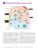
Innate immunity
(a) The complement pathway
Classical pathway
MBL pathway
Alternative pathway
C1q can be activated by IgM or
IgG immune complexes, CRP
and some bacterial cell wall
components. It is able to cleave
and activate C4 and C2
Activated by mannose-binding lectin,
which binds to mannose-containing
carbohydrates on bacteria or viruses.
MBL forms a complex with MASP-1 and
MASP-2 which can activate C4 and C2
Constitutively activated. Lack of complement
inhibitors on the surface of a pathogen results
in activation of terminal complement
components. Host cells protected by
inhibitors which terminate activation
C1
MBL-MASP1-MASP2
Factor D
C3
C4, C2
C4bC2a
C3bBb
Activation of the
classical pathway
leads to low C4 and C3
Anaphylotoxins
(b) Phagocytes
– neutrophils and macrophages
C9 C9 C9 C9
C9
C9
C9
C9
C9
C9
C6
C7 C8 C9
C5a
C5
C5b
C5b
Membrane
attack
complex
(ii) Response to infection
Pathogen opsonised
by IgG or CRP
FcγRindependent
phagocytosis
(i) Phagocyte entry into
sites of inflammation
Chemokine:
Chemokine
receptor
interaction
Activation of the alternative pathway
leads to low C3 with normal C4 levels
C3a
C3b
Neutrophil
Factor B
Release of
proteases as
neutrophil
disposes
of pathogen
Pathogen
FcγR-mediated
phagocytosis
MIP-2
Pathogen internalised
to phago-lysosome and
broken down
Neutrophil
CXCR2
Integrin:
Adhesion
molecule
interaction
PAMP recognition
via TLR
MAC-1
FcγR-mediated
phagocytosis
ICAM-1
Mannose
receptor-mediated
endocytosis
MR
Complement
receptor-mediated
phagocytosis
C3b
Monocyte
Release of
pro-inflammatory
cytokines
(e.g. Il-6, TNF-α)
and chemokines
CR
Macrophage
(iii) Response to tissue injury
Signal 1
TLR stimulation
via DAMP or PAMP
Signal 2
Intravascular
space
Endothelial
cell
DAMP
NFκB
pro IL-1β
ATP
Caspase 1
HSP DAMP-R
HMGB1
Uric
acid
Inflammasome
activation
pro IL-1β
Caspase 1
Macrophage
Transplantation at a Glance, First Edition. Menna Clatworthy, Christopher Watson, Michael Allison and John Dark.
22 © 2012 John Wiley & Sons, Ltd. Published 2012 by John Wiley & Sons, Ltd.
IL-1β
Release of IL-1β
The role of the immune system is to identify and remove invading
microorganisms before they cause harm to the host. This is
achieved by a rapid, non-specific innate immune response that is
followed by a more finely tuned, targeted, adaptive immune
response. The innate immune system is comprised of components
that directly recognise and destroy pathogens (the complement
system), a number of ‘flags’ known as opsonins (e.g. C-reactive
protein [CRP], C3b, natural IgM antibody), which make pathogens more easily recognised by immune cells such as phagocytes
(neutrophils and macrophages), which engulf and kill internalised
pathogens, and natural killer (NK) cells, which can detect and
destroy virus-infected cells.
The complement system
The complement system is a series of proteases, which are sequentially activated and culminate in the formation of the membrane
attack complex (MAC). The MAC forms a hole in the membrane
of the cell into which it is inserted (pathogen or host), disrupting
membrane integrity and causing cell lysis. The complement system
can be activated in three ways:
• the classical pathway
• the alternative pathway
• the mannose binding pathway.
IgM or immune complexed IgG activate the classical pathway.
The alternative pathway is constitutively active, while the mannose
binding pathway is activated by carbohydrates present on pathogens. The net result of activating any of the three pathways is the
formation of a C3 convertase (either C4bC2a or C3bBb), which
cleaves C3. The resulting C3b cleaves C5 and activates a final
common pathway resulting in MAC formation. Complement activation also leads to the production of anaphylotoxins (C3a and
C5a), which activate neutrophils and mast cells, promoting inflammation. In addition, C3b can opsonise pathogens for uptake by
complement receptors CR1 and CR3 on phagocytes.
Pentraxins
These are a family of proteins with a pentameric structure that
include CRP and serum amyloid protein (SAP). CRP and SAP are
synthesised in the liver and rapidly released into the bloodstream
in response to inflammation and are therefore called acute phase
proteins. Pentraxins bind to phosphorylcholine found on the
surface of pathogens and can fix complement (via the classical
pathway) and opsonise pathogens for uptake by phagocytes
through binding to surface Fc-gamma receptors (FcγRs). Pentraxins can also bind to apoptotic cells, facilitating their disposal.
Phagocytes
Phagocytes (from the Greek word ‘phagein’ – ‘to eat’) are cells
that ingest debris, pathogens and dying cells. There are two main
types of phagocyte, neutrophils (which circulate in the blood until
they are called into tissues), and macrophages, which are resident
in tissues and act as immune sentinels. The circulating monocyte
is the precursor to tissue macrophages. Neutrophils are the most
abundant circulating leucocyte and can be identified by their
multi-lobed nucleus and the presence of numerous granules within
their cytoplasm, which contain proteases (for example myeloperoxidase) and other bacteriocidal substances. Neutrophils move
into tissues by virtue of surface molecules called integrins (for
example MAC-1), which bind to adhesion molecules that are upregulated on vascular endothelium in inflamed tissue (for example
ICAM-1).
Phagocytes detect pathogens via membrane receptors, which
recognise repeating surface motifs on microbes, so-called pathogen-associated molecular patterns (PAMPs). These innate receptors include the toll-like receptors (TLRs) and the mannose
receptors. Phagocytes can also internalise opsonised pathogens via
complement receptors and FcγRs. Once internalised, the microbe
will be destroyed within the phagolysosome by proteases and by
the generation of oxygen and nitrogen free radicals. Tissueresident macrophages secrete pro-inflammatory cytokines such as
tumour necrosis factor (TNF)-α and interleukin (IL)-6, which lead
to changes in vascular permeability, and in the molecules expressed
on vascular endothelial cells. They also produce chemicals that
attract neutrophils and monocytes (known as chemokines). These
changes facilitate the entry of neutrophils and monocytes from the
circulation into the site of infection and result in the cardinal signs
of inflammation (calor, dolor, rubor and tumor, i.e. heat, pain,
redness and swelling).
Macrophages can also be activated by danger/damageassociated molecular patterns (DAMPs), for example heat shock
proteins (HSPs) or ATP, which are release by damaged or dying
host cells. This leads to activation of the inflammasome and the
production of IL1-β and IL18.
In addition, macrophages have the capacity to process and
present antigen (see Chapter 8).
Mast cells
Mast cells are large tissue-resident cells found mainly in the skin
and at mucosal surfaces. They are packed with granules containing
vasoactive amines (e.g. histamine) and heparin. Mast cell degranulation may be induced by trauma or UV light, and by binding of
IgE antibodies to Fc-epsilon receptors found on the surface of
mast cells. Mast cells play an important role in allergy and
anaphylaxis.
Natural killer cells
Natural killer cells express surface receptors (killer-cell immunoglobulin-like receptors [KIRs]), which bind to and assess cell
surface major histocompatibility complex (MHC) class I molecules. If non-self or altered self-antigen is detected on class I molecules, e.g. in virally infected cells or tumour cells, then the NK
cell will destroy this cell by the release of perforin (punches holes
in cells), granzyme (poisons cells) or the induction of apoptosis. In
addition, NK cells express FcγRs and can therefore be activated
against antibody-opsonised cells. This is known as antibodydependent cellular cytotoxicity (ADCC).
Innate immunity Immunology of organ transplantation 23