Ebook Hematologic problems in the critically ill: Part 1
Bạn đang xem bản rút gọn của tài liệu. Xem và tải ngay bản đầy đủ của tài liệu tại đây (1.04 MB, 62 trang )
Giorgio Berlot · Gabriele Pozzato
Editors
Hematologic Problems
in the Critically lll
123
Hematologic Problems
in the Critically Ill
Giorgio Berlot • Gabriele Pozzato
Editors
Hematologic Problems
in the Critically Ill
Editors
Giorgio Berlot
Anesthesia and Intensive Care
University of Trieste
University Hospital
Trieste
Italy
Gabriele Pozzato
Haematology
University of Trieste
University Hospital
Trieste
Italy
ISBN 978-88-470-5300-7
ISBN 978-88-470-5301-4 (eBook)
DOI 10.1007/978-88-470-5301-4
Springer Milan Heidelberg New York Dordrecht London
Library of Congress Control Number: 2014952789
© Springer-Verlag Italia 2015
This work is subject to copyright. All rights are reserved by the Publisher,
whether the whole or part of the material is concerned, specifically the rights
of translation, reprinting, reuse of illustrations, recitation, broadcasting, reproduction on microfilms or in any other physical way, and transmission or information storage and retrieval, electronic adaptation, computer software, or by
similar or dissimilar methodology now known or hereafter developed.
Exempted from this legal reservation are brief excerpts in connection with
reviews or scholarly analysis or material supplied specifically for the purpose
of being entered and executed on a computer system, for exclusive use by the
purchaser of the work. Duplication of this publication or parts thereof is permitted only under the provisions of the Copyright Law of the Publisher's location, in its current version, and permission for use must always be obtained
from Springer. Permissions for use may be obtained through RightsLink at
the Copyright Clearance Center. Violations are liable to prosecution under
the respective Copyright Law.
The use of general descriptive names, registered names, trademarks, service
marks, etc. in this publication does not imply, even in the absence of a specific
statement, that such names are exempt from the relevant protective laws and
regulations and therefore free for general use.
While the advice and information in this book are believed to be true and
accurate at the date of publication, neither the authors nor the editors nor the
publisher can accept any legal responsibility for any errors or omissions that
may be made. The publisher makes no warranty, express or implied, with
respect to the material contained herein.
Printed on acid-free paper
Springer is part of Springer Science+Business Media (www.springer.com)
Contents
1 Introduction . . . . . . . . . . . . . . . . . . . . . . . . . . . . . . .
Giorgio Berlot and Gabriele Pozzato
1
2 Anemia . . . . . . . . . . . . . . . . . . . . . . . . . . . . . . . . . . .
Gabriele Pozzato
3
3 Anemia in the Critically Ill Patient . . . . . . . . . . . .
Giorgio Berlot and Perla Rossini
21
4 Leukopenia in the Critically Ill Patient . . . . . . . .
Giorgio Berlot, Barbara Presello,
and Antoinette Agbedyro
37
5 Leukocytosis in the Critically Ill Patient . . . . . . .
Giorgio Berlot, Antoinette Agbedyro,
and Barbara Presello
47
6 The Critically Ill Patient with Abnormal
Platelet Count . . . . . . . . . . . . . . . . . . . . . . . . . . . . .
Luca G. Mascaretti and Paola Pradella
59
7 Adverse Transfusion Reactions
in Critically Ill Patients . . . . . . . . . . . . . . . . . . . . . .
Federica Tomasella and Luca G. Mascaretti
81
8 Drugs and Blood Cells . . . . . . . . . . . . . . . . . . . . . .
Federico Pea and Pier Giorgio Cojutti
111
v
Chapter 1
Introduction
Giorgio Berlot and Gabriele Pozzato
Three o’clock a.m. You just sit down and drink a cup of coffee
when the phone rings. It is the ED: 10 min ago a man was admitted with hypotension, fever and leukopenia associated with low
platelet count and abnormal coagulation tests. More or less an
hour ago you visited another patient with ever-decreasing hemoglobin values in whom the most common sources of bleeding
have been excluded. You are blaming yourself because you
failed to buy a textbook of hematology you saw at a congress a
couple of weeks ago and the hospital administration because a
hematologist will be available only after 9.00 a.m. In the meanwhile, you are expected to keep these patients alive till someone
with a more in-depth knowledge of hematological disease will
arrive to help you and your colleagues.
Actually, the presence of hematological alterations is very
common in critically ill patients just for the kind of diagnosis of
G. Berlot ( )
Anesthesia and Intensive Care,
University of Trieste, University Hospital, Trieste, Italy
e-mail:
G. Pozzato
Haematology, University of Trieste, University Hospital, Trieste, Italy
e-mail:
G. Berlot, G. Pozzato (eds.), Hematologic Problems in the Critically Ill,
DOI 10.1007/978-88-470-5301-4_1, © Springer-Verlag Italia 2015
1
2
G. Berlot and G. Pozzato
admitted cases, that is, severe traumas, car crashes, septic
shocks, severe respiratory distress and so on. In these patients,
the finding of anemia or leukocytosis is an expected feature of
the acute event and does not alert doctors and nurses. The
requests of hematological counseling occur when there are discrepancies between the clinical situation and the main hematological parameters: for example, sepsis is improving and
leukocyte level is still increasing or there is a worsening anemia
without evidence of blood loss.
In these critical patients, the traditional tools for evaluating
the nature of the hematological diseases are not feasible: the
family and the personal history of the patients are often unavailable, and other anamnestic features like changes in stool habits
or dietary history are irrelevant and useless. Even to perform the
physical examination is often difficult, given the common presence of several medical devices (nasogastric tube, central vein
catheters, endotracheal tube, invasive hemodynamic monitoring) and the absence of patient cooperation. Therefore, to identify the cause of the hematological alterations, there is the need
of several key laboratory tests.
Obviously, a different approach is indicated in case of cytopenias (anemia, thrombocytopenia, leukopenia) and in the case
of thrombocytosis, leukocytosis or, rarely, of erithrocytosis.
These hematological alterations could be mixed in different
ways with regard of the several acute and chronic pathological
conditions present in the same critical patient. However, for
didactic reasons, the main hematological conditions requiring
counseling will be separately discussed. Since the most common hematological problem in the critically ill patient is anemia, the opening chapter will discuss this pathological
condition.
Chapter 2
Anemia
Gabriele Pozzato
Anemia is not a disease by itself but a condition that is a
consequence of acquired or genetic abnormalities. Functionally,
anemia is defined as an insufficient red cell mass to deliver
adequate amount of oxygen to organs and peripheral tissues, and,
for practical reasons, an Hb concentration less than 14.0 g/dL
for men and 12.0 g/dL for women. At present, Hb concentration,
as well as other red cell parameters, is determined by electronic
cell counters able to deliver the results in few minutes. In most
patients, blood determination of Hb levels is useful for assessing
anemia, but there are some limitations that must be recognized:
1. Hb changes may reflect altered plasma volume, not a change
in red cell mass. In pregnancy, for example, the increased
plasma volume decreases the Hb concentration and, in fact,
total red cell mass is increased but to a lesser degree than
plasma volume. Likewise, very often the critically ill patient
is hyper-hydrated to avoid dangerous hypotension or shock;
G. Pozzato
Department of Hematology, University of Trieste,
University Hospital, Piazza Ospedale 1, Trieste 34100, Italy
e-mail:
G. Berlot, G. Pozzato (eds.), Hematologic Problems in the Critically Ill,
DOI 10.1007/978-88-470-5301-4_2, © Springer-Verlag Italia 2015
3
4
G. Pozzato
this common therapeutic approach determines an increase of
plasma volume and reduces Hb concentration and the degree
of anemia may appear severe. Conversely, burn patients,
through the injured skin, lose plasma and not red cells; therefore, Hb concentration appears normal or even high while the
red cell mass could be decreased.
2. Several abnormal Hb have altered ability to bind and to
release the oxygen and this is associated with different Hb
concentrations. The carriers of Hb with high affinity for
oxygen show levels of Hb higher than normal, while the
carriers of Hb with decreased oxygen affinity (and better
oxygen delivering to tissues) have lower than normal Hb
levels.
3. There are several pathological conditions that determine a
compensatory increase of red cell mass, the most common
are the emphysema (and similar pulmonary diseases) or the
right-to-left cardiac shunt (often unknown). These patients
have abnormally elevated Hb levels; therefore, a normal Hb
level may represent an “anemia” since tissue oxygenation is
impaired. Conversely, the patients with hypothyroidism
(decreased oxygen needs) may have low Hb level with adequate oxygen delivery to tissues.
4. Acute blood loss is another example of the problem of evaluating anemia by the Hb concentration. In fact, immediately
after blood loss, the Hb is normal because the compensatory
response to acute hemorrhage is the vasoconstriction.
Therefore, the decrease of the Hb concentration begins after
4–6 h. The recognition of this situation is generally easy for
the patients recovered in intensive care units since they are
monitored in a continuous fashion.
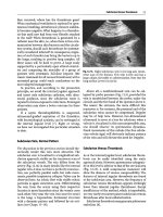
Once the diagnosis of anemia is defined, the cause of this
condition must be identified. The classification of the anemia is
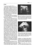
not simple, but a useful approach could be to ask several
questions stepwise (Fig. 2.1).
2
Anemia
5
Is anemia
associated
with other
hematologic
abnormalities ?
YES
NO
Bone
marrow
examination
Reticulocyte
count
High
reticulocyte
count
Low
reticulocyte
count
Red cell
mean
corpuscolar
volume
Evidence of
hemolysis ?
YES
NO
Check
causes of
hemolysis
Check
causes of
hemorrhagia
MCV < 80
Fig. 2.1 Diagnostic algorythm for anemia
MCV 80–
100
MCV > 100
6
G. Pozzato
The first question is whether anemia is associated with other
hematological abnormalities such as low platelet levels and/or
low leukocyte counts and/or presence of abnormal leukocytes
(blasts) on blood smear. If this is the case, the presence of bone
marrow failure (aplastic anemia) or of malignant hematological
disorders such as acute leukemias or myelodysplastic syndromes
is likely. In these cases, the bone marrow biopsy and the
appropriate cytometric studies of marrow and peripheral blood
are mandatory.
The second question is whether anemia determined is
associated with an appropriate reticulocyte response. The
reticulocyte count is important to evaluate the new red cell
production and is very helpful in determining the marrow
response to anemia. Very often the reticulocyte count is lacking
for the evaluation of the anemic conditions, while this test has
a crucial role in the diagnostic process. Until a few years ago,
the red blood cells were stained with brilliant cresyl blue,
which allows the visualization of ribosomes and reticulin
network, thereafter the blood smear was examined by microscope with manual count of stained cells. This method was
time-consuming and often the responses were delayed, thus
reducing the clinical impact of the test. Lately, automated
reticulocyte analyzers are available; these counters have a
higher degree of precision than can be achieved manually and,
in addition, the responses are immediate. These automated
reticulocyte counters may show errors in few rare conditions as
the case of presence of Heinz or Howell-Jolly bodies inside red
cells. Much more important than the percentage of reticulocytes is their absolute count, which can be easily determined
starting from the red cell count: absolute reticulocytes count = %
of reticulocytes × red cells count/L3. The value over 100 × 109/L
is indicative of a bone marrow responding normally to hemolysis or blood loss. If the anemia is associated with a poor reticulocyte count (less than 25 × 109/L), an impaired red cell
production is likely.
2
Anemia
2.1
7
Anemias with High Reticulocyte Count
In the case of high reticulocyte count, the subsequent question
is: Is there evidence of hemolysis or not? The laboratory tests
used to identify a hemolytic process are available easily in any
hospital: Serum unconjugated bilirubin, serum lactic dehydrogenase (LDH), and serum aptoglobin. These tests are related to
the red cell increased destruction rate and, in most patients, are
indicative of a hemolytic process, but in critically ill patients
may be misleading. An increased level of total and unconjugated bilirubin is a common finding in intensive care units for
several reasons: prolonged fasting or artificial nutrition, hypotension or shock with reduced liver blood flow, heart failure or
tamponade with secondary liver venous stasis, hepatosplenic
blood flow modification by endotoxemia or peritonitis, portal
thrombosis, preexisting chronic liver diseases, and other less
common causes. LDH is an enzyme not specific to the red cells,
and it can be found in any organ and tissue; therefore, any cytolitic process is able to increase LDH serum levels. In critically
ill patients, high of very high level of serum LDH can be found
very easily due to crush syndrome with muscle necrosis, lung
inflammatory processes, chronic and acute viral liver diseases
or acute cholestasis, fatty liver, sepsis, myocardial ischemia,
bone fractures, and others. In addition, high LDH levels without
evidence of disease can be found in about 3 % of normal people.
The LDH isoenzymes could be useful for determining the
involved tissue, but this test is not available in most hospitals
and it is used for research purposes only. In conclusion, LDH is
not trustworthy in the context of the critically ill patient. The
haptoglobin is a protein synthesized by the liver, and it is able to
bind to Hb when this molecule is released in the plasma (like
occurs in hemolysis). The complex haptoglobin-Hb is removed
by the hepatocytes. Despite the presence of haptoglobin in
serum only, this protein decreases or becomes undetectable in
8
G. Pozzato
case of both intravascular and extravascular hemolysis. Serum
haptoglobin determination is useful in the diagnostic path of the
majority of patients, but in the intensive care units the interpretation of its levels is complicated and its diagnostic power is
significantly reduced. In fact, haptoglobin is an acute-phase
protein, therefore, its synthesis increases in response to inflammation, infections, or malignant diseases. Taking into account
these characteristics, in critically ill patients, the increased synthesis of this protein due to sepsis, infections, inflammatory
states of various etiologies, may overcome the decrease induced
by hemolytic process. Conversely, abnormal low levels of haptoglobin can be found in the absence of hemolysis in the case of
malnutrition or of the other clinical situations characterized by
abnormal protein loss like occurs after extensive burns or for
nephritic syndrome; by preexisting chronic liver disease; or by
the impossibility of a normal aliment absorption like occurs in
large intestine resections for vascular disease or for accident
perforation, events not uncommon in the intensive care units. In
conclusion, the usual laboratory tests used to identify a hemolytic process are have a limited diagnostic value in the intensive
care setting and, often, additional tests and a careful follow-up
of the patient are needed for a correct diagnosis. Even the diagnosis of posthemorrhagic anemia may be difficult in these
patients. In fact, after an acute blood loss, the plasma volume
and red cell mass are reduced in proportional amount; consequently, the Hb concentration does not change. Therefore, the
amount of blood loss can be underestimated by the degree of
anemia, especially early. In the days following the blood loss,
the reticulocyte count is normal and increases only after 6–10
days; in this “window,” even the iron stores are unmodified, and
mean corpuscular volume is still normal. An external hemorrhage sufficient to determine anemia is usually evident, but
internal bleeding may be less apparent. If the hemorrhage
occurs in retroperitoneal space, into a body cavity or in a cyst,
the decrease of Hb level may be a diagnostic problem. In
2
Anemia
9
addition, the breakdown and the absorption of red cell in the
tissues are able to increase indirect bilirubinemia, and this picture, along with high reticulocyte count, can be confused with a
hemolytic anemia. Therefore, a careful follow-up of the patient
and appropriate tests are mandatory for a correct diagnosis.
If repeated tests confirm high reticulocyte counts (in the
absence of blood loss) and a possible hemolytic process is suspected, the main causes of hemolysis should be carefully
checked. Since in the adult patients the most common acquired
hemolytic disorders are the immune-mediated processes, the
direct anti-globulin test (Coomb’s test) should be determined.
Thereafter, the diagnostic process can be separated for the
patients with positive and negative direct anti-globulin test.
2.1.1
Patients Positive for Direct
Anti-globulin Test
These cases have presumably an immune-hemolytic anemia and
can undergo immediate glucocorticoids therapy, which remains
the treatment of choice of this immune disorder. Intravenously
administered doses of 1.0 mg/kg b.w. of methyl-prednisolone
daily are efficacious in most cases. The response may not be
evident for several days and an increase of Hb level can be
noticeable only after 7 days of treatment. A further delay in the
response is expected in critically ill patients since many acute
factors may interfere in the red cell production like prolonged
fasting or artificial nutrition, hypotension, reduced liver blood
flow, acute renal failure with reduced erythropoietin production,
endotoxemia or other acute stress situations. In the rare cases of
lack of response or in the case of worsening of the hemolytic
process, high-dose i.v. immunoglobulin administration (1 g/kg
b.w.) can be useful in decreasing the clearance of the red cells
by the monocyte macrophage system. This therapy can be
repeated after 1 or 2 weeks if required.
G. Pozzato
10
2.1.2
Patients Negative for Direct
Anti-globulin Test
In these cases, the clinical history (when available) is helpful
to exclude the exposure to chemical or physical agents; thereafter, some infections (malaria, leishmaniasis, trypanosomiasis, bartonellosis) should be taken into consideration in white
people back from recent adventure travels in the third world or
in people shortly after arriving from Africa or from other
underdeveloped countries. In critically ill patients, the septicemia of Clostridium perfrigens should be taken into consideration, in fact it may occur after traumatic wound infections,
necrotizing enterocolitis, genitourinary or gastrointestinal
surgery, and other acute severe conditions. In this case, a
severe, often-fatal, hemolytic anemia occurs with a massive
hemolysis, and hemoglobin concentration may fall to a very
low level in a matter of hours. The diagnosis is suspected when
high fever, jaundice, and anemia occur together in a patient of
the intensive care unit. The clostridial infection responds well
to antibiotics therapy but the treatment must be started as
quickly as possible, even before the blood culture results are
available.
After the exclusion of these infective causes with appropriate
tests, the other causes of nonimmune hemolytic anemia should
be considered. For the diagnosis of the most common diseases,
a few laboratory investigations are needed:
1.
2.
3.
4.
Hb electrophoresis
Osmotic fragility test
Red cell enzyme determination
Blood smear examination
The Hb electrophoresis may indicate the presence of genetic
diseases like sickle cell anemia, or thalassemia or of the rare
conditions associated with abnormal Hb (Hb C, SC, D, SD, and
2
Anemia
11
E). The osmotic fragility test is able to discover the spherocytic
anemia and related disorders, and, finally, the enzyme determination is useful to detect the glucose-6-phosphate deficiency
(G6PD), known as favism, or pyruvate kinase deficiency. All
these conditions are inherited diseases; some of these are common in Italy like thalassemias or favism, while others are very
rare in Europe, like sickle cell anemia or the unstable Hb diseases. All these diseases worsen the degree of anemia in patients
in critical medical conditions and should be recognized to avoid
unnecessary support treatments or delay in discharging the
patient fearing covert bleeding.
The blood smear examination by microscope is a disregarded
tool, which, on the contrary, is able to give important information on the etiology of many hematological disorders even in the
setting of the intensive care units. In the case of patients with
overt hemolysis and negative for the direct anti-globulin test, the
blood smear is very important for the diagnosis of the so-called
fragmentation hemolysis, a relatively common condition in the
critically ill patient.
When the red blood cells are subjected to physical trauma, as
occurs in the alterations of heart or for the appearance of microvascular thrombi in small vessels, they may undergo fragmentation, thereby resulting in hemolytic anemia. In these cases, the
blood smear shows characteristic fragmented red blood cells
named schistocytes; these cells have a crescent shape or take the
form of triangles or helmets or other bizarre forms. The identification of the presence of schistocytes is very important since
usually there are not other diagnostic tools to recognize the
clinical condition characterized by the fragmentation hemolysis.
The main causes of red cell fragmentation are indicated in
Table 2.1. As shown, only a fraction of the pathological conditions indicated in the table are associated with acute diseases
that can be found in the intensive care units; in the following
paragraphs only these conditions will be discussed, since the
others are outside the scope of this book.
12
G. Pozzato
Table 2.1 Clinical condition associated with fragmentation hemolysis
Heart and great vessels abnormalities
Synthetic valvular prostheses (especially aortic)
Unoperated valve diseases (especially aortic stenosis)
Teflon patch repair of atrio-ventricular defects
Ruptured chordae tendineae
Valve porcine xenografts or homografts or xenobioprostheses
Coarctation of aorta
Small vessel diseases (microangiopathic hemolytic anemias)
Thrombotic thrombocytopenic purpura (Moshkowitz’s disease)
Hemolytic uremic syndrome
Disseminated malignant disease
Transplant-associated microangiopathy
Malignant hypertension
Disseminated intravascular coagulation
Giant hemangiomas and liver hemoangioendothelioma
March hemoglobinuria
Pregnancy-associated thrombotic microangiopathy
HELLP syndrome
Pregnancy-associated thrombotic thrombocytopenic purpura and
hemolytic uremic syndrome
Autoimmune diseases
Lupus erythematosus
Wegener granulomatosis
2.2
Heart and Great Vessels Abnormalities
Many patients, after open-heart surgery, are recovered in the
intensive care units; therefore, in the management of these
patients, medical staff should be able to recognize the laboratory
signs of fragmentation hemolysis. In fact, some patients, soon
after surgery, develop anemia of different severity. The incidence of hemolysis is reported to be variable ranging from 5 to
25 %. This great variability depends on the method used for
detecting hemolysis, lower if only haptoglobin level is determined, higher if more sophisticated methods, like red cell
2
Anemia
13
survival, are available. Several mechanisms are involved in the
hemolysis, but all are referable to high turbulence. When the
lumen of the aortic prosthesis is small relatively to the stroke
volume, a shearing stress higher than 3,000 dyn/cm2 can easily
be generated and this determines mechanical hemolysis. The
presence of a severe fragmentation hemolysis with anemia
requiring transfusions immediately after open-heart surgery
often indicates malfunction of valvular prosthesis. Since this
condition does not improve spontaneously, a prompt surgery
and valve replacement is indicated. Awaiting the surgery, the
patients must be kept at bed rest since hemolysis becomes worse
after even slight physical activity.
2.3
Thrombotic Thrombocytopenic
Purpura (TTP)
This disease is characterized by disseminated microvascular
thrombi in small vessels and by a syndrome including hemolytic
anemia, severe thrombocytopenia, neurological symptoms,
renal dysfunction, and fever. At the time of presentation, the
clinical conditions of the affected patients can be critical; therefore, they are often recovered in intensive care units. Excluding
the very rare inherited forms (Upshaw-Shulman syndrome) that
appear during childhood, TTP has a peak of incidence between
30 and 40 years. Like most autoimmune diseases, TTP is more
common in women than in men (ratio of 2:1). The pathogenesis
of the TTP has been clarified in the past years. The von
Willebrand Factor (vWF) is a multimeric protein synthesized
and stored as ultra-large multimers in endothelial cells, and
released at constant rate in circulation. The ultra-large multimers of vWF are immediately cleaved by a metalloprotease present on surface of the endothelial cells and in plasma. This
enzyme, known as ADAMTS13, is able to cut the ultra-large
14
G. Pozzato
vWF in small multimers necessary for normal platelet adhesion.
If the ADAMTS13 does not work for either inherited disease or
for antibodies, the ultra-large vWF multimers bind to platelets,
promoting platelet agglutination and aggregation, and, at the
end, coagulation activation and disseminated microthrombi formation. These microthrombi may be found throughout the body,
but they are seen most commonly in brain (especially cortical
grey matter), kidney, pancreas, spleen, heart, and cortical
glands. The hemolytic anemia is related to the red cell damage
for the interaction with fibrin networks and microthrombi in the
small vessels, this interaction produces the schistocytes evident
on blood smear. Schistocytes have a short life span since the
spleen rapidly removes them.
At the presentation of TTP, the neurologic symptoms are the
most common, while, despite severe thrombocytopenia, the
hemorrhagic problems are not remarkable. The neurologic
symptoms include headache, cranial nerve palsies, paresis, dysphasia, aphasia, and confusion; these symptoms are transient but
recurrent and, if the disease is not recognized, may progress
shortly to stupor, seizures, and coma. Fever and the symptoms
of a rapid-onset anemia are present in 50 % of the cases. Less
common symptoms are abdominal pain (due to pancreatitis),
acute respiratory distress symptoms, cardiac conduction abnormalities, and infarcts.
In addition to anemia and thrombocytopenia, the main laboratory findings are those of a hemolytic process, that is, elevated
unconjugated bilirubin, undetectable haptoglobin, and very high
level of LDH, usually more than 1,000 U/L. The LDH increase
may be the expression of not only red cells’ destruction but even
of disseminated tissue damage.
The diagnosis of TTP is clinically easy since the presentation, at the same time, of neurologic symptoms associated with
hemolytic anemia and thrombocytopenia is uncommon in other
diseases. However, the presence of some comorbidities like
preexisting neurologic problems or liver cirrhosis or other
2
Anemia
15
diseases able to lower platelet levels, may confound the clinical
picture. In these cases, there are no diagnostic tools to confirm
the diagnosis of TTP outside blood smear examination for
detecting schistocytes. At present, commercial kits to determine
ADAMTS13 activity as well as the presence of anti-ADAMTS13
antibodies are available, but these tests are troublesome and cannot be used in an emergency since responses are delayed for
weeks. Conversely, we need to confirm the clinical suspect of
TTP as soon as possible, since the treatment should start immediately to avoid fatal neurologic complications. Therefore, the
detection of schistocytes at the microscopic examination of
blood smear remains the stronghold of the diagnosis.
The treatment of TTP is based on aggressive plasma
exchange. If the treatment starts shortly after the diagnosis, the
survival rate is more than 80 %. Before the introduction of this
procedure, TTP was fatal in over 80 % of the cases within 3
months and only less than 10 % of the patients survived more
than 12 months. Plasma exchange determines a favorable outcome even in the presence of renal failure or advanced neurologic complications. The infusion of large amount of fresh
plasma, containing intact ADAMTS13, can be considered only
as a temporary therapy in the case of delay of the plasma
exchange. In fact, in a controlled prospective therapeutic trial
comparing plasma infusion and plasma exchange, the latter
demonstrated significantly better outcomes. The extraordinary
effect of plasma exchange is due to the removal of
anti-ADAMTS13 antibodies and of ultra-large vWF multimers
together with the replacement of the fresh enzyme, able to
cleave residual abnormal vWF multimers. The response is often
dramatic; the neurologic complications disappear within a few
hours and main laboratory alterations improve in a short time.
The procedure should be performed daily until the platelet count
is normal and hemolysis is minimal. Since the disease is due to
autoantibodies, traditionally patients receive, in addition to
plasma exchange, high-dose corticosteroids. Immunosuppressive
G. Pozzato
16
treatment seems more useful to prevent early relapse than to
reduce the specific antibodies levels, thus increasing significantly the serum ADAMTS13 activity.
2.4
Hemolytic Uremic Syndrome
Hemolytic uremic syndrome (HUS) is a rare disease characterized by three primary symptoms: hemolytic anemia (with schistocytes), low platelet count, and acute renal failure. HUS is
classified into two primary types: (1) HUS due to infections,
often associated with diarrhea; and (2) HUS related to complement abnormalities—such HUS is also known as “atypical
HUS” and is not diarrhea associated. The HUS associated to
infection is common in children aged 1–5 years, at least in
Europe and North America. The disease is due to a toxin (Shiga
toxin) produced by some bacteria: Escherichia coli is the most
commonly involved species; Shigella Dysenteriae type I and
Citrobacter freundii have been less frequently observed. The
toxin, produced in the gut, is absorbed and, in target organs
(e.g., kidney and gut) it binds to glycolipid receptors on the cell
surface, then the toxin is endocytosed and transported to the
Golgi apparatus and the endoplasmic reticulum, it is later translocated to the cytosol where it inactivates ribosomes and causes
cell death. HUS is a pediatric disease and diagnosis is relatively
easy in cases of typical presentation with watery diarrhea, followed by bloody diarrhea and abdominal cramps. In the following days, the symptoms are related to severe anemia, hemolysis,
and renal failure. The diagnosis of the atypical HUS, or notinfectious HUS, is much more complicated since the diarrhea is
absent and a trigger of the disease cannot be found. The atypical
HUS is a very rare event (0.2 cases/100,000/year) and more than
70 % of cases are in pediatric age, since the disease is related to
inherited abnormalities of some complement factors or of the
2
Anemia
17
cobalamin metabolism. In children, the age of onset, family
history, and clinical presentation are useful for a correct differential diagnosis, while in adults, autoimmune diseases, pregnancy, transplantation, and drugs are causes of atypical HUS. In
adult patients with thrombocytopenia and hemolysis presenting
renal involvement, the presence of atypical HUS should be suspected and the blood smear examination for schistocytes is
mandatory. The diagnosis might be performed as soon as possible since a prompt treatment avoids the progression of the
renal failure. The treatment is the fresh frozen plasma infusions,
and, when disease activity is not controlled, the plasma
exchange should be performed. Prophylactic antibiotics should
be administered because infections can trigger relapse.
2.5
Disseminated Intravascular
Coagulation (DIC)
This disease is a relatively common problem in the intensive
care units, and DIC still remains a diagnostic and therapeutic
challenge. The clinical features of DIC are bleeding manifestations, often very serious and of abrupt onset, therefore, the
anemia is due more easily to hemorrhages than to hemolysis.
However, clinicians should be aware of the possibility of the
presence of a hemolytic process proportional to the severity
of the coagulation abnormalities, this to avoid unnecessary
tests and/or treatment delay. The prognosis of DIC depends
on its etiology and on the possibility to remove or to treat the
trigger of the process; the most common causes of DIC are
reported in As shown, some hematological diseases can determine DIC, and, in rare cases the beginning of the disease
could be a DIC.
Among these cases, the promyelocytic leukemia is the most
common: a sudden and severe bleeding often of serious magnitude
18
G. Pozzato
can be the onset of the disease. To recognize immediately the
presence of this leukemia is very important since the treatment
must start immediately and, consequently, the prognosis is very
good with a predicted long-term survival of more than 90 % even
avoiding chemotherapy. In these cases, together with the clinical
and laboratory signs of the DIC, abnormal leukocyte count with
immature cells are present on peripheral blood, therefore the
diagnosis is easy. The age-adjusted incidence rate of acute
myeloid leukemia (AML) in adults is about 3.7 per 100,000/year
for both sexes, and promyelocytic leukemia represents the
10–5 % of all AML, therefore promyelocytic leukemia has an
incidence ranging from 0.4 to 0.2 cases/100,000/year.
Even rarer is the paroxysmal nocturnal hemoglobinuria
(PNH) whose incidence is still unknown, but the data collection
from different sources has given quotes of about 0.5
cases/100,000/year. Since the disease is underdiagnosed, its
incidence may be higher. This disease, due to the mutations of
the gene PIG-A placed on X chromosome, shows a complex
pathogenesis and it may present mild hemolytic anemia associated with recurrent hemoglobinuria, mainly during the night, or
the features of an aplastic anemia, and finally a thrombotic
syndrome. In rare cases, the onset of the disease is a severe
hemolytic episode; these attacks are associated with general
malaise, fever, headache, and abdominal and lumbar pains.
Since in PNH the hemolysis is intravascular, a massive hemolytic episode is able to activate coagulation cascade and to
initiate the DIC. In these very rare cases, the diagnosis is particularly difficult: only the more or less massive hemoglobinuria
may suggest PNH, while the other laboratory features of the
disease are not specific. In addition, the diagnosis underlies on
the demonstration of the lack of CD59 and CD55 expression on
red cells, granulocytes, and platelets on flow cytometry, not
available in any hospital.
Sickle cell anemia is an inherited disease due to the substitution of a single mutation (GAG vs. GTB) in the sixth codon of
2
Anemia
19
the β gene; this determines a substitution of valine instead of
glutamine in the sixth position of the β chain in the Hb (HbS).
This also determines a decrease of the Hb solubility of Hb when
deoxygenated with formation of HbS polymers inside red cells
and subsequent erythrocyte deformation. These “sickled” erythrocytes have poor deformability and patients develop a diffuse
veno-occlusive disease and, consequently, with acute events like
painful crisis; stroke; acute chest syndromes; priapism; and
chronic organ damage, especially bones and joints, cardiovascular system, kidney, pulmonary system, liver, and eyes. The disease has its highest prevalence in tropical Africa; in several
countries about 45 % of the population has sickle trait. In USA,
about 8 % of Afro-Americans are carriers of the sickle gene. In
Europe, sickle cell anemia is present only in the countries of the
Mediterranean basin (Italy, Greek) with a very low incidence. In
some cases, even in carriers of the trait only, life-threatening
hyper-hemolytic crisis may occur with abrupt anemia; the massive hemolysis (like in PNH) is able to activate the coagulation
and a DIC may appear. The hyper-hemolytic crisis may be triggered by infections or by exposition to cold temperature and by
strenuous physical exercise. The diagnosis of sickle cell anemia
should be suspected in any African or Afro-American patient,
and, since there are an increasing number of immigrants in any
European country, the doctors, especially those working in
intensive care units, must be able to recognize the disease. The
diagnosis is straightforward since a simple electrophoresis using
cellulose acetate is rapid, inexpensive, and effective to separate
the normal Hb from variants and the method is available in any
hospital. The presence of other inherited Hb diseases, like
β-thalassemia, may complicate the diagnostic path and additional tests are required like isoelectric focusing or
HPLC. However, the presence of HbS on electrophoresis must
be considered as the hallmark of the disease, also if other Hb
electrophoretic abnormalities are found. Therefore, in addition
to the therapy of the DIC, in patients with hyper-hemolytic crisis